30 Respiratory and Circulatory Systems
30 Respiratory and Circulatory Systems
30 Respiratory and Circulatory Systems

You also want an ePaper? Increase the reach of your titles
YUMPU automatically turns print PDFs into web optimized ePapers that Google loves.
<strong>30</strong><br />
CHAPTER<br />
<strong>Respiratory</strong><br />
<strong>and</strong> <strong>Circulatory</strong><br />
<strong>Systems</strong><br />
KEY CONCEPTS<br />
<strong>30</strong>.1 <strong>Respiratory</strong> <strong>and</strong> <strong>Circulatory</strong> Functions<br />
The respiratory <strong>and</strong> circulatory systems bring oxygen <strong>and</strong><br />
nutrients to the cells.<br />
<strong>30</strong>.2 Respiration <strong>and</strong> Gas Exchange<br />
The respiratory system exchanges oxygen <strong>and</strong> carbon dioxide.<br />
<strong>30</strong>.3 The Heart <strong>and</strong> Circulation<br />
The heart is a muscular pump that moves the blood through<br />
two pathways.<br />
<strong>30</strong>.4 Blood Vessels <strong>and</strong> Transport<br />
The circulatory system transports materials throughout the body.<br />
<strong>30</strong>.5 Blood<br />
Blood is a complex tissue that transports materials.<br />
<strong>30</strong>.6 Lymphatic System<br />
The lymphatic system provides another type of circulation in<br />
the body.<br />
BIOLOGY<br />
CLASSZONE.COM<br />
BIOLOGY<br />
View animated chapter<br />
concepts.<br />
• How You Breathe<br />
• How the Heart Pumps Blood<br />
• Blood Typing<br />
• Build the <strong>Circulatory</strong> <strong>and</strong><br />
<strong>Respiratory</strong> <strong>Systems</strong><br />
Keep current with biology news.<br />
• News feeds<br />
• Strange Biology<br />
• Careers<br />
RESOURCE CENTER<br />
Get more information on<br />
• <strong>Circulatory</strong> System<br />
• Blood<br />
• Lymphatic System<br />
908 Unit 9: Human Biology
What can take<br />
your breath away<br />
This photograph shows a resin cast of<br />
the thous<strong>and</strong>s of vessels that supply<br />
blood <strong>and</strong> air to your lungs. As you take a<br />
breath every three to five seconds, this<br />
network takes in oxygen <strong>and</strong> expels carbon<br />
dioxide. Pollution, disease, or injury<br />
can damage this intricate network, literally<br />
taking your breath away.<br />
Connecting<br />
CONCEPTS<br />
Nervous System The heart<br />
shown to the left is a model<br />
that has been x-rayed to<br />
reveal the inner chambers.<br />
Your heart is a muscular<br />
pump that moves blood<br />
through your body, bringing<br />
carbon dioxide to the lungs<br />
<strong>and</strong> picking up oxygen. When<br />
your body’s needs for oxygen<br />
change, signals from the<br />
nervous system speed up or<br />
slow down your heartbeat<br />
<strong>and</strong> breathing rate.<br />
Chapter <strong>30</strong>: <strong>Respiratory</strong> <strong>and</strong> <strong>Circulatory</strong> <strong>Systems</strong> 909
<strong>30</strong>.1 <strong>Respiratory</strong> <strong>and</strong><br />
<strong>Circulatory</strong> Functions<br />
KEY CONCEPT The respiratory <strong>and</strong> circulatory systems bring oxygen <strong>and</strong> nutrients to the cells.<br />
MAIN IDEAS<br />
• The respiratory <strong>and</strong> circulatory systems work<br />
together to maintain homeostasis.<br />
• The respiratory system moves gases into <strong>and</strong> out<br />
of the blood.<br />
• The circulatory system moves blood to all parts<br />
of the body.<br />
VOCABULARY<br />
circulatory system,<br />
p. 910<br />
respiratory system,<br />
p. 910<br />
trachea, p. 911<br />
lung, p. 911<br />
alveoli, p. 911<br />
diaphragm, p. 912<br />
heart, p. 912<br />
artery, p. 913<br />
vein, p. 913<br />
capillary, p. 913<br />
Connect You have thous<strong>and</strong>s of kilometers of blood vessels in your body <strong>and</strong><br />
several hundred million tiny air sacs in your lungs. Blood circulates constantly<br />
through the vessels, while air continually fills <strong>and</strong> empties from the tiny air sacs.<br />
Your heart keeps beating <strong>and</strong> your lungs keep working 24 hours a day, every day<br />
of your life. Even more amazing, everything works without your having to think<br />
about it.<br />
TAKING NOTES<br />
Use a supporting main ideas<br />
strategy to help you remember<br />
the respiratory <strong>and</strong> circulatory<br />
structures <strong>and</strong> their functions.<br />
respiratory system<br />
–brings gases into <strong>and</strong> out of the body<br />
nose, sinuses<br />
–warm <strong>and</strong> moisten air<br />
trachea<br />
MAIN IDEA<br />
The respiratory <strong>and</strong> circulatory systems work<br />
together to maintain homeostasis.<br />
Every cell in your body needs nutrients <strong>and</strong> oxygen to function <strong>and</strong> needs to<br />
get rid of its waste products. The circulatory system is the body system that<br />
transports blood <strong>and</strong> other materials. It brings vital supplies to the cells <strong>and</strong><br />
carries away their wastes. The blood vessels of the circulatory system also keep<br />
oxygen-poor blood from mixing with oxygen-rich blood. The respiratory<br />
system is the body system in which gas exchange takes place. You can think of<br />
your respiratory system as a major supply depot where the blood can pick up<br />
oxygen (O 2 ) <strong>and</strong> deposit excess carbon dioxide (CO 2 ). The lungs of the respiratory<br />
system are the only place in your body where gases in the blood are<br />
exchanged with gases from the atmosphere.<br />
The respiratory <strong>and</strong> circulatory systems work closely together to maintain<br />
homeostasis in the face of constant change. Every time you exercise, lie down<br />
to rest, or simply st<strong>and</strong> up, you change your needs for oxygen <strong>and</strong> nutrients.<br />
As a result, your heart speeds up or slows down <strong>and</strong> you breathe faster or<br />
slower, depending on your activity. This section gives you an overview of the<br />
major structures of the respiratory <strong>and</strong> circulatory systems <strong>and</strong> their functions.<br />
Sections <strong>30</strong>.2 to <strong>30</strong>.5 provide a closer look at the organs of each system,<br />
how they work, <strong>and</strong> what can damage them.<br />
Apply When you st<strong>and</strong> up after lying down, why do your heart rate <strong>and</strong> breathing<br />
rate increase<br />
910 Unit 9: Human Biology
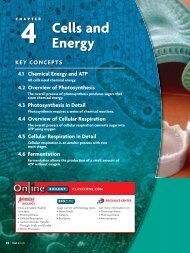
FIGURE <strong>30</strong>.1 <strong>Respiratory</strong> Organs <strong>and</strong> Tissues<br />
Specialized structures move air into <strong>and</strong> out of the body.<br />
sinus<br />
nose<br />
mouth<br />
bronchus<br />
bronchiole<br />
epiglottis<br />
trachea<br />
alveoli<br />
bronchiole<br />
lungs<br />
Infer How do the structures in the lungs increase their surface area<br />
MAIN IDEA<br />
The respiratory system moves gases into <strong>and</strong> out<br />
of the blood.<br />
The function of the respiratory system is to bring O 2 into the body <strong>and</strong> to<br />
expel CO 2 <strong>and</strong> water vapor. The structures of this system bring the gases in<br />
close contact with the blood, which absorbs O 2 . The circulatory system then<br />
carries O 2 to all of the body’s cells <strong>and</strong> transports CO 2 from the rest of the<br />
body to the lungs, where it is exhaled.<br />
The specialized structures of the respiratory system are shown in FIGURE <strong>30</strong>.1.<br />
The nose <strong>and</strong> mouth are the entry points to the system. When air enters the<br />
nose, mucus that lines the nasal passages warms <strong>and</strong> moistens the air. The<br />
mucus <strong>and</strong> tiny hairs called cilia help filter dust <strong>and</strong> pathogens from the air. At<br />
the back of the throat, a small piece of tissue, the epiglottis, regulates airflow<br />
into the trachea, or windpipe. The trachea (TRAY-kee-uh) is a long structure<br />
made of soft tissue reinforced with C-shaped rings of cartilage. It resembles<br />
the hose of a vacuum cleaner. When you swallow, the epiglottis closes the<br />
entrance to the trachea to keep food or saliva from entering the airways. The<br />
trachea divides into the two bronchi, with one branch going to each lung.<br />
The lungs are the organs that absorb O 2 from the air you inhale. Inside the<br />
lungs, the bronchi divide into smaller <strong>and</strong> smaller branches that resemble the<br />
limbs <strong>and</strong> twigs of a tree. The smallest branches, the bronchioles, end in<br />
clusters of tiny air sacs called alveoli (al-VEE-uh-LY). One air sac is called an<br />
alveolus. The lungs have a huge number of alveoli—from <strong>30</strong>0 to 600 million.<br />
Connecting<br />
CONCEPTS<br />
Cellular Respiration You learned<br />
in Chapter 4 that eukaryotic cells<br />
require a constant supply of<br />
oxygen to produce ATP, which is<br />
the main energy source for cells.<br />
Chapter <strong>30</strong>: <strong>Respiratory</strong> <strong>and</strong> <strong>Circulatory</strong> <strong>Systems</strong> 911
Air inhaled.<br />
Air exhaled.<br />
BIOLOGY<br />
Explore how<br />
you breathe at<br />
ClassZone.com.<br />
Muscles contract <strong>and</strong><br />
rib cage exp<strong>and</strong>s.<br />
Muscles <strong>and</strong> rib<br />
cage relax.<br />
FIGURE <strong>30</strong>.2 When you inhale,<br />
movements of the rib cage <strong>and</strong><br />
diaphragm produce lower pressure<br />
in the lungs, <strong>and</strong> air flows in.<br />
When you exhale, rib cage <strong>and</strong><br />
diaphragm movements produce<br />
higher pressure in the lungs, <strong>and</strong><br />
air flows out.<br />
VOCABULARY<br />
The word diaphragm is based<br />
on the Latin diaphragma,<br />
which means “midriff.” The<br />
midriff extends from below the<br />
breast to the waist. The diaphragm<br />
is located in this area.<br />
Diaphragm flattens<br />
<strong>and</strong> moves downward.<br />
Diaphragm relaxes<br />
<strong>and</strong> rises.<br />
This huge number of alveoli gives the lungs a massive surface area for absorbing<br />
O 2 <strong>and</strong> releasing CO 2 <strong>and</strong> water vapor. Lung tissue is spongy <strong>and</strong> elastic,<br />
which allows the lungs to exp<strong>and</strong> <strong>and</strong> contract as you breathe. Lung mucus<br />
<strong>and</strong> cilia help trap <strong>and</strong> remove foreign materials <strong>and</strong> pathogens.<br />
The mechanics of breathing involve the muscles of the rib cage <strong>and</strong> the<br />
diaphragm, as FIGURE <strong>30</strong>.2 shows. The diaphragm is a dome-shaped muscle at<br />
the base of the rib cage. When you inhale, the muscles of the rib cage contract,<br />
causing the rib cage to exp<strong>and</strong>. The diaphragm then flattens <strong>and</strong> moves<br />
downward. The volume of your lungs increases, <strong>and</strong> the air pressure decreases,<br />
falling below the air pressure outside your body. Gases move from areas of<br />
greater pressure to areas of lower pressure, so air flows into the lungs.<br />
When you exhale, the rib cage muscles relax, <strong>and</strong> the rib cage becomes<br />
smaller. The diaphragm also relaxes, causing it to rise <strong>and</strong> regain its domelike<br />
shape. Now the air pressure inside your lungs is greater than the air pressure<br />
outside your body, so air flows out.<br />
Predict How might damaged alveoli affect the oxygen level in the blood<br />
MAIN IDEA<br />
The circulatory system moves blood to all parts<br />
of the body.<br />
The function of the circulatory system is to transport O 2 <strong>and</strong> nutrients to<br />
body cells <strong>and</strong> to carry oxygen-poor blood <strong>and</strong> CO 2 back to the heart <strong>and</strong><br />
lungs. To do its job, the system must keep blood constantly circulating.<br />
The main parts of the circulatory system are the heart, the blood, <strong>and</strong> the<br />
blood vessels. The heart is a muscular pump, about the size of your fist, that<br />
keeps the blood moving to every part of the body. The blood circulates<br />
through a closed system—that is, blood in the circulatory system stays inside<br />
the vessels. The average adult body contains about 5 liters (more than 5 qt) of<br />
blood. On average, your blood circulates from your heart, throughout your<br />
body, <strong>and</strong> back to your heart about every 60 seconds.<br />
912 Unit 9: Human Biology
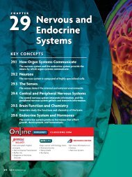
The circulatory system has three types of blood vessels: arteries, veins, <strong>and</strong><br />
capillaries. Arteries are blood vessels that carry blood away from the heart to<br />
the rest of the body. Veins are blood vessels that carry blood from the rest of<br />
the body back to the heart. As illustrated in FIGURE <strong>30</strong>.3, arteries carry oxygenrich<br />
blood (red) <strong>and</strong> veins carry oxygen-poor blood (blue). Blue is used for<br />
illustration purposes only. In your body, oxygen-poor blood is not actually<br />
blue but a darker red color. You can think of arteries <strong>and</strong> veins as a system of<br />
roads. Large arteries <strong>and</strong> veins are like major highways. Smaller arteries <strong>and</strong><br />
veins are like streets that route traffic through local neighborhoods.<br />
Arteries <strong>and</strong> veins are connected by a system of capillaries. Capillaries are<br />
the tiny blood vessels that transport blood to <strong>and</strong> from the cells of the body.<br />
These vessels are so small that blood cells must move through them in single<br />
file. The walls of these tiny blood vessels are only one cell thick. Materials can<br />
easily diffuse into <strong>and</strong> out of them.<br />
In addition to transporting vital supplies to the cells, the circulatory system<br />
performs two other important functions that maintain homeostasis.<br />
• The circulatory system collects waste materials produced by digestion <strong>and</strong><br />
cell metabolism, <strong>and</strong> delivers them to the liver <strong>and</strong> kidneys to be filtered<br />
out of the body. For example, muscle cell activity produces a waste product<br />
known as urea. As blood moves past the muscle cells, urea is moved into<br />
the bloodstream <strong>and</strong> carried to the kidneys to be excreted.<br />
• The circulatory system helps maintain body temperature by distributing<br />
the heat that cells produce in the muscles <strong>and</strong> internal organs. When you<br />
are active, your organs <strong>and</strong> muscles produce more heat. The heart pumps<br />
harder, <strong>and</strong> the blood vessels dilate to bring excess heat to the skin, where it<br />
can escape. In cold weather, the blood vessels constrict to conserve heat.<br />
The heart, the blood vessels, <strong>and</strong> the blood are described in more detail in<br />
Sections <strong>30</strong>.3 to <strong>30</strong>.5.<br />
Infer If a person has a weak heart, how might his or her ability to maintain a stable<br />
body temperature be affected<br />
heart<br />
FIGURE <strong>30</strong>.3 The circulatory<br />
system is composed of the heart,<br />
arteries carrying oxygen-rich<br />
blood (red), veins carrying oxygenpoor<br />
blood (blue), <strong>and</strong> capillaries.<br />
<strong>30</strong>.1 ASSESSMENT<br />
ONLINE QUIZ<br />
ClassZone.com<br />
REVIEWING<br />
MAIN IDEAS<br />
1. How do the respiratory <strong>and</strong><br />
circulatory systems help maintain<br />
homeostasis in the body<br />
2. List the main parts <strong>and</strong> functions of<br />
the respiratory system.<br />
3. Describe the basic parts <strong>and</strong> functions<br />
of the circulatory system.<br />
CRITICAL THINKING<br />
4. Apply Why can’t you breathe<br />
through the mouth while you are<br />
swallowing food What would<br />
happen if you could do this<br />
5. Infer Arteries <strong>and</strong> veins are equally<br />
distributed throughout the body.<br />
How does this arrangement help<br />
to maintain the functions of each<br />
cell<br />
Connecting CONCEPTS<br />
6. Science <strong>and</strong> Technology A<br />
mechanical ventilator breathes<br />
for a paralyzed person. During<br />
inhalation, the machine forces<br />
air under pressure into the<br />
lungs. During exhalation, the<br />
pressure drops <strong>and</strong> air moves<br />
out of the lungs. How does<br />
this machine compare with<br />
natural breathing<br />
Chapter <strong>30</strong>: <strong>Respiratory</strong> <strong>and</strong> <strong>Circulatory</strong> <strong>Systems</strong> 913
<strong>30</strong>.2<br />
Respiration <strong>and</strong><br />
Gas Exchange<br />
KEY CONCEPT The respiratory system exchanges oxygen <strong>and</strong> carbon dioxide.<br />
MAIN IDEAS<br />
• Gas exchange occurs in the alveoli<br />
of the lungs.<br />
• <strong>Respiratory</strong> diseases interfere with<br />
gas exchange.<br />
VOCABULARY<br />
red blood cell, p. 915<br />
hemoglobin, p. 915<br />
emphysema, p. 916<br />
asthma, p. 916<br />
Review<br />
alveoli, lung, capillary,<br />
diffusion<br />
Connect Nearly every winter, newspapers carry stories of people killed by<br />
carbon monoxide (CO) gas in their homes. This colorless, odorless gas escapes<br />
from leaks in furnaces that burn fossil fuels. What makes CO so deadly Your<br />
body readily absorbs it into the blood, which means less O 2 is absorbed. Within a<br />
short time, your cells become oxygen starved. You must quickly get to an area<br />
where you can breathe fresh air.<br />
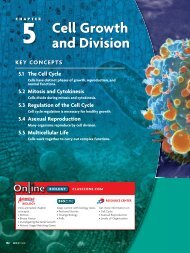
FIGURE <strong>30</strong>.4 This micrograph<br />
shows a bronchiole <strong>and</strong> several<br />
alveoli. Alveoli walls are about one<br />
cell thick, which allows O 2 <strong>and</strong><br />
CO 2 to diffuse easily across them.<br />
(colored SEM; magnification 150)<br />
alveoli<br />
bronchiole<br />
MAIN IDEA<br />
Gas exchange occurs in the alveoli of the lungs.<br />
Recall that the cells in your body carry out cellular respiration, which requires<br />
O 2 <strong>and</strong> produces CO 2 as a waste product. Thus, every cell in the body needs O 2<br />
<strong>and</strong> must get rid of CO 2 . However, the alveoli <strong>and</strong> their capillaries are the only<br />
places where gas exchange with the atmosphere occurs. The lungs bring in a<br />
steady supply of O 2 <strong>and</strong> expel excess CO 2 . Gas exchange in the lungs is based<br />
on three principles:<br />
• O 2 <strong>and</strong> CO 2 are carried by the blood.<br />
• Gases move by diffusion—that is, they move from an area of higher<br />
concentration to an area of lower concentration.<br />
• The lining of the alveoli must be moist to help gases diffuse.<br />
Diffusion of O 2 <strong>and</strong> CO 2<br />
In the alveoli, the respiratory <strong>and</strong> circulatory systems come together in the<br />
process of gas exchange. When you inhale, air flows from the bronchi to the<br />
bronchioles <strong>and</strong> finally to the alveoli. A cross-section of a bronchiole <strong>and</strong><br />
several alveoli is shown in FIGURE <strong>30</strong>.4. Each alveolus is about the size of a grain<br />
of s<strong>and</strong>, but all of the alveoli together give the lungs a surface area of about<br />
100 square meters. Without this huge area for gas exchange, the lungs would<br />
be unable to extract enough O 2 from the air to keep you alive.<br />
A complex network of capillaries surrounds <strong>and</strong> penetrates the alveoli, as<br />
shown in FIGURE <strong>30</strong>.5. Blood entering these capillaries contains a lower concentration<br />
of O 2 than does the air in the alveoli. As a result, the O 2 diffuses from<br />
an area of high concentration in the alveoli to an area of low concentration in<br />
914 Unit 9: Human Biology
FIGURE <strong>30</strong>.5 Gas Exchange in the Alveoli<br />
Diffusion of gases into <strong>and</strong> out of the alveoli<br />
maintains O 2 <strong>and</strong> CO 2 homeostasis.<br />
ALVEOLI<br />
GAS EXCHANGE<br />
alveolus<br />
capillary<br />
CO 2 diffuses<br />
into alveolus.<br />
CO 2<br />
O 2<br />
O 2 diffuses<br />
into blood.<br />
capillaries<br />
Predict How might a sudden rise in CO 2 in the blood<br />
affect the gas exchange process<br />
the capillaries. The blood in the capillaries contain red blood cells, a type of<br />
cell that picks up oxygen in the lungs <strong>and</strong> delivers it to all of the body’s cells.<br />
In red blood cells, most of the O 2 molecules bind to an iron-rich protein<br />
called hemoglobin (HEE-muh-GLOH-bihn). Each molecule of hemoglobin<br />
binds with four O 2 molecules. The iron in hemoglobin is what gives blood its<br />
reddish color. Blood becomes bright red only when it absorbs oxygen. The<br />
blood leaving the alveoli carries almost three times the amount of O 2 that it<br />
had coming into the lungs.<br />
In contrast, CO 2 concentrations are higher in the blood than in the alveoli.<br />
As a result, CO 2 diffuses into the alveoli. The higher concentration of CO 2 in<br />
the blood is due to the fact that every cell produces CO 2 <strong>and</strong> water as waste<br />
products. The CO 2 <strong>and</strong> water combine in the blood to form the compound<br />
carbonic acid. The more carbonic acid there is in the blood, the more acidic<br />
the blood becomes. When carbonic acid diffuses into the alveoli, the compound<br />
separates into CO 2 <strong>and</strong> water, which are exhaled.<br />
Gas Exchange <strong>and</strong> the Nervous System<br />
Gas exchange is so critical to the body that it is an autonomic function regulated<br />
by the medulla <strong>and</strong> pons in the brain stem. These centers monitor<br />
dissolved gases in the blood, particularly CO 2 concentrations. As you become<br />
more active, CO 2 levels increase <strong>and</strong> the blood becomes more acidic. Sensors<br />
in the respiratory <strong>and</strong> circulatory systems signal this change to the brain stem.<br />
The medulla sends messages through the nervous <strong>and</strong> endocrine systems that<br />
stimulate the diaphragm <strong>and</strong> rib cage muscles to work harder. The medulla<br />
regulates how often <strong>and</strong> how deeply you breathe based on your activity.<br />
Analyze How does the alveoli’s structure relate to the function of gas exchange<br />
Connecting<br />
CONCEPTS<br />
Nervous System As you read in<br />
Chapter 29, the brain stem is<br />
located at the base of the brain.<br />
The brain stem is involved in<br />
regulating breathing <strong>and</strong> other<br />
autonomic functions that help<br />
maintain homeostasis.<br />
Chapter <strong>30</strong>: <strong>Respiratory</strong> <strong>and</strong> <strong>Circulatory</strong> <strong>Systems</strong> 915
MAIN IDEA<br />
<strong>Respiratory</strong> diseases interfere with gas exchange.<br />
FIGURE <strong>30</strong>.6 Healthy lung tissue<br />
is free of any deposits. When a<br />
person smokes for several years,<br />
black tar deposits first invade<br />
<strong>and</strong> then choke the tissue, <strong>and</strong><br />
greatly reduce gas exchange.<br />
(LM; magnification 250)<br />
Healthy lung tissue<br />
Smoker’s lung tissue<br />
<br />
<br />
For more information on the<br />
respiratory system, go to<br />
scilinks.org.<br />
Keycode: MLB0<strong>30</strong><br />
Damage to the respiratory system makes gas exchange more difficult, which in<br />
turn affects every cell in the body. Smoking is the leading cause of respiratory<br />
diseases such as lung cancer <strong>and</strong> emphysema. Tobacco smoke contains more<br />
than 4800 chemicals that can paralyze cilia, damage alveoli, <strong>and</strong> cause genetic<br />
mutations leading to cancer. In FIGURE <strong>30</strong>.6, you can see the effects of smoking<br />
on lung tissue. When a person smokes for several years, the lung<br />
tissue is slowly coated by tars <strong>and</strong> other chemicals. Eventually, the<br />
tissue becomes almost solid black. The sooner people stop smoking,<br />
the sooner such damage to the lungs can be reversed.<br />
Emphysema (EHM-fih-SEE-muh) is a lung disorder caused<br />
mainly by smoking. Over time, many alveoli are destroyed. This<br />
process gradually reduces the surface area for gas exchange, <strong>and</strong><br />
not enough oxygen can enter the blood. People with advanced<br />
emphysema must use supplemental oxygen, but eventually their<br />
lungs fail. At present, this disease has no cure. The best way to prevent<br />
emphysema is to refrain from smoking.<br />
Asthma (AZ-muh) causes the bronchioles to constrict due to<br />
muscle spasms. This condition makes it hard to move air in <strong>and</strong><br />
out of the lungs. A person having a severe asthma attack can die<br />
from lack of oxygen. Attacks may be triggered by allergies, stress,<br />
exposure to smoke <strong>and</strong> chemicals, or exercise. The attacks can be<br />
relieved by drugs that relax the bronchioles.<br />
Cystic fibrosis (CF) is a genetic disease that causes the lungs to produce a<br />
thick, sticky mucus. This mucus blocks the airways <strong>and</strong> allows microorganisms<br />
to thrive in the lungs. People with CF have frequent, sometimes fatal,<br />
lung infections. Treatments focus on preventing the mucus from building up.<br />
Synthesize How does smoking affect gas exchange<br />
<strong>30</strong>.2 ASSESSMENT<br />
ONLINE QUIZ<br />
ClassZone.com<br />
REVIEWING<br />
MAIN IDEAS<br />
1. Explain how diffusion allows gases to<br />
move into <strong>and</strong> out of the alveoli of<br />
the lungs. Use the term red blood<br />
cell in your explanation.<br />
2. In what ways can respiratory<br />
diseases reduce the level of O 2<br />
in the blood<br />
CRITICAL THINKING<br />
3. Synthesize Explain how your<br />
breathing rate would change if your<br />
blood became more acidic.<br />
4. Apply People poisoned by CO are<br />
often given 100 percent O 2 in a<br />
room with two to three times<br />
normal atmospheric pressure.<br />
Explain why more oxygen would<br />
enter their blood under these<br />
conditions.<br />
Connecting CONCEPTS<br />
5. Forensic Science When police<br />
find a body in a lake or river,<br />
they must determine if the<br />
person was drowned or was<br />
killed in some other way <strong>and</strong><br />
then thrown into the water.<br />
How would examining the<br />
lungs of the person help them<br />
to solve the mystery<br />
916 Unit 9: Human Biology
<strong>30</strong>.3<br />
The Heart <strong>and</strong> Circulation<br />
KEY CONCEPT The heart is a muscular pump that moves the blood through two pathways.<br />
MAIN IDEAS<br />
• The tissues <strong>and</strong> structures of the<br />
heart make it an efficient pump.<br />
• The heart pumps blood through<br />
two main pathways.<br />
VOCABULARY<br />
atrium, p. 917<br />
ventricle, p. 917<br />
valve, p. 917<br />
pacemaker, p. 918<br />
pulmonary circulation,<br />
p. 920<br />
systemic circulation,<br />
p. 920<br />
Review<br />
heart, artery, vein<br />
Connect Lub-dub, lub-dub. This is the sound of your heart beating. The lub<br />
sound occurs when the valves between the upper <strong>and</strong> lower chambers of the<br />
heart snap shut. The dub sound is made by valves closing the two arteries that<br />
carry blood out of the heart. If a valve does not close properly <strong>and</strong> allows blood<br />
to leak backward, the sound of the heart changes. A heart with a leaky valve<br />
might sound like this: Lub-dub-shhh, lub-dub-shhh. The sounds your heart<br />
makes can tell a physician a great deal about how it is performing.<br />
MAIN IDEA<br />
The tissues <strong>and</strong> structures of the heart make it<br />
an efficient pump.<br />
Each day your heart beats about 100,000 times, circulating blood through<br />
nearly 96,000 kilometers of blood vessels—roughly one-quarter of the distance<br />
to the moon. Over 70 years, your heart will beat about 2.5 billion times.<br />
How can it keep going One reason is that cardiac muscle tissue, unlike<br />
skeletal muscle tissue, can work continuously<br />
FIGURE <strong>30</strong>.7 HEART CHAMBERS AND VALVES<br />
without becoming tired. Also, the structures of<br />
the heart make this organ an efficient pump.<br />
pulmonary<br />
valve<br />
right atrium<br />
tricuspid valve<br />
right ventricle<br />
aortic valve<br />
left atrium<br />
mitral valve<br />
left<br />
ventricle<br />
septum<br />
Structures of the Heart<br />
The largest structures in your heart are the four<br />
chambers. As shown in FIGURE <strong>30</strong>.7, the two<br />
smaller chambers are the right atrium <strong>and</strong> left<br />
atrium (plural, atria), <strong>and</strong> the two larger chambers<br />
are the right <strong>and</strong> left ventricles. The ventricles<br />
are separated by the septum, a thick wall of<br />
tissue. The heart valves are flaps of tissue that<br />
prevent blood from flowing backward. They open<br />
when the atria or ventricles contract, <strong>and</strong> close<br />
when the atria or ventricles relax.<br />
After blood fills a chamber, the cardiac muscle<br />
contracts <strong>and</strong> pumps the blood out of the chamber.<br />
The heart is an amazingly powerful pump.<br />
Chapter <strong>30</strong>: <strong>Respiratory</strong> <strong>and</strong> <strong>Circulatory</strong> <strong>Systems</strong> 917
SA node<br />
(pacemaker)<br />
AV node<br />
FIGURE <strong>30</strong>.8 An electrical signal<br />
from the SA node causes both<br />
atria to contract. The AV node<br />
then picks up the signal <strong>and</strong> transmits<br />
it to both ventricles, causing<br />
them to contract.<br />
The reason has to do with the small size of the heart, which allows the strong<br />
cardiac muscles to exert a great deal of force on the chamber. The combination<br />
of small size <strong>and</strong> large force results in a powerful pumping action. The<br />
heart is also an efficient, self-regulating pump. It can respond to signals from<br />
the nervous system to change the speed <strong>and</strong> force of its pumping action. For<br />
example, if you increase your level of activity, your heart will pump faster.<br />
The Heartbeat<br />
The heartbeat consists of two contractions: the first takes place in the atria <strong>and</strong><br />
the second in the ventricles. The contractions occur partly because the cardiac<br />
muscle fibers of the chambers have a unique property. Whenever one fiber is<br />
stimulated to contract, all of the fibers contract at the same time.<br />
The first contraction of the heart begins in the right atrium at a signal<br />
from the sinoatrial (SA) node, shown in FIGURE <strong>30</strong>.8. The SA node is known as<br />
the heart’s pacemaker because the cells in this node generate an electrical<br />
signal that starts the wave of contractions. Once the atria have contracted, the<br />
electrical signal spreads through conducting fibers to the atrioventricular (AV)<br />
node, located in the wall of the right ventricle. The AV signal stimulates both<br />
ventricles to contract at the same time.<br />
If the SA node is seriously damaged by injury or disease, it can be replaced<br />
with an artificial pacemaker that is implanted into the heart. This device, like<br />
the SA node, sends electrical signals to the muscle fibers of the atria.<br />
Blood Flow in the Heart<br />
Once you know the basic structures <strong>and</strong> actions of the heart, you can follow<br />
how oxygen-rich <strong>and</strong> oxygen-poor blood flow through this organ. Study<br />
FIGURE <strong>30</strong>.9, which illustrates this pathway. Notice that blood always enters the<br />
heart through an atrium <strong>and</strong> leaves the heart through a ventricle. The contractions<br />
of the atria <strong>and</strong> then of the ventricles keep blood moving in this sequence.<br />
VOCABULARY<br />
The word pulmonary comes<br />
from the Latin root pulmo,<br />
meaning “lung.” The suffix<br />
-ary means “belonging to or<br />
connected with.” Therefore,<br />
pulmonary means something<br />
“belonging to or connected<br />
with the lung.”<br />
1 Oxygen-poor blood from the body enters the right atrium. The SA node<br />
signals the atria to contract, <strong>and</strong> blood flows into the right ventricle.<br />
2 When the AV node signals the ventricles to contract, blood is pumped<br />
from the right ventricle into the pulmonary artery. This artery, which<br />
goes to the lungs, is the only artery in the body that carries oxygen-poor<br />
blood. The blood enters the lungs, where CO 2 <strong>and</strong> water vapor diffuse<br />
into the alveoli <strong>and</strong> O 2 diffuses into the blood.<br />
3 Oxygen-rich blood returns to the heart through the pulmonary vein <strong>and</strong><br />
enters the left atrium. This is the only vein in the body that carries<br />
oxygen-rich blood. As the atria contract, blood is pumped into the left<br />
ventricle, the largest chamber in the heart.<br />
4 When the ventricles contract, blood is pumped from the left ventricle<br />
into a large artery, the aorta, <strong>and</strong> is circulated to the rest of the body.<br />
After oxygen has been delivered to the cells, the oxygen-poor blood returns<br />
through the veins to the heart, <strong>and</strong> the sequence begins again.<br />
Analyze The left ventricle is the largest chamber of the heart. How is its size<br />
related to its function<br />
918 Unit 9: Human Biology
FIGURE <strong>30</strong>.9 Blood Flow in the Heart<br />
The structures of the heart keep oxygen-poor blood<br />
separated from oxygen-rich blood.<br />
BIOLOGY<br />
See how the heart<br />
pumps blood at<br />
ClassZone.com.<br />
Oxygen-poor blood<br />
Oxygen-rich blood<br />
FROM BODY<br />
TO LUNGS<br />
FROM LUNGS<br />
TO BODY<br />
1<br />
2<br />
The right atrium<br />
receives oxygenpoor<br />
blood from the<br />
body <strong>and</strong> pumps it<br />
to the right ventricle.<br />
The right ventricle<br />
pumps oxygen-poor<br />
blood to the lungs.<br />
3<br />
4<br />
The left atrium receives<br />
oxygen-rich blood from<br />
the lungs <strong>and</strong> pumps it<br />
to the left ventricle.<br />
The left ventricle pumps<br />
oxygen-rich blood to all<br />
parts of the body.<br />
NORMAL HUMAN HEART<br />
CRITICAL<br />
VIEWING<br />
If the valves in the right ventricle do not close properly,<br />
where in the body might circulation be affected the most<br />
Chapter <strong>30</strong>: <strong>Respiratory</strong> <strong>and</strong> <strong>Circulatory</strong> <strong>Systems</strong> 919
MAIN IDEA<br />
The heart pumps blood through two main<br />
pathways.<br />
PULMONARY<br />
SYSTEMIC<br />
FIGURE <strong>30</strong>.10 The circulatory<br />
system has two general pathways.<br />
Pulmonary circulation moves<br />
blood between the heart <strong>and</strong> the<br />
lungs. Systemic circulation moves<br />
blood between the heart <strong>and</strong> the<br />
rest of the body.<br />
Circulating blood follows two separate pathways that meet at the heart, as<br />
shown in FIGURE <strong>30</strong>.10. These pathways are called the pulmonary <strong>and</strong> systemic<br />
circulation. All of your blood travels through both of these pathways.<br />
Pulmonary circulation (PUL-muh-NEHR-ee) occurs only between the heart <strong>and</strong><br />
the lungs. The main function of this circulation is to carry oxygen-poor blood<br />
to the lungs, where it picks up O 2 , expels excess CO 2 <strong>and</strong> water, <strong>and</strong> carries<br />
oxygen-rich blood back to the heart. Each lung is supplied by its own pulmonary<br />
artery <strong>and</strong> pulmonary vein. Systemic circulation (sihs-STEHM-ihk)<br />
occurs between the heart <strong>and</strong> the rest of the body, except for the lungs. The<br />
main function of this circulation is to carry oxygen-rich blood to all cells <strong>and</strong><br />
transport oxygen-poor blood back to the heart. Systemic circulation begins<br />
when blood leaves the left ventricle, the largest chamber of the heart. The<br />
blood then circulates through the torso, arms, legs, <strong>and</strong> head, <strong>and</strong> then returns<br />
to the heart.<br />
As the body’s need for oxygen changes, sensors in the walls of major<br />
arteries in the pulmonary <strong>and</strong> systemic pathways send information to the<br />
medulla in the brain stem. The medulla coordinates this information with<br />
signals from the respiratory system. Homeostasis is maintained by matching<br />
heart rate <strong>and</strong> respiration rate with the oxygen needs of the body.<br />
In extreme conditions, such as severe cold, the pulmonary <strong>and</strong> systemic<br />
circulation systems serve another vital function—making sure the body’s<br />
brain, heart, <strong>and</strong> other major organs remain at a constant temperature. When<br />
the body is exposed for any length of time to a cold environment, blood vessels<br />
to the arms <strong>and</strong> legs begin to constrict. The blood flow to the arms <strong>and</strong> legs is<br />
reduced in order to keep the torso <strong>and</strong> head warm. Once you reach a warmer<br />
environment, these blood vessels dilate <strong>and</strong> normal circulation resumes.<br />
Infer Why is it important to have two separate pathways for circulation<br />
<strong>30</strong>.3 ASSESSMENT<br />
ONLINE QUIZ<br />
ClassZone.com<br />
REVIEWING<br />
MAIN IDEAS<br />
1. What structures make the heart<br />
an efficient pump In your answer,<br />
describe the direction of blood<br />
flow into <strong>and</strong> out of the heart.<br />
2. Briefly describe the pulmonary<br />
<strong>and</strong> systemic circulation<br />
pathways.<br />
CRITICAL THINKING<br />
3. Predict Explain how leaky heart<br />
valves might damage the heart<br />
over time.<br />
4. Predict How might a high fever<br />
affect a person’s heart <strong>and</strong> breathing<br />
rates Explain your answer.<br />
Connecting CONCEPTS<br />
5. Animals Unlike a human<br />
heart, an amphibian heart<br />
has two atria but only a<br />
single ventricle. How might<br />
living in a watery environment<br />
help reduce the work that an<br />
amphibian heart needs to do<br />
920 Unit 9: Human Biology
CHAPTER <strong>30</strong><br />
MATERIALS<br />
• 100-mL graduated<br />
cylinder<br />
• 250-mL beaker<br />
• 800 mL water<br />
• eyedropper<br />
• bromothymol blue<br />
solution<br />
• straw<br />
• 50 mL 0.4% sodium<br />
hydroxide solution<br />
• clock with second h<strong>and</strong><br />
PROCESS SKILLS<br />
• Observing<br />
• Measuring<br />
• Analyzing<br />
INVESTIGATION<br />
Carbon Dioxide <strong>and</strong> Exercise<br />
In this lab you will examine the effects of exercise on<br />
how much carbon dioxide is released by your<br />
respiratory system. Bromothymol blue turns yellow in<br />
the presence of carbon dioxide when sodium<br />
hydroxide is added to the solution. The more sodium<br />
hydroxide that needs to be added to turn the<br />
bromothymol blue solution yellow, the more carbon<br />
dioxide there is in the solution.<br />
PROBLEM How does exercise affect the release of<br />
carbon dioxide from the lungs<br />
PROCEDURE<br />
1. Fill the graduated cylinder with 100 mL of water <strong>and</strong><br />
pour the water into a beaker.<br />
2. Add four drops of bromothymol blue solution<br />
to the water.<br />
3. Place one end of the straw in the water <strong>and</strong> blow<br />
into the water for one minute. Caution: Do not<br />
inhale from the straw.<br />
4. Add one drop at a time of the sodium hydroxide solution to the water. Swirl the<br />
water while adding the drops. Count the number of drops needed for the<br />
solution in the cylinder to turn yellow <strong>and</strong> remain yellow for one minute.<br />
5. Record your data in a table similar to the one below.<br />
6. Empty the beaker. Repeat steps 1 <strong>and</strong> 2.<br />
7. Perform three types of exercise during your experiment: one that is low impact,<br />
such as walking; one that is medium impact, such as running in place; <strong>and</strong> one that<br />
is high impact, such as jumping jacks.<br />
8. Perform each type of exercise for two minutes. At the end of each two-minute<br />
exercise period, repeat steps 3 through 6.<br />
TABLE 1. THE EFFECT OF EXERCISE ON CARBON DIOXIDE RELEASE<br />
Exercise<br />
Number of 0.4% Sodium Hydroxide<br />
Solution Drops<br />
Rest<br />
Exercise 1 (walking)<br />
Exercise 2 (running in place)<br />
Exercise 3 (jumping jacks)<br />
ANALYZE AND CONCLUDE<br />
1. Graph Data Construct a graph that represents your data.<br />
2. Analyze How does the amount of carbon dioxide exhaled change with different<br />
types of exercise What mechanisms in the body explain these results<br />
3. Hypothesize Suppose you had subjects exercise for 15 minutes, <strong>and</strong> every 5<br />
minutes they increased their rate of exertion. If you measured the amount of<br />
carbon dioxide exhaled every 5 minutes, what results would you expect Explain<br />
your answer.<br />
Chapter <strong>30</strong>: <strong>Respiratory</strong> <strong>and</strong> <strong>Circulatory</strong> <strong>Systems</strong> 921
<strong>30</strong>.4<br />
Blood Vessels <strong>and</strong> Transport<br />
KEY CONCEPT The circulatory system transports materials throughout the body.<br />
MAIN IDEAS<br />
• Arteries, veins, <strong>and</strong> capillaries transport<br />
blood to all parts of the body.<br />
• Lifestyle plays a key role in circulatory<br />
diseases.<br />
VOCABULARY<br />
blood pressure, p. 923<br />
systolic pressure, p. 923<br />
diastolic pressure, p. 923<br />
Review<br />
artery, vein, capillary,<br />
ventricle<br />
Connect In the 1600s, most scientists thought that the lungs, not the heart,<br />
moved the blood, <strong>and</strong> that blood was consumed <strong>and</strong> produced by the internal<br />
organs. William Harvey, court physician to the king of Engl<strong>and</strong>, challenged these<br />
ideas. He showed that the heart was the true pump for the blood <strong>and</strong> that blood<br />
circulated in two pathways: one between the heart <strong>and</strong> the lungs, <strong>and</strong> another<br />
between the heart <strong>and</strong> the rest of the body. Harvey’s work on circulation is<br />
regarded as one of the greatest advances in the history of medicine.<br />
MAIN IDEA<br />
Arteries, veins, <strong>and</strong> capillaries transport blood to<br />
all parts of the body.<br />
As you read in Section <strong>30</strong>.1, the circulatory system includes three types of<br />
blood vessels—arteries, veins, <strong>and</strong> capillaries—that act as transportation<br />
networks for the blood. Each of the three vessels has its own structure <strong>and</strong><br />
function, as illustrated in FIGURE <strong>30</strong>.11.<br />
TAKING NOTES<br />
A two-column chart can help<br />
you organize your notes about<br />
different blood vessels <strong>and</strong> circulatory<br />
pathways.<br />
arteries<br />
- Thicker, more<br />
muscular than<br />
veins<br />
- Blood under<br />
greater<br />
pressure<br />
Arteries<br />
Arteries need to be strong <strong>and</strong> flexible because the blood they carry from the<br />
heart is under great pressure. An artery’s thick wall is composed of three<br />
layers. The innermost layer consists of endothelium coated with a protein that<br />
prevents blood from clotting. The middle layer is a thick b<strong>and</strong> of smooth<br />
muscle <strong>and</strong> elastic fibers. The outer layer consists of connective tissue <strong>and</strong><br />
elastic fibers. The elastic fibers allow the arterial walls to exp<strong>and</strong> <strong>and</strong> contract<br />
to help move blood through the arteries. Arterioles, or smaller arteries, contain<br />
the same three layers, but the outer <strong>and</strong> middle layers are much thinner.<br />
Veins<br />
The structures of veins reflect the fact that blood is under much less pressure<br />
when it is returning to the heart. Veins have larger diameters <strong>and</strong> thinner walls<br />
than do arteries <strong>and</strong> contain valves that prevent blood from flowing backwards.<br />
Veins do not have a thick layer that exp<strong>and</strong>s <strong>and</strong> contracts to keep blood<br />
moving. Instead, they need the activity of skeletal muscles to help maintain<br />
circulation. For example, as you walk, skeletal muscles in your legs push against<br />
the veins. The valves open, <strong>and</strong> blood moves toward the heart. If you sit for too<br />
long, the lack of exercise makes it harder for the blood to move upward.<br />
Venules are small veins that join larger veins to capillaries.<br />
922 Unit 9: Human Biology
FIGURE <strong>30</strong>.11 Three Types of Blood Vessels<br />
Arteries, veins, <strong>and</strong> capillaries transport the blood to every cell.<br />
endothelium<br />
valve<br />
smooth muscle<br />
connective tissue<br />
ARTERY<br />
VEIN<br />
CAPILLARIES<br />
arteriole<br />
venule<br />
Analyze Explain why arteries do not need valves to keep blood<br />
moving in one direction.<br />
Capillaries<br />
Capillary walls are made of epithelium, but they contain no muscle cells or<br />
elastic fibers. The thinness of capillary walls allows materials to diffuse into <strong>and</strong><br />
out of the blood quickly <strong>and</strong> easily. In areas of high metabolic activity, such as<br />
the lungs, kidneys, <strong>and</strong> liver, capillaries form dense networks called capillary<br />
beds. These beds move a great deal of blood into <strong>and</strong> out of these organs.<br />
Circulation <strong>and</strong> Blood Pressure<br />
Blood pressure is the force with which blood pushes against the wall of an<br />
artery. A healthy resting blood pressure for a young adult is around 120/70<br />
mm Hg (read as “120 over 70 millimeters of mercury”). The top, <strong>and</strong> higher,<br />
number is known as the systolic pressure (sih-STAHL-ihk). This is the amount<br />
of pressure on the walls of an artery when the left ventricle contracts to pump<br />
blood through the body. The bottom, <strong>and</strong> lower, number is known as the<br />
diastolic pressure (DY-uh-STAHLihk).<br />
This is the pressure in the artery<br />
when the left ventricle relaxes.<br />
Blood pressure depends on how<br />
elastic <strong>and</strong> unblocked the arteries are<br />
<strong>and</strong> on the strength of the heart<br />
contraction. The less elastic the<br />
arteries <strong>and</strong> the more blockages that<br />
reduce blood flow, the harder the<br />
heart must pump. As a result, blood<br />
pressure rises. Blood pressure also<br />
VISUAL VOCAB<br />
Systolic pressure occurs when the left<br />
ventricle contracts. Diastolic pressure<br />
occurs when the ventricle relaxes. You<br />
can write these numbers as a fraction in<br />
which systolic pressure is always on top.<br />
120<br />
systolic = numerator<br />
70 diastolic = denominator<br />
rises naturally with activity, stress, <strong>and</strong> strong emotions, but it should drop<br />
again with rest. If the pressure remains high, there could be a problem in the<br />
circulatory system.<br />
Connecting<br />
CONCEPTS<br />
Differentiated Cells In Chapter<br />
28, you learned that epithelial<br />
cells line most organs <strong>and</strong> structures<br />
in the body. These cells<br />
provide a protective layer that<br />
helps each organ or structure to<br />
do its job.<br />
Chapter <strong>30</strong>: <strong>Respiratory</strong> <strong>and</strong> <strong>Circulatory</strong> <strong>Systems</strong> 923
People with permanently high blood pressure have a condition called<br />
hypertension, which can lead to a heart attack or stroke. A heart attack occurs<br />
when the arteries to the heart muscle are damaged or blocked. A stroke can<br />
occur when blood flow to the brain is interrupted. Most people can lower<br />
their blood pressure through weight loss, proper diet, <strong>and</strong> exercise. If these<br />
remedies fail, people can use medications to reduce blood pressure.<br />
Infer Why do you think that blood moving from the heart to the lungs must be<br />
carried by an artery <strong>and</strong> not by a vein<br />
MAIN IDEA<br />
Lifestyle plays a key role in circulatory diseases.<br />
artery wall<br />
plaque<br />
FIGURE <strong>30</strong>.12 This micrograph<br />
clearly shows fatty deposits, called<br />
plaque, building up on an artery<br />
wall. If such deposits block blood<br />
flow, they can cause a heart attack<br />
or stroke. (LM; magnification 25)<br />
Lifestyle choices strongly influence the health of your circulatory system. Smoking,<br />
lack of exercise, excessive weight, long-term stress, <strong>and</strong> a diet low in fruits<br />
<strong>and</strong> vegetables but high in saturated fats are all linked to an increased risk of<br />
developing circulatory diseases. These diseases mainly affect the heart <strong>and</strong> the<br />
arteries. For example, in arteriosclerosis (ahr-TEER-ee-oh-skluh-ROH-sihs), the<br />
artery walls become thick <strong>and</strong> inflexible. In atherosclerosis (ATH-uh-roh-skluh-<br />
ROH-sihs), blood flow is partially or fully blocked by sticky material, called<br />
plaque, that collects on the walls of the arteries, as FIGURE <strong>30</strong>.12 shows. High<br />
blood pressure is often the only warning sign of these problems.<br />
Both diseases can lead to a heart attack, stroke, or kidney damage. Some<br />
blocked arteries supplying the heart muscle can be opened using a surgical<br />
technique known as a balloon angioplasty (AN-jee-uh-PLAS-tee). A device is<br />
threaded into the artery <strong>and</strong> then inflated so that it squeezes the obstruction<br />
against the artery wall. If this procedure does not work, bypass surgery may be<br />
necessary. In this operation, a healthy blood vessel from another part of the<br />
body (usually the leg) is attached to the artery on either side of the blockage.<br />
Blood can then bypass the obstruction.<br />
To reduce the risk of circulatory diseases, physicians urge people either not<br />
to smoke or to quit smoking, to maintain a healthy weight, <strong>and</strong> to exercise<br />
regularly. Medications can also help reduce the risks of heart disease.<br />
Provide Examples How can lifestyle choices affect the function of the arteries<br />
<strong>30</strong>.4 ASSESSMENT<br />
ONLINE QUIZ<br />
ClassZone.com<br />
REVIEWING<br />
MAIN IDEAS<br />
1. How do the structures of arteries,<br />
veins, <strong>and</strong> capillaries relate to their<br />
functions<br />
2. How can lifestyle choices help<br />
reduce the risk of heart disease<br />
CRITICAL THINKING<br />
3. Infer People who smoke often<br />
have cold h<strong>and</strong>s <strong>and</strong> feet. What<br />
might explain this condition in<br />
terms of blood flow<br />
4. Apply Explain why narrowing of<br />
the arteries decreases blood flow<br />
but increases blood pressure.<br />
Connecting CONCEPTS<br />
5. Arthropods The hard exoskeleton<br />
of an arthropod exerts<br />
pressure on the animal’s<br />
circulatory system. In what way<br />
does the exoskeleton serve the<br />
same function as the heart<br />
does in mammals<br />
924 Unit 9: Human Biology
DATA ANALYSIS<br />
Age Group <strong>and</strong> Disease<br />
FORMING A NULL HYPOTHESIS<br />
When scientists investigate some type of phenomenon, such as when they are trying to<br />
determine the cause of a disease, they often need to rule out variables that may or may<br />
not be important. This is especially helpful when many factors might play some role in<br />
the phenomenon, as is often the case, in the causes of disease.<br />
The formation of a null hypothesis is useful during these types of investigations.<br />
The null hypothesis states that there is no difference among study groups for the<br />
independent variable being tested. The null hypothesis is always stated in the<br />
negative: one variable does not have an effect on the other variable. The null<br />
hypothesis is accepted or rejected based on the data. If the investigation shows<br />
that the one variable does affect the other, the null hypothesis is rejected. If the<br />
investigation shows that the one variable does not affect the other, then the<br />
null hypothesis is accepted.<br />
EXAMPLE<br />
A scientist investigates the rate of death from heart disease among different age<br />
groups. The null hypothesis for this investigation would be, “There is no<br />
difference in the rate of death from heart disease among different age groups.”<br />
Consider the results listed below for the rate of death from heart disease per<br />
100,000 people.<br />
• Rate for ages 55–64 is 246.9<br />
• Rate for ages 65–74 is 635.1<br />
• Rate for ages 75–84 is 1725.7<br />
DATA ANALYSIS<br />
ClassZone.com<br />
In this case, the null hypothesis would be rejected because there is an obvious<br />
difference in the rate of death due to heart disease among different age groups.<br />
As people get older, the rate of death increases.<br />
Exercise is an important factor<br />
in preventing heart disease.<br />
ACCEPT OR REJECT THE NULL HYPOTHESIS<br />
The graph at right shows the results of an investigation<br />
about differences in the rate of asthma based<br />
on age.<br />
1. Hypothesize Form a null hypothesis for this<br />
investigation.<br />
2. Evaluate Explain whether you accept or reject<br />
the null hypothesis, based on the data.<br />
GRAPH 1. RATES OF ASTHMA BY AGE GROUP<br />
<br />
<br />
<br />
<br />
<br />
<br />
<br />
<br />
<br />
Source: National Health Interview Survey,<br />
National Center for Health Statistics<br />
Chapter <strong>30</strong>: <strong>Respiratory</strong> <strong>and</strong> <strong>Circulatory</strong> <strong>Systems</strong> 925
<strong>30</strong>.5<br />
Blood<br />
KEY CONCEPT Blood is a complex tissue that transports materials.<br />
MAIN IDEAS<br />
• Blood is composed mainly of cells,<br />
cell fragments, <strong>and</strong> plasma.<br />
• Platelets <strong>and</strong> different types of<br />
blood cells have different functions.<br />
VOCABULARY<br />
platelet, p. 926<br />
plasma, p. 926<br />
ABO blood group, p. 927<br />
Rh factor, p. 928<br />
white blood cells, p. 928<br />
Review<br />
hemoglobin, red blood cell<br />
Connect The adult human body contains about 5 liters (more than 5 qt) of<br />
blood. This fluid supplies your organs with gases <strong>and</strong> nutrients, helps you keep<br />
warm or cool off, <strong>and</strong> gets rid of waste products from your cells. Blood also has<br />
other components that help fight infections <strong>and</strong> control bleeding from damaged<br />
blood vessels. How can one substance accomplish all of these functions<br />
plasma<br />
red blood cells,<br />
white blood cells,<br />
<strong>and</strong> platelets<br />
FIGURE <strong>30</strong>.13 Whole blood<br />
is composed of several parts<br />
that help to fight infections,<br />
control bleeding, <strong>and</strong> transport<br />
gases, nutrients, waste products,<br />
<strong>and</strong> hormones.<br />
MAIN IDEA<br />
Blood is composed mainly of cells, cell<br />
fragments, <strong>and</strong> plasma.<br />
When you look at blood with the naked eye, it appears to be a single substance.<br />
Whole blood is actually a sticky mixture of cells, cell fragments, <strong>and</strong><br />
fluid, along with particles of fat, other nutrients, <strong>and</strong> dissolved gases. If you<br />
put blood in a test tube <strong>and</strong> spin it in a centrifuge, it will separate into two<br />
main parts, as shown in FIGURE <strong>30</strong>.13. At the bottom, a reddish-brown b<strong>and</strong><br />
contains red blood cells, white blood cells, <strong>and</strong> platelets. Platelets are cell<br />
fragments, produced in bone marrow, that help in blood clotting.<br />
At the top of the tube is plasma, a clear pale-yellow fluid that makes up<br />
about 55 percent of the blood. Plasma is roughly 90 percent water. Many types<br />
of molecules dissolve in plasma <strong>and</strong> can be transported throughout the body.<br />
These molecules include amino acids, glucose, hormones, vitamins, salts, <strong>and</strong><br />
waste products.<br />
Why is plasma important The concentration of molecules dissolved in<br />
plasma determines which substances will diffuse into <strong>and</strong> out of the blood<br />
that moves through the capillaries. The movement of water, gases, nutrients,<br />
<strong>and</strong> ions between the capillaries <strong>and</strong> the cells plays a critical role in maintaining<br />
homeostasis. For instance, as the concentration of glucose increases in the<br />
capillaries, it moves outward to an area of lower concentration <strong>and</strong> eventually<br />
enters the cells.<br />
Plasma proteins such as albumin, fibrinogen, <strong>and</strong> immune proteins also<br />
help maintain homeostasis. Albumin, the same substance as in egg white, is<br />
the most abundant plasma protein. Its main role is to stabilize blood volume<br />
so that fluid in the blood does not leak out of the vessels. Fibrinogen is a<br />
clotting factor that works with platelets to stop the bleeding after an injury.<br />
926 Unit 9: Human Biology
A group of specialized proteins made by the immune system fights infection<br />
or attacks foreign materials in the blood. You will learn more about these<br />
proteins in Chapter 31.<br />
Predict What do you think might happen to your blood if you become dehydrated<br />
MAIN IDEA<br />
Platelets <strong>and</strong> different types of blood cells have<br />
different functions.<br />
Blood contains red blood cells, several types of white blood<br />
cells, <strong>and</strong> platelets, as the photograph in FIGURE <strong>30</strong>.14 shows.<br />
These three blood components are manufactured mostly in the<br />
bone marrow. Each one has a specialized shape <strong>and</strong> function.<br />
red blood cell<br />
Red Blood Cells<br />
Red blood cells make up 40 to 45 percent of all cells in the<br />
blood. Mature red blood cells are shaped like an inner tube<br />
with a solid center. They are produced from stem cells in bone<br />
marrow. As these cells mature, they gradually fill with hemoglobin<br />
<strong>and</strong> lose their nuclei <strong>and</strong> other organelles. Without<br />
platelet<br />
nuclei, they cannot undergo cell division. Red blood cells<br />
circulate through the body for about 120 days before they begin<br />
to degrade. Degraded cells are carried to the liver <strong>and</strong> spleen,<br />
which break up the cells <strong>and</strong> recycle them.<br />
The most important function of red blood cells is to transport O 2 to the<br />
cells <strong>and</strong> carry CO 2 away from them. As you read in Section <strong>30</strong>.3, O 2 binds to<br />
the hemoglobin in red blood cells <strong>and</strong> is transported to all cells. When blood<br />
is returning to the heart, it picks up CO 2 <strong>and</strong> carries it to the lungs.<br />
If red blood cells are damaged or misshapen, they cannot transport O 2<br />
effectively. In sickle cell disease, for example, red blood cells are distorted into<br />
crescent shapes, as shown in FIGURE <strong>30</strong>.15. They transport less O 2 , last only 10 to<br />
20 days, <strong>and</strong> tend to clump in blood vessels. This genetic disorder is most<br />
commonly found in people of African descent.<br />
white blood cell<br />
ABO Blood Group <strong>and</strong> Rh Factors<br />
Red blood cells have surface protein markers that define your blood type.<br />
Blood type is very important when people give or receive blood for transfusions.<br />
If you receive blood with a protein marker different from your own,<br />
your immune system will attack the foreign blood cells, causing them to<br />
clump. The clumped blood can block vital blood vessels <strong>and</strong> result in death.<br />
Protein markers exist for about 26 different blood types. The most common<br />
markers are A <strong>and</strong> B, which produce four blood types: A, B, AB, <strong>and</strong> O,<br />
also known as the ABO blood group. Type O has no protein marker <strong>and</strong> can<br />
be donated to a person with any other blood type. Type AB blood has both<br />
protein markers <strong>and</strong> can accept any type of blood. People with Type A <strong>and</strong><br />
Type B blood can receive only their own blood type or type O blood.<br />
FIGURE <strong>30</strong>.14 Red blood cells<br />
transport gases, white blood cells<br />
defend the body against pathogens<br />
<strong>and</strong> foreign materials, <strong>and</strong><br />
platelets help seal wounds. (colored<br />
SEM; magnification 2500)<br />
sickle cell<br />
FIGURE <strong>30</strong>.15 Sickle cell anemia<br />
is an inherited blood disease in<br />
which hemoglobin proteins clump<br />
together. This causes red blood<br />
cells to stiffen <strong>and</strong> curl into a<br />
crescent shape. (colored SEM;<br />
magnification 2500)<br />
Chapter <strong>30</strong>: <strong>Respiratory</strong> <strong>and</strong> <strong>Circulatory</strong> <strong>Systems</strong> 927
QUICK LAB<br />
OBSERVING<br />
Blood Cells<br />
In this lab, you will examine different types of blood cells under<br />
the microscope.<br />
PROBLEM What are the different characteristics of blood cells<br />
PROCEDURE<br />
1. Examine the slide under low power <strong>and</strong> high power on the<br />
microscope. Identify a red blood cell, a white blood cell, <strong>and</strong> a<br />
platelet. Notice the proportion of each type of cell on your slide.<br />
2. Draw each type of cell <strong>and</strong> label its structures.<br />
ANALYZE AND CONCLUDE<br />
1. Explain What is the general shape of a red blood cell How is<br />
this shape related to the function of a red blood cell<br />
2. Infer Based on the proportion of each type of cell on your slide,<br />
which type of cell is the most numerous in the blood of a healthy<br />
person Which is least numerous<br />
MATERIALS<br />
• slide of blood cells<br />
• microscope<br />
Connecting<br />
CONCEPTS<br />
Genetics In Chapter 7, you read<br />
about the alleles that produce<br />
the different phenotypes in the<br />
ABO blood group.<br />
Another blood protein, known as the Rh factor, is also critical in making a<br />
successful transfusion. People either are Rh positive (Rh + ) <strong>and</strong> have this<br />
protein or are Rh negative (Rh – ) <strong>and</strong> do not have it. Anyone can receive Rh –<br />
blood without harm. However, if you are Rh – <strong>and</strong> receive Rh + blood, your<br />
immune system will make proteins that cause the Rh + blood cells to swell <strong>and</strong><br />
burst. As a result, blood must be matched for both the ABO group <strong>and</strong> the Rh<br />
group. The possible ABO/Rh blood combinations are shown in FIGURE <strong>30</strong>.16.<br />
FIGURE <strong>30</strong>.16 ABO Rh BLOOD COMBINATIONS<br />
BLOOD TYPE CAN DONATE TO CAN RECEIVE FROM<br />
A A, AB A, O<br />
B AB, B B, O<br />
AB AB A, B, AB, O<br />
O A, B, AB, O O<br />
Rh FACTOR CAN DONATE TO CAN RECEIVE FROM<br />
Rh + factor Rh + Rh + , Rh –<br />
Rh – factor Rh + , Rh – Rh –<br />
White Blood Cells<br />
White blood cells, which contain no hemoglobin, are cells that defend the<br />
body against infection <strong>and</strong> that remove foreign material <strong>and</strong> dead cells.<br />
Different kinds of white blood cells defend the body in different ways. Some<br />
surround <strong>and</strong> ingest microorganisms. Others produce proteins that act to<br />
destroy pathogens. Unlike red blood cells, white blood cells are not limited to<br />
the circulatory system. Some of these cells are able to pass through capillary<br />
928 Unit 9: Human Biology
walls into the lymphatic system <strong>and</strong> attack pathogens in<br />
the body’s tissues. For this reason, white blood cells are<br />
also considered part of the immune system.<br />
Platelets <strong>and</strong> Blood Clotting<br />
Platelets are cell fragments that help form clots that<br />
control bleeding. When you cut or tear a blood vessel,<br />
platelets quickly cluster around the wound.<br />
fibrin<br />
Repairing injuries At the site of an injury, platelets form<br />
red blood cell<br />
spiky extensions that intertwine into a complex net. The<br />
platelets then release proteins known as clotting factors,<br />
which begin the process of repair. One of the factors converts<br />
prothrombin, a plasma protein, into thrombin. Thrombin, in turn,<br />
converts fibrinogen into fibrin. Sticky threads of fibrin form a web that<br />
traps platelets <strong>and</strong> white blood cells, as the top photo shows in FIGURE <strong>30</strong>.17.<br />
The bottom photo shows how the tangle of fibrin, platelets, <strong>and</strong> blood<br />
cells has grown to form a plug, or clot, on the blood vessel. The clot seals<br />
the wound <strong>and</strong> prevents any further loss of blood. The steps in blood<br />
clotting are a good example of a positive feedback loop. The body increases<br />
the rate of change in clotting until the wound is sealed. Once the injury<br />
heals, other chemicals are released that dissolve the clot.<br />
Blood clotting disorders Blood clots can also form inside blood vessels<br />
<strong>and</strong> present serious risks to a person’s health. For example, clots that block<br />
arteries to the heart or brain can cause a heart attack or stroke. Medications that<br />
thin the blood or dissolve clots can help prevent these circulatory problems.<br />
The inability to form clots can be equally serious. For example, hemophilia<br />
is a genetic disorder in which a key clotting factor is missing in the blood. For<br />
people with hemophilia, even a minor cut can cause life-threatening bleeding.<br />
As a result, they must guard against the slightest scrape or bruise. When<br />
injured, they must have the missing clotting factor injected into their blood to<br />
help seal the wound.<br />
Apply Why might it be important for white blood cells to be part of a clot that<br />
seals an injury<br />
platelets<br />
blood vessel<br />
clot<br />
white blood cell<br />
FIGURE <strong>30</strong>.17 The top photograph<br />
shows platelets clustering at<br />
the site of a wound. Fibrin threads<br />
trap more cells until a plug, or<br />
clot, forms (bottom) <strong>and</strong> stops<br />
the bleeding from a blood vessel.<br />
(colored SEMs; magnifications: platelets<br />
<strong>and</strong> fibrin 6000; clot 1900)<br />
<strong>30</strong>.5 ASSESSMENT<br />
ONLINE QUIZ<br />
ClassZone.com<br />
REVIEWING MAIN IDEAS<br />
1. List some of the substances dissolved<br />
in plasma <strong>and</strong> describe how they<br />
help maintain homeostasis.<br />
2. What are the primary roles of red<br />
blood cells, white blood cells, <strong>and</strong><br />
platelets<br />
CRITICAL THINKING<br />
3. Apply What would happen if a<br />
person with type A Rh blood<br />
were transfused with type<br />
A Rh blood<br />
4. Infer Some people must take medications<br />
that interfere with clotting<br />
factors. How might they need to<br />
change their activities<br />
Connecting CONCEPTS<br />
5. Chemistry Water is the most<br />
abundant component in<br />
human blood. What characteristic<br />
of water allows glucose,<br />
hormones, <strong>and</strong> many other<br />
materials to dissolve into it<br />
Chapter <strong>30</strong>: <strong>Respiratory</strong> <strong>and</strong> <strong>Circulatory</strong> <strong>Systems</strong> 929
<strong>30</strong>.6<br />
Lymphatic System<br />
KEY CONCEPT The lymphatic system provides another type of circulation in the body.<br />
MAIN IDEAS<br />
• Lymph is collected from tissues <strong>and</strong> returned to<br />
the circulatory system.<br />
• The lymphatic system is a major part of the<br />
immune system.<br />
VOCABULARY<br />
lymphatic system, p. 9<strong>30</strong><br />
lymph, p. 9<strong>30</strong><br />
node, p. 9<strong>30</strong><br />
lymphocyte, p. 931<br />
Connect Your body has two transport networks that circulate fluids. The first is<br />
the circulatory system, which brings gases <strong>and</strong> nutrients to every cell. The second<br />
is the lymphatic system. While this system also helps to distribute nutrients, its<br />
main jobs are to absorb excess fluid, to fight disease, <strong>and</strong> to carry waste products<br />
away from the cells. These two systems work so closely together that almost<br />
everywhere there are blood vessels, there are also lymph vessels.<br />
tonsils<br />
thymus<br />
heart<br />
lymph<br />
vessels<br />
spleen<br />
lymph<br />
nodes<br />
FIGURE <strong>30</strong>.18 The lymphatic system<br />
collects fluid that leaks from<br />
the blood vessels <strong>and</strong> returns it to<br />
the heart. The spleen recycles old<br />
red blood cells; white blood cells<br />
mature in the thymus.<br />
MAIN IDEA<br />
Lymph is collected from tissues <strong>and</strong> returned to<br />
the circulatory system.<br />
The lymphatic system (lihm-FAT-ihk) consists of a complex network of<br />
organs, vessels, <strong>and</strong> nodes throughout the body, as shown in FIGURE <strong>30</strong>.18. The<br />
system collects excess fluid that leaks out of the blood capillaries into the area<br />
between the cells. This fluid, called interstitial (IHN-tuhr-STIHSH-uhl) fluid,<br />
brings nutrients to the cells <strong>and</strong> removes their wastes.<br />
Although 90 percent of the fluid returns to the capillaries, up to 3 liters<br />
(3 qt) per day remain outside the blood vessels. Without the lymphatic system,<br />
your body would begin to swell as more fluid becomes trapped in your tissues.<br />
The system prevents this problem through a two-step process:<br />
• It collects the fluid <strong>and</strong> filters it to remove dead cells <strong>and</strong> microorganisms.<br />
• It returns the cleaned fluid to the circulatory system.<br />
Lymphatic circulation begins when the fluid between the cells enters the<br />
lymphatic capillaries, where it becomes known as lymph. The lymph then<br />
flows into larger vessels within the lymphatic system. Without a heart to pump<br />
the fluid, the vessels rely on contractions of skeletal <strong>and</strong> smooth muscles to<br />
circulate the lymph. One-way valves similar to those in veins keep the fluid<br />
from flowing backwards.<br />
From the vessels, lymph collects in small rounded structures called lymph<br />
nodes. The nodes filter the lymph <strong>and</strong> trap bacteria, viruses, fungi, <strong>and</strong> cell<br />
fragments. Specialized immune cells ingest <strong>and</strong> destroy this organic material.<br />
Vessels then carry the lymph out of the nodes. In the last stage of the journey,<br />
the lymph returns to the circulatory system. Two large vessels, one on either<br />
side of the body, enter veins located just under the collarbones. The lymph is<br />
returned to the blood <strong>and</strong> becomes part of the circulatory system again.<br />
9<strong>30</strong> Unit 9: Human Biology
When lymphatic tissues <strong>and</strong> nodes are damaged or removed,<br />
lymph cannot drain normally from that area. The result is swelling as<br />
fluid accumulates. The swelling can be controlled by exercise, pressure<br />
b<strong>and</strong>ages or garments, <strong>and</strong> massage. These treatments exert pressure<br />
on the tissues <strong>and</strong> nodes to “push” lymph into the vessels.<br />
Predict How would sitting for a long time affect the lymphatic circulation<br />
MAIN IDEA<br />
The lymphatic system is a major part of the<br />
immune system.<br />
Three structures in the lymphatic system—the tonsils, thymus, <strong>and</strong><br />
spleen—also function as part of the immune system. Each of these<br />
structures has specialized functions that help the body to defend itself. The<br />
tonsils are lymph nodes set in the back of the throat on either side. These<br />
nodes help to filter out bacteria <strong>and</strong> viruses that have escaped the body’s outer<br />
defenses. When too many pathogens collect in the tonsils, these nodes become<br />
swollen <strong>and</strong> infected <strong>and</strong> may have to be removed.<br />
The thymus, located behind the breastbone, is important in developing<br />
certain types of white blood cells known as lymphocytes (LIHM-fuh-SYTS).<br />
These cells help the body fight pathogens, parasites, <strong>and</strong> other types of foreign<br />
organisms. Some immature lymphocytes migrate from the bone marrow to<br />
the thymus, where they develop the ability to recognize specific microorganisms.<br />
Most of these cells leave the thymus <strong>and</strong> circulate through the lymphatic<br />
<strong>and</strong> circulatory systems to protect the body.<br />
The spleen is the largest organ in the lymphatic system. Its main job is to<br />
filter <strong>and</strong> clean the lymph of cell fragments <strong>and</strong> abnormal tissue. This organ<br />
also contains many lymphocytes <strong>and</strong> other white blood cells that destroy<br />
harmful bacteria <strong>and</strong> foreign organisms.<br />
Predict If the spleen is removed, how might the immune <strong>and</strong> lymphatic systems<br />
be affected<br />
FIGURE <strong>30</strong>.19 Doctors check<br />
your lymph nodes during a routine<br />
physical examination.<br />
Enlarged or lumpy nodes might<br />
indicate that your body is fighting<br />
an infection.<br />
Connecting<br />
CONCEPTS<br />
Immune System You will read<br />
more in Chapter 31 about how<br />
the body uses lymphocytes <strong>and</strong><br />
other types of cells to fight<br />
pathogens.<br />
<strong>30</strong>.6 ASSESSMENT<br />
ONLINE QUIZ<br />
ClassZone.com<br />
REVIEWING<br />
MAIN IDEAS<br />
1. Describe the main organs <strong>and</strong><br />
functions of the lymphatic<br />
system.<br />
2. Give three examples of organs that<br />
are part of both the immune <strong>and</strong><br />
lymphatic systems, <strong>and</strong> briefly<br />
describe their functions.<br />
CRITICAL THINKING<br />
3. Compare <strong>and</strong> Contrast How are the<br />
structures of an artery <strong>and</strong> a vein<br />
different from or similar to the<br />
structure of a lymph vessel<br />
4. Infer The circulatory system of the<br />
blood is a closed system. Is<br />
the lymphatic system a closed or<br />
an open system Explain your<br />
answer.<br />
Connecting CONCEPTS<br />
5. Immunology Mononucleosis is<br />
a disease that causes the body<br />
to greatly increase the number<br />
of lymphocytes circulating in<br />
the blood <strong>and</strong> lymph. If you<br />
felt the spleen <strong>and</strong> looked at<br />
the tonsils of someone with<br />
mononucleosis, what might<br />
you observe <strong>and</strong> why<br />
Chapter <strong>30</strong>: <strong>Respiratory</strong> <strong>and</strong> <strong>Circulatory</strong> <strong>Systems</strong> 931
CHAPTER <strong>30</strong><br />
OPTIONS FOR INQUIRY<br />
Use these inquiry-based labs <strong>and</strong> online activities to deepen your<br />
underst<strong>and</strong>ing of how the lungs respond when you exercise.<br />
INVESTIGATION<br />
Making <strong>and</strong> Using a Respirometer<br />
In this lab you will make a model respirometer, an<br />
instrument that measures your lung capacity. You will<br />
use your model to measure your lung capacity before<br />
<strong>and</strong> after different types of exercise.<br />
SKILL Modeling<br />
PROBLEM How might exercise affect a person’s lung<br />
capacity<br />
PROCEDURE<br />
1. Use the materials to construct a model like the one<br />
shown. In your finished model, you should be able to<br />
breathe into the straw <strong>and</strong> raise the cup.<br />
2. Construct a data table in which to<br />
record your data.<br />
3. Measure your lung capacity at rest.<br />
Take a deep breath <strong>and</strong> blow into<br />
the straw until the bottom of the<br />
cup reaches between 2 cm <strong>and</strong> 6<br />
cm. Your partner will time how long<br />
you can keep the cup between<br />
these two points.<br />
4. Decide on three types of exercises<br />
to perform during your investigation.<br />
You will perform each exercise for<br />
two minutes. After two minutes,<br />
MATERIALS<br />
• 1 liter bottle with a hole in<br />
the bottom<br />
• scissors<br />
• white paper<br />
• marker<br />
• metric ruler<br />
• 15 cm clear tape<br />
• small paper cup<br />
• plastic straw that bends<br />
• clock with a second h<strong>and</strong><br />
immediately blow into your respirometer. Have your partner time how long you can<br />
keep the bottom of the cup between the 2 cm <strong>and</strong> 6 cm mark. Rest for three minutes,<br />
then start the next exercise.<br />
ANALYZE AND CONCLUDE<br />
1. Graph Data Construct a bar graph to represent your data. What differences occurred in<br />
your lung capacity before <strong>and</strong> after you exercised<br />
2. Contrast What were the differences in your lung capacity after each type of exercise<br />
3. Predict How would you expect a person’s lung capacity to change one week, two<br />
weeks, <strong>and</strong> three weeks after recovering from a minor cold<br />
EXTEND YOUR INVESTIGATION<br />
Ask a sprinter <strong>and</strong> a long-distance runner, someone who plays a trumpet or trombone,<br />
<strong>and</strong> someone who is a singer to do this experiment. Which person would you expect<br />
to have the greatest lung capacity After conducting the experiment, form a<br />
hypothesis based on your observations for further testing.<br />
932 Unit 9: Human Biology
INVESTIGATION<br />
Stimuli <strong>and</strong> Heart Rate<br />
In this lab, you will test how different stimuli affect<br />
heart rate. You will expose the subject to two<br />
different stimuli—one that is intense <strong>and</strong> exciting<br />
<strong>and</strong> one that is calming <strong>and</strong> relaxing. You will then<br />
measure how the heart rate is affected before <strong>and</strong><br />
after each stimulus.<br />
SKILL Designing Experiments<br />
BIOLOGY<br />
CLASSZONE.COM<br />
VIRTUAL LAB<br />
Blood Typing<br />
How does a doctor find out the blood type<br />
of a blood sample In this interactive lab,<br />
you will learn how to identify blood types,<br />
<strong>and</strong> then work on your own to identify the<br />
blood types of several samples.<br />
PROBLEM How would the heart rate respond to<br />
two different types of stimuli<br />
MATERIALS<br />
• CD player<br />
• VCR or DVD player<br />
• videos, DVDs, or CDs<br />
• clock with a second h<strong>and</strong> (or stopwatch)<br />
PROCEDURE<br />
1. Determine a procedure for your experiment. You<br />
may want to compare the difference in results<br />
between music with a fast beat <strong>and</strong> music that is<br />
more relaxing. Or you may want to compare the<br />
difference between a video or DVD of an exciting<br />
sports competition <strong>and</strong> a video or DVD that<br />
shows beautiful scenery.<br />
2. Identify your independent <strong>and</strong> dependent<br />
variables. How long will you expose the subject to<br />
each stimulus How will you measure heart rate<br />
At what points in the experiment will you<br />
measure the subject’s heart rate<br />
3. Predict how the independent variables will affect<br />
the dependent variable. Construct a data table in<br />
which to record your data.<br />
4. Have your teacher approve your procedure. Carry<br />
out your experiment.<br />
ANALYZE AND CONCLUDE<br />
1. Analyze Construct a graph to represent your data.<br />
What is the relationship between the<br />
independent <strong>and</strong> dependent variables Was your<br />
prediction supported by the data<br />
2. Evaluate How could you improve the design of<br />
your experiment to obtain more accurate results<br />
What variables could you control better<br />
ANIMATED BIOLOGY<br />
Build the <strong>Circulatory</strong> <strong>and</strong><br />
<strong>Respiratory</strong> <strong>Systems</strong><br />
The respiratory system <strong>and</strong> circulatory<br />
system are closely connected. Build the two<br />
systems to get a better underst<strong>and</strong>ing of<br />
how they work together.<br />
WEBQUEST<br />
Asthma rates are increasing steadily across<br />
the country. Complete the WebQuest to<br />
learn what causes asthma, what triggers an<br />
episode, <strong>and</strong> why people with asthma can<br />
still live healthy, active lives.<br />
Chapter <strong>30</strong>: <strong>Respiratory</strong> <strong>and</strong> <strong>Circulatory</strong> <strong>Systems</strong> 933
CHAPTER<br />
<strong>30</strong><br />
@ CLASSZONE.COM<br />
KEY CONCEPTS Vocabulary Games Concept Maps Animated Biology Online Quiz<br />
<strong>30</strong>.1 <strong>Respiratory</strong> <strong>and</strong> <strong>Circulatory</strong> Functions<br />
The respiratory <strong>and</strong> circulatory systems bring<br />
oxygen <strong>and</strong> nutrients to the cells. These two<br />
systems work together to maintain homeostasis.<br />
The respiratory system moves gases into <strong>and</strong> out<br />
of the blood. The circulatory system transports<br />
blood to all parts of the body.<br />
inhale<br />
exhale<br />
<strong>30</strong>.2 Respiration <strong>and</strong> Gas Exchange<br />
The respiratory system exchanges oxygen <strong>and</strong><br />
carbon dioxide. Gas exchange occurs in the<br />
alveoli of the lungs, where oxygen <strong>and</strong> carbon<br />
dioxide diffuse into <strong>and</strong> out of the blood. <strong>Respiratory</strong><br />
diseases such as emphysema <strong>and</strong> asthma<br />
interfere with gas exchange.<br />
<strong>30</strong>.3 The Heart <strong>and</strong> Circulation<br />
The heart is a muscular pump that moves the<br />
blood through two pathways. The tissues <strong>and</strong><br />
structures of the heart make it an efficient pump<br />
<strong>and</strong> allow it to work continuously. The heartbeat<br />
consists of two contractions that move blood<br />
from the atria to the ventricles. Blood circulates<br />
through the pulmonary <strong>and</strong> systemic pathways.<br />
Synthesize Your Notes<br />
Venn Diagram Use a Venn diagram to help you compare<br />
structures in the respiratory <strong>and</strong> circulatory systems.<br />
<strong>30</strong>.4 Blood Vessels <strong>and</strong> Transport<br />
The circulatory system transports materials<br />
throughout the body. Arteries, veins, <strong>and</strong> capillaries<br />
transport blood to all the cells. The force<br />
with which blood pushes against the wall of an<br />
artery is known as blood pressure. The health of<br />
the circulatory system can be supported or<br />
harmed by lifestyle choices.<br />
<strong>30</strong>.5 Blood<br />
Blood is a complex tissue that transports<br />
materials. Blood is composed mainly of cells,<br />
platelets, <strong>and</strong> plasma. Red blood cells transport<br />
gases, white blood cells help fight diseases, <strong>and</strong><br />
platelets help seal wounds. Proteins in blood<br />
determine blood type <strong>and</strong> Rh+ <strong>and</strong> Rh- factors.<br />
The ABO group is the most commonly used of all<br />
the blood grouping systems.<br />
<strong>30</strong>.6 Lymphatic System<br />
The lymphatic system provides another type<br />
of circulation in the body. The lymphatic system<br />
collects excess fluid between the cells, filters it,<br />
<strong>and</strong> returns it to the circulatory system. The lymphatic<br />
system is also an important part of the<br />
immune system.<br />
Concept Map A concept map is a good way to organize<br />
your notes on topics such as the components of blood.<br />
ARTERIES<br />
VEINS<br />
blood<br />
thicker, more<br />
muscular<br />
• same three tissue<br />
layers<br />
• part of closed<br />
system<br />
thinner,<br />
with valves<br />
red blood<br />
cells<br />
is composed of<br />
platelets<br />
934 Unit 9: Human Biology
Chapter Assessment<br />
Chapter Vocabulary<br />
<strong>30</strong>.1 circulatory systems, p. 910<br />
respiratory systems, p. 910<br />
trachea, p. 911<br />
lung, p. 911<br />
alveoli, p. 911<br />
diaphragm, p. 912<br />
heart, p. 912<br />
artery, p. 913<br />
vein, p. 913<br />
capillaries, p. 913<br />
<strong>30</strong>.2 red blood cell, p. 915<br />
hemoglobin, p. 915<br />
emphysema, p. 916<br />
asthma, p. 916<br />
<strong>30</strong>.3 atrium, p. 917<br />
ventricle, p. 917<br />
valve, p. 917<br />
pacemaker, p. 918<br />
pulmonary circulation, p. 920<br />
systemic circulation, p. 920<br />
<strong>30</strong>.4 blood pressure, p. 923<br />
systolic pressure, p. 923<br />
diastolic pressure, p. 923<br />
<strong>30</strong>.5 platelet, p. 926<br />
plasma, p. 926<br />
ABO blood group, p. 927<br />
Rh factor, p. 928<br />
white blood cells, p. 928<br />
<strong>30</strong>.6 lymphatic system, p. 9<strong>30</strong><br />
lymph, p. 9<strong>30</strong><br />
node, p. 9<strong>30</strong><br />
lymphocyte, p. 931<br />
Reviewing Vocabulary<br />
Term Relationships<br />
For each pair of terms below, write a sentence that<br />
describes or explains a relationship between the two<br />
terms. For example, oxygen, carbon dioxide: All cells<br />
use oxygen <strong>and</strong> produce carbon dioxide as a waste<br />
product.<br />
1. vein, capillary<br />
2. atrium, ventricle<br />
3. artery, heart<br />
4. systemic circulation, pulmonary circulation<br />
5. systolic pressure, diastolic pressure<br />
6. platelet, plasma<br />
7. lung, trachea<br />
8. lymph, lymphocyte<br />
Word Origins<br />
For each term below, describe how its Latin or Greek<br />
meaning relates to its definition.<br />
9. Alveolus comes from the Latin term alveus, which means<br />
“small hollow space.”<br />
10. Emphysema comes from the Greek term emphysan,<br />
which means “to inflate.”<br />
11. Hemoglobin is a combination of the Greek term haima,<br />
which means “blood,” <strong>and</strong> the word globin, which refers<br />
to a protein.<br />
12. Plasma comes from the Greek term plassein, which<br />
means “to mold or spread thin.”<br />
Reviewing MAIN IDEAS<br />
13. When you exercise, your need for O 2 rises. How do your<br />
respiratory <strong>and</strong> circulatory systems react to maintain<br />
homeostasis in your body<br />
14. Describe how the diaphragm <strong>and</strong> muscles of the rib cage<br />
help bring air into <strong>and</strong> out of the lungs.<br />
15. Explain how, in normal respiration, CO 2 <strong>and</strong> O 2 are able<br />
to diffuse in opposite directions through the alveolar<br />
<strong>and</strong> capillary walls.<br />
16. How does damage to the alveoli from injury or disease<br />
affect the exchange of gases in the lungs<br />
17. Describe how the structures of the heart make it an<br />
efficient pump.<br />
18. Explain how the circulatory system keeps oxygen-poor<br />
blood separate from oxygen-rich blood.<br />
19. Compare the functions of arteries <strong>and</strong> veins.<br />
20. Advice for maintaining a healthy circulatory system<br />
always includes proper diet <strong>and</strong> exercise. Explain what<br />
impact these two factors can have on the arteries.<br />
21. Describe the main components of blood <strong>and</strong> the<br />
function of each component.<br />
22. Explain what happens after a blood vessel is torn.<br />
23. Why might it be important to know your blood type<br />
24. Explain what the lymphatic system is <strong>and</strong> why it is<br />
considered part of the immune system.<br />
Chapter <strong>30</strong>: <strong>Respiratory</strong> <strong>and</strong> <strong>Circulatory</strong> <strong>Systems</strong> 935
Critical Thinking<br />
25. Infer When people lift or push a heavy weight, their<br />
veins often puff up <strong>and</strong> become visible under their skin.<br />
Why do you think this happens<br />
26. Synthesize A person who is exposed to high levels of<br />
smoke during a fire may be taken to a hospital for a test<br />
to determine whether he or she has enough oxygen in<br />
the blood. Would you test the blood from an artery or a<br />
vein in this case Explain your answer.<br />
27. Analyze In volcanic areas, certain depressions in the<br />
ground are filled with high levels of CO 2 . Animals<br />
w<strong>and</strong>ering into these areas die so quickly that they have<br />
no chance of getting out. Use your knowledge of<br />
diffusion <strong>and</strong> circulation to explain why death might<br />
occur so quickly.<br />
28. Apply If someone has a heavy weight on his or her<br />
chest, explain why after a short time it becomes difficult<br />
to breathe, even though the person’s mouth <strong>and</strong> nose<br />
are not affected.<br />
29. Predict A stroke occurs when the blood supply to the<br />
brain is interrupted. What immediate impact would this<br />
event have on the brain cells<br />
Interpreting Visuals<br />
Use the diagram of the heart to answer the next three<br />
questions.<br />
to body<br />
from body<br />
to lungs<br />
to lungs<br />
Analyzing Data<br />
A scientist is investigating the relationship between<br />
age <strong>and</strong> rates of smoking—light, medium, or heavy.<br />
Use the graph below about age <strong>and</strong> smoking rates to<br />
answer the next two questions about accepting or<br />
rejecting a null hypothesis.<br />
AGE GROUPS AND RATES OF SMOKING<br />
<br />
<br />
<br />
<br />
<br />
<br />
<br />
<br />
<br />
<br />
<br />
33. Evaluate One null hypothesis for this investigation<br />
might be: “There is no relationship between age <strong>and</strong><br />
smoking rates.” Based on the data, would you accept or<br />
reject this null hypothesis Explain your answer.<br />
34. Apply Form another null hypothesis for this<br />
investigation based on age <strong>and</strong> smoking rates. Explain<br />
whether the data would cause you to accept or reject<br />
your hypothesis.<br />
from lungs<br />
right atrium<br />
right ventricle<br />
from lungs<br />
left atrium<br />
left ventricle<br />
Connecting CONCEPTS<br />
35 . Write a Proposal Suppose you are asked to make an<br />
animated film about the journey of an oxygen molecule<br />
from the time it is inhaled until it reaches a cell. Write a<br />
proposal describing your concept for the film. Include in<br />
your description the names of structures in the<br />
circulatory <strong>and</strong> respiratory systems, such as trachea,<br />
alveoli, atrium, ventricle, <strong>and</strong> so on.<br />
from body<br />
to body<br />
<strong>30</strong>. Identify Describe the path of blood in the heart.<br />
31. Infer Why does the heart need two openings leading<br />
into the right atrium<br />
32. Analyze Which chamber(s) <strong>and</strong> valve(s) do you think<br />
would be under the most stress from high blood<br />
pressure Explain.<br />
36. Synthesize The heart <strong>and</strong> lungs work very efficiently,<br />
but they can be strengthened by exercise to work even<br />
better. For instance, the heart rate of an athlete at rest is<br />
actually lower than the resting heart rate of a person<br />
who does not regularly exercise. Why do you think this<br />
is so Use your knowledge of the muscular, circulatory,<br />
<strong>and</strong> respiratory systems in your answer.<br />
936 Unit 9: Human Biology
For more test practice,<br />
go to ClassZone.com.<br />
1.<br />
Blood Type Compatibility<br />
Blood Type Can Receive<br />
Blood From<br />
A<br />
A or O<br />
B<br />
B or O<br />
AB<br />
A, B, AB, O<br />
O<br />
O<br />
This chart shows compatibility between<br />
different blood types. What would happen if a<br />
person with type O blood received a blood<br />
transfusion of any other blood type<br />
A There would be no complications because any<br />
blood type can be exchanged for another.<br />
B Complications would result as the immune<br />
system attacks the foreign blood cells.<br />
C A larger amount of the different blood type<br />
would be needed to supply the missing protein.<br />
D There would be complications only if type AB<br />
were given.<br />
2. The circulatory <strong>and</strong> respiratory systems work<br />
together to provide cells with oxygen <strong>and</strong><br />
nutrients <strong>and</strong> remove waste products such as<br />
carbon dioxide. When you need more oxygen,<br />
how does the circulatory system respond<br />
A More blood is sent to the lungs <strong>and</strong> less to the<br />
rest of the body.<br />
B The blood vessels to the arms <strong>and</strong> legs<br />
constrict to conserve oxygen.<br />
C The heart beats faster to match the rise in<br />
breathing rate.<br />
D Blood moves more slowly through the organs<br />
to carry away more wastes.<br />
3. In order for the body to maintain homeostasis,<br />
the intake of oxygen into the lungs must be<br />
followed by<br />
A an increase in blood pressure.<br />
B the exhalation of carbon dioxide.<br />
C a decrease in gas exchange.<br />
D a decrease in blood flow.<br />
4.<br />
alveolus<br />
higher O 2<br />
concentration<br />
lower CO 2<br />
concentration<br />
CO 2<br />
O 2<br />
capillary<br />
lower O 2<br />
concentration<br />
higher CO 2<br />
concentration<br />
As shown above, gas exchange occurs between<br />
the alveoli <strong>and</strong> the capillaries of the lungs. What<br />
process is this diagram illustrating<br />
A circulation<br />
B absorption<br />
C diffusion<br />
D exhalation<br />
5. At higher elevations, the air has a lower<br />
concentration of oxygen. What effect would<br />
living at higher elevations have on a person’s<br />
lung capacity<br />
A It would be larger to take in more oxygen.<br />
B It would be smaller because the lungs need<br />
less oxygen.<br />
C It would be larger because there is more<br />
carbon dioxide.<br />
D It would be the same regardless of elevation.<br />
THINK THROUGH THE QUESTION<br />
Think about what concentration means in this<br />
context. At higher elevations, a st<strong>and</strong>ard breath of<br />
air would contain less oxygen than would the<br />
same volume of air at lower elevations.<br />
6. The circulatory system helps to maintain a<br />
stable body temperature as you exercise. Which<br />
statement best describes this process<br />
A Heat produced by the muscles is absorbed by<br />
the surrounding blood, where it cools.<br />
B Blood carries excess heat from the muscles to<br />
the skin, which allows heat to escape.<br />
C The circulatory <strong>and</strong> lymphatic systems work<br />
together to remove heat from the muscles.<br />
D Excess heat from the muscles is transported to<br />
the heart <strong>and</strong> lungs, where it cools.<br />
Chapter <strong>30</strong>: <strong>Respiratory</strong> <strong>and</strong> <strong>Circulatory</strong> <strong>Systems</strong> 937