neuro-oncology
neuro-oncology
neuro-oncology

Create successful ePaper yourself
Turn your PDF publications into a flip-book with our unique Google optimized e-Paper software.
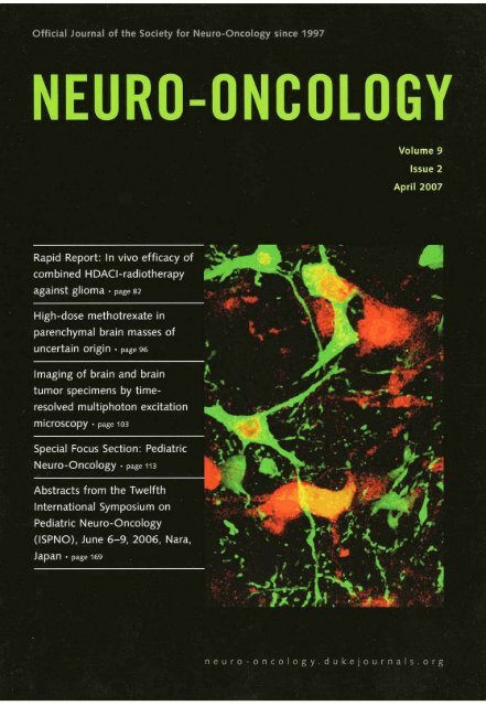
Official Journal of the Soclety for Neuro-Oncology since 1997<br />
NEURO-ONCOLOGY<br />
Volume 9<br />
lssue 2<br />
April 2007<br />
Rapid Report: In vivo efficacy of<br />
com bined H DACI-radiotherapy<br />
against glioma page 82<br />
Ik<br />
parenchymal brain masses of<br />
uncertain origin page 96<br />
lmaging of brain and brain<br />
tumor specirnens by time-<br />
, resolved multiphoton excitation<br />
.<br />
microscopy Page 103<br />
Special Focus Section: Pediatric<br />
Neuro-Oncology page 793<br />
Abstracts from the Twelfth<br />
International Symposium on<br />
Pediatrk Neuro-Oncology<br />
(ISPNO), June 6-9, 2006, Nara,<br />
Japan . page ~ 6s
lmaging of brain and brain tumor specimens<br />
by time-resolved multiphoton excitation<br />
microscopy ex vivol<br />
Sven R. Kantelhardt, lan Leppert, lochen Krajewski, Nadine Petkus, Erich Reusche,<br />
Volker M. Tronnier, Gereon Hüttmann, and Alf Giese2<br />
Department of Neurosurgery, Georg-August-Universiiy of Göttingen, 37075 Gditingen, Gerrnany (5. R. K.,<br />
N. E, A. G.); Departrnent of Neurosurgery, University Hospital Schleswig-Holstein, 23538 Luebeck, Germany<br />
(I. L., J. K., WM. T.); Institute for Biomedical Optics and Medical Laser Center, University Luebeck, 23538<br />
Luebedr; Germany (G. H.); Department o f Neuropathology, University Hospital Schleswig Holstein, 23538<br />
Luebedc, Germany (E. R.)<br />
Multiphoton wrcitation fluorescent micmscopy is a laserbased<br />
technology that allows subdufar resolution of<br />
native tissues in situ. We have recently applied this t&-<br />
noIogy to the smictural and photochemical imaging of<br />
cultured glioma cells and experimental gliomas ex vivo.<br />
We demonstrated that high microanatomical definitiw<br />
of the tumor, invasion zone, and normal adjacmt' brain<br />
can be obtained down to single-cd resolution in unprocessed<br />
tissue blocks. In this shrdy, we used multiphoton<br />
excitatiw and four-dimensionai microscopy to generah<br />
fluoresmce lifetime maps of the murine brain anatorny,<br />
experimentaI g1ioma tissue, and biopsy specimens of<br />
human glial tumors, in mun~e brain, celhlr and nonellular<br />
elements of the normal anatomy were identified<br />
Ditinct excitation profles aad lifetirnes of endogenous<br />
0uorophores were identiaed for specific brain regions.<br />
Received March 24,2006; accepted September 21,2006.<br />
'This study was supported by grants of the Univenib Hospital of<br />
Schleswig-Holsteln, Campus Labeck (A.G., J.L., and N.P.), the Kreitz<br />
Foundation (1.1. and A.G.}, and the Future Investment Program of<br />
Schleswig-Hokteln and ihe Deutsche Fo~chungsgemeinschaft<br />
(A.G. and G.H.).<br />
Wdress correspondence to Alf Ciese, M.D., Department of<br />
Neurosurgery, Georg-August-Univercity of Gattingen. Robert-<br />
Koch-Straw 40,37075 GBttingen, Germany (alf.gleseOmed<br />
.uni-goettingen.de).<br />
Intrauanial grafcs of human glioma ceii liaes in mouse<br />
brain were used ta stady the excitauon pmüles and fluorescence<br />
lifetimes of tumor celIs and adjacent host brain.<br />
These studies demonstrated that normal brain and<br />
mmor could be distingaished on the basis of ffuorescence<br />
intensiv and fluorescence lifnime Profiles. Human brain<br />
specimens and brain tumor biopsies were also analyzed<br />
by mdtiphoton miaoswpy, which demomted distinct<br />
excitation and lifetime profiies in gtioma specimens and<br />
tumor-adjacent brain. This shidy demonstrates that multiphoton<br />
excitation of autofluorescence can distimguish<br />
mmor tissue and normal biain based w the intens& and<br />
lifetime of fluorescence. Further t ecbl deve1op~wts<br />
in this technology may provide a means for in situ tissue<br />
analysis, which might be used to detect residd tumor<br />
at the resecrion edge. Neuro- Oncology 9, 703-112,2007<br />
{Posted to Neuro- Oncology [serial onlinel, Doc. D06-<br />
00049, Februar y 26,2007. URL http://<strong>neuro</strong>-<strong>oncology</strong><br />
. dukejournals.org; DOI: 70.1275/75228517-2006-034)<br />
Keywords: glioma, gIioma invasion, fluorescence Iifetime<br />
imaging, four-dimensional microscopy, multiphotan<br />
excitation fluorescence microscopy<br />
M<br />
ultiphoton microscopy uses near-infrared<br />
femtosecond laser pulses to excite endogenous<br />
intra- and extracellular fluorophores<br />
in a femtoliter target volume (König, 2000). The fluorescence<br />
of the excited endogenous fluorophores can be<br />
Copyright 2007 by the Society for Neuro-Oncology
Kantelhardt et al.: Muläphoton mlcrnscopy of brain and brain tumo~<br />
detected by a photomultiplier and may be reconstructed<br />
into three-dimensional intensity images of native target<br />
tissues at a subcellular resolution without the need for<br />
conmst-enhancing markers. In a conceptual study using<br />
experimental gliomas, we have recently demonstrated<br />
high anatomicai definirion of the tumor parenchyma, the<br />
invasion zone, and normal adjacent brain in unprocessed<br />
tissue blocks by multiphoton excitation autofluorescence<br />
microscopy (Leppert et aL, 2006). Morphologicai daracteristics<br />
of individuaI cell rypes could be identi6ed ar a<br />
singie-cell lwel down to resolution of ceIluiar organeiies.<br />
This technology, however, is not limited to anatomical<br />
and structural imaging. Picosecond time-resolvd detection<br />
of the photons trnitted from multiphotoa-excited<br />
fluorophores may be used to analyze the Iifetime of the<br />
autofluorescence, which is the average time between<br />
excitation and ernission of the fluorescence (Becker et<br />
al., 2001; Xu et al., 1996a, 1996b). Using specific excitation<br />
wavelengths, fluorescence lifetime imaging (fourdimensional<br />
rnicrosmpy) can selehvely excite and derect<br />
endogen~us molecular fluorophores by their excitation<br />
spectra and tbeir fluorescence lifetime. Such biochemical<br />
imaging by multiphoton microscopy has been shown to<br />
discinguish extracellular matrix componentc such as dastic<br />
fibers from collagen in human skm (König et al., 2005)<br />
and has faciiitated selective excitation of rnelanin (Teuchner<br />
et at., 1999). Recently, our analysis of the rdationship<br />
between the laser excitation wavelength and the lifetime<br />
of excitable endogenous fluorophores in cells derived<br />
from tumors of different histotypes has suggested individual<br />
fluorescence lifetime profiles for distinct cell types.<br />
We have further shown that time-resoived measurements<br />
of fluorescence lifetimes distinguish turnor cdls from<br />
normal brain parenchyma (Leppert et al., 2006).<br />
In Lhe present study, we used multiphoton excitation<br />
to generate color-coded ff uorescence Iifetime images of<br />
the murine brain anatomy, experimental gliorna tissue,<br />
and biopsy specimens of human glial tumors. In murine<br />
brain, cellular and noncelluIar elements of the normal<br />
brain anatomy were identifred, which showed distinct<br />
excitation profiles of endogenous fluorophores and a disthct<br />
spectrum of fluorescence lifetimes. We used intracranial<br />
grafts of human glioma ceIl lines in rnouse brain<br />
to smdy the excitation profiles and fluorescence lifetimes<br />
of tumor cells and the adjacent host brain. These studies<br />
demonstrated that normal brain and turnor could be distinguished<br />
based on fluorescence incensity and distinct<br />
excitationllifetime profiles. Unprocessed tissue b lds of<br />
human brain specimens and brain tumor biopsy specimens<br />
analyzed by multiphoron excitation also dernonstrated<br />
distinct excitationllifetime profiles in glioma<br />
spccimens compared with normal brain.<br />
Materials and Methods<br />
For multiphoton excitation of endogenous fluorophores<br />
in experimental gliarnas, we ucd the DermaInspect in<br />
viv0 imging system (JenLab, Jena, Germany). The sys-<br />
tem contains a solid-state, mode-locked 80-MHz tiraniumsapphire<br />
laser (MaiTai, Spectra Physics, Darmstadt,<br />
Germany) with a tuning raage of 710-920 nm, a<br />
mean laser output of >900 rnW at 800 nm, and a 75-fs<br />
pulse width. The scanning module conrains a motorized<br />
beam attenuator, a shutter, and a two-axis galvoscanner.<br />
A piezo-driven 40x focusing optic with a 1.3 numerical<br />
aperture and 140- km working distance (Plan Meofluar,<br />
Zeiss, Göttingen, Germany) was used to study native<br />
brain and tumor tissue. The autofluorescence signai was<br />
detected by a photomultiplier tube module (H7732-01,<br />
Hamamatsu, Herrsching, Gerrnany) after passing a<br />
beam splitter and a short-pass filter (BG39, Schott,<br />
Mainz, Germany).<br />
Fluorescence lifetime images were measured by timecorreiared<br />
single-photon counting (Fig. 11, A photomuIriplier<br />
modute (PMH-100-0, Becker & Hickl, Berlin,<br />
Germany) detected the fluorescence photons ernitted<br />
by rhe tissue. The start signal for the phatomultipIier<br />
and stop signaIs provided by the laser were processed<br />
by a PC-based single-photon counting board (SPC 830,<br />
Becker & Hickl), which allowed Count rates of up to<br />
8 X 10⁶ photonsls. The single-photon counting board<br />
was synchronized with the spatial beam position, which<br />
was calculated from signals of the galvoscanner. Spatially<br />
resolved autofluorescence decay curves were recorded<br />
for 256 X 256 pixels per image field, which typically<br />
was 150 Pm. The depth of the excitation volume typically<br />
was less than 1 Pm. Curve fitting of a single exponential<br />
decay curve, including a deconvolution with the<br />
time response of the system (SPCImage 26, Becker &<br />
Hickl), was used to calculate a mean fluorescence iifetime<br />
for each pixel, which was displayed in color-coded<br />
images (Becker et al., 2001). The accuracy of the measurements<br />
can be judged by the scattering of the measured<br />
values. Under optimal conditions when only the<br />
shot noise of the photons determines rhe relative mor of<br />
the measured lifetimes, it is approximated by 2 divided<br />
by the square root of thc numbex of detected photons,<br />
which was between 100 and a few rhousand per pixei<br />
during the measurements (Köllner and Wolfrum, 1992).<br />
Therefom, errors of up ta 10% are expeaed. To analyze<br />
the fluorescence Iifetimes of endogenous fluorophores<br />
within specific ceIIular compartments, regions of interest<br />
were defined and the analysis was performed in at.<br />
least three regions of similar cornpartments. The fluorescence<br />
lifetime for each region of interest was determined.<br />
The data are reported as the means of triplicate<br />
determinations, and the fluorescence lüetimes are plotted<br />
as a function of the excitation wavekngrh.<br />
Wot& Gliw Mouse Mo&&<br />
Tmr Spedmau, arid Hcstology<br />
The human glioblastoma-derived cdl lines G-28, G-112,<br />
and U87 were- grown in minimum essential medium containing<br />
10% fetal calf serum. For intracranial implantation<br />
in nude NMRI mice, cells were expanded and bar-<br />
NEURO-OHCOLOGY A P R I L 2 007
Kantelhardt et al.: Mukiphoton microscopy of brain and brain tumors<br />
rnuliiphoton microscopy<br />
TCSPC 19fetlme imaging<br />
I 1 pie~ CMJ<br />
X munter<br />
I<br />
native tissue block<br />
Fig. 1. Schematic presentation of rnultiphoton mictoscopy of native centrai nervous system tissue and glioma tissue.<br />
vested in log-phase growth by trypsinization. Cells were<br />
washed in PBS three times and then resuspended at a concentration<br />
of 2 X 10⁴/~1. All ~rocedures were uerformed<br />
in accordance with kgulatilns of the ~nirnai Care and<br />
Use Cornmittee of the University Hospital af Schleswig-<br />
Hoisrein (tierrnit 30101031. Mice were anesrbetized bv<br />
1s<br />
peritoneal injection of ket&ninefxylazine solution (20b<br />
rng ketamine and 20 mg xyiazine in 17 ml saline) at<br />
0.15 mgllO g body weight; the cranium was fixed in a<br />
stereotacric frame (TSE Systems, Bad Homburg, Germany).<br />
A 4-mm bur hole was drilled 3 mm lateral to the<br />
bregrna, and a stereotactic implanration of 3 pI of cell<br />
suspension injected over 3 min was placed in an area<br />
coriesponding to the internal capsule 0.5 rnm below the<br />
fiber tracts of the corpus callosum. After implantation,<br />
50 mglkg novaminsulfone was adrninistexed subcutaneously,<br />
and 1 rnglml novaminsulfone was added to the<br />
drinking water for three days. Four weeks after implantation,<br />
tumor-bearing brains were explanted following a<br />
lerhal intraperitoneal injection of SO mglkg xylazine and<br />
350 mglkg ketamine. The specimens were processed on<br />
ice. and the brains wem divided into ewo tissue blocks<br />
at a coronal plane using a scalpel. The tissue samples<br />
were placed in a humidified biopsy chamber (MiniCeM,<br />
JenLab) adherent to a 0.17-W Cover glass and imaged.<br />
Following the imaging studies, the specimens were fixed<br />
in formalin, and the rissue blocks were smioned parallel<br />
to the optical imaging plane and embedded in parafh;<br />
5-prn sections were cur and stained with hernatoxylia<br />
and eosin.<br />
Recently, we have shawn that glioma cells in culture<br />
and cells dwived from different histotypes differ in their<br />
rnultiphoton excitationffluorescence lifetime profile, suggesting<br />
that muitiphoton microscopy and fluorescence<br />
lifetime imaging may provide some cell-type specificity<br />
and a means of identifying glioma celh in brain tissue<br />
(Leppert er aL, 2006). We therefore imaged the normal<br />
anatomy of native rnouse brain tissue blocks and analyzed<br />
the excitability of fluorescence lifetime as a function<br />
of the excitation wavelength.<br />
From specific areas of interest, lifetime images were<br />
obtained using increasing excitation wavelengths from<br />
720 to 780 nm at iacrements of 10 nm. The disrribution<br />
of fluorescence lifetime was color coded using a continuous<br />
spectrum of red (short lived) to blue (long lived). The<br />
lifetime of hornogeneous areas, cells, or organelles such<br />
as the nucleus or highly autofiuorescent granula were<br />
analyzed separately in some specimens. On the basis of<br />
these Parameters, graphs were plotted displaying the<br />
lifetime of specific regions of interest as a function of<br />
the excitauon wavelength (Fig. 2).<br />
Normal rnouse brain contained several anatomical<br />
and microanatomical sttuctures readily identified by<br />
rnultiphoton microscopy. Fluorescence intensity imaging<br />
demonstrated that metabolically highly active cells
Kantelhardt et d.: Multiphoton microscopy of brain and brain tumors<br />
Fig. 2. Multiphoton microscopy of normai mouse basal ganglia. (A) Intensity image of the autofiuorescence signal, demonstrating cells of<br />
intense fluorescence with low-intensity nuclei. Near these intensely fluorescing cells. nuciei of cells with low-signal-intensity cytoplasm<br />
could be identified. Acellular areas of the parenchyma were selected hsed on thfee-dimensional stacks of images demonstrating no cellular<br />
nuclei above or below the plane of analysis. (B) Corresponding color-coded fluorescence Iifetime image generated at an excitation wavelength<br />
of 750 nm. (C) Color-coded distribution of fluorescence lifetimes at 750 nrn within the whole image fmme, shown in picoseconds.<br />
(D) Excitation/lifetime profile of different areas of interest framed in A. This analysis demonstrated that high-intensity fluorescence cells<br />
not only showed a longer fluorescence lifetime than did IOW-intensity cells but also showed distinct excitation/lifetime profiles.<br />
and tissucs, such as the ependyma, choroid plexus, or<br />
vascular endorhelial cells, tended to show high signal<br />
intensity. When analyzed by fluorescence lifetirne irnaging,<br />
these structures also shawed the longest lifetirnes of<br />
endogenous fluorophores (>I700 ps) excited at 750 nm,<br />
In conuast, excitabk fluorophores within erythrocyres<br />
were the shortest lived (900 I 72 ps). In gray and white<br />
matter, the brain parenchyma showed an intermediate<br />
fluorescence lifetime (1380 i 23 ps and 1360 i 33 ps,<br />
respecrively) (Fig. 3). Generally, tbe nuclei of glia showed<br />
low fluorescmce intensity. The cytoplasm of glia cells<br />
frequently contained granules of high fluorescence intensiry<br />
and relatively short fluorescence Iifetime. Confirming<br />
our previous observations, the fluorescence lifetime<br />
of the nuclei inueased with inueasing excitation wavelengths<br />
(Leppere et al., 2006). The nuclei of hippocampal<br />
<strong>neuro</strong>ns also showed low fluorescence inrensity (Fig. 4).<br />
The fluorescence lifetimes of endogenous fluorophores<br />
within hippocampal <strong>neuro</strong>ns showed a linear increase<br />
from 720 um to 770 nm excitation, which was signifi-<br />
cantly different from the brain parenchyma neighbming<br />
the groups of <strong>neuro</strong>m. These dktinct excitation/lifearne<br />
profiles of cellular and subcellular structures reflea the<br />
photochemical cornposition of these regions of interest<br />
(Xu et al., 1996a, 1996b).<br />
Obviously, multiphoton exciration microscopy and fluorescence<br />
lifetime imaging have the potential of providing<br />
cell-type-specific or tissue-specific informarion. We<br />
therefore used an intracranial tumor traasplantation<br />
model in NMRI mice to study the relationsbip of fluorescence<br />
intensity and fluorescence lifetime af humanglioma-derived<br />
ceils and the murine host brain.<br />
Tumor-bearing mouse brains were obtained as<br />
described in Materials and Methods. Coronal sections<br />
(2 gm) were cut at the levd of the implantation<br />
site, and the native tissue was subjected to multiphoton
Kantelhardt et d.: Multrphoton rnimm~ of braln and braln tumors<br />
\ '"I -1<br />
umld pkls<br />
*<br />
l,<br />
~ ~ ~ - - - ~<br />
mmrmmmrn<br />
Fig. 3. Microanatomical structures of normal mouse brain were analyzed by multiphoton excitation intensity irnaging, color-coded fluorescence<br />
Iifetirne irnaging (at750 nrn excitation), and conventional light rnicroswpy of histological sections stalned with hematoxylin and eosin.<br />
The excitation/lifetlme profile of regions of interest within specific brain areas was calculated from at least three analyses (right panel). A representative<br />
region of lnterest for each anatornlcal site is lllustrnted by a framed area. Metabolically highly active cells of the ependyma, droroid<br />
plexus, and vascular endothelid cells showed high fluorescence on intensity irnaging and tended to show long fluoreccenw lifetirnes.<br />
Fig. 4. Fluorescence lntensity images of hippocampal <strong>neuro</strong>ns in mouse brain. The ins& shows the corresponding fluorescence lifetime<br />
irnage at an excitation wavelength of 750 nm. The graphs show the excltation/llfetime pmf es for specific &ons if interest (red, hippocampal<br />
<strong>neuro</strong>ns; yeliow and black, adjacertt parenchyrna). The nuclei of the hippocampal <strong>neuro</strong>ns show a characteristlc excitation/lifetime<br />
profile with a positive correlation of excitation wavelength to fluorescence lifetirne.
Kantelhardt et al.: Multiphoton mimscopy of brain and brain tumors<br />
rnicroscopy immediately. Intensity images allowed easy<br />
identification of the tumor rranspIants because of a profoundly<br />
increased signal intensity of the turnor cells at<br />
750 nm excitation over normal cells of the white or gay<br />
marter and the surrounding brain parenchyma (Fig. SA).<br />
Although our analysis of normal brain identified several<br />
highly autofluorescing ce11 types, these could be distinguished<br />
from tumor based on their disrribution and specific<br />
morphoIogy (compare Figs. 3 and 4). On intensity<br />
images, tumor-adjacent white matrer showed a low density<br />
of low-signal-intensity nuclei. In contrast, the tumor<br />
transplants were highly celiular, with low-fluorescenceinrensity<br />
nudei and a high-signal-intensity cytoplasm.<br />
Continuous-spectrum color-coded fluorescence Lfetime<br />
irnaging of turnor and adjacent h in demonstrated that<br />
tumor tissue showed longer mean fiuorescence lifetimes<br />
than did normal white matter or normal cortical gray<br />
matter (1780 i 43 ps and 1540 * 30 ps, respectively)<br />
(Fig. SB and C). U87 cells implanted into mouse brain<br />
typically form a well-defined tumor-to-brain interface<br />
with few single invasive cells. Discrete color coding of<br />
lifetime ranges adapted to a region of interest in U87<br />
tumors therefore resulted in an exact reproduction of<br />
the anatomical tumor-to-brain interface based on fluorescence<br />
lifetimes (Fig. SD). Aithough fluorescence<br />
Iifetimes differed among experimental tumors derived<br />
from the h ee ceil lines that we studied, rhe fluorescence<br />
lifetime was always significantly longer (ranging from<br />
approximately 1640 to 1800 ps) than for t umor-adjacent<br />
brain (about 1510-1580 ps) ar 750 nm excitation. The<br />
analysis of excitation/fluorescence lifetime profiles of<br />
U87 tumors and adjacent brain for increasing excitation<br />
wavelengths resulted in similar biphasic lifetime profiles<br />
for both tumor and adjacent brain (Fig. 6). However, the<br />
fluorescence lifetimes of tumor tissue were significantly<br />
bnger ar any excitation wavelength than those for brain<br />
tissue. A biphasic excitarion/fluorescence lifetime profile<br />
with a maxbum ar approximately 750 nm excitation<br />
was observed in al experimental tumors derived from<br />
the three human glioma cell lines used (G-28, G-112, and<br />
US7) (data not shown). These findings are consistent<br />
with our previously reported excitationlfluorescence<br />
lifetime profiles of G-28, G-112, and U87 cells in monolayer<br />
culture (Leppert et al., 2006). Inrerestingly, the<br />
mean Iifetimes of turnor-adjacent brain (1540 ps) at<br />
750 nrn excitation tended to be langer than the mean<br />
fluorescence Iifetimes of brain more distant from the<br />
tumor or the mean lifetimes of white matter obtained<br />
frorn non-turnor-bearing animals, which generally were<br />
1300-1400 ps. Fluorescence intensity images of tumoradjacent<br />
brain and normal brain shawed no differente.<br />
To determine wherher fluorescence lifetime imaging<br />
could delineate adjacent brain and brain tumors in clinical<br />
specimens, tumor-adjacent brain and brain tumor<br />
biopsies were obtained at surgery and immediately subjected<br />
to multiphoton miuoscopy. Following the analysis,<br />
the tissues were fixed in formalin and processed for<br />
routine diagnostic histopathology.<br />
L';<br />
FIg. 5. (A) Fluoremnce intensity image of the tumor-brain interface in an experimental U87 glioma in NMRl mouse brain. (B and C) Continuous<br />
color-coded lifetime image of the tumor-mouse brain interface. (B) Histogram of the fluorescence lifetime dlstributlon in a region<br />
of lnterest (white frame) wrrespondingto tumor-adjacent brain. (C) Fluorescence lifetirne distributions In a region of lnterest mrresponding<br />
to U87 glioma tissue, demonrtrating the significantly longer Ruorescence Iifetimes of tumor tissue compared with adjacent rnouse brain. (D)<br />
Two-color-coded Image of the tumor-brain interface. The lifetime ranges for red and green were selected based on the peak distributions of<br />
the Iifetirne histagram. (E} Color-coded gating of fluorescence Iifetimes allowed discrimination of tumor. and adjacent bmin demonstrated<br />
a well-defined tumor-brain interface in U87 giiomas. All images shown here were obtained at an excitation wavelength of 750 nm.<br />
NEURO-ONCOLOGY . A P R I L 2 00 7
Kantelhardt et al.: Multiphoton microxopy of brain and brain tumors<br />
Fig. 6. The continuous-spcctrum color-coded fluorescence Iifetime images of U87 grioma cells in mouse brain (A) and discrete color-wded<br />
spectrum for optimal discrimination of tumor and normal brain (B). Longer fluorescence lifetimes of tumor cells are coded blue. The intensity<br />
images show the microanatomy of the corresponding turnor area (C). Tumor cells showed higher fluorescence intensity and a prolonged<br />
fluorescence lifetime compared with surrounding brain. The analysis of individual tumor cells and adjacent brain showed that at all excitation<br />
wavelengths the fluorescence lifetimes exceeded those of normal brain parenchyma. The nuclei of tumor cells and cytoplasmic areas<br />
of tumor cells showed distinct excitation/lifetime profiles.<br />
Tumor-adjacent human brain specimens showed and rhe low-grade astrocytoma (1740 t 44 ps). In these<br />
structures similar ro those in the murine brain speci- spechens, the excitation/fluorescence lifetime profiles<br />
mens. Multiphoton intensiry imaging dernonsrrated few demonstrated that, in both the anaplastic astrocytoma<br />
nuclei per tissue volume and some vascular elements and the gIioblastoma, the fluorescence lifetimes were<br />
identified as capillaries. In conrrasr to normal mouse decreased at excitation wavelengths greater rhan 750<br />
brain, human tumor-adjacent brain specimens con- nm (Fig. 7).<br />
tained a larger number of cells with granuIated cytoplasm<br />
(Fig. 7). The iniracellular granules were highly<br />
autofluorescinp; at an excitation wavelenmh of 750 nrn.<br />
Discussion<br />
Fluoreccence lifetime imaging showed very short-lived<br />
fiuorophores within these intracellular compartments,<br />
Neu-infrared multiphocon excitation laser scanning<br />
with mean lifetimes of 560 22 ps (whire arrow, Fi. 7).<br />
microscopes can potenrialIy bt employed as novel noninvasive<br />
biomedical tools for three-dimensionally and<br />
Hisrohgically, these cells cortesponded to CD68' macmphages<br />
(data not shown). The parenchyma of<br />
time-resolved imaging of fluorophores in optical tissue<br />
these<br />
brain specimens showed fluorescence lifetimes ranging<br />
diagnostics. This includes structuml imaging at the subfrom<br />
1400 to 1750 ps at 750 nm excitation. A menincelluIar<br />
level, as well as the photocbemical characterizauon<br />
of living tissues and functional irnaging of solid<br />
gioma specimen showed high fluorescence intensity of<br />
tissues.<br />
the cytoplasm af tumor cells with low-signal-intensity<br />
The first biomedical applications in experimental<br />
nuclei. The excirationlfluorescence lifetime profile of the<br />
meningioma specimen showed a tendency to increased<br />
dermatology have already demonstrated the potential<br />
of this technology, and the DermaEnspecr multiphoton<br />
lifetimes at increased excitation wavelengths, but with<br />
microscope used in this study is now cammercially availgenerally<br />
longer lifetimes of endogenaus fluorophores<br />
than for tumor-adjacent brain. We also analyzed<br />
able for dinical applications in dumatology (König and<br />
three<br />
gliomas, a WH0 grade I1 astrocytoma, an anaplastic<br />
Rieman, 2003). We have recenrly applied this technology<br />
to structural arid<br />
astrocymma, and a WH0 grade IV glioblastoma. Fluophotochemical<br />
irnaging of cultured<br />
glioma celIs and experimental gliomas ex vivo.<br />
rescence lifetime imaging at 750 nrn demonstrated longer<br />
man fluorescence lifetimes for the<br />
This conceptual study demonstrated that high microtumor<br />
parenchyma<br />
anaromical definition of<br />
of all three glioma specimens than for<br />
rhe tumor parenchyma, invatumor-adjacent<br />
brain. Strikingiy, the glioblasuima specimen showed the<br />
sion zone, and normal adjacenr brain can be obtained<br />
in<br />
longest mean lifetime of autofluorescence (2110 2 73<br />
unprocessed tissue blocks. In an intracranial mouse<br />
model, fluorescence intensity images<br />
ps), followed by<br />
allowed delineation<br />
the anaplastic glioma (1870 z 40 ps)
Kantelhardt et al.: MulphoSon mlawcopy of brain and bin<br />
tumors<br />
Fig. 7. Multlphoton excitaäon fluoreseence intemity Irnaging and fluorercence lifeüme imaging (at 750 nm) of human turnor-adjacent brain<br />
and specimens of human brairt tumors. For Ruorescence Iifetime Imaging of the cytoplasrnic area of turnor cells, ldentlcai paramaters of<br />
conünuous-npectrurn mlor coding were used for alt specimens. The analysis of fluorescence Cfetirnes at inming excitaäon waveiengths<br />
demortstrated distinct excitaüon/lifetime profilesfor normal brain and malignmt gliomas.<br />
of singIe tumor ceIls invading murint brain (Leppert et<br />
al., 2006). However, the characterization of individual<br />
cells was not limited to structural anatomical imaging.<br />
Fluorescence lifetime imaging of cultured glioma cells<br />
in vivo demonstrated that subceliular compartmeats<br />
showed different excitabilitv and fi uorescence lifetimes<br />
of endogenous fluorophores.'~mong the fluorophores for<br />
which fluorescence lifetime specua have already been<br />
chuacterixed are NADPH, flavines, lipofuscin, eiastin,<br />
collagen, and melanin (König and Rieman, 2003). Howwer,<br />
no studies have investigated specific fluorophores<br />
in the brain.<br />
Fluorescence lifetime images alIowed delineation of<br />
the peripheral and perinuclear cytoplasm, intracellular<br />
granules, and rhe nucleus. Owing to the limited number<br />
of animals used in this study, we were not abie to give<br />
quantitative values for the sensitivity and specifxiry, The<br />
aim of this study was to show that we can detect spectroscopic<br />
differences (cbaracteristic dependence of the<br />
fluorescence lifetime on tbe excitation wavelength). The<br />
hardware-dependent Iimiration of the time resolution,<br />
as described in Materials and Methods, were found to<br />
be errors of up m 10% of the measured lifetimes. TypicaIly,<br />
the differences in the lifetimes of the different rissue<br />
components were found to be 20%-50%. Therefore,<br />
ar least 200 photons have to be detec4ed to separate cell<br />
types by two standard deviations of the measured decay<br />
time. With a typical Count rate of 50,000 photonds, 250<br />
pixelds can be measured.<br />
The analysis of the relatianship between the laser<br />
excitation wavelength and the lifetime of endogenous<br />
fluorophores showed characterisuc profiles for intracellular<br />
cornpartments in cultured glioma cells. Interestingly,<br />
these excitationlfluorescence lifetime profiles of<br />
cultured glioma ceIl lines and primary cultures of gliomas<br />
differed from profiles obtained from cultured cells<br />
derived from othex tumor types (Leppert et al., 2006).<br />
This suggesrs that fluorescence lifetime spectroscopy<br />
may differentiate historypes of cells based on the excitability<br />
of ceI1-type-specific expression of endogenous<br />
chromophores, their chemical states, or their interaction<br />
wirh other biomolecules. Whether fluorescence lifetime
Kantelhardt et al.: Multiphoton mlcroscopy of brain and braln turnon<br />
spectroscopy may be extended to a discrimination of<br />
functional ceIlular states in cultured glioma cells is currently<br />
under investigation.<br />
Our aim in the present study was ro analyze whether<br />
fluorescence lifetime imaging may be able to identify glioma<br />
cells in situ. Such anatysis requires rhe characterization<br />
of fluorescence intensities and fluorescence lifetimes<br />
within normal h in and brain tumors. Therefore, we<br />
used an intracranial rnodel system in NMRI mice using<br />
transplaatable human glioma cell lines (Brockmann et<br />
al., 2006). This study demonstrated that rnultiphoton<br />
microscopy in native tissue blocks allows a detailed display<br />
of microanatomical brain structures without the<br />
need of contrasting techniques.<br />
FIuorescence lifetime imaging showed that gray and<br />
white matter represent areas of l-tomogeneous distribution<br />
of fiuorescence 1iferim-s at any excitarion wavelength<br />
between 720 and 770 nm. Per tissue volurne, few nudei<br />
and cytoplasmic suucrures of resident ceIk couId be identi6ed.<br />
Within this homogeneous background, the mmox<br />
transplants could be readily identified, because of high<br />
ffuorescence intensity of the cytoplasmic areas of tumor<br />
cells, which contrasred with the low-intensity nudei. At<br />
any excitation wavelength, the tumors derived from the<br />
three different cell lines showed markedly longer fluorescence<br />
Iifetimes than did adjacent gray or white matter.<br />
Interestingly, the fluorescence lifetime of tumor-adja-<br />
Cent brain was wnsistently longer than that of normal<br />
white matter. Whether this is a consequence of tissue<br />
edema or ingress of celtular eiements responding to the<br />
tumor stimulus remains open. However, several microanatomical<br />
structures were identified within normal brain<br />
hat by multiphown excitation showed intense autofluorescence<br />
and long -fluorescence lifetimes of excited fluorophores.<br />
nie lifetimes of the ventricular ependyma, fm<br />
example, reached values similar to those for tumor ceiis.<br />
Interestingly, the elemems of normal brain showing long<br />
fluorescence lifetimes were composed of rnetabolicaily<br />
highiy active cell types. Fmher examples were cells of the<br />
epithelium of the choroid plexus and endothelial cdls of<br />
capillaries and larger blood vessels. Because of their specific<br />
morphology, these anatomical structures and normal<br />
cells cxiuld be easily distinguished from tumor.<br />
In the deep basal ganglia, however, single celIs of<br />
high fluorescence intensity and long fluorescence lifetime<br />
were observed in normal brain specimens (compare<br />
Fig. 2). These individual ceIls showed excitation spectra<br />
similar to those of rumor transplants, This may suggest,<br />
on the basis of the Parameters analyzed here, hat multiphoton<br />
excitation fluorescence intensity irnaging and<br />
fluorescence lifetime spectroscopy offer no nunor specificit<br />
y but rather rnay identify meta bolically highiy active<br />
rissues. This is further supported by a recent observation<br />
(using sirnilar detection parameters) hat the intestinal<br />
endothelium shows intense autofluo~escence and<br />
long fluorescence Iifetimes (Gebert et aI., manuscript in<br />
preparation).<br />
Nevertheless, fluorescence lifetime imaging of rnalignant<br />
human giioma specimens showed tbar the fluorescence<br />
lifetimes and the excitation/iifetime profiles of<br />
tumor specimens were significantly different from those<br />
of tumor-adjacent brain. These data would suggesr that<br />
multiphoton excitation of aurofi uorescence theoreticauy<br />
provides means for a tissue analysis in situ, which could<br />
be used, for example, to detect residual turnor at the<br />
resection tdge. In contrasr ro conventional one-photon<br />
laser scanning microscopy, femtosecond pulsed laser<br />
microscopy of living specimens can be performed ar<br />
peak intensities of 200 GWlcm2 with no sign of structural<br />
or functional photodarnage. This has been dernonstrated<br />
for cells in monolayer culture as wetl as for mammalian<br />
embryos and human skin (Masters et al., 1997,<br />
1998; Oehring et al., 2000; Squirrell et al., 1999; Tyrell<br />
and Keyse, 1990). Therefore, inrraoperative in vivo multiphoton<br />
microscopy of brain tissue conceivably couM<br />
provide a high-resolution, noninvasive diagnostic tool.<br />
Recent developmwts of this technobgy have introduced<br />
miniaturized Scanner probes connected to optic fibers<br />
that have been used for in viv0 irnaging of the rnouse<br />
central nervous system over extended periods of time<br />
{Kim et aI., 2004). Such probes glaced in direct contact<br />
with the target tissue rnay offer future solutions to<br />
high-resolution opticaI imaging of a target volume that<br />
foIIows the respiratory and arterial cycle, such as the<br />
brain.<br />
gecker, W., Bergmann, A., KBnig, K., and firlapur, U. (2001) Picosmnd<br />
fluorescence iifetlrnc rnlcroscopy by TCSP Imaging. Pmc. SPIE4262.<br />
Brockmann, M.A., Ulmer, S., Leppert, 1.. Nadrowitz. R., Wuestenberg, U.,<br />
Ndte, I,, Petersen, D., Groden, C., Glese,A., and Gottschalk, 5. (2006)<br />
Analysls of mowe brain using a cllnical 1.5 tesla xanner and a standard<br />
mall loop surface coil. Brain RH. 1068.138-142.<br />
Kim, D.. Klm, K.H., Yazdanfar, S., and So. P.T.C. (20941 Hlgh-speed handhtld<br />
multiphoton mulblfocl mlcmxripy. Proc. SPIE5323.267-272.<br />
Kallner. M., end Wolfrum, J. (1992) How many photons are necessary for<br />
fluorexence-lifetirne meawrements Chem. Phys. Lett. 2W), 499.<br />
Kdnfg, K. (2000) Multiphotwi mlcroscopy In llfe sclence. J. Mimt. 200.<br />
83-104.<br />
Konig, K., and Riernann, 1. (2003) Hlgh-resolution muftfphoton tomography<br />
of human skin with subcellular spatial resolution and picosecond<br />
time resolution. I. Biomed. Opt. 8,432-439.<br />
Kbnlg, K., Schenke-Layland, K., Riernann. I., and Stock, U.A. (2005) Multlphoton<br />
autofl uorescence imaging of Intratlaue elatic fi bers. Blomaterials<br />
26,495-500.<br />
Leppert,].. Krafewski, J., Kantelhardt, S.R., SchlaffPr.5.. Petkus, N., Reusche,<br />
E., Huttmann, C., and Ciese, A. (2006) Multiphton excltatlon fluorescence<br />
rnlwisiripy of glioma tissue. Neumsurgery 58,7!39-767.
Kantelhardt et al.: Multiphoton mlcrnxopy of brain and brain P u m<br />
Masters, B.R., So, P.T., and Cratton, E. (1997) Multiphotdn mitation RUOrescence<br />
rnicmwpy and spectroscopy of In vivo human skln. Biophys.<br />
J. 72,2405-2412.<br />
Masters, B.R.. So, P.T., and Grattofi, E. (1998) Multiphobn excltation<br />
rnicmcopy of In vlvo human skn. Functional and morphologid optlcal<br />
biopsy based on three-dimensional imaglng, lktime mmiurernents<br />
and flwmscence spectroxopy. Ann. N. Y. kad. Scl. 838,5867,<br />
Oehring, H., Riemann, I., Flxher, P., Halbhuber, K.J., and Könlg, K. (2000)<br />
Ultrastructure and reproduction behavtour of slngie CHO-KI cells<br />
exposed to near infrared femtosernnd laser pulses. Scanning 22,263-<br />
270.<br />
Squirfell, ).M., Wokesln, D.L., WhHe, J.G., and Bavlster, B.D. (199$) Long.<br />
km two-photon Ruomnce irnaging of mamrnallan embryos without<br />
compmmising vlablllty. Nat Biotechml. 47,763-767.<br />
Teuchner, K.. Frtyer, W., Leupold, D., Volkmer, A., Birch, D.J., Altrneyer,<br />
P., Stucker, M., and Hoffmann, K. (1999) Femtosecond two-photon<br />
mited fluorrxem of rnelanln. Photochem. Phobbiol. 70, 146-151.<br />
Tyrefl, R.M., and Keyse, S.M, (7990) New trends in pttotobiology: The<br />
interacolon of UVA radlation with cultured cells. J. Photohem. Photobid.<br />
4,349-361.<br />
Xu, C., Wllllarns, R.M., Zipfel, W., and Webb, W.W. (1996a) Mvltiphoton<br />
excitation cross-rectiiom d rnolecular fluorophores. Biolmaging4,<br />
198-2W.<br />
Xu, C., Zlpfel, W.. Shear, J.B„ WIIHarns, R.M, md Webb, W.W. (1996b)<br />
Multiphotan Ruorescence excitation: New spectrai wlndows for Mo-<br />
iogicai nonlinear mlcroseopy. Pmc. Ne#. Acad. Sci, U. S. A. 93, 10763-<br />
1m6a.<br />
NEURO-ONCOLOGY A P R f L 2 o 07