Pelvic Brochure 06 COVER - AGES
Pelvic Brochure 06 COVER - AGES
Pelvic Brochure 06 COVER - AGES

Create successful ePaper yourself
Turn your PDF publications into a flip-book with our unique Google optimized e-Paper software.
Australian<br />
Gynaecological<br />
Endoscopy<br />
Society Ltd<br />
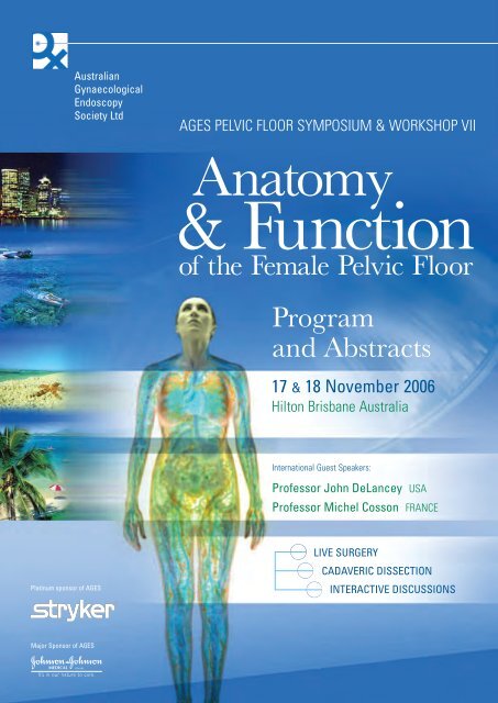
<strong>AGES</strong> PELVIC FLOOR SYMPOSIUM & WORKSHOP VII<br />
Anatomy<br />
& Function<br />
of the Female <strong>Pelvic</strong> Floor<br />
Program<br />
and Abstracts<br />
17 & 18 November 20<strong>06</strong><br />
Hilton Brisbane Australia<br />
International Guest Speakers:<br />
Professor John DeLancey USA<br />
Professor Michel Cosson FRANCE<br />
Platinum sponsor of <strong>AGES</strong><br />
LIVE SURGERY<br />
CADAVERIC DISSECTION<br />
INTERACTIVE DISCUSSIONS<br />
Major Sponsor of <strong>AGES</strong>
Sponsorship and Exhibition<br />
<strong>AGES</strong> gratefully acknowledges the following companies for their support of this meeting:<br />
Platinum Sponsor of <strong>AGES</strong><br />
Major Sponsor of <strong>AGES</strong><br />
Major Sponsors of<br />
<strong>AGES</strong> <strong>Pelvic</strong> Floor<br />
Symposium & Workshop VII<br />
Sponsors of invited speakers<br />
Johnson & Jonhson Medical<br />
American Medical Systems<br />
Femcare<br />
Bard Australia<br />
Cook Australia<br />
Arrow Pharmaceuticals<br />
GE Ultrasound<br />
Boston Scientific<br />
Medtronic Australasia<br />
Exhibitors<br />
Olympus<br />
Experien<br />
Scanmedics<br />
B Braun Australia<br />
Gyrus ACMI<br />
Smith and Nephew<br />
ConMed Linvatec<br />
Hoyland Medical<br />
Sydmed<br />
Cytyc (Australia)<br />
InSight Oceania<br />
Device Technologies<br />
Medtel
Contents<br />
Sponsorship and Exhibition<br />
inside cover<br />
Faculty and Committee Members 2<br />
Welcome Message 3<br />
Conference Program 4<br />
Social Program 7<br />
Abstracts – Friday 17 November 8<br />
Abstracts – Saturday 18 November 16<br />
PR&CRM POINTS - CPD POINTS<br />
The Conference has been approved as a RANZCOG Approved<br />
O&G Meeting and eligible Fellows of the College will earn<br />
points for attendance as follows:<br />
Full attendance: 16 CPD points in the Meetings category<br />
Attendance one day: 8 points<br />
Delegates will be required to sign the attendance sheet prior<br />
to morning tea on both Friday 17 and Saturday 18 November.<br />
Pre- and Post- Questionnaires<br />
The College approved Pre- and Post- Questionaires are<br />
comprised of a number of multiple choice questions from<br />
lectures to be given on Friday 17 and Saturday 18 November.<br />
The Pre- Questionnaire is to be handed in at morning tea on<br />
Friday 17 November. The Post- Questionnaire is to be handed<br />
in at the close of the Meeting. No exceptions can be made to<br />
these deadlines.<br />
<strong>AGES</strong> PELVIC FLOOR SYMPOSIUM & WORKSHOP VII<br />
Anatomy & Function<br />
of the Female <strong>Pelvic</strong> Floor<br />
1
CONFERENCE COMMITTEE<br />
INTERNATIONAL FACULTY<br />
Assoc. Professor Chris Maher<br />
Dr Anusch Yazdani<br />
Assoc. Professor Alan Lam<br />
Dr Lewis Lander<br />
Chairman<br />
Co-Chairman<br />
Professor John DeLancey<br />
Professor Michel Cosson<br />
Assoc. Professor Kaven Baessler<br />
USA<br />
France<br />
Germany<br />
Dr Melissa Buttini<br />
Dr Stephen Cook<br />
Dr David Baartz<br />
Dr Julie Lindstrom<br />
Ms Janelle Greitschus<br />
AUSTRALIAN FACULTY<br />
Dr David Baartz<br />
Dr Melissa Buttini<br />
Dr Marcus Carey<br />
Queensland<br />
Queensland<br />
Victoria<br />
Dr Greg Cario<br />
New South Wales<br />
<strong>AGES</strong> BOARD<br />
Dr Robert O’Shea<br />
Assoc. Professor Alan Lam<br />
Dr Jim Tsaltas<br />
Dr Geoffrey Reid<br />
Dr Greg Cario<br />
Dr Jenny Cook<br />
Professor David Healy<br />
Dr Krish Karthigasu<br />
Assoc. Professor Chris Maher<br />
Dr Anusch Yazdani<br />
President<br />
Vice President<br />
Hon. Secretary<br />
Treasurer<br />
Dr Barton Clarke<br />
Dr Jenny Cook<br />
Dr Stephen Cook<br />
Dr Eva De Cuyper<br />
Assoc. Professor Peter Dietz<br />
Assoc. Professor Peter Dwyer<br />
Dr Bruce Farnsworth<br />
Assoc. Professor Malcolm Frazer<br />
Professor Judith Goh<br />
Ms Janelle Greitschus<br />
Assoc. Professor Bernie Haylen<br />
Professor David Healy<br />
Queensland<br />
South Australia<br />
Queensland<br />
Queensland<br />
New South Wales<br />
Victoria<br />
New South Wales<br />
Queensland<br />
Queensland<br />
Queensland<br />
New South Wales<br />
Victoria<br />
Professor Paul Hodges<br />
Queensland<br />
<strong>AGES</strong> PELVIC FLOOR SYMPOSIUM & WORKSHOP VII<br />
<strong>AGES</strong> SECRETARIAT<br />
– CONFERENCE ORGANISER<br />
Michele Bender, Director<br />
Conference Connection<br />
Phone: +61 2 9967 2928<br />
Fax: +61 2 9967 2627<br />
Mobile: +61 4 1111 0464<br />
E-mail: conferences@ages.com.au<br />
282 Edinburgh Road<br />
CASTLECRAG NSW 2<strong>06</strong>8 AUSTRALIA<br />
MEMBERSHIP OF <strong>AGES</strong><br />
Membership application forms are available from the <strong>AGES</strong><br />
website, www.ages.com.au, or from the <strong>AGES</strong> Secretariat.<br />
Dr Krish Karthigasu<br />
Dr Hannah Krause<br />
Assoc. Professor Alan Lam<br />
Dr Lewis Lander<br />
Dr Julie Lindstrom<br />
Assoc. Professor Christopher Maher<br />
Assoc. Professor Peter Maher<br />
Dr David Molloy<br />
Dr Peter Mactaggart<br />
Dr Rob O’Shea<br />
Dr Damien Peterson<br />
Professor Ajay Rane<br />
Dr Geoff Reid<br />
Dr Richard Reid<br />
Dr Anna Rosamilia<br />
Ms Ruth Sapsford<br />
Dr Jim Tsaltas<br />
Dr Anusch Yazdani<br />
Western Australia<br />
Queensland<br />
New South Wales<br />
Queensland<br />
Queensland<br />
Queensland<br />
Victoria<br />
Queensland<br />
Queensland<br />
South Australia<br />
Queensland<br />
Queensland<br />
New South Wales<br />
New South Wales<br />
Victoria<br />
Queensland<br />
Victoria<br />
Queensland<br />
Anatomy & Function<br />
of the Female <strong>Pelvic</strong> Floor<br />
2
Welcome<br />
– <strong>AGES</strong> President<br />
and Conference Chairmen<br />
Dear Colleagues<br />
On behalf of the Australian Gynaecological Endoscopy Society, we would<br />
like to warmly welcome you to the 7 th <strong>AGES</strong> <strong>Pelvic</strong> Floor Meeting.<br />
We are proud to welcome our keynote speaker, the world’s foremost pelvic<br />
floor anatomist, Professor John DeLancey from Ann Arbour, Michigan,<br />
USA, on his first visit to Australia.<br />
<strong>AGES</strong> PELVIC FLOOR SYMPOSIUM & WORKSHOP VII<br />
Anatomy<br />
& Function<br />
of the Female <strong>Pelvic</strong> Floor<br />
Our other international speaker, Professor Michel Cosson from France, is a<br />
leading researcher of in vivo and clinical evaluation of prosthetic materials<br />
in pelvic floor surgery.<br />
The theme of the meeting, ‘Anatomy and Function of the Female <strong>Pelvic</strong><br />
Floor’, has been carefully selected to meet the current needs of our<br />
members and to complement the strengths of our international visitors.<br />
We invite you to interact with international and national experts to<br />
rediscover the anatomy of the pelvic floor using cadaveric dissection,<br />
ultrasound and MRI, and to debate the relative merits of new diagnostic<br />
modalities. The evidence-based literature and surgical approaches to each<br />
compartment will be presented. We have allocated ample time for<br />
interaction between delegates and experts.<br />
Finally, enjoy our social program cruising the Brisbane River at sundown to<br />
the award winning Watts Fine Dining in Newfarm for an evening of<br />
exceptional food, wine and friendship. We are certain that our surprise<br />
dinner guest speaker will keep you entertained.<br />
Again, we extend the warmest of welcomes to Brisbane for the 7 th annual<br />
<strong>AGES</strong> <strong>Pelvic</strong> Floor Meeting.<br />
Assoc. Professor Christopher Maher Dr Anusch Yazdani Dr Rob O’Shea<br />
Conference Chairman Co-Chairman <strong>AGES</strong> President<br />
Anatomy & Function<br />
of the Female <strong>Pelvic</strong> Floor<br />
<strong>AGES</strong> PELVIC FLOOR SYMPOSIUM & WORKSHOP VII<br />
3
<strong>AGES</strong> PELVIC FLOOR<br />
SYMPOSIUM &<br />
WORKSHOP VII<br />
Anatomy<br />
& Function<br />
FRIDAY 17 NOVEMBER<br />
BALLROOM<br />
HILTON BRISBANE<br />
Program<br />
<strong>AGES</strong> PELVIC FLOOR SYMPOSIUM & WORKSHOP VII<br />
Anatomy & Function<br />
of the Female <strong>Pelvic</strong> Floor<br />
4<br />
0730 – 0800 Conference Registration<br />
0800 – 0805 WELCOME AND OPENING A Yazdani<br />
0805 – 0815 PR&CRM: Pre-questionnaire J Cook<br />
0815 – 0920 SESSION 1<br />
How to Assess our Patients<br />
Chair: R O’Shea, K Karthigasu<br />
Sponsored by tyco<br />
0815 – 0830 History & examination: cheap and effective<br />
K Baessler<br />
0830 – 0845 Ultrasound: what’s behind the bulge<br />
P Dietz<br />
0845 – 0900 Magnetic Resonance Imaging: what are<br />
the defects<br />
J DeLancey<br />
0900 – 0920 Experts on the spot<br />
0920 – 0955 SESSION 2<br />
Upper Vaginal Prolapse<br />
Chair: B Clarke, J Cook<br />
Sponsored by Stryker<br />
0920 – 0940 Anatomy of upper vaginal prolapse<br />
J DeLancey<br />
0940 – 0955 Literature review of upper vaginal prolapse<br />
E DeCuyper<br />
0955 – 1025 Morning Tea and Trade Exhibition<br />
1025 – 1255 SESSION 2 (Continued)<br />
Chair: P Maher, A Rosamilia<br />
Sponsored by Stryker<br />
1025 – 1215 Live Surgery<br />
Laparoscopic Sacral colpopexy<br />
C Maher<br />
Vaginal prolift mesh M Cosson<br />
1215 – 1225 Vaginal Apogee repair: Video and analysis<br />
A Rane<br />
1225 – 1235 Transvaginal uterosacral repair: Video and analysis<br />
P Dwyer<br />
1235 – 1255 Grill the experts Panel<br />
1255 – 1345 Lunch and Trade Exhibition<br />
1345 – 1415 SESSION 3<br />
Plenary Lecture<br />
Chair: J Tsaltas, A Yazdani<br />
Sponsored by Johnson & Johnson Medical<br />
1345 – 1410 Biomaterials in POP surgery: Past, present and<br />
the future:<br />
M Cosson<br />
1410 – 1415 Questions<br />
1415 – 1500 SESSION 4<br />
Anterior Compartment Prolapse<br />
Chair: G Cario, R O’Shea<br />
Sponsored by Stryker<br />
1415 – 1435 Anatomy of the anterior compartment<br />
J DeLancey<br />
1435 – 1450 Literature review of anterior compartment prolapse<br />
K Baessler<br />
1450 – 1500 Laparoscopic paravaginal repair: Video and analysis<br />
A Lam<br />
1500 – 1530 Afternoon Tea and Trade Exhibition<br />
1530 – 1700 SESSION 4 (Continued)<br />
Sponsored by Stryker<br />
1530 – 1600 Live Surgery<br />
Synthetic mesh repair perigee A Rane<br />
1600 – 1610 No mesh thanks! <strong>Pelvic</strong>ol<br />
Video and analysis<br />
1610 – 1620 Anterior prolift: Video and analysis<br />
J Goh<br />
B Farnsworth<br />
1620 – 1630 No place for native tissue repair<br />
C Maher<br />
1630 – 1640 A challenge for the panel<br />
1640 – 1700 Questions<br />
1700 – 1800 Welcome Cocktail Reception<br />
Tropicana Deck<br />
Level 8, Hilton Brisbane<br />
1830 – 2300 Brisbane River Cruise and Gala Conference Dinner<br />
1830 Meet in Hilton Brisbane lobby for short stroll<br />
to CityCat wharf, Riverside Terminal,<br />
Eagle Street, Brisbane<br />
Cruise to Watt Modern Dining<br />
On the Riverside of the Brisbane Powerhouse
of the Female<br />
<strong>Pelvic</strong> Floor<br />
SATURDAY 18 NOVEMBER<br />
BALLROOM<br />
HILTON BRISBANE<br />
0800 – 1015 SESSION 5<br />
Posterior Compartment Prolapse<br />
Chair: G Reid, S Cook<br />
Sponsored by Stryker<br />
0800 – 0820 Anatomy of the posterior compartment<br />
J DeLancey<br />
0820 – 0835 Literature review of posterior compartment prolapse<br />
H Krause<br />
0835 – 0850 Native tissue repair for all: Video and analysis<br />
C Maher<br />
0850 – 0905 Colorectal approach: Video and analysis<br />
D Peterson<br />
0905 – 0940 Live Surgery<br />
No mesh posterior wall: how about SIS<br />
L Lander<br />
0940 – 0955 Laparoscopic approach to minimize dyspareunia:<br />
Video and analysis<br />
A Lam<br />
0955 – 1015 Questions and Panel Discussion<br />
1015 – 1045 Morning Tea and Trade Exhibition<br />
1045 – 1200 SESSION 6<br />
The <strong>Pelvic</strong> Floor Endemic<br />
Chair: L Lander, R Sapsford<br />
Sponsored by American Medical Systems<br />
1045 – 1115 Parturition ageing and the pelvic floor endemic<br />
J DeLancey<br />
1115 – 1135 Preventing the endemic: The physiotherapist role<br />
P Hodges<br />
1135 – 1200 Embracing the endemic: The sex therapist view<br />
G Morrissey<br />
1200 – 1300 Lunch and Trade Exhibition<br />
1300 – 1405 SESSION 7<br />
Overactive Bladder<br />
Chair: D Healy, J Lindstrom<br />
Sponsored by Fisher & Paykel Healthcare<br />
1300 – 1310 Aetiology and epidemiology H Krause<br />
1310 – 1320 Bladder retraining and lifestyle changes<br />
J Greitschus<br />
Program<br />
1320 – 1335 Current & new medical treatments<br />
A Rosamilia<br />
1335 – 1345 New therapies (Botox therapy)<br />
P Dwyer<br />
1345 – 1355 Neuromodulation M Carey<br />
1355 – 1405 Questions and Panel Discussion<br />
1405 – 1535 SESSION 8<br />
Continence Surgery<br />
Chair: D Molloy, M Buttini<br />
Sponsored by Johnson & Johnson Medical<br />
1405 – 1420 Anatomy of continence J DeLancey<br />
1420 – 1435 Evidence for continence surgery<br />
B Haylen<br />
1435 – 1445 Laparoscopic colposuspension: dead & buried<br />
M Carey<br />
1445 – 1455 Are all tapes the same M Frazer<br />
1455 – 1505 Urologist view: rectus sheath slings for all<br />
P Mactaggart<br />
1505 – 1515 Safyre adjustable sling R Reid<br />
1515 – 1535 Panel Discussion<br />
1535 – 1600 Afternoon Tea and Trade Exhibition<br />
1600 – 1645 SESSION 9<br />
Challenging Cases for the Panel<br />
Chair: D Baartz, C Maher<br />
Panel: M Cosson, M Frazer, A Rane, G Cario,<br />
A Rosamilia, J Goh<br />
1645 – 1655 PR&CRM: Post-questionnaire and answers<br />
J Cook<br />
1655 – 1700 Close C Maher<br />
<strong>AGES</strong> PELVIC FLOOR SYMPOSIUM & WORKSHOP VII<br />
Anatomy & Function<br />
of the Female <strong>Pelvic</strong> Floor<br />
5
Social Program<br />
WELCOME RECEPTION<br />
Friday 17 November<br />
1700 – 1800<br />
Tropicana Deck<br />
Level 8<br />
Hilton Brisbane<br />
BRISBANE RIVER CRUISE & GALA CONFERENCE DINNER<br />
Friday 17 November<br />
1830 – 2300<br />
Watt Modern Dining<br />
On the Riverside of the Brisbane Powerhouse<br />
119 Lamington Street, New Farm<br />
This informal gathering provides a perfect opportunity to<br />
network with colleagues and sponsors while enjoying canapés<br />
and a selection of Australia’s fine wines. Join us on the<br />
Tropicana Deck at the Hilton Brisbane and relax at the end of<br />
the first day of the conference.<br />
The evening begins at 6:30pm with a short stroll from the<br />
conference hotel to the CityCat wharf. Following a relaxing 30<br />
minute cruise on the picturesque Brisbane River, pre-dinner drinks<br />
will be served on the riverbank.<br />
Watt, on the boardwalk in front of the recently restored<br />
Powerhouse arts venue, has one of the most beautiful outlooks<br />
of any Brisbane restaurant, across the Brisbane River to the<br />
Norman Park hills.<br />
Winner of the Restaurant and Caterers’ inaugural ‘Best Al fresco<br />
Restaurant 2004’ category, Watt Modern Dining embodies<br />
Brisbane’s well-earned ‘lifestyle’ tag.<br />
MELBOURNE AUSTRALIA<br />
11-14 MARCH 2008<br />
10TH<br />
Our vision:<br />
WORLD CONGRESS<br />
ON ENDOMETRIOSIS<br />
The energy of <strong>AGES</strong> in Surgery, Science<br />
and Patient Care in the 21st Century.<br />
Our theme:<br />
ART AND SCIENCE OF ENDOMETRIOSIS<br />
Clinical acumen, surgical flare and<br />
biomedical advances unite to engage<br />
endometriosis: do not miss this event!<br />
<strong>AGES</strong> PELVIC FLOOR SYMPOSIUM & WORKSHOP VII<br />
Artwork: Fiona Hall born Australia 1953 | Paradisus Terrestris Entitled: Miwulngini (Ngan’gikurunggurr) / Nelumbo nucifera / lotus (1996) | aluminium and tin 24.6 x 12.1 x 3.6 cm | Purchased through The Art Foundation<br />
of Victoria with the assistance of the Rudy Komon Fund, Governor, 1997 | National Gallery of Victoria, Melbourne. | Fiona Hall is a leading Australian contemporary artist with a formidable career spanning three decades.<br />
ART & SCIENCE<br />
OF ENDOMETRIOSIS<br />
WCE 2008<br />
<strong>AGES</strong> President: Dr Rob O’Shea<br />
Chairman: Prof. David Healy<br />
Organizer: Mrs Michele Bender<br />
Platinum Sponsor<br />
World<br />
Endometriosis<br />
Society<br />
Australian<br />
Gynaecological<br />
Endoscopy<br />
Society<br />
Anatomy & Function<br />
of the Female <strong>Pelvic</strong> Floor<br />
7
PROGRAM ABSTRACTS<br />
Anatomy & Fucntion of the Female <strong>Pelvic</strong> Floor<br />
<strong>AGES</strong> PELVIC FLOOR SYMPOSIUM & WORKSHOP VII<br />
Anatomy & Function<br />
of the Female <strong>Pelvic</strong> Floor<br />
History & examination: cheap and effective<br />
Friday 17 November / Session 1 / 0815 – 0830<br />
Baessler K<br />
The ideal assessment of a patient’s problems is cheap, effective,<br />
non-invasive, painless and quick.<br />
<strong>Pelvic</strong> floor symptoms like incontinence, incomplete bladder and<br />
bowel emptying and prolapse sensation can significantly impair<br />
a woman’s quality of life. When a patient seeks professional help<br />
we have to assess her symptoms, their severity and<br />
bothersomeness and their impact on her quality of life. Not all<br />
patients admit to all symptoms they might have; this is<br />
particularly true for sexual dysfunction. Therefore, pelvic floor<br />
function including bladder, bowel and sexual function and<br />
prolapse symptoms should be explored.<br />
There are validated self-administered disease-specific<br />
incontinence questionnaires available that also assess quality of<br />
life. However, in order to assess bladder, bowel, prolapse and<br />
sexual symptoms, several questionnaires have to be applied<br />
which is time-consuming and might strain the patient’s patience.<br />
In daily routine, the history is often taken employing a<br />
standardised questionnaire. There is only one pelvic floor<br />
questionnaire that assesses all aspects of pelvic floor function as<br />
well as quality of life. This questionnaire will be presented in more<br />
detail. It is suitable for routine clinics and also for research and is<br />
available as a self and interviewer administered questionnaire.<br />
Past medical and surgical history, significant family, neurological<br />
and drug history and allergies have to be recorded.<br />
Physical examination should be guided by the patient’s symptoms<br />
but have to include an abdominal, brief neurological and vaginal<br />
examination. In case of defaecatory symptoms and significant<br />
posterior vaginal wall prolapse, a rectal exam should also be<br />
performed. On inspection, scars, atrophy, anatomical anomalities,<br />
pelvic organ prolapse and any urinary leakage are noted. A cough<br />
stress test and pelvic organ prolapse quantification according to<br />
the ICS standardisation during maximum straining ensures<br />
systematic exploration of all compartments and allows<br />
comparisons over time and before and after treatment. Reflex<br />
activity of the pelvic floor muscle and S3-S5 is evaluated with the<br />
bulbocavernosus and anal reflexes. <strong>Pelvic</strong> floor tone and defects<br />
and palpated at rest. <strong>Pelvic</strong> floor contraction strength can be<br />
assessed using the modified Oxford scale (0-5). <strong>Pelvic</strong><br />
examination should also check for any masses, pain, uterus<br />
enlargement, strictures, vaginal capacity, urethral diverticula or<br />
cysts. Rectal palpation can localise the exact position of an<br />
anterior rectocele (above or involving the anal sphincter,<br />
perineocele, high rectocele etc.) and is also helpful in evaluation<br />
of the sensation of incomplete bowel evacuation.<br />
Ultrasound imaging is necessary to visualise any pathological<br />
masses or anal sphincter defects. Although bladder neck mobility<br />
and cystoceles can be nicely imaged, they are also easily<br />
detected on vaginal inspection. <strong>Pelvic</strong> and perineal ultrasound<br />
should be performed after failed incontinence surgery especially<br />
to assess the position of suburethral tapes and other implants.<br />
Urodynamic studies are only necessary before incontinence and<br />
prolapse surgery and in cases of inconclusive history and<br />
examination and failed treatment.<br />
A complete history and examination alone might yield a diagnosis<br />
and will guide any further investigations.<br />
Author address: Kaven Baessler, MD. Charite University Hospital,<br />
Berlin, Germany<br />
<strong>Pelvic</strong> floor ultrasound: What’s behind<br />
the bulge<br />
Friday 17 November / Session 1 / 0830 – 0845<br />
Dietz HP<br />
Over the last 20 years, ultrasound has largely replaced<br />
radiological methods in the investigation of pelvic floor disorders.<br />
Transrectal, transvaginal/ introital and transperineal/ translabial<br />
methods have been investigated, with the transperineal/<br />
translabial approach currently the most widespread due to ease<br />
of use and the fact that equipment is available almost universally.<br />
Position and mobility of the bladder neck, bladder wall thickness,<br />
pelvic floor muscle activity and pelvic organ prolapse can be<br />
quantified, and Color Doppler may be used to document stress<br />
urinary incontinence. As a consequence, both preoperative<br />
assessment and audit activities in the field of pelvic<br />
reconstructive surgery have been markedly enhanced. This has<br />
been most evident in the evaluation of new synthetic slings and<br />
implants which usually are highly echogenic, making ultrasound<br />
the imaging method of choice for the evaluation of such grafts.<br />
Most recently, 3D translabial and transvaginal ultrasound have<br />
given access to the axial plane, allowing imaging of paravaginal<br />
spaces, the levator hiatus and the pubovisceral muscle, i.e., the<br />
inferior aspects of the levator ani. The observation of manoeuvres<br />
-such as a pelvic floor contraction or a Valsalva- in the three<br />
orthogonal planes, rendered 3D volumes and 4D volume cine clips<br />
opens up new possibilities for the assessment of functional<br />
anatomy. With recent advances in soft- and hardware, pelvic floor<br />
ultrasound has now reached the spatial resolution of MR in all<br />
three dimensions, while providing far superior temporal<br />
resolution. It has become clear that there is a wide range of<br />
‘normality’ in pelvic organ mobility and the dimensions of the<br />
levator ani and the hiatus itself. Against the background of such<br />
variability, childbirth has a distinct effect on the dimensions of<br />
8
FRIDAY 17 AUGUST<br />
Anatomy & Fucntion of the Female <strong>Pelvic</strong> Floor<br />
the hiatus and can cause significant soft- tissue trauma that<br />
may or may not be apparent at the time of delivery. Major<br />
levator trauma is common (20-30% of vaginally parous women),<br />
associated with age at first delivery, and very likely constitutes<br />
the missing link between childbirth and prolapse. It seems to be<br />
a risk factor for prolapse recurrence and may well require new<br />
surgical approaches. Even more importantly, these recent<br />
discoveries open up entirely new opportunities for primary and<br />
secondary prevention.<br />
Author address: HP Dietz, Sydney<br />
Magnetic Resonance Imaging: What are<br />
the defects<br />
Friday 17 Nevember / Session 1 / 0845 – 0900<br />
DeLancey J<br />
The pelvic floor is a dynamic unit. Support of the pelvic organs<br />
comes from the combined actions of the levator ani muscles<br />
which close the pelvic floor and the action of the connective<br />
tissue supports that attach the organs to the pelvic sidewalls. The<br />
connective tissues hold the organs in proper alignment so that the<br />
muscles can have their supporting effect. Although it has been<br />
traditional, especially for surgeons, to assume that pelvic organ<br />
support is entirely provided by connective tissue, this is clearly<br />
only a part of the overall picture.<br />
The connective tissues that attach the cervix and vagina to the<br />
pelvic sidewalls can be roughly divided into three regions: 1) The<br />
upper one-third of the vagina and the cervix are supported by<br />
vertical fibers of the cardinal ligaments and dorsally directive<br />
fibers of the uterosacral ligament. These allow upward mobility of<br />
the cervix and the vagina while restraining their downward<br />
descent. 2) The mid portion of the vagina is attached in the<br />
anterior compartment by the arcus tendineus fascia pelvis and in<br />
the posterior compartment by the posterior arcus. The distal<br />
portion of the vagina is fused to its surrounding structures.<br />
Laterally it attaches directly to the levator ani muscles and<br />
perineal membrane while dorsally it is fused with the perineal<br />
body. Anteriorly it is related to the urethra and attachments of the<br />
urethra to the pubic bones. Dynamic MRIs will be used to<br />
demonstrate how these supports influence pelvic organ support.<br />
References:<br />
DeLancey JO. Anatomic aspects of vaginal eversion after<br />
hysterectomy. Am J Obstet Gynecol. 1992 Jun;166(6 Pt 1):1717-24;<br />
discussion 1724-8. PMID: 1615980<br />
Chen L, Ashton-Miller JA, Hsu Y, DeLancey JO. Interaction among<br />
apical support, levator ani impairment, and anterior vaginal wall<br />
prolapse. Obstet Gynecol. 20<strong>06</strong> Aug;108(2):324-32<br />
Author address: John O. L. DeLancey, MD. Norman F. Miller<br />
Professor of Obstetrics and Gynecology. Director of <strong>Pelvic</strong> Floor<br />
Research. University of Michigan Ann Arbor, Michigan, USA<br />
Anatomy of upper vaginal support<br />
Friday 17 November / Session 2 / 0920 – 0940<br />
DeLancey J<br />
One of the key elements to support the upper third of the vagina is<br />
the cardinal and uterosacral ligaments. While most of our<br />
observations about these ligaments are made in supine individuals<br />
where the uterus is being elevated, the anatomy of these<br />
connections is best understood in the upright posture. When seen<br />
in the standing position, the cardinal ligaments are vertical in<br />
orientation and attached to the cervix and vagina to the pelvic<br />
sidewalls. These follow the lines of the internal iliac vessels. A<br />
more dorsally directed attachment comes from the uterosacral<br />
ligaments. The functional view of these must consider them in the<br />
standing position with the uterus being pulled downward.<br />
The uterosacral ligament that is visible laparoscopically is a small<br />
portion of this supportive complex. It is primarily smooth muscle<br />
structure in this region. Down below the peritoneum and attaching<br />
to the upper third of the vagina is a very well defined band of<br />
connective tissue that is critical to posterior vaginal wall support.<br />
Downward traction on this in a cadaver can be shown to attach<br />
the upper vagina to the area of the sacrospinous ligament<br />
and sacrum.<br />
As will be demonstrated in the sections on individual<br />
compartments, support of the apex is critically important to both<br />
anterior and posterior compartment support. Details of these<br />
connections and their role in cystocele and rectocele will be<br />
described in the sections on those compartments.<br />
References:<br />
DeLancey JO. Anatomic aspects of vaginal eversion after<br />
hysterectomy. Am J Obstet Gynecol. 1992 Jun;166(6 Pt 1):1717-24;<br />
discussion 1724-8. PMID: 1615980<br />
Umek WH, Morgan DM, Ashton-Miller JA, DeLancey JO. Quantitative<br />
analysis of uterosacral ligament origin and insertion points by magnetic<br />
resonance imaging. Obstet Gynecol. 2004 Mar;103(3):447-51. PMID:<br />
14990404<br />
Bartscht KD, DeLancey JO. A technique to study the passive supports<br />
of the uterus. Obstet Gynecol. 1988 Dec;72(6):940-3. PMID: 3186104<br />
Author address: John O. L. DeLancey, MD. Norman F. Miller<br />
Professor of Obstetrics and Gynecology. Director of <strong>Pelvic</strong> Floor<br />
Research. University of Michigan Ann Arbor, Michigan, USA<br />
<strong>AGES</strong> PELVIC FLOOR SYMPOSIUM & WORKSHOP VII<br />
Anatomy & Function<br />
of the Female <strong>Pelvic</strong> Floor<br />
9
PROGRAM ABSTRACTS<br />
Anatomy & Fucntion of the Female <strong>Pelvic</strong> Floor<br />
<strong>AGES</strong> PELVIC FLOOR SYMPOSIUM & WORKSHOP VII<br />
Anatomy & Function<br />
of the Female <strong>Pelvic</strong> Floor<br />
10<br />
Surgical treatment of upper vaginal prolapse:<br />
literature review<br />
Friday 17 November / Session 2 / 0940 – 0955<br />
De Cuyper E<br />
The aim of surgical treatment of pelvic organ prolapse remains<br />
the restoration of normal vaginal topography while maintaining or<br />
restoring bowel, bladder and sexual function. Upper vaginal<br />
prolapse includes uterine and vaginal vault prolapse and is<br />
caused by failure of the uterosacral and cardinal ligament<br />
complex. A huge array of abdominal and vaginal procedures have<br />
been described to correct upper vaginal prolapse. Three<br />
randomized controlled trials are available comparing the<br />
abdominal sacral colpopexy and vagina sacrospinous colpopexy 1-3<br />
and the Cochrane review meta-analysis concluded that the<br />
abdominal sacral colpopexy was associated with lower recurrent<br />
prolapse but longer operating time, length of admission, morbidity<br />
and cost than the vaginal sacrospinous colpopexy 4 .<br />
Today over 40 years after the abdominal sacral colpopexy was<br />
first described by Lane 5 many unanswered questions remain. In<br />
an attempt to reduce hospital stay, postoperative pain and<br />
recovery time the laparoscopic approach has been proposed. In a<br />
retrospective case control study Parisio et al have demonstrated<br />
that the laparoscopic approach was as successful as the open<br />
approach with prolonged operating time but significantly reduced<br />
blood loss and hospital stay 6 . Further evaluation of the<br />
laparoscopic approach is required.<br />
Many clinicians have routinely performed hysterectomy<br />
concomitantly at time of sacral colpopexy 1,2,7 . Recently we have<br />
become aware that the risk of postoperative mesh erosion into the<br />
vagina is increased 5-7 times if hysterectomy is performed at time<br />
of sacral colpopexy 8,9 . Alternative abdominal surgical options for<br />
those with uterine prolapse include a subtotal hysterectomy or<br />
sacral mesh hysteropexy where the cervical stump or cervix,<br />
respectively are suspended from the sacrum. For those requiring<br />
hysterectomy the vaginal approach is suitable and effective.<br />
Roovers et al in a randomized surgical trial comparing the sacral<br />
mesh hysteropexy and vaginal hysterectomy and vault suspension<br />
in those with uterine prolapse found that the vaginal approach<br />
was superior 10 . Perhaps the sacral colpopexy should be reserved<br />
for those with post hysterectomy vaginal vault prolapse as was<br />
initially described by Lane over 40 years ago.<br />
The vaginal approach to uterine and vaginal vault prolapse<br />
remains a viable alternative especially, but not limited to, those<br />
undergoing hysterectomy, and where abdominal approach may<br />
be contraindicated including the elderly, infirm, those with<br />
multiple previous abdominal surgeries and those not suitable for<br />
general anesthesia. A wide variety of vaginal vault suspending<br />
procedures are available including sacrospinous or<br />
iliococcygeus ligament fixation, high uterosacral ligament<br />
suspension or McCall culdoplasty 11 all of which are appropriate<br />
alternatives depending upon the training and outcomes of the<br />
surgery for the individual clinician.<br />
The posterior intravaginal slingplasty (PIVS) uses a multifilament<br />
polypropylene mesh to suspend the upper vagina via a novel<br />
transgluteal approach and was first described by Petros in 1997 12 .<br />
Meschia and colleagues compared the vaginal sacrospinous<br />
colpopexy and PIVS and at 2 years found both to be equally<br />
effective at correcting the prolapse. The PIVS was quicker to<br />
perform but associated with a 9% mesh erosion complication rate.<br />
New Prosthetic Systems:<br />
Following the success of mid urethral tapes for continence<br />
surgery, Eglins work in using the transobturator approach for<br />
securing the mesh in the anterior compartment and Petros’s work<br />
in securing the vaginal vault with the transgluteal PIVS two new<br />
procedures have been introduced: Anterior, Posterior or total<br />
Prolift (Gynecare, Ethicon, Sommerville, USA) and Apogee/<br />
Perogee (American Medical Systems, Minnetonka, MN, USA).<br />
Both employ polypropylene mesh secured with 2 arms through the<br />
obturator foramen anteriorly and secured around the ischial spine<br />
at the vault via a transgluteal approach. Despite the widespread<br />
use of these devices in everyday clinical practice there is a very<br />
significant paucity of data available on the efficacy and safety.<br />
Cosson et al reported a 95% success rate using the Prolift system<br />
in 687 patients at 3.6 month follow-up with a mesh erosion rate of<br />
6.7% and mesh shrinkage of 2.8%. The authors stressed the<br />
importance of avoiding simultaneous hysterectomy and minimizing<br />
the length of vaginal incisions to decrease complications such as<br />
mesh erosions and granulomas.<br />
The abdominal sacral colpopexy remains an excellent procedure for<br />
the management of upper vaginal prolapse. Vaginal vault procedures<br />
including sacrospinous, iliococcygeus, and high uterosacral<br />
ligament suspensions remain viable alternatives. As hysterectomy at<br />
time of sacral colpopexy is associated with high rates of mesh<br />
erosions perhaps the sacral colpopexy should remain as a vault<br />
suspending procedure as initially described by Lane in 1962 with<br />
primary repairs being performed vaginally using native tissue<br />
repairs. Vaginal mesh repairs require significant further evaluation.<br />
References:<br />
1. Benson JT, Lucente V, McClellan E. Vaginal versus abdominal<br />
reconstructive surgery for the treatment of pelvic support<br />
defects: a prospective randomized study with long-term<br />
outcome evaluation. Am J Obstet Gynecol 1996;175(6):1418-21.<br />
2. Lo TS, Wang AC. Abdominal colposacropexy and<br />
sacrospinous ligament suspension for severe uterovaginal<br />
prolapse; A comparison. Journal of Gynecologic Surgery<br />
1998;14(2):59-64.<br />
3. Maher CF, Qatawneh A, Dwyer PL, Carey MP, Cornish A,<br />
Schluter P. Abdominal sacral colpopexy or vaginal<br />
sacrospinous colpopexy for vaginal vault prolapse. A<br />
prospective randomized trial. Am J Obstet Gynecol<br />
2004;190:20-6.<br />
4. Maher CF, Baessler K, Glazener C, Adams E, Hagan S.<br />
Surgical management <strong>Pelvic</strong> organ prolapse in women.<br />
The Cochrane databases of systemic reviews. 2004(4).
FRIDAY 17 AUGUST<br />
Anatomy & Fucntion of the Female <strong>Pelvic</strong> Floor<br />
5. Lane F. Repair of post hysterectom vaginl vault prolapse.<br />
Obstet Gynecol 1962;89:501-5<strong>06</strong>.<br />
6. Paraiso MFR, Walters MD, Rackley RR, Melek S, Hugney C.<br />
Laparoscopic and abdominal sacral colpopexies: a<br />
comparative cohort study. American Journal of Obstetrics &<br />
Gynecology 2005;192(5):1752-8.<br />
7. Brizzolara S, Pillai Allen A. Risk of mesh erosion with sacral<br />
colpopexy and concurrent hysterectomy. Obstet Gynecol<br />
2003;102(2):3<strong>06</strong>-10.<br />
8. Wu JM, Wells EC, Hundley AF, Connolly A, Williams KS, Visco<br />
AG. Mesh erosion in abdominal sacral colpopexy with and<br />
without concomitant hysterectomy. American Journal of<br />
Obstetrics & Gynecology 20<strong>06</strong>;194(5):1418-22.<br />
9. Bensinger G, Lind L, Lesser M, Guess M, Winkler HA.<br />
Abdominal sacral suspensions: analysis of complications<br />
using permanent mesh. American Journal of Obstetrics &<br />
Gynecology 2005;193(6):2094-8.<br />
10. JP Roovers CHV, JG. Bom, JH Schagen van Leeuwen, PC<br />
Scholten, APM Heintz . A randomised controlled trial<br />
comparing abdominal and vaginal prolapse surgery: effects<br />
on urogenital function. BJOG 2004;111(1):50-6.<br />
11. Colombo M, Milani R. Sacrospinous ligament fixation and<br />
modified McCall culdoplasty during vaginal hysterectomy for<br />
advanced uterovaginal prolapse. American Journal of<br />
Obstetrics & Gynecology 1998;179(1):13-20.<br />
12. Petros PE. New ambulatory surgical methods using an<br />
anatomical classification of urinary dysfunction improve<br />
stress, urge and abnormal emptying. International<br />
Urogynecology Journal 1997;8(5):270-7.<br />
Author address: Dr. Eva M. J. De Cuyper. Fellow Urogynaecology. Royal<br />
Brisbane and Mater Hospitals<br />
Bilateral extraperitoneal uterosacral<br />
suspension for post-hysterectomy vaginal<br />
vault prolapse<br />
Friday 17 November / Session 2 / 1225 – 1235<br />
Dwyer P<br />
The post-hysterectomy vaginal vault is normally suspended to the<br />
pelvic wall by the ligamentous complex of the paracolpos and<br />
lateral cervical-uterosacral complex. In post-hysterectomy vaginal<br />
vault prolapse there is detachment of these ligamentous supports.<br />
The sacrospinous ligament and the iliococcygeal fascia have both<br />
been used as anchor points to suspend the vaginal vault but both<br />
procedures have been found to have a high rate of recurrence<br />
particularly of the anterior compartment. The uterosacral ligament<br />
complex can be used for vault suspension and can be approached<br />
either transperitoneally as described by Shull or extraperitoneally.<br />
These ligaments provide strong natural support for the vault and<br />
give the vagina a normal axis.<br />
The transvaginal extraperitoneal uterosacral ligament vault<br />
suspension has been our main operation for post-hysterectomy<br />
for vault prolapse over the last 4 years. In women with complete<br />
vaginal eversion a midline incision is made extending from the<br />
urethra anteriorly onto the vault and down the posterior wall to<br />
the perineum. Little or no vagina needs to be excised. The bladder,<br />
enterocele sac and rectum are dissected off the vagina and the<br />
uterosacral ligaments are identified, and are usually present high<br />
on the lateral pelvic side walls. Midline fascial repairs are<br />
performed on the anterior and posterior compartments, reinforced<br />
where necessary with polypropylene mesh. Two sutures of 0 PDS<br />
are placed into each ligament bilaterally and the vagina to<br />
suspend the vault.<br />
Our experience using this procedure over the last 4 years will be<br />
discussed and a video of the procedure will be shown.<br />
Anatomy of anterior compartment<br />
Friday 17 November / Session 4 / 1415 – 1435<br />
DeLancey J<br />
The anterior compartment is bounded anteriorly by the pubic<br />
bones, laterally by the pelvic sidewalls, and posteriorly by the<br />
vaginal wall. The downward descent of the anterior<br />
compartment occurs because of the descent of the anterior<br />
vaginal wall. This supportive layer is relatively trapezoidal in<br />
shape. The narrow portion of the trapezoid is the attachment of<br />
the arcus tendineus fascia pelvis to the pubic bone in the front<br />
and the attachment of vagina to the arcus tendineus on the<br />
sides. The primary support comes from the upper elevation of<br />
the broad area of the trapezoid near the cervix by the cardinal<br />
ligaments. Anterior compartment descent occurs primarily<br />
because of downward descent of the vaginal apex. Recent<br />
research shows that 50% of cystocele size is directly<br />
determined by apical descent (Summers 20<strong>06</strong>). This apical<br />
descent results in a widening of the gap between the vagina<br />
and the arcus tendineus fascia pelvis. Therefore, there is a<br />
direct relationship between apical descent and the development<br />
of paravaginal defect. Support of the mid and distal portions of<br />
the anterior vaginal wall is also influenced by the actions of the<br />
levator ani muscle. Contraction of the levator ani muscles<br />
elevates the anterior vaginal wall in this area. The integrity of<br />
levators, as will be discussed below, is a critical element of<br />
pelvic organ prolapse and cannot be completely separated from<br />
an analysis of connective tissue support any more than the<br />
action of the quadriceps muscle can be separated from the<br />
muscles connection to the bone by the muscle tendon.<br />
References:<br />
DeLancey JO. Fascial and muscular abnormalities in women with<br />
urethral hypermobility and anterior vaginal wall prolapse. Am J Obstet<br />
Gynecol. 2002 Jul;187(1):93-8. PMID: 12114894<br />
Summers A, Winkel LA, Hussain HK, DeLancey JO. The relationship<br />
between anterior and apical compartment support. Am J Obstet<br />
Gynecol. 20<strong>06</strong> May;194(5):1438-43. Epub 20<strong>06</strong> Mar 30. PMID: 16579933<br />
<strong>AGES</strong> PELVIC FLOOR SYMPOSIUM & WORKSHOP VII<br />
Anatomy & Function<br />
of the Female <strong>Pelvic</strong> Floor<br />
11
PROGRAM ABSTRACTS<br />
Anatomy & Fucntion of the Female <strong>Pelvic</strong> Floor<br />
Author address: John O. L. DeLancey, MD. Norman F. Miller Professor of<br />
Obstetrics and Gynecology. Director of <strong>Pelvic</strong> Floor Research. University<br />
of Michigan Ann Arbor, Michigan, USA<br />
Laparoscopic paravaginal repair in<br />
anterior compartment<br />
<strong>AGES</strong> PELVIC FLOOR SYMPOSIUM & WORKSHOP VII<br />
Surgical management of anterior vaginal<br />
wall prolapse<br />
Friday 17 November / Session 4 / 1435 – 1450<br />
Baessler K, MD<br />
The aim of this review is to summarize the available literature on<br />
surgical management of anterior compartment prolapse. A<br />
Medline search from 1966 to 20<strong>06</strong> and a hand-search of<br />
conference proceedings of the International Continence Society<br />
and International Urogynecological Association from 2001 to 20<strong>06</strong><br />
was performed. The success rates for the anterior colporrhaphy<br />
vary widely between 37% and 100%. Augmentation with<br />
absorbable mesh (polyglactin) significantly increases the success<br />
rate for cystoceles. For other mesh overlays, the review of single<br />
studies with rather small numbers revealed heterogeneous data:<br />
anterior colporrhaphy with and without <strong>Pelvic</strong>ol overlay<br />
demonstrated a significantly higher success rate for the <strong>Pelvic</strong>ol<br />
group with one <strong>Pelvic</strong>ol rejection requiring surgical removal.<br />
Success rates were similar for anterior colporrhaphy with and<br />
without Tutoplast (solvent dehydrated cadaveric fascia lata) and<br />
also for Prolene Soft or <strong>Pelvic</strong>ol overlay. Dyspareunia occurred in<br />
30% in the Prolene group and in 14% in the <strong>Pelvic</strong>ol group; mesh<br />
erosions in 8% and 3%, respectively. <strong>Pelvic</strong>ol was superior to<br />
Vicryl overlay in one trial with failure rates at 10% and 31%.<br />
Abdominal sacrocolpopexy combined with paravaginal repair<br />
significantly reduced the risk for further cystocele surgery<br />
compared to anterior colporrhaphy and sacrospinous<br />
colpopexy. The abdominal and vaginal paravaginal repair has<br />
success rates between 76% and 100% but no randomized trials<br />
have been performed.<br />
The reviewed studies were too diverse in their inclusion criteria,<br />
surgical techniques, meshes used, outcome variables and length<br />
of follow up to recommend the routine use of any graft in primary<br />
repairs. Complications like mesh erosion, infection and rejection<br />
have to be tempered against anatomical success rates.<br />
Author address: Kaven Baessler, MD. Charite University Hospital,<br />
Berlin, Germany<br />
Friday 17 November / Session 4 / 1450 - 1500<br />
Lam A<br />
In the anterior pelvic compartment, the vagina is normally<br />
attached to the lateral pelvic wall by connecting tissue or<br />
endopelvic fascia called pubocervical fascia. The line of<br />
attachment, called the arcus tendineous pelvic fascia (white line),<br />
runs along a line from behind the pubic symphysis to the ischial<br />
spine. Detachment of this vaginal lateral suspension results in loss<br />
of the vaginal sulcus and formation of paravaginal defects.<br />
The objective of paravaginal defect repair is to reattach the<br />
anterolateral vaginal sulcus to the arcus tendineus pelvic<br />
fascia. This can be achieved either by vagina, abdominal or<br />
laparoscopic approach.<br />
In this presentation, the objectives will be:<br />
• To identify the anatomical landmarks in the anterior pelvic<br />
compartment as they are related to paravaginal repair<br />
• To learn the surgical principles of laparoscopic<br />
paravaginal repair<br />
• To analyze the outcomes, adequacy, deficiency and<br />
potential complications of this technique<br />
Author address: Alan Lam. Clinical Associate Professor, Royal North<br />
Shore Hospital, Northern Clinical School, University of Sydney<br />
Anatomy & Function<br />
of the Female <strong>Pelvic</strong> Floor<br />
12
FRIDAY 17 AUGUST<br />
Anatomy & Fucntion of the Female <strong>Pelvic</strong> Floor<br />
No Mesh Thanks! <strong>Pelvic</strong>ol<br />
Friday 17 November / Session 4 / 1600 – 1610<br />
Farnsworth B<br />
Why do the surgery<br />
<strong>Pelvic</strong>ol and Pelvisoft are used as an alternative to polypropylene<br />
mesh in prolapse surgery. <strong>Pelvic</strong>ol is a natural cross linked<br />
collagen material with a three dimensional structure that<br />
facilitates ingrowth. <strong>Pelvic</strong>ol acellular matrix acts as a scaffold for<br />
host tissue and is modified with HMDI (hexamethylene diisocyanate)<br />
in order to resist enzymatic attack, preserve its<br />
structure and volume, and reduce antigenicity. The graft remains<br />
intact and is separate but infiltrated by the surrounding tissue and<br />
effects a permanent repair 1 .<br />
<strong>Pelvic</strong>ol is an implant that provides both immediate support for<br />
repair and longterm reinforcement, and a scaffold for ingrowth of<br />
the patient’s cells and for angiogenesis. The acellular collagen<br />
matrix can be cut, shaped, stretched, sutured and stapled to<br />
match a specific anatomical configuration. Test results<br />
demonstrate that the tensile and suture pull through strengths of<br />
<strong>Pelvic</strong>ol tissue, Prolene mesh, and human dermal allografts are<br />
similar 2 . Explanted grafts showed greater tensile strength than the<br />
surrounding host tissue.<br />
Patient selection:<br />
<strong>Pelvic</strong>ol can be used as a level 1 ligamentous replacement<br />
material where strips of material are reattached to the pelvic<br />
brim or as a Level 2 fascial replacement when sheets of<br />
material are implanted behind the vaginal epithelium to recreate<br />
the fascial layers.<br />
<strong>Pelvic</strong>ol can be used as an alternative prosthesis in patients<br />
where there is poor quality epithelium or a contraindication to the<br />
use of synthetic mesh. Younger patients may benefit from the use<br />
of <strong>Pelvic</strong>ol in order to avoid long term complications from<br />
synthetic mesh. Elderly patients may benefit from biological mesh<br />
where epithelial quality is poor and durability less of an issue.<br />
Contraindications:<br />
There are few contraindications to the use of <strong>Pelvic</strong>ol. The only<br />
absolute contraindication is a sensitivity to porcine material.<br />
Complications:<br />
Pelvisoft is the most recent development of <strong>Pelvic</strong>ol. Clinicians<br />
using <strong>Pelvic</strong>ol soon recognised that in a small number of patients<br />
failure of integration led to seroma formation and early disruption<br />
of the prosthesis. By creating a pattern of small incisions in the<br />
<strong>Pelvic</strong>ol tissue integration is facilitated because the surface area<br />
available for integration is increased 3 . In addition, there is greater<br />
vascular reaction, and a greater tensile strength of the fibrous<br />
tissue is achieved. Early studies have shown that rates of<br />
infection, vaginal wound dehiscence and operation revision are<br />
less using Pelvisoft compared to <strong>Pelvic</strong>ol 4 .<br />
References:<br />
1. Harper C. Permacol: clinical experience with a new<br />
biomaterial. Hospital Medicine 2001;62:90-95<br />
2. British Journal of Plastic Surgery (1982) 35: 519-523<br />
3. Macleod TM et al. The diamond CO2 laser as a method of<br />
improving the vascularisation of a permanent collagen<br />
implant. Burns. 204 Nov;30(7):704-12<br />
4. Dell JR. O’Kelley KR. Pelvisoft BioMesh augmentation of<br />
rectocoele repair: the initial clinical experience in 35 patients.<br />
Int Urogynecol J <strong>Pelvic</strong> Floor Dysfunction 2005 Jan-<br />
Feb;16(1):44-7<br />
Author address: Dr Bruce Farnsworth. Centre for <strong>Pelvic</strong> Reconstructive<br />
Surgery Sydney Adventist Hospital, Sydney, Australia<br />
No place for native tissue repair<br />
Friday 17 November / Session 4 / 1620 – 1630<br />
Maher C<br />
The surgical management of anterior compartment prolapse<br />
remains controversial. The surgeon is exposed to a huge variety<br />
of native surgical techniques and biological, absorbable and<br />
non-absorbable synthetic grafts to correct the defects.<br />
As early as 1909 Ahlfelt stated that the only problem left<br />
unresolved in plastic Gynecology was the permanent cure of<br />
cystocele. Today the surgical treatment of anterior vaginal<br />
compartment remains problematic. The traditional anterior<br />
colporrhaphy involves the central plication of paravesical<br />
tissue. The success rate of anterior colporrhaphy (AC) in case<br />
series ranges from 80-100% 1, 2 . After more rigorous evaluation<br />
in randomized control trials Weber et al 3 and Sand et al 4<br />
reported the AC to be successful in the management of anterior<br />
compartment prolapse in only 42% and 57% respectively. While<br />
no women in either study required further surgery to correct<br />
anterior compartment prolapse these results were met with<br />
widespread concern. Interestingly, in 1996 when Benson<br />
published his RCT comparing the sacral colpopexy and<br />
sacrospinous colpopexy the ideal success rate of the sacral<br />
colpopexy was lower than that reported for anterior<br />
colporrhaphy above and was meet with widespread acclaim.<br />
The reoperation in Benson’s study was over 10% at 2 years 5 .<br />
Following the success of synthetic mesh at continence surgery<br />
and at sacral colpopexy many clinicians have employed biologic<br />
or synthetic grafts in an attempt to improve the surgical<br />
outcome of anterior compartment prolapse surgery. Julian et al<br />
<strong>AGES</strong> PELVIC FLOOR SYMPOSIUM & WORKSHOP VII<br />
Anatomy & Function<br />
of the Female <strong>Pelvic</strong> Floor<br />
13
PROGRAM ABSTRACTS<br />
Anatomy & Fucntion of the Female <strong>Pelvic</strong> Floor<br />
<strong>AGES</strong> PELVIC FLOOR SYMPOSIUM & WORKSHOP VII<br />
Anatomy & Function<br />
of the Female <strong>Pelvic</strong> Floor<br />
14<br />
demonstrated in a prospective case control study that in women<br />
who had undergone at least 2 previous vaginal repairs the<br />
overlaying of Marlex (Bard, Billerica, MA, USA) mesh to the<br />
anterior colporrhaphy reduced the recurrence rate of cystocele<br />
from 33% to 0%. The Marlex mesh was associated with a mesh<br />
erosion rate of 25% 12 . Many authors have described the use of<br />
tensionless polypropylene mesh with a success rate of over<br />
90% and mesh erosion rates of between 6-13% 13-15 .<br />
Eglin et al was the first to describe fixing the polypropylene<br />
mesh through the obturator membrane with an Emmet needle in<br />
103 consecutive cases 16 . The recurrence rate at 18 months was<br />
only 3% and the mesh erosion rate was 5%. De Tayrac et al 17<br />
pursued the theme of fixing the mesh through the obturator<br />
membrane and used a specifically designed low weight<br />
polypropelene mesh coated in an absorbable hydrophilic film to<br />
minimize acute inflammation of the pelvic viscera on 132 women<br />
with anterior compartment prolapse. At 1-year the recurrence<br />
rate was 6.8% and the vaginal erosion rate was 6.3%. The<br />
authors claimed the hydrophilic coating reduced early postoperative<br />
local morbidity.<br />
The use of absorbable or biologic grafts has arisen from a<br />
desire to obtain the benefit of the permanent synthetic grafts<br />
without the morbidity. Two well conducted randomized control<br />
trials (RCT) 3, 4 have evaluated the safety and efficacy of<br />
absorbable Polyglactin 910 mesh (Vicryl, Ethicon,<br />
Sommerville,USA) and although the results were conflicting<br />
meta-analysis from the Cochrane review 18 concluded the<br />
Polyglactin 910 was effective in reducing the rate of recurrent<br />
cystocele as compared to the traditional anterior colporrhaphy.<br />
Donor allograft and xenografts material including Porcine<br />
dermis (<strong>Pelvic</strong>ol) and small intestine submucosa (SIS) have<br />
been favored as they may reduce the risk of vaginal erosion but<br />
have a potential risk of prion or viral transmission. Gandhi et al 19<br />
in a RCT demonstrated that augmenting the anterior<br />
colporrhaphy with solvent dehydrated cadaveric fascia lata<br />
(2x4cm Tutoplast) failed to be effective in minimizing recurrent<br />
anterior wall prolapse (16/76) as compared to AC alone (23/78).<br />
Similarly, SIS overlay in a small case control study failed to<br />
demonstrate any reduction in anterior compartment prolapse as<br />
compared to the AC alone 20 . The assumption that allografts and<br />
xenografts cause little morbidity is challenged by a<br />
retrospective cohort study that demonstrated no benefit from<br />
the use of predominately biologic grafts (SIS, <strong>Pelvic</strong>ol and<br />
cadaveric fascia lata) as compared with AC. The graft infection<br />
rate was 18% and granulation tissue was seen in 39% 21 .<br />
Alternatively Meschia et al 22 demonstrated, in a large well<br />
conducted RCT, that augmenting the AC with Porcine skin<br />
dermis (<strong>Pelvic</strong>ol TM ) significantly decreased the rate of anterior<br />
compartment recurrence (7/98) as compared to anterior<br />
colporrhaphy alone (20/103) (RR 0.37 95% CI 0.16-0.83). Morbidity<br />
was similar between the groups with one case of graft erosion<br />
that required oversewing.<br />
References:<br />
1. MACER GA. Transabdominal repair of cystocele, a 20 year<br />
experience, compared with the traditional vaginal approach.<br />
Am J Obstet Gynecol 1978;131:203-7.<br />
2. WALTER S, OLESEN KP, HALD T, JENSEN HK, PEDERSEN PH.<br />
Urodynamic evaluation after vaginal repair and<br />
colposuspension. Br J Urol 1982;54:377-80.<br />
3. WEBER AM, WALTERS MD, PIEDMONTE MR, BALLARD LA.<br />
Anterior colporrhaphy: a randomized trial of three surgical<br />
techniques. Am J Obstet Gynecol 2001;185:1299-304.<br />
4. SAND PK, KODURI S, LOBEL RW, et al. Prospective<br />
randomized trial of polyglactin 910 mesh to prevent<br />
recurrence of cystoceles and rectoceles. Am J Obstet<br />
Gynecol 2001;184:1357-62.<br />
5. BENSON JT, LUCENTE V, MCCLELLAN E. Vaginal versus<br />
abdominal reconstructive surgery for the treatment of pelvic<br />
support defects: a prospective randomized study with long-term<br />
outcome evaluation. Am J Obstet Gynecol 1996;175:1418-21.<br />
6. BRUCE RG, EL GALLEY RE, GALLOWAY NT. Paravaginal defect<br />
repair in the treatment of female stress urinary incontinence<br />
and cystocele. Urology 1999;54:647-51.<br />
7. RICHARDSON AC, EDMONDS PB, WILLIAMS NL. Treatment of<br />
stress urinary incontinence due to paravaginal fascial defect.<br />
Obstet Gynecol 1981;57:357-62.<br />
8. SHULL BL, BENN SJ, KUEHL TJ. Surgical management of<br />
prolapse of the anterior vaginal segment :An analysis of<br />
support defects, operative morbidity, and anatomical<br />
outcome. Am J Obstet Gynecol 1994;171.<br />
9. WHITE GR. An anatomic operation for the cure of cystocele.<br />
Am J Obstet Dis Women Children 1912;65:286-90.<br />
10. MALLIPEDDI PK, STEELE AC, KOHLI N, KARRAM MM.<br />
Anatomic and functional outcome of vaginal paravaginal<br />
repair in the correction of anterior vaginal wall prolapse. Int<br />
Urogynecol J <strong>Pelvic</strong> Floor Dysfunct 2001;12:83-8.<br />
11. YOUNG SB, DAMAN JJ, BONY LG. Vaginal paravaginal repair:<br />
one-year outcomes. Am J Obstet Gynecol 2001;185:1360-6.<br />
12. JULIAN TM. The efficacy of Marlex mesh in the repair of<br />
severe, recurrent vaginal prolapse of the anterior midvaginal<br />
wall. Am J Obstet Gynecol 1996;175:1472-5.<br />
13. DE TAYRAC R, GERVAISE A, CHAUVEAUD A, FERNANDEZ H.<br />
Tension-free polypropylene mesh for vaginal repair of anterior<br />
vaginal wall prolapse. Journal of Reproductive Medicine<br />
2005;50:75-80.<br />
14. MILANI R, SALVATORE S, SOLIGO M, PIFAROTTI P, MESCHIA<br />
M, CORTESE M. Functional and anatomical outcome of<br />
anterior and posterior vaginal prolapse repair with prolene<br />
mesh.[see comment]. BJOG: An International Journal of<br />
Obstetrics & Gynaecology 2005;112:107-11.<br />
15. DWYER PL, O'REILLY BA. Transvaginal repair of anterior and<br />
posterior compartment prolapse with Atrium polypropylene<br />
mesh. BJOG: An International Journal of Obstetrics &<br />
Gynaecology 2004;111:831-6.
FRIDAY 17 AUGUST<br />
Anatomy & Fucntion of the Female <strong>Pelvic</strong> Floor<br />
16. EGLIN G, SKA JM, SERRES X. [Transobturator subvesical<br />
mesh. Tolerance and short-term results of a 103 case<br />
continuous series]. Gynecologie, Obstetrique & Fertilite<br />
2003;31:14-9.<br />
17. DE TAYRAC R, DEVOLDERE G, RENAUDIE J, et al. prolapse<br />
repair by the vaginal route using a new protected low-weight<br />
polypropelene mesh; 1-year functional and antomical<br />
outcome in prospective multicentre study. Int Urogynecol J<br />
<strong>Pelvic</strong> Floor Dysfunct 20<strong>06</strong>;(epub ahead of print).<br />
22. MESCHIA M, PIFFAROTTI P, MAGATTI F, BERNASCONI F,<br />
KOJANCIC E. Porcine Skin Collagen Implant (<strong>Pelvic</strong>ol TM) to<br />
prevent anterior vaginal wall prolapse recurrence; A<br />
randomized trial. Neurourol Urodyn 2005;24:587-88.<br />
Author address: Associate Professor Christopher Maher. Mater, Royal<br />
Women’s and Wesley Urogynaecology, Brisbane<br />
18. MAHER C, BAESSLER K, GLAZENER CM, ADAMS EJ, HAGEN<br />
S. Surgical management of pelvic organ prolapse in women.<br />
Cochrane Database Syst Rev 2004:Cd004014.<br />
19. GANDHI S, GOLDBERG RP, KWON C, et al. A prospective<br />
randomized trial using solvent dehydrated fascia lata for the<br />
prevention of recurrent anterior vaginal wall prolapse.<br />
American Journal of Obstetrics & Gynecology 2005;192:1649-54.<br />
20. CHALIHA C, KHALID U, CAMPAGNA L, DIGESU A, AJAY B,<br />
KHULLAR V. SIS graft for anterior vaginal wall prolapse repair<br />
- a case control study. Int Urogynecol J <strong>Pelvic</strong> Floor Dysfunct<br />
20<strong>06</strong>:(Epub prior to print).<br />
21. VAKILI B, HUYNH T, LOESCH H, FRANCO N, CHESSON RR.<br />
Outcomes of vaginal reconstructive surgery with and without<br />
graft material. American Journal of Obstetrics & Gynecology<br />
2005;193:2126-32.
PROGRAM ABSTRACTS<br />
Anatomy & Fucntion of the Female <strong>Pelvic</strong> Floor<br />
<strong>AGES</strong> PELVIC FLOOR SYMPOSIUM & WORKSHOP VII<br />
Anatomy & Function<br />
of the Female <strong>Pelvic</strong> Floor<br />
Anatomy of the posterior compartment<br />
Saturday 18 November /Session 5 / 0800 – 0820<br />
DeLancey J<br />
The posterior compartment can be roughly considered as a boxshaped<br />
area occupied by the cul-de-sac and rectum. The bottom<br />
of the box is the perineal body and closure of the anus by the anal<br />
sphincter. The front of the box is the posterior vaginal wall and the<br />
lateral sides and back of the box are formed by the levator ani<br />
muscles and levator plate. The box is opened superiorly to receive<br />
the sigmoid colon in the posterior cul-de-sac. Unlike the anterior<br />
compartment where the ventral and lateral sides are formed by<br />
the pelvic sidewalls that don’t move, in the posterior compartment<br />
the sides and posterior part formed by the levator ani muscle<br />
which are movable structures.<br />
There are two broad categories of posterior compartment failure.<br />
These are rectocele and enterocele. An enterocele occurs<br />
because of loss to the apical supports of the posterior vaginal wall<br />
where the vaginal apex comes to lie anteriorly and caudally. In<br />
this instance, either the upper rectum or the cul-de-sac descend<br />
and form either a high rectocele or enterocele. The rectocele that<br />
occurs in the mid and lower portion of the vagina occurs due to<br />
failure of the perineal body or posterior vaginal wall in this region<br />
to restrain the rectum in a normal position.<br />
There is an important interaction between the activity of the<br />
levator ani muscle and the apical supports in the determination of<br />
enterocele. The flap-valve closure mechanism is a part of normal<br />
pelvic support formed by the dorsal connections of the vagina<br />
through the vaginal portion of the uterosacral ligaments to the<br />
inside of the sacrum that hold the vagina over the levator plate.<br />
When damage to the levators happens, the levator plate tips<br />
downwards and when damage to the cardinal and uterosacral<br />
ligaments happens, the vagina moves forward. This disturbs this<br />
relationship and results in progressive loss of posterior vaginal<br />
wall support in the area of the upper posterior vaginal wall. The<br />
interplay between muscle and connective tissue is therefore<br />
critically important to posterior wall support.<br />
References:<br />
DeLancey JO. Structural anatomy of the posterior pelvic compartment<br />
as it relates to rectocele. Am J Obstet Gynecol. 1999 Apr;180(4):815-23.<br />
PMID: 10203649<br />
Author address: John O. L. DeLancey, MD. Norman F. Miller Professor of<br />
Obstetrics and Gynecology. Director of <strong>Pelvic</strong> Floor Research. University<br />
of Michigan Ann Arbor, Michigan, USA<br />
Literature review of posterior compartment<br />
prolapse<br />
Saturday 18 November / Session 5 / 0820 – 0835<br />
Krause H<br />
The posterior vaginal compartment consists of the perineum,<br />
posterior vagina, anterior rectum and all intervening tissues.<br />
Posterior vaginal prolapse may be asymptomatic, or the woman<br />
may present with symptoms of a vaginal lump, pelvic heaviness<br />
and defecatory dysfunction.<br />
Management options of the posterior compartment prolapse<br />
include conservative management or surgery. Conservative<br />
options include vaginal pessary, pelvic floor rehabilitation and<br />
conservative treatment of anorectal symptoms.<br />
There are a number of surgical treatments for the woman with a<br />
posterior vaginal prolapse.<br />
• Transvaginal repair<br />
- Midline placation<br />
- Site specific<br />
- With or without augmentation using synthetic or<br />
biological grafts<br />
• Transanal repair<br />
• Abdominal repair<br />
• Transperineal repair<br />
Current literature indicate that for the transvaginal repair using<br />
native tissue, the midline repair has superior results compared to<br />
the site-specific approach. The transanal repair does not reduce<br />
the sexual dysfunction compared to the transvaginal approach but<br />
has a higher rate of recurrence of prolapse.<br />
Author address: Dr Hannah Krause. Suite 5a, Greenslopes Private<br />
Hospital Brisbane<br />
Native tissue repair for all: Video & analysis<br />
Saturday 18 November / Session 5 / 0835 – 0850<br />
Maher C<br />
At posterior vaginal compartment prolapse the rectum and small<br />
bowel underlie the protruding vaginal skin with the perineum<br />
frequently being deficient. The traditional posterior colporrhaphy<br />
(PC) described by Francis and Jeffcoate 1 plicated the levator ani<br />
muscle and was highly effective but associated with unacceptably<br />
high rates of dyspareunia 1-3 . To minimize dyspareunia many<br />
Gynaecologist performed a midline fascial plication as seen in<br />
16
SATURDAY 18 AUGUST<br />
Anatomy & Fucntion of the Female <strong>Pelvic</strong> Floor<br />
Figure 4 4, 5 while others performed defect specific repairs of the<br />
fascia 6-8 both reporting success rates between 80-100% without<br />
the dyspareunia of the levator-ani plication. Abramov et al<br />
retrospectively compared the 2 techniques and found a<br />
significantly higher recurrence rate of rectoceles following the<br />
discrete site-specific repair (32%) as compared to the midline<br />
fascial plication (13%) (P=0.015) 9 .<br />
As the success rate of native tissue repair in the posterior<br />
compartment is significantly higher than the anterior compartment<br />
and coupled with concerns regarding increased dyspareunia and<br />
erosion of synthetic mesh into the rectum many clinicians are<br />
apprehensive when considering graft placement in the posterior<br />
vaginal compartment. Polyglactin 910 (Vicryl, Ethicon,<br />
Sommerville, USA) does not decrease the recurrence rate of<br />
posterior compartment prolapse as compared to posterior<br />
colporrhaphy alone 10 . At 3 year review the anatomical and<br />
functional results of porcine dermis (<strong>Pelvic</strong>ol) graft were<br />
disappointing with 41% recurrence rate and 50% recurrence of<br />
incomplete bowel emptying 11 . Efficacy of polypropylene mesh<br />
gafts have been described in retrospective case series. Milani<br />
found that the use of prolene mesh overlay produced satisfactory<br />
anatomical outcomes but the morbidity was unacceptably high<br />
including mesh complications in 10%, sexual activity decreasing<br />
by 12% and dyspareunia increasing in 63% 12 . Atrium mesh overlay<br />
and lateral extension to the pelvic side wall and to the<br />
sacrospinous ligament also resulted in a 90% anatomical success<br />
rate with a 9% erosion rate and one rectovaginal fistula 13 .<br />
De Tayrac used the same low weight polypropylene mesh with an<br />
absorbable hydrophilic film in the posterior compartment as<br />
described above in the anterior compartment. At 1 year the failure<br />
rate was 2.6% with a vaginal erosion rate of 6.3% and de novo<br />
dyspareunia rate of 12% 14 . Altman et al 11 reported disappointing<br />
anatomical and functional outcomes following <strong>Pelvic</strong>ol overlay in<br />
the posterior compartment.<br />
Gynaecologist can feel confident that the native tissue fascial<br />
plication remains an excellent option in the management of<br />
posterior compartment prolapse.<br />
References:<br />
1. FRANCIS WJA, TNA. J. Dyspareunia following vaginal<br />
operations. J Obstet gynaecol Br Comnwlth 1961;68:1-10.<br />
2. KAHN MA SS. Posterior colporrhaphy:its effects on bowel<br />
and sexual function. Br J Obstet Gynaecol 1997;104:82-6.<br />
3. MELLGREN A, ANZEN B, NILSSON BY, et al. Results of rectocele<br />
repair. A prospective study. Dis Colon Rectum 1995;38:7-13.<br />
4. MAHER CF, QATAWNEH AM, BAESSLER K, SCHLUTER PJ.<br />
Midline rectovaginal fascial plication for repair of rectocele<br />
and obstructed defecation. Obstet Gynecol 2004;104:685-9.<br />
5. SINGH K, CORTES E, REID WM. Evaluation of the fascial<br />
technique for surgical repair of isolated posterior vaginal wall<br />
prolapse. Obstet Gynecol 2003;101:320-4.<br />
6. KENTON K, SHOTT S, BRUBAKER L. Outcome after rectovaginal<br />
fascia reattachment for rectocele repair. Am J Obstet Gynecol<br />
1999;181:1360-3.<br />
7. PORTER WE, STEELE A, WALSH P, KOHLI N, M. K. The<br />
anatomic and functional outcomes of defect-specific<br />
rectocele repair. Am J Obstet Gynecol 1999;181:1353-9.<br />
8. RICHARDSON AC. The rectovaginal septum revisited: its<br />
relationship to rectocele and its importance in rectocele<br />
repair. Clin Obstet Gynecol 1993;36:976-83.<br />
9. ABRAMOV Y, GANDHI S, GOLDBERG RP, BOTROS SM,<br />
KWON C, SAND PK. Site-specific rectocele repair<br />
compared with standard posterior colporrhaphy. Obstet<br />
Gynecol 2005;105:314-8.<br />
10. SAND PK, KODURI S, LOBEL RW, et al. Prospective<br />
randomized trial of polyglactin 910 mesh to prevent<br />
recurrence of cystoceles and rectoceles. Am J Obstet<br />
Gynecol 2001;184:1357-62.<br />
11. ALTMAN D, ZETTERSTROM J, MELLGREN A, GUSTAFSSON C,<br />
ANZEN B, LOPEZ A. A three-year prospective assessment of<br />
rectocele repair using porcine xenograft. Obstetrics &<br />
Gynecology 20<strong>06</strong>;107:59-65.<br />
12. MILANI R, SALVATORE S, SOLIGO M, PIFAROTTI P, MESCHIA<br />
M, CORTESE M. Functional and anatomical outcome of<br />
anterior and posterior vaginal prolapse repair with prolene<br />
mesh.[see comment]. BJOG: An International Journal of<br />
Obstetrics & Gynaecology 2005;112:107-11.<br />
13. DWYER PL, O'REILLY BA. Transvaginal repair of anterior and<br />
posterior compartment prolapse with Atrium polypropylene<br />
mesh. BJOG: An International Journal of Obstetrics &<br />
Gynaecology 2004;111:831-6.<br />
14. DE TAYRAC R, DEVOLDERE G, RENAUDIE J, et al. prolapse<br />
repair by the vaginal route using a new protected low-weight<br />
polypropelene mesh; 1-year functional and antomical outcome<br />
in prospective multicentre study. Int Urogynecol J <strong>Pelvic</strong> Floor<br />
Dysfunct 20<strong>06</strong>;(epub ahead of print).<br />
Author address: Christopher Maher. Mater, Royal Women’s and Wesley<br />
Urogynaecology Brisbane<br />
Laparoscopic approach to minimize<br />
dyspareunia<br />
Saturday 18 November / Session 5 / 0940 - 0955<br />
Lam A<br />
Dyspareunia is potential complication of any pelvic<br />
reconstructive surgical technique. The actual incidence of this<br />
complication is unknown but in one series the reported<br />
incidence was up to 27%.<br />
This complication is predictable but may not be always<br />
preventable. Dyspareunia may result from undue narrowing of<br />
the introitus, painful scar formation, vaginal stenosis, chronic<br />
inflammation or recurrent infection secondary to foreign<br />
body reaction.<br />
As the trend in pelvic reconstructive surgery moves towards the<br />
increasing use of permanent suture materials and mesh to achieve<br />
higher anatomical cure rates, one should be mindful of the<br />
increasing potential of dyspareunia as a complication which can<br />
cause major disruption and disharmony to patient’s sexual life.<br />
<strong>AGES</strong> PELVIC FLOOR SYMPOSIUM & WORKSHOP VII<br />
Anatomy & Function<br />
of the Female <strong>Pelvic</strong> Floor<br />
17
PROGRAM ABSTRACTS<br />
Anatomy & Fucntion of the Female <strong>Pelvic</strong> Floor<br />
The objectives of this presentation are:<br />
• To study the variety of laparoscopic techniques to pelvic<br />
floor repair<br />
• To examine the mesh materials currently employed<br />
• To analyse and determine if, how and which technique<br />
and material should be considered in order to<br />
minimize dyspareunia<br />
• To look at some case examples of how dyspareunia arose<br />
and how the problems were managed<br />
Author address: Alan Lam. Clinical Associate Professor. Royal North<br />
Shore Hospital, Northern Clinical School, University of Sydney<br />
DeLancey JO. The hidden epidemic of pelvic floor dysfunction:<br />
achievable goals for improved prevention and treatment. Am J Obstet<br />
Gynecol. 2005 May;192(5):1488-95. Review. PMID: 15902147<br />
Author address: John O. L. DeLancey, MD. Norman F. Miller Professor of<br />
Obstetrics and Gynecology. Director of <strong>Pelvic</strong> Floor Research. University<br />
of Michigan Ann Arbor, Michigan, USA<br />
Preventing the endemic:<br />
The physiotherapist role<br />
Saturday 19 November / Session 6 / 1115-1135<br />
Parturition, aging and pelvic floor endemic<br />
Hodges P<br />
<strong>AGES</strong> PELVIC FLOOR SYMPOSIUM & WORKSHOP VII<br />
Anatomy & Function<br />
of the Female <strong>Pelvic</strong> Floor<br />
Saturday 18 November / Session 6 / 1045 – 1115<br />
DeLancey J<br />
There is no hour during a woman’s life when the pelvic floor is at<br />
greater risk for injury than during the second stage of labor. This<br />
critical moment is directly under the supervision of obstetricians,<br />
gynecologists and midwives. As a team we are responsible for<br />
safeguarding the mother and the infant. Substantial improvements<br />
have been made in general maternal and fetal safety during labor,<br />
but there has been relatively little scientific research that allows<br />
us to understand injury mechanisms during vaginal birth. Changes<br />
that occur in the pelvic floor, as a result of vaginal delivery, are<br />
responsible for an eight-fold increase in pelvic organ prolapse.<br />
Recent research in our unit has resulted in discovery of damage to<br />
the levator ani muscles that occur as a result of vaginal birth. This<br />
involves the pubic portion of the levator ani muscle and its<br />
attachments to the vagina, perineal body, and anal sphincters. Loss<br />
of this muscle’s activities can result in a loss of pelvic organ support.<br />
Obstetrical factors that are associated with increases in levator ani<br />
muscle injury include forceps delivery, advanced maternal age,<br />
prolonged second stage, and are also associated with increases in<br />
other perineal lacerations such as 3 rd and 4 th degree tears.<br />
Computer simulations have shown that the mechanism of injury<br />
seem to be over lengthening of the levator ani muscles.<br />
Remarkable changes in pelvic floor soft tissue occur during birth<br />
that stretch muscles far beyond the usual rupture threshold.<br />
Those portions of the muscle that are seen to be ruptured on<br />
magnetic resonance images are those portions of the muscle that<br />
undergo the greatest degree of stretch. There is also elongation of<br />
the pelvic nerves as well which may be responsible for<br />
neuromuscular dysfunction.<br />
References:<br />
Patel DA, Xu X, Thomason AD, Ransom SB, Ivy JS, DeLancey JO.<br />
Childbirth and pelvic floor dysfunction: an epidemiologic approach to<br />
the assessment of prevention opportunities at delivery. Am J Obstet<br />
Gynecol. 20<strong>06</strong> Jul;195(1):23-8. Epub 20<strong>06</strong> Mar 30. PMID: 16579934<br />
There is increasing evidence for the efficacy of physiotherapy<br />
management of stress urinary incontinence (SUI). A recent<br />
systematic review suggests that 73% of women achieve<br />
significant improvement in symptoms with interventions that target<br />
retraining the function of the pelvic floor muscles (Neumann et al,<br />
20<strong>06</strong>). While the interventions are effective, recent studies<br />
highlight the need to consider the role of muscles that surround<br />
the abdominal cavity in addition to the muscles of the pelvic floor<br />
in women with SUI. Activity of the abdominal and diaphragm<br />
muscles influences the continence mechanism as their activity<br />
increases intra-abdominal pressure (IAP), which in turn increases<br />
intra-vesicle pressure and challenges the control of the position of<br />
the pelvic floor and base of the bladder. Thus, activity of these<br />
muscles increases the demand on the pelvic floor muscles to<br />
maintain continence and may lead to compromise.<br />
Recent data suggest that activity of the superficial abdominal<br />
muscles is increased in women with SUI (Smith et al, 20<strong>06</strong>),<br />
particularly those with moderate/severe symptoms. In these studies<br />
women either caught a mass in a bucket or performed arm<br />
movements to challenge postural control. <strong>Pelvic</strong> floor and<br />
abdominal muscle activity is initiated in association with these<br />
challenges to postural stability to increased IAP to control the<br />
spine. As abdominal muscle activity was not increased in women<br />
with mild symptoms, the data indicate that increased activity of the<br />
abdominal muscles may contribute to the severity of symptoms of<br />
SUI. Recent data also point to increased activity of the abdominal<br />
muscles during the voluntary contractions of the pelvic floor in<br />
symptomatic women who depressed the bladder base compared to<br />
control subjects (Thompson et al, 20<strong>06</strong>). Furthermore, pelvic floor<br />
muscle activity is modified by posture of the lumbar region. Taken<br />
together these data suggest that exercise management of stress<br />
urinary incontinence requires consideration of the entire lumbopelvic<br />
system to ensure optimal function.<br />
Interestingly the changes in abdominal muscle activity that have<br />
been observed on women with moderate-severe SUI are similar to<br />
that identified in women with low back pain. This may provide an<br />
18
SATURDAY 18 AUGUST<br />
Anatomy & Fucntion of the Female <strong>Pelvic</strong> Floor<br />
explanation for increased incidence of low back pain in women<br />
with incontinence (Smith et al, 20<strong>06</strong>) and the increased risk for<br />
future development of low back pain in women with existing<br />
incontinence (Smith et al, unpublished data). Theoretically this<br />
association may be explained physiologically due to the<br />
competition between the roles of the trunk muscles in continence<br />
and control of the stability of the spine.<br />
In summary, physiotherapy retraining of pelvic floor muscle<br />
function requires consideration of the interaction between the<br />
activity of these muscles and the other muscles that surround the<br />
abdominal wall.<br />
References:<br />
Hodges, P.W., Sapsford, R., Pengel, L.H.M. (20<strong>06</strong>) Postural and<br />
respiratory functions of the pelvic floor muscles. Neurourol Urodyn,<br />
in press.<br />
Patricia B Neumann, P.B., Grimmer, K.A., Deenadayalan, Y. (20<strong>06</strong>)<br />
<strong>Pelvic</strong> floor muscle training and adjunctive therapies for the treatment<br />
of stress urinary incontinence in women: a systematic review. BMC<br />
Women's Health 20<strong>06</strong>, 6:11<br />
Sapsford RR, Richardson CA, Stanton WR. Sitting posture affects<br />
pelvic floor muscle activity in parous women: an observational study.<br />
Aust J Physiother 20<strong>06</strong>;52(3):219-22.<br />
Smith, M. D., Russell, A., Hodges, P.W. (20<strong>06</strong>) Disorders of breathing<br />
and continence have a stronger association with back pain than<br />
obesity and physical activity. Aus J Physiother, 52:11-16.<br />
Smith, M. D., Coppieters, M., Hodges, P.W. (20<strong>06</strong>) Postural response of<br />
the pelvic floor and abdominal muscles in women with and without<br />
incontinence. Neurourol Urodyn, in press.<br />
Thompson JA, O'Sullivan PB, Briffa NK, Neumann P. Differences in<br />
muscle activation patterns during pelvic floor muscle contraction and<br />
Valsalva maneuver. Neurourol Urodyn 20<strong>06</strong>;25(2):148-55<br />
Author address: Paul W Hodges, Division of Physiotherapy, The<br />
University of Queensland, Brisbane, Qdl 4072<br />
Aetiology and epidemiology of<br />
overactive bladder<br />
Saturday 18 November / Session 7 / 1300 – 1310<br />
Krause H<br />
Overactive bladder is associated with symptoms of urgency with<br />
or without urge incontinence, frequency and nocturia. It is a<br />
common condition with approximately 17% of women suffering<br />
from this condition. The prevalence increases with age.<br />
The main causes of overactive bladder symptoms are neurogenic,<br />
obstructive voiding and idiopathic. This overview will deal with<br />
idiopathic detrusor overactivity.<br />
There are several theories for the aetiology of detrusor<br />
overactivity. The 2 main current theories are:<br />
1. Altered levels of neurotransmitters and purinergic fibres<br />
• Higher concentrations of non-adrenergic<br />
non-cholinergic fibres<br />
• Altered levels of ATP<br />
• Deficiency of various purinoceptor subtypes within<br />
bladder smooth muscle<br />
2. Abnormal function of bladder interstitial cells<br />
• Aberrant pace-maker signals generated by these cells<br />
• Abnormal electrical coupling between these cells<br />
These 2 concepts will be discussed in more detail.<br />
Author address: Dr Hannah Krause. Suite 5a Greenslopes Private<br />
Hospital Brisbane<br />
Bladder retraining and lifestyle changes<br />
Saturday 18 November / Session 7 / 1310 – 1320<br />
Greitschus J<br />
The overactive bladder (OAB) presents as symptoms of urinary<br />
frequency, nocturia, urgency +\- urge urinary incontinence<br />
(Ouslander 2004).<br />
Recommendations for first line management of this condition are<br />
conservative and fall into 2 categories: pharmacological and<br />
behavioural. (Burgio 2000, Madersbacher 2004)<br />
I will address the components of the Behavioural Management for<br />
Overactive Bladder.<br />
Loss of bladder control has a marked effect on quality of life and<br />
can cause sufferers to severely restrict their social outings.<br />
Behavioural treatments are particularly empowering for patients<br />
giving a great sense of achievement. Burgio et al (1998) when<br />
comparing behaviour therapy for the management of OAB to drug<br />
therapy reported the highest perceived improvement and<br />
satisfaction with progress in the behavioural group.<br />
Behavioural therapy involves a multifaceted approach to<br />
management including:<br />
• Bladder Retraining<br />
• <strong>Pelvic</strong> Floor Muscle Exercises<br />
• Urge Control Strategies<br />
• Life-style modifications<br />
Bladder Retraining is a systematic approach of deferring voiding<br />
to increase the functional bladder capacity and hence reduce the<br />
symptoms of frequency and urgency (Wallace 2004). Voiding<br />
diaries are used to record the time of void, voided volume, degree<br />
of perceived urgency and episodes of incontinence. Fluid intake is<br />
also recorded. Voiding diaries allow for a clear comparison<br />
<strong>AGES</strong> PELVIC FLOOR SYMPOSIUM & WORKSHOP VII<br />
Anatomy & Function<br />
of the Female <strong>Pelvic</strong> Floor<br />
19
PROGRAM ABSTRACTS<br />
Anatomy & Fucntion of the Female <strong>Pelvic</strong> Floor<br />
<strong>AGES</strong> PELVIC FLOOR SYMPOSIUM & WORKSHOP VII<br />
Anatomy & Function<br />
of the Female <strong>Pelvic</strong> Floor<br />
between the patient’s records and normal voiding parameters<br />
providing immediate feedback to the patient.<br />
<strong>Pelvic</strong> Floor muscle (PFm) training has been found to be beneficial<br />
for improving the outcome of patients with OAB (Hay-Smith 20<strong>06</strong>).<br />
Activity in the pelvic floor muscles has an inhibitory effect on the<br />
detrusor (Mahoney 1977) and can increase intra-urethral closing<br />
pressure hence preventing urge incontinence (Burgio 1998).<br />
Individual assessment of PF muscle activity should be performed<br />
to ensure for correct exercise technique. The most common<br />
assessment used by physiotherapists is a digital vaginal palpation.<br />
EMG biofeedback or ultrasound imagings using either a<br />
transabdominal or translabial approach are also suitable methods.<br />
A home program of pelvic floor muscle exercises is then given.<br />
PFm contractions can then be implemented as an urge control<br />
strategy. This is especially successful to suppress the onset of<br />
urgency at predictable times eg when turning on a tap or arriving<br />
at the front door.<br />
Other useful urge control strategies include cognitive control,<br />
perineal pressure and foot intrinsic muscle contraction.<br />
High caffeine intake and being a current or ex-smoker have been<br />
associated with urge urinary incontinence. (Jayna 2004) There is<br />
also some debate as to the influence of alcohol on urinary<br />
symptoms. The usual recommendations are for patients to<br />
significantly reduce their intake of caffeine and alcohol and<br />
embark on a quit smoking program. Obesity is correlated with<br />
higher levels of both stress and urge incontinence. Weight loss<br />
programmes should be recommended for obese patients.<br />
Success with behavioural therapy requires a motivated, compliant<br />
patient but has no negative side effects and is associated with a<br />
high level of satisfaction and willingness to continue the program<br />
long term (Burgio 1998). There is some suggestion that bladder<br />
retraining may result in better reduction of frequency than drug<br />
therapy and that drug therapy may be more beneficial in reducing<br />
episodes of incontinence (Ghei 20<strong>06</strong>) so it follows that combining<br />
both treatments may improve patient outcomes. The BE-DRI study<br />
(Kraus 20<strong>06</strong>) is a trial of combination behavioural and drug therapy<br />
with the aim of withdrawing drug management but sustaining the<br />
treatment effect.<br />
Clinically combination therapy is how we manage many clients.<br />
The anticholinergic effect helps the compliance and success of<br />
bladder retraining to normalise voiding parameters. Once normal<br />
volumes and voiding frequency are gained and incontinence<br />
ceases, the medication is weaned. Good voiding habits have been<br />
re-established and can hopefully be maintained in the long term.<br />
References:<br />
1. Burgio K et al (2003) Predictors of Outcome in the behavioural<br />
treatment of urinary incontinence in women. Obstetrics &<br />
Gynecology 2003;102:940-947<br />
2. Burgio K et al (1998) Behavioural vs Drug Treatment for Urge<br />
Urinary Incontinence in OlderWomen. A randomised<br />
controlled trial JAMA 1998;280:1995-2000<br />
3. Ghei M et al (20<strong>06</strong>) Case series data to encourage randomized<br />
trials of bladder retraining compared to antimuscarinic<br />
agents. J Urol 20<strong>06</strong>;Apr;175(4):1411-5<br />
4. Goode PS et al (2002) Urodynamic changes associated with<br />
behavioural and drug treatment of urge incontinence in older<br />
women. J Am Geriatr Soc. 2002 May;50(5):808-16<br />
5. Hay-Smith EJ, Dumoulin C (20<strong>06</strong>) <strong>Pelvic</strong> floor muscle training<br />
versus no treatment, or inactive control treatments, for<br />
urinary incontinence in women. Cochrane Database Syst Rev<br />
20<strong>06</strong> Jan 25;(1):CD005654<br />
6. Kraus (20<strong>06</strong>) Design of the Behavior Enhances Drug Reduction of<br />
Incontinence (BE-DRI) study. Contemp Clin Trials 20<strong>06</strong>;Jun 18.<br />
7. Madersbacher H (2004) Overactive bladder – a practical<br />
approach to evaluation and management. J Med Liban 2004<br />
Oct-Dec;52(4):220-6<br />
8. Ouslander J (2004) Management of Overactive Bladder. Drug<br />
Therapy 2004; vol 350(8)786-799<br />
9. Wallace S et al (2004) Bladder training for urinary<br />
incontinence in adults. Cochrane Database Syst Rev.<br />
2004;(1):CD001308<br />
Author address: Janelle Greitschus (Physiotherapist). Royal Brisbane<br />
and Women’s Hospital<br />
Current and new medical treatments<br />
Saturday 18 November / Session 7 / 1320 – 1335<br />
Rosamilia A<br />
The prevalence of overactive bladder is approximately 17% of the<br />
populaton over 40 years with one third having urge incontinence.<br />
Pharmaceutical company derived data suggest that there are<br />
approximately 22,000 patients per month taking antimuscarinics in<br />
Australia. Up until 2005 the two options in Australia were<br />
probanthine and oxybutynin (ditropan). In that year tolterodine<br />
(detrusitol) was made available as was oral DDAVP (Minirin). In<br />
20<strong>06</strong> the available options now also include transdermal<br />
oxybutynin (oxytrol) and the most recent introduction of<br />
solifenacin(vesicare) and darifenacin( enablex). An overview of<br />
the major strengths and weaknesses of these alternatives will be<br />
presented including prescribing information, side effect profile<br />
and current cost in Australia.<br />
Oxybutynin is the most commonly prescribed drug currently; it is<br />
well established with a known side effect profile of constipation,<br />
dry mouth etc which limit dosage and are a associated with<br />
discontinuation. It has Pharmaceutical Benefits Scheme listing.<br />
Transdermal oxybutynin has similar efficacy but avoids many of<br />
these side effects; patch irritation occurs in about 15 %.<br />
Tolterodine immediate release twice daily is available in Australia<br />
for approximately $55-65 per month; it has a reduced side effect<br />
20
SATURDAY 18 AUGUST<br />
Anatomy & Fucntion of the Female <strong>Pelvic</strong> Floor<br />
profile compared with oxybutynin but may not be quite as<br />
efficacious. Extended release tolterodine is not yet available in<br />
Australia but is the market leader in many countries.<br />
Solifenacin has been compared to placebo, tolterodine immediate<br />
and extended release with favourable results. It is associated with<br />
a ‘dry rate’ in urge incontinence of over 50% in 12 weeks and 60%<br />
over 12 month usage. Five mg daily has a favourable side effect<br />
profile but costs approximately $ 48 per month. About 40% of<br />
patients in trial settings request an increase of the dose to 10mg<br />
which increases the dry rate by about 10% but increases side<br />
effects such as constipation.<br />
Darifenacin is also prolonged release once daily dose and has<br />
similar reduction in incontinence episodes of over 60% over 12<br />
weeks. In all these studies the placebo response is between 35<br />
and 55% reduction.<br />
There has been a boom recently in the options available for<br />
treatment of bladder overactivity as the pharmaceutical industry<br />
acknowledges the extent of the problem of bladder overactivity<br />
and position themselves to increase market share. Further<br />
experience of clinicians locally will continue to inform us as to<br />
how effective and tolerable these medications are.<br />
Botox therapy for overactive bladder<br />
Saturday 18 November / Session 7 / 1335 – 1345<br />
Dwyer P<br />
Botulinum toxin, the product of the bacteria Clostridium botulinum,<br />
was first isolated in 1897 and remains today one of the most<br />
powerful neurotoxins known to man. A number of serotypes have<br />
been isolated from type A to G. Types A and B are in clinical use<br />
with only type A used in urogynaecological practice, as it has a<br />
longer duration of action and less side effects. Type A is available<br />
as Botox (Allergan) or Dysport (Ipsen). Botox is administered in a<br />
standard dosage of 100 units and Dysport in 500 units. Botulinum<br />
toxin (BTX) is a neurotoxin which inhibits release of acetylcholine<br />
(ACH) from motor nerve neuromuscular junctions causing<br />
relaxation and chemical denervation of skeletal and smooth<br />
muscle. If also inhibits release of transmitters of sensory nerves.<br />
These affects are temporary but can last between 6 and 12<br />
months. BTX has been used in the treatment of Strabismus,<br />
spastic neurological diseases and diseases of the autonomic<br />
nervous system. In female urology it has been used for<br />
neurogenic and non-neurogenic overactive bladder, detrusor<br />
sphincter dyssynergia and urinary retention and the painful<br />
bladder syndrome.<br />
In this presentation I will review its use for treatment of the<br />
neurogenic and non-neurogenic overactive bladder.<br />
BTX is normally administered as an intramuscular or submucosal<br />
injection into the posterior bladder wall in the form of 20 - 30<br />
aliquots injections of diluted BTX, avoiding the trigone. Injection or<br />
near the ureters is avoided because of the fear of creating<br />
ureteric obstruction or reflux. BTX has also been administered in<br />
an intravesical infusion. Published studies on the use of BTX have<br />
mainly been retrospective in nature and uncontrolled. Reitz et al,<br />
in a large multicentre study of 231 patients with neurogenic<br />
detrusor overactivity were treated with Botox 300 units. They<br />
found an objective on urodynamic follow-up that there was a<br />
significant improvement in bladder function with a low incidence<br />
of complications.<br />
Ropp et al reported a series of 35 patients with non-neurogenic<br />
refractory overactive bladder who had 300 units of BTX injected at<br />
30 sites. At six months follow-up 34% reported that their OAB<br />
symptoms had resolved, in 26 were improved and 40% were the<br />
same at six month follow-up.<br />
More recent prospective randomised studies will also be<br />
presented. Popat et al compared 44 patients with neurogenic<br />
detrusor overactivity (NDO) with 31 patients with idiopathic<br />
detrusor overactivity (IDO). Owing to the increased risk of needing<br />
clean intermittent self-catheterization reported in previous<br />
studies, a lower dose of 200 u of Botox (20 injections of 1 ml) was<br />
used in the IDO group compared with 300 u (30 injections) in the<br />
NDO group. Urodynamic assessment at 4 and 16 weeks was<br />
carried out alongside bladder diaries. Both groups showed an<br />
excellent response to the treatment, with improved urodynamic<br />
parameters and reduced daytime frequency, urgency and<br />
episodes of urine leak. No significant difference was noted<br />
between the groups. At 4 weeks, 25 of 39 patients with NDO<br />
(64.1%) and 13 of 24 patients with IDO (54.2%) were dry. At 16<br />
weeks these figures were 55.2% for patients with NDO and 57.1%<br />
for those with IDO. All but two patients reported at least an<br />
improvement in number of incontinence episodes at both visits.<br />
Botox has shown considerable promise for the short term<br />
treatment of neurogenic and non-neurogenic overactive bladder.<br />
However, potential risks include urinary retention can occur so<br />
some authors suggest a lower dose if there is urodynamics<br />
evidence of voiding dysfunction or the use of simultaneous<br />
injection of BTX into the urethral sphincter. There are few<br />
prospective randomised studies using validated instruments to<br />
assess effectiveness that have been published to date.<br />
References:<br />
Reitz A et al (2004) European experience of 200 cases treated with<br />
Botulinum – A toxin injections into the detrusor muscle for urinary<br />
incontinence due to neurogenic detrusor overactivity. EUR<br />
Urol 45:510<br />
Ropp et al Urol 2004<br />
Popat R, Apostolidis A, Kalsi V, et al. A comparison between the<br />
response of patients with idiopathic detrusor overactivity and<br />
neurogenic detrusor overactivity to the first intradetrusor injection of<br />
botulinum-A toxin. J Urol 2005; 174:984–989.<br />
Author address: Associate Professor Peter Dwyer, Urogynaecology<br />
Department, Mercy Hospital for Women, Melbourne<br />
<strong>AGES</strong> PELVIC FLOOR SYMPOSIUM & WORKSHOP VII<br />
Anatomy & Function<br />
of the Female <strong>Pelvic</strong> Floor<br />
21
PROGRAM ABSTRACTS<br />
Anatomy & Fucntion of the Female <strong>Pelvic</strong> Floor<br />
<strong>AGES</strong> PELVIC FLOOR SYMPOSIUM & WORKSHOP VII<br />
Anatomy & Function<br />
of the Female <strong>Pelvic</strong> Floor<br />
22<br />
Sacral Neuromodulation (SNS)<br />
Saturday 18 November / Session 7 / 1345 – 1355<br />
Carey MP<br />
Sacral nerve stimulation (SNS) has become established therapy<br />
for the management of severe and refractory over active bladder<br />
syndromes (urge incontinence, urgency-frequency syndrome) and<br />
idiopathic urinary retention. More recently, SNS has been used for<br />
interstitial cystitis and neuropathic faecal incontinence. The<br />
precise mechanism of action of SNS remains unknown. The<br />
implanted sacral nerve stimulator device comprises a pulse<br />
generator, extension cable and lead with quadripolar electrodes.<br />
Recent lead modifications have seen a tread towards a two<br />
staged implant procedure using small skin incisions. These recent<br />
modifications allow for surgery to be completed under local<br />
anaesthesia. This new minimal access surgical approach to SNS<br />
implantation is likely to result in more accurate patient screening<br />
and reduced wound morbidity.<br />
Anatomical Considerations:<br />
The third sacral nerve root is the target for SNS. This sacral nerve<br />
root has a width of 3 to 4 mm and exits from the third sacral<br />
foramen. Occasionally, needle insertion into S3 can result in<br />
vascular and nerve damage. This damage can be minimized by<br />
employing a lateral entry into foramen and by ensuring the needle<br />
enters the foramen at an acute angle rather than vertically. The<br />
sacral nerves provide many branches to the pelvis and lower<br />
limbs. The pudendal nerve, which is the main sensory and motor<br />
nerve to the pelvic floor, receives contributions from S2, S3 and<br />
S4. Stimulation of S3 results in both a motor and sensory<br />
responses. The motor response includes contraction of the<br />
levator ani muscle complex (‘bellows response’) and flexion of<br />
the toes via stimulation of the tibial branch of the sciatic nerve.<br />
The sensory response includes a sensation of ‘tingling’ in the<br />
vagina, rectum and labia majora. In clinical practice, accurate<br />
placement of electrodes into the third sacral foramen is<br />
confirmed by the appropriate motor and sensory responses and<br />
by fluoroscopy (if available).<br />
The most easily identified surface anatomy landmark of the S3<br />
foramen is the greater sciatic notch. The S3 foramen is located<br />
medial to the upper edge of the greater sciatic notch and a middle<br />
finger’s breadth from the spine of the sacrum (midline).<br />
Mechanism of action of SNS:<br />
The precise mechanism of action of SNS is unclear and a number<br />
of theories have been advanced. Sacral nerve neuromodulation<br />
stimulates the afferent somatic nerve fibres responsible for the<br />
modulation of sensory processing and the micturition reflex in the<br />
spinal cord. It has been postulated that SNS depends on the<br />
electrical stimulation of afferent nerve fibres in the spinal roots<br />
that, in turn, modulate voiding and continence reflex pathways in<br />
the central nervous system.<br />
SNS may cause suppression of bladder over activity by the<br />
neuromodulation of several reflex mechanisms. Firstly, direct<br />
inhibition of bladder preglangionic neurons suppresses unstable<br />
bladder contractions. Secondly, inhibition of unstable bladder<br />
contractions by suppression of interneuronal transmission in the<br />
afferent limb of the micturition reflex. SNS does not interfere with<br />
voluntary voiding mediated by descending excitatory efferent<br />
pathways from the brain to the sacral parasympathetic<br />
preganglionic neurons.<br />
Efficient bladder emptying relies on the ability of brain pathways<br />
to turn off urethral sphincter guarding reflexes. SNS may act by<br />
switching off excitatory outflow to the urethral sphincter, thereby<br />
promoting bladder emptying in patients with urinary retention.<br />
Clinical Indications for SNS:<br />
In the United States, SNS has FDA approval for urge incontinence,<br />
urge-frequency syndrome and voiding difficulty. The cost of SNS<br />
is around $AUS13,000 and surgical revisions are required in about<br />
30% of cases. SNS is therefore reserved for refractory lower<br />
urinary tract dysfunction.<br />
Thorough clinical assessment, including neurological evaluation,<br />
is mandatory prior to considering SNS. Appropriate investigations<br />
are also required prior to SNS to establish a precise diagnosis and<br />
exclude neurological disorders (e.g. multiple sclerosis). Often<br />
urodynamic studies, cystoscopy and various imaging techniques<br />
(MRI; MRI scanning is contraindicated once SNS has been<br />
implanted) are performed prior to SNS. Psychiatric assessment is<br />
appropriate in some cases.<br />
SNS should be considered as an alternative to major urology<br />
procedures such as augmentation cystoplasty and urinary diversion.<br />
Results of SNS:<br />
Recent studies by Schmidt et al (J Urol 1999), Hassouna et al (J<br />
Urol 2000) and Jonus et al (J Urol2001) reported the results of SNS<br />
for refractory lower urinary tract disorders. These studies<br />
demonstrated SNS to be effective, safe and reversible therapy for<br />
the treatment urge incontinence, urgency-frequency syndrome<br />
and voiding difficulty.<br />
Surgical revision is reported in 6% to 50% of cases. The largest<br />
RCT evaluating SNS is the MDT-103 study. This study involved 633<br />
patients: 210 with urge incontinence; 229 with urgency-frequency<br />
syndrome; and 194 with urinary retention. Repositioning of the<br />
electrode or extension lead was required in 24.4% of patients. A<br />
further 21.1% of patients required repositioning or replacement of<br />
the implanted pulse generator.<br />
Recent lead modifications and the trend towards a two staged<br />
implantation procedure with a minimal assess surgical approach<br />
are likely to improve the outcomes for patients undergoing SNS.
SATURDAY 18 AUGUST<br />
Anatomy & Fucntion of the Female <strong>Pelvic</strong> Floor<br />
Conclusion:<br />
SNS is effective therapy for refractory over active bladder<br />
syndromes and idiopathic urinary retention. Emerging indications<br />
include interstitial cystitis, perineal pain syndromes, and<br />
neuropathic faecal incontinence. Currently, the high cost of SNS<br />
and its restriction to refractory lower urinary tract disorders limits<br />
the use of SNS to specialist tertiary centers.<br />
Author address: Marcus P. Carey, Royal Women’s Hospital, Melbourne<br />
Anatomy of urinary continence<br />
Saturday 18 / Session 8 / 1405 – 1420<br />
DeLancey J<br />
There is a great deal of research which has been carried out<br />
during the last 50 years concerning the cause of stress urinary<br />
incontinence. Because of the empirical success of urethral<br />
suspension, most theories have suggested that it is urethral<br />
support which is the most important determinant of stress urinary<br />
incontinence. From a scientific standpoint, however, this turns out<br />
not to be true. Recent analyses in case control studies in our unit<br />
have shown that the maximum urethral closure pressure is a far<br />
more important parameter in establishing whether an individual is<br />
stress incontinent or not than is urethral support. This is not to say<br />
that urethral support plays no role in urinary continence but it<br />
does establish that urethral function is a more important<br />
parameter than we have previously anticipated.<br />
Urethral support is determined by both connective tissue and<br />
muscle. The connective tissues involve attachments of the fiber<br />
muscular layer of the anterior vaginal wall usually referred to as<br />
endopelvic fascia to the arcus tendineus fascia pelvis. These<br />
tissues are also laterally attached to the levator ani muscles. This<br />
attachment allows for normal mobility in the upper third of the<br />
urethra. It is a plausible paradigm that increases in intraabdominal<br />
pressure force the urethra against these underlying<br />
tissues to compress them closed. There is also a cough<br />
associated contraction of both the urethra and the levator ani<br />
muscles which stabilize the tissues in this region.<br />
The urethra itself is composed of a number of muscle layers. The<br />
striated urogenital sphincter muscle acts to constrict and<br />
compress the urethra. Our recent research has shown that there<br />
is a substantial loss of striated muscle with increasing age which<br />
roughly parallels the changes in decrease in maximum urethral<br />
closure pressure. Ongoing work is needed to understand all<br />
factors associated with urethral function but certainly the<br />
occurrence of low maximum urethral closure pressure in some<br />
nulliparous women early in life means that there may be a genetic<br />
predisposition to urinary incontinence in women born with a<br />
relatively poorly functioning urethra.<br />
Although there has been considerable effort in understanding<br />
the success rate of different surgical procedures for stress<br />
urinary incontinence, there is a substantial opportunity to<br />
develop new forms of treatment now that we recognize that<br />
urethral closure pressure is also a critical determinant of stress<br />
urinary incontinence.<br />
References:<br />
DeLancey JO. Correlative study of paraurethral anatomy. Obstet<br />
Gynecol. 1986 Jul;68(1):91-7. PMID: 3725263<br />
Chou Q, DeLancey JO. A structured system to evaluate urethral<br />
support anatomy in magnetic resonance images. Am J Obstet<br />
Gynecol. 2001 Jul;185(1):44-50. PMID: 11483902<br />
Miller JM, Umek WH, Delancey JO, Ashton-Miller JA. Can women<br />
without visible pubococcygeal muscle in MR images still increase<br />
urethral closure pressures Am J Obstet Gynecol. 2004 Jul;191(1):171-<br />
5. PMID: 15295360<br />
Perucchini D, DeLancey JO, Ashton-Miller JA, Galecki A, Schaer GN.<br />
Age effects on urethral striated muscle. II. Anatomic location of<br />
muscle loss. Am J Obstet Gynecol. 2002 Mar;186(3):356-60. PMID:<br />
11904591<br />
Perucchini D, DeLancey JO, Ashton-Miller JA, Peschers U, Kataria T.<br />
Age effects on urethral striated muscle. I. Changes in number and<br />
diameter of striated muscle fibers in the ventral urethra. Am J Obstet<br />
Gynecol. 2002 Mar;186(3):351-5. PMID: 11904590<br />
Umek WH, Kearney R, Morgan DM, Ashton-Miller JA, DeLancey JO.<br />
The axial location of structural regions in the urethra: a magnetic<br />
resonance study in nulliparous women. Obstet Gynecol. 2003<br />
Nov;102(5 Pt 1):1039-45. PMID: 14672484<br />
Miller JM, Perucchini D, Carchidi LT, DeLancey JO, Ashton-Miller J.<br />
<strong>Pelvic</strong> floor muscle contraction during a cough and decreased<br />
vesical neck mobility. Obstet Gynecol. 2001 Feb;97(2):255-60.<br />
PMID: 11165591<br />
Howard D, Delancey JO, Tunn R, Ashton-Miller JA. Racial differences<br />
in the structure and function of the stress urinary continence<br />
mechanism. Obstet Gynecol. 2000 May;95(5):713-7. PMID: 10775735<br />
Howard D, Miller JM, Delancey JO, Ashton-Miller JA. Differential<br />
effects of cough, valsalva, and continence status on vesical neck<br />
movement. Obstet Gynecol. 2000 Apr;95(4):535-40. PMID: 10725485<br />
Author address: John O. L. DeLancey, MD. Norman F. Miller Professor of<br />
Obstetrics and Gynecology. Director of <strong>Pelvic</strong> Floor Research. University<br />
of Michigan Ann Arbor, Michigan, USA<br />
<strong>AGES</strong> PELVIC FLOOR SYMPOSIUM & WORKSHOP VII<br />
Anatomy & Function<br />
of the Female <strong>Pelvic</strong> Floor<br />
23
PROGRAM ABSTRACTS<br />
Anatomy & Fucntion of the Female <strong>Pelvic</strong> Floor<br />
<strong>AGES</strong> PELVIC FLOOR SYMPOSIUM & WORKSHOP VII<br />
Anatomy & Function<br />
of the Female <strong>Pelvic</strong> Floor<br />
24<br />
Evidence for continence surgery<br />
Saturday 18 November / Session 8 / 1420 – 1435<br />
Haylen BT, Chetty N<br />
Evidence is defined (Oxford Concise English Dictionary, 1995) as<br />
‘available facts, circumstances etc, supporting or otherwise a<br />
belief, proposition, etc, or indicating whether or not a thing is true<br />
or valid’.<br />
Clinical evidence for continence surgery should be as a minimum<br />
the presence of the symptom and sign of stress incontinence.<br />
There is little evidence for whether urodynamics made a<br />
difference to the chance of incontinence after subsequent<br />
treatment. The value of urodynamics is, however, the ability to<br />
separate the group for whom surgery is effective from those<br />
where it is not effective or contra-indicated or where<br />
management needs to be altered. Women undergoing<br />
urodynamics are more likely to receive drug or surgical treatment.<br />
There is no evidence that physiotherapy provides an effective<br />
curative alternative to surgical treatment for stress incontinence.<br />
Many continence surgeries have been subject to Cochrane<br />
studies. Summary statements can be made on most of these.<br />
Anterior Vaginal Repair: Less effective than open<br />
retropubic suspension.<br />
Bladder Neck Suspensions: Similar performance to anterior<br />
vaginal repair.<br />
Open Retropubic Colposuspension: Effective continence<br />
procedure, more invasive than midurethral slings.<br />
Laparoscopic colposuspension: As effective as open<br />
colposuspension (short-term) with less perioperative morbidity /<br />
hospitalization though more costly.<br />
Traditional Suburethral Sling: Too little data to allow<br />
comparative studies<br />
Periurethral Injection Therapy: Very little randomized data.<br />
The most popular continence procedure, the midurethral sling<br />
(tape) procedure has been subject to non-Cochrane studies. Cure<br />
rates of 81% - 93% for most studies; 63% vs 51% (open colpo) in<br />
randomized controlled trial at 2 years. Similar efficacy retropubic<br />
vs trans-obturator approach except severely incontinent women<br />
where retropubic approach may be more effective.<br />
Author address: Bernard T. Haylen, Naven Chetty. St Vincent’s Clinic &<br />
Hospital, Sydney. Australia<br />
Laparoscopic colposuspension: dead<br />
and buried<br />
Saturday 18 November / Session 8 / 1435 – 1445<br />
Carey M<br />
The open Burch colposuspension (OBC) appears to be an effective<br />
and durable anti-incontinent procedure. The laparoscopic Burch<br />
colposuspension (LBC), first described in the early 1990’s, has the<br />
proposed advantages of no laparotomy incision, less postoperative<br />
pain, shorter time in hospital and a quicker return to<br />
normal activities. LBC has been performed for over a decade with<br />
a relatively small number of reported prospective randomised trials<br />
(Moehrer B, Carey M, Wilson D. Laparoscopic colposuspension: a<br />
systematic review. Br J Obstet Gynaecol 2003; 110:230-235). Most<br />
of the trials comparing surgical outcomes have been complicated<br />
by different surgical techniques for the open and laparoscopic<br />
approaches, such as different number or type of sutures.<br />
In our unit, we performed an RCT of 200 women with urodynamic<br />
stress incontinence randomly allocated to either LBC or OBC<br />
(Carey MP, Goh J et al. Laparoscopic versus open Burch<br />
colposuspension: a randomised controlled trial. BJOG 20<strong>06</strong> Sep;<br />
113(9): 999-10<strong>06</strong>). We attempted to treat both groups in an identical<br />
fashion, with the only difference being the laparoscopic or low<br />
transverse incisions for surgical approach. Our data showed no<br />
difference in short and long term cure rates assessed by<br />
symptoms, self-completed questionnaires and urodynamics.<br />
We demonstrated that LBC has several advantages over OBC: less<br />
blood loss, less pain and earlier return to activities of normal daily<br />
living. The role of LBC in the treatment of urinary stress<br />
incontinence has changed with the introduction of the tension-free<br />
vaginal tape (TVT) procedure and other mid-urethral tapes. From<br />
our data, rates of cure for stress incontinence at 6-month postoperative<br />
urodynamics were 78% for OBC and 72% for LBC. These<br />
were comparable to rates of cure for stress incontinence at 6-<br />
month post-operative urodynamics for a multicentre randomised<br />
controlled trial comparing OBC (67%) and TVT (81%) in greater than<br />
300 subjects (Ward K, Hilton P. Prospective multicentre randomised<br />
trial of tension-free vaginal tape and colposuspension as primary<br />
treatment for stress incontinence. BMJ 2002; 325 (7355): 67-70).<br />
Presently, there are only a few small, randomised controlled trials<br />
comparing LBC and TVT with relatively small numbers and short<br />
follow-up times. TVT is also a minimally invasive procedure that is<br />
relatively quick to perform, requiring little equipment, and having a<br />
shorter learning curve than LBC. As more evidence is accumulated<br />
about the long-term success rates of TVT, it has become the first<br />
line choice for stress incontinence surgery in many centres.<br />
In my practice, LBC is now usually performed on women<br />
presenting with failed mid-urethral tape procedures.<br />
Author address: Marcus Carey. Royal Women’s Hospital, Melbourne
NOTES<br />
Anatomy & Function<br />
of the Female <strong>Pelvic</strong> Floor<br />
<strong>AGES</strong> PELVIC FLOOR SYMPOSIUM & WORKSHOP VII<br />
29
CONFERENCE INFORMATION AND CONDITIONS<br />
DEPOSITS AND FINAL PAYMENTS:<br />
All costs are payable in advance. If, for any reason, your<br />
entire payment has not been received by the due date, we<br />
reserve the right to treat your booking fee as cancelled<br />
and will apply the appropriate cancellation fee.<br />
CANCELLATION POLICY:<br />
Should you or a member of your party be forced to cancel,<br />
you should advise the Conference Organisers in writing.<br />
Single Meeting Registrations: The <strong>AGES</strong> cancellation<br />
policy for workshops and courses allows a cancellation<br />
fee of $100.00 of registration fees for cancellations<br />
received 8 weeks’ prior to the first day of the Meeting and<br />
of 50% of registration fees for cancellations 4 weeks’ prior<br />
to the Meeting. No refund will be made after this time.<br />
Multiple meeting registrants: No refunds apply.<br />
Hotels and other suppliers of services, depending on date<br />
of cancellation, may also impose cancellation charges.<br />
Accommodation payments will be forfeited if the room is<br />
not occupied on the requested check-in date. Please note<br />
that a claim for reimbursement of cancellation charges<br />
may fall within the terms of travel insurance you effect.<br />
<strong>AGES</strong> reserves the right to cancel any workshop or course<br />
if there are insufficient registrations.<br />
INSURANCE:<br />
Registration fees do not include insurance of any kind.<br />
Insurance is strongly recommended to cover: loss of<br />
payments as a result of cancellation of your participation<br />
in the Conference, or through cancellation of the<br />
Conference itself, loss of airfares for any reason, loss or<br />
damage to personal property, additional expenses and<br />
repatriation should travel arrangements need to be<br />
altered, medical expenses, or any other related losses.<br />
PRICING POLICY:<br />
It is impossible to predict increases to cost elements such<br />
as government taxes and other service provider tariffs. In<br />
the event of such fluctuations or increases affecting the<br />
price of the Conference tour, we reserve the right to adjust<br />
our tour prices as may be necessary at any time up to and<br />
including the day of departure, even though the balance<br />
payment may have been made. If we are forced to change<br />
your booking or any part of it for any reasons beyond our<br />
control, for instance, if an airline changes its schedule - we<br />
reserve the right to vary your itinerary and will give you, or<br />
cause to be given to you, prompt notice thereof.<br />
COSTS DO NOT INCLUDE:<br />
Insurance, telephone calls, laundry, food and beverage<br />
except as itemised in the brochure, items of a personal<br />
nature.<br />
TRAVEL AND ACCOMMODATION:<br />
<strong>AGES</strong> and Conference Connection are not itself carriers or<br />
hoteliers nor do we own aircraft, hotels, or coaches. The<br />
flights, coach journeys, other travel and hotel<br />
accommodation herein are provided by reputable carriers<br />
and hoteliers on their own conditions. It is important to note,<br />
therefore, that all bookings with the Conference Organisers<br />
are subject to the terms and conditions and limitations of<br />
liability imposed by hoteliers and other service providers<br />
whose services we utilise, some of which limit or exclude<br />
liability in respect of death, personal injury, delay and loss or<br />
damage to baggage.<br />
OUR RESPONSIBILITY:<br />
<strong>AGES</strong> and Conference Connection cannot accept any liability<br />
of whatever nature for the acts, omissions or default, whether<br />
negligent or otherwise of those airlines, coach operators,<br />
shipping companies, hoteliers, or other persons providing<br />
services in connection with your tour pursuant to a contract<br />
between themselves and yourself (which may be evidenced in<br />
writing by the issue of a ticket, voucher, coupon or the like)<br />
and over whom we have no direct and exclusive control.<br />
<strong>AGES</strong> and Conference Connection do not accept any<br />
liability in contract or in tort (actionable wrong) for any<br />
injury, damage, loss, delay, additional expense or<br />
inconvenience caused directly or indirectly by force<br />
majeure or other events which are beyond our control, or<br />
which are not preventable by reasonable diligence on our<br />
part including but not limited to war, civil disturbance, fire,<br />
floods, unusually severe weather, acts of God, act of<br />
Government or any authorities, accidents to or failure of<br />
machinery or equipment or industrial action (whether or<br />
not involving our employees and even though such action<br />
may be settled by acceding to the demands of a labour<br />
group. Please note that the prices quoted are subject to<br />
change without notice.<br />
PRIVACY ACT 1988, Corporations Act 2001: Collection,<br />
maintenance and disclosure of certain personal<br />
information are governed by legislation included in these<br />
Acts. Please note that your details may be disclosed to the<br />
parties mentioned in this brochure.