Vision research for the future - Bascom Palmer Eye Institute
Vision research for the future - Bascom Palmer Eye Institute
Vision research for the future - Bascom Palmer Eye Institute

You also want an ePaper? Increase the reach of your titles
YUMPU automatically turns print PDFs into web optimized ePapers that Google loves.
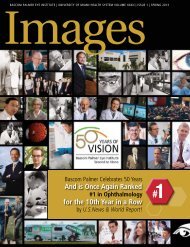
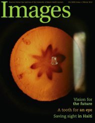
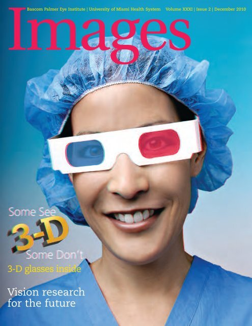
Images<br />
<strong>Bascom</strong> <strong>Palmer</strong> <strong>Eye</strong> <strong>Institute</strong> | University of Miami Health System Volume XXXI | Issue 2 | December 2010<br />
3-D glasses inside<br />
<strong>Vision</strong> <strong>research</strong><br />
<strong>for</strong> <strong>the</strong> <strong>future</strong>
<strong>Bascom</strong> <strong>Palmer</strong> <strong>Eye</strong> <strong>Institute</strong>’s mission is to enhance <strong>the</strong><br />
quality of life by improving sight, preventing blindness, and<br />
advancing ophthalmic knowledge through compassionate<br />
patient care and innovative vision <strong>research</strong>.<br />
Collaboration: The Key to <strong>Vision</strong> Research 2<br />
Meet our Scientists 6<br />
3-D: Some see it,<br />
some don’t. Do you 11<br />
10<br />
Announcements 22<br />
Awards and Honors 24<br />
<strong>Bascom</strong> <strong>Palmer</strong> Worldwide 26<br />
Profiles in Philanthropy 32<br />
23<br />
30<br />
Editor’s note: This edition of Images contains three dimensional (3-D) photographs, which may<br />
appear blurry without <strong>the</strong> use of <strong>the</strong> enclosed special glasses.<br />
<strong>Bascom</strong> <strong>Palmer</strong>’s advances on 3-D imaging to better diagnose and treat eye disease,<br />
coupled with <strong>the</strong> recent popularity of 3-D movies, television and magazines, were <strong>the</strong> exciting<br />
impetus <strong>for</strong> this issue. Special thanks to Dr. Douglas Anderson, who <strong>for</strong> fifty-plus years has<br />
been a world-renowned expert in <strong>the</strong> fields of glaucoma and stereo/3-D vision, and Derek<br />
Nankivil of <strong>Bascom</strong> <strong>Palmer</strong>’s Ophthalmic Biophysics Center, who fabricated a stereo-vision<br />
tripod adapter that was utilized to make <strong>the</strong> 3-D photographs in this issue possible.<br />
Your glasses are inside — put <strong>the</strong>m on and enjoy. — Marla Bercuson<br />
On <strong>the</strong> cover:<br />
Wendy Lee, M.D., M.S., oculoplastic specialist and assistant professor of clinical ophthalmology
Dear Friends and Colleagues:<br />
2010 has been a year of growth and achievement <strong>for</strong> <strong>Bascom</strong> <strong>Palmer</strong> <strong>Eye</strong> <strong>Institute</strong>. In this issue<br />
of Images, we are pleased to introduce you to Dr. Vittorio Porciatti, our new director and vice chair of<br />
<strong>research</strong> along with some members of his scientific team who are working to better understand <strong>the</strong><br />
underlying causes of eye disease, prevent blindness, and maintain vision in older patients. <strong>Bascom</strong> <strong>Palmer</strong><br />
has a long history of leadership in ophthalmology to advance patient care, medical education and vision<br />
<strong>research</strong>. For example:<br />
From <strong>the</strong> Chairman<br />
n Our uveitis service, under <strong>the</strong> superb leadership of Dr. Janet Davis,<br />
was recently recognized as generating two of <strong>the</strong> ten most important<br />
papers on uveitis (an inflammatory disease of <strong>the</strong> eye) available <strong>for</strong><br />
United States resident training in 2009.<br />
n Dr. Stephen Schwartz was elected president of <strong>the</strong> Florida Society of<br />
Ophthalmology (FSO); Dr. David Samimi, one of our talented third-year<br />
residents, won first place <strong>for</strong> his <strong>research</strong> presentation at <strong>the</strong> annual<br />
FSO meeting; and Dr. David Goldman was recently elected to <strong>the</strong><br />
presidency of <strong>the</strong> Palm Beach County Ophthalmology Society.<br />
I am joined by Dr. Akef El-Maghraby (center), chairman of <strong>the</strong><br />
Magrabi Hospitals and Clinics, and <strong>Bascom</strong> <strong>Palmer</strong>’s Dr. David Tse<br />
(right). We are excited to conduct teleconferences <strong>for</strong> ophthalmic<br />
education and training with international institutions such as <strong>the</strong><br />
Magrabi group in <strong>the</strong> Middle East.<br />
n Two of our investigators received prestigious Research to Prevent<br />
Blindness Awards: Dr. Abigail Hackam — <strong>the</strong> Ernest & Elizabeth<br />
Althouse Scholar Award; and Dr. Jeffrey Goldberg — <strong>the</strong> Walt and Lilly<br />
Disney Award <strong>for</strong> Amblyopia Research. Dr. Goldberg has had his work<br />
published in Science and was named <strong>the</strong> Scientist of <strong>the</strong> Year by Hope<br />
<strong>for</strong> <strong>Vision</strong>.<br />
n Dr. Victor Perez continued to receive accolades <strong>for</strong> his pioneering surgery in <strong>the</strong> United States of a<br />
modified osteo-odonto keratopros<strong>the</strong>sis (artificial cornea).<br />
<strong>Bascom</strong> <strong>Palmer</strong> is dedicated to providing each of our patients with <strong>the</strong> best possible care. This includes<br />
our conversion to UChart – a new streamlined electronic medical records system. The first phase of<br />
UChart, including patient scheduling, registration and billing has been implemented and we expect <strong>the</strong><br />
system to be fully installed by October 2011.<br />
I am especially proud of this current issue of Images, which is ano<strong>the</strong>r example of <strong>the</strong> imagination,<br />
creativity and innovative thinking that is <strong>the</strong> hallmark of <strong>Bascom</strong> <strong>Palmer</strong>. Marla Bercuson, Images editor and<br />
<strong>Bascom</strong> <strong>Palmer</strong>’s director of marketing and communications, has published this superlative magazine that<br />
combines science and medicine with contemporary culture. The article “3-D: Some See It, Some Don’t”<br />
and its accompanying spectacular 3-D photographs explain how 3-D imaging is used by our physicians to<br />
diagnose and treat our patient’s eye disease. I hope that you will enjoy, and share, this unique issue.<br />
During this holiday season, I want to thank our many patients, friends and donors who make all of<br />
<strong>Bascom</strong> <strong>Palmer</strong>’s good work possible. I wish you and your loved ones a happy, healthy and prosperous 2011.<br />
Sincerely,<br />
Eduardo C. Alfonso, M.D.<br />
Kathleen and Stanley J. Glaser Chair in Ophthalmology<br />
Chairman, <strong>Bascom</strong> <strong>Palmer</strong> <strong>Eye</strong> <strong>Institute</strong><br />
1<br />
BASCOM PALMER EYE INSTITUTE
Research<br />
Collaboration<br />
is key to success<br />
Meet Vittorio Porciatti, D.Sc., <strong>Bascom</strong> <strong>Palmer</strong>’s<br />
Director and Vice Chair of Research<br />
Vittorio Porciatti, D.Sc., tenured <strong>research</strong> professor of<br />
ophthalmology, was recently named director and<br />
vice chair of <strong>research</strong> <strong>for</strong> <strong>Bascom</strong> <strong>Palmer</strong> <strong>Eye</strong> <strong>Institute</strong>.<br />
A neuroscientist, electrophysiologist and biophysicist,<br />
his current <strong>research</strong> focuses on prevention of glaucoma.<br />
BASCOM PALMER EYE INSTITUTE<br />
2<br />
Porciatti, who joined <strong>Bascom</strong> <strong>Palmer</strong>’s faculty in 2001, received his education<br />
and training at <strong>the</strong> University of Pisa, Italy. He served <strong>for</strong> many years as senior<br />
scientist and a member of <strong>the</strong> scientific committee at <strong>the</strong> <strong>Institute</strong> of<br />
Neurophysiology at <strong>the</strong> Italian Research Council. He has published and<br />
lectured extensively.<br />
What is your long-term vision <strong>for</strong> <strong>research</strong> at <strong>Bascom</strong> <strong>Palmer</strong><br />
<strong>Bascom</strong> <strong>Palmer</strong> has been <strong>the</strong> number one eye institute on <strong>the</strong> clinical side <strong>for</strong><br />
many years. In <strong>research</strong>, we are moving up <strong>the</strong> ladder, in terms of both studies<br />
and grants, but we are not yet number one. In <strong>the</strong> past few years, we have<br />
increased in size from half a dozen scientific <strong>research</strong>ers to more than 25 in our<br />
program based at <strong>Bascom</strong> <strong>Palmer</strong>’s Evelyn F. and William L. McKnight <strong>Vision</strong><br />
Research Center. That’s a major advance, just in terms of growth. Now, we are<br />
considered one of <strong>the</strong> top 10 ophthalmology <strong>research</strong> laboratories in <strong>the</strong><br />
country. My vision is <strong>for</strong> us to continue moving up in <strong>the</strong> national rankings as<br />
we contribute to better understanding of ophthalmic diseases and disorders<br />
and <strong>the</strong> ways to treat or cure <strong>the</strong>m.<br />
What challenges do you face<br />
With our recent growth, we have virtually exhausted <strong>the</strong> existing laboratory<br />
space in <strong>the</strong> McKnight Research Center. This limits our investigators to expand<br />
<strong>the</strong>ir programs and hinders our ability to hire new investigators. Under <strong>the</strong><br />
direction of Eduardo C. Alfonso, M.D., chair of <strong>Bascom</strong> <strong>Palmer</strong>, we are in <strong>the</strong><br />
process of converting administrative offices into much-needed laboratory space<br />
which we will need to equip and staff.
How and from where do <strong>Bascom</strong> <strong>Palmer</strong>’s<br />
<strong>research</strong>ers receive funding<br />
Getting funds during an economic downturn is<br />
challenging, but we are successfully increasing our<br />
<strong>research</strong> grants and donor funding. The National<br />
<strong>Institute</strong>s of Health (NIH) is a major source of funding<br />
<strong>for</strong> our <strong>research</strong> projects. We are also supported by<br />
various private foundations and individual donors. In<br />
addition to external funding, we, as <strong>the</strong> department<br />
of ophthalmology, invest in our own <strong>research</strong>, such as<br />
covering a portion of a young investigator’s salary. Or,<br />
we may contribute to <strong>the</strong> purchase and maintenance<br />
of expensive equipment that is beyond <strong>the</strong> reach of<br />
individual investigators. This internal support is critical<br />
to our success.<br />
What are your <strong>research</strong> priorities<br />
Our physicians are very skilled in <strong>the</strong>ir clinical fields.<br />
They have accomplished remarkable feats in repairing<br />
<strong>the</strong> eye and restoring vision, even in patients with very<br />
difficult visual conditions. Our laboratory <strong>research</strong>ers<br />
work to better understand <strong>the</strong> underlying causes of<br />
eye diseases to prevent blindness and maintain useful<br />
vision in older patients. For example, using <strong>the</strong> tools<br />
of 21st century medicine – including genetics, cellular<br />
biology, molecular diagnostics and advanced imaging–<br />
we are trying to understand why <strong>the</strong> eye may become<br />
susceptible to diseases like glaucoma or macular<br />
degeneration, and how biotechnologies may help in<br />
preventing such conditions.<br />
Could you expand on that goal<br />
Pascal Goldschmidt, M.D., dean of <strong>the</strong> University of<br />
Miami Miller School of Medicine, noted recently in his<br />
blog that <strong>for</strong> most chronic illnesses, humans are born<br />
with genetic susceptibilities. At some point in life –<br />
perhaps influenced by environmental factors, such as<br />
nutrition, smoking or stress – <strong>the</strong> disease may reach a<br />
“tipping point,” when <strong>the</strong> body can no longer repair<br />
that cell and tissue damage; <strong>the</strong> disease becomes<br />
symptomatic and progresses at a fast rate. By that<br />
time, various <strong>the</strong>rapies can ease <strong>the</strong> patient’s<br />
symptoms and stabilize <strong>the</strong> condition, but cannot turn<br />
back <strong>the</strong> clock. In many of our studies at <strong>Bascom</strong><br />
<strong>Palmer</strong>, we are seeking to identify <strong>the</strong> biomarkers that<br />
may indicate whe<strong>the</strong>r or not an individual has reached<br />
that tipping point. In that way, we may be able to<br />
intervene clinically at an earlier stage and prevent<br />
damage to vision.<br />
3<br />
BASCOM PALMER EYE INSTITUTE
4Research<br />
BASCOM PALMER EYE INSTITUTE<br />
“I look<br />
<strong>for</strong>ward to<br />
building upon<br />
<strong>Bascom</strong><br />
<strong>Palmer</strong>’s<br />
tradition of<br />
collaboration<br />
and<br />
continuing to<br />
strive toward<br />
unparalleled<br />
ophthalmic<br />
discoveries.”<br />
— Vittorio Porciatti, D.Sc.<br />
Looking ahead, what progress in<br />
<strong>research</strong> do you see in <strong>the</strong> next<br />
five to ten years<br />
We are making substantial progress,<br />
although it takes considerable time to<br />
move from basic science at <strong>the</strong><br />
laboratory “bench,” through clinical<br />
models, to actual trials on patients. First<br />
of all, gene <strong>the</strong>rapy is now a reality in<br />
humans. As we learn more and more<br />
about genomics – our genetic blueprint<br />
– we will be able to recognize potential<br />
medical problems and develop new<br />
tools <strong>for</strong> treatment. Second, we are<br />
currently conducting clinical trials involving growth<br />
factors. By injecting <strong>the</strong>se compounds into <strong>the</strong><br />
eye, our investigators have been able to regrow<br />
damaged photoreceptors – a very important<br />
advance, according to <strong>the</strong> NIH, which is funding<br />
<strong>the</strong> <strong>research</strong>. Third, a better understanding of<br />
genetics will influence <strong>the</strong> diagnosis and<br />
treatment of macular degeneration. We will be<br />
able to better understand an individual’s prognosis<br />
and prescribe effective treatments. Fourth, we are<br />
studying <strong>the</strong> use of stem cells to regrow tissue in<br />
clinical models, and finally, we are looking at<br />
nanotechnology <strong>for</strong> tagging cells and steering<br />
<strong>the</strong>m to specific locations affected by <strong>the</strong> disease<br />
process. We expect that this may become a<br />
powerful tool in treating some diseases at <strong>the</strong><br />
cellular level.<br />
What is <strong>the</strong> difference between basic<br />
science and clinical science<br />
In medicine, basic science focuses on understanding<br />
biological processes at <strong>the</strong> root level,<br />
such as cellular, genetic and chemical interactions.<br />
Clinical science is concerned with translating <strong>the</strong><br />
breakthroughs at <strong>the</strong> laboratory level into new<br />
diagnostic and treatment <strong>the</strong>rapies. Traditional<br />
clinicians typically do not conduct basic science.<br />
However, we are <strong>for</strong>tunate to have a number of<br />
medical doctors at <strong>Bascom</strong> <strong>Palmer</strong> who have also<br />
been trained in basic <strong>research</strong>. They find it natural<br />
to think in terms of both basic and clinical<br />
<strong>research</strong>, and spend some days seeing patients<br />
and <strong>the</strong> o<strong>the</strong>r days working in <strong>the</strong> laboratory.<br />
What about translational <strong>research</strong> and how<br />
it may affect patient care<br />
Translational <strong>research</strong> aims to bring laboratory<br />
findings into <strong>the</strong> physician’s practice. At <strong>Bascom</strong><br />
<strong>Palmer</strong>, this is a two-way process, often sparked by<br />
our physicians’ clinical questions. They may ask:<br />
Why does a disease progress in a certain way Is<br />
<strong>the</strong>re a genetic predisposition to certain disorders<br />
How do <strong>the</strong> biochemical interactions within <strong>the</strong><br />
eye’s structure influence loss of vision That often<br />
leads our scientists in <strong>the</strong> laboratory to begin<br />
generating new in<strong>for</strong>mation, ideas and ways of<br />
thinking about a condition. Translational <strong>research</strong> is<br />
not just developing new biotechnology or<br />
identifying a problematic molecule – it’s changing<br />
<strong>the</strong> way clinicians think about a disease.<br />
What is unique about <strong>Bascom</strong> <strong>Palmer</strong>’s<br />
<strong>research</strong> program<br />
We have <strong>the</strong> strongest clinical program in <strong>the</strong><br />
nation, exceptional residency, fellowship and <strong>for</strong>eign<br />
observer training programs that attract physicians<br />
from around <strong>the</strong> world, and a large volume of<br />
patients. This ideal combination provides a steady<br />
flow of <strong>research</strong> ideas and helps generate a great<br />
number of translational <strong>research</strong> opportunities in<br />
areas like glaucoma, macular degeneration, corneal<br />
disease, retinal disorders, neuro-ophthalmology and<br />
o<strong>the</strong>r fields. Ano<strong>the</strong>r exceptional area that sets us<br />
apart is our Ophthalmic Biophysics Center, which<br />
has a stellar history of creating devices, instruments<br />
and technologies that help advance patient care.<br />
Are <strong>the</strong>re examples of that synergy<br />
Yes. When Dr. John R. Guy joined our faculty two<br />
years ago as professor of ophthalmology and basic<br />
<strong>research</strong>er on mitochondrial DNA, he was able to<br />
access a large pool of patients with Leber hereditary<br />
optic neuropathy who may be candidates <strong>for</strong> gene<br />
<strong>the</strong>rapy. There are many o<strong>the</strong>r examples of that<br />
crossover between clinical care and basic <strong>research</strong>.<br />
Dr. Jinhua (Jay) Wang, associate professor of<br />
ophthalmology and one of <strong>the</strong> world’s <strong>for</strong>emost dry<br />
eye <strong>research</strong>ers, is developing hardware <strong>for</strong> better<br />
imaging and quantifying optical coherence tomography<br />
images of eye structures. This will provide<br />
novel diagnostic tools to help doctors detect and<br />
monitor eye diseases and <strong>the</strong> effect of treatments.
Why is <strong>Bascom</strong> <strong>Palmer</strong>’s<br />
commitment to<br />
collaboration in <strong>research</strong>,<br />
bioengineering and<br />
clinical care so important<br />
Having clinical specialists in<br />
many disciplines of<br />
ophthalmology toge<strong>the</strong>r in one<br />
place leads to a continual<br />
sharing of ideas and insights. In<br />
addition to spurring basic<br />
scientific <strong>research</strong>, it leads to advances in medical<br />
technology. A prime example is <strong>the</strong> creation of a<br />
visual pros<strong>the</strong>sis made with a patient’s canine tooth<br />
or “eyetooth.” You may recall that last year, Dr.<br />
Victor Perez per<strong>for</strong>med <strong>the</strong> first surgery of its kind<br />
in <strong>the</strong> United States when he restored a woman’s<br />
sight using her eyetooth. That remarkable<br />
procedure required his expertise as an ophthalmic<br />
surgeon, complemented by an oral surgeon, our<br />
bioengineering team led by Dr. Jean-Marie Parel,<br />
who designed <strong>the</strong> pros<strong>the</strong>tic lens, and our <strong>research</strong><br />
expertise in cellular biology and optics. Without that<br />
combination, this clinical advance would not have<br />
been possible.<br />
How are you building awareness of<br />
<strong>Bascom</strong> <strong>Palmer</strong>’s <strong>research</strong><br />
Our <strong>research</strong>ers participate in a number of<br />
medical education programs, including our weekly<br />
grand rounds at <strong>the</strong> hospital, our continuing<br />
medical education programs and <strong>research</strong> seminars.<br />
Our scientists also travel around <strong>the</strong> world to<br />
present <strong>the</strong>ir findings and take an active role in <strong>the</strong><br />
Association <strong>for</strong> Research in <strong>Vision</strong> and<br />
Ophthalmology, which is <strong>the</strong> world’s largest<br />
organization devoted to sight-saving <strong>research</strong>. In<br />
addition, <strong>Bascom</strong> <strong>Palmer</strong>’s website is continually<br />
updated to display our investigators’ projects. We<br />
have found that <strong>research</strong>ers around <strong>the</strong> world can<br />
visit bascompalmer.org to see what we are working<br />
on, and tap into that expertise if appropriate. And,<br />
of course, we are certainly open to collaboration<br />
with o<strong>the</strong>r institutes.<br />
Tell us about your own <strong>research</strong> interests<br />
and activities.<br />
My focus is on <strong>the</strong> early detection of glaucoma –<br />
determining an individual’s susceptibility to this<br />
disease be<strong>for</strong>e <strong>the</strong> “tipping point” is reached. We<br />
have tools <strong>for</strong> evaluating <strong>the</strong> cells of <strong>the</strong> retina and<br />
measuring if <strong>the</strong> electrical activity<br />
<strong>the</strong>y generate is normal or not.<br />
This is similar to using a treadmill<br />
<strong>for</strong> a stress test in cardiology<br />
patients. My primary goal is to<br />
prevent <strong>the</strong> onset of glaucoma,<br />
improve <strong>the</strong> patient’s quality of<br />
life and reduce costs to <strong>the</strong><br />
overall healthcare system. My<br />
mo<strong>the</strong>r, Anna, is virtually blind<br />
from glaucoma, and this is a<br />
personal quest <strong>for</strong> me.<br />
Can you provide more details on your<br />
<strong>research</strong><br />
I received a $1.9 million grant from <strong>the</strong> NIH/<br />
National <strong>Eye</strong> <strong>Institute</strong> <strong>for</strong> <strong>the</strong> project, “Reversible<br />
Dysfunction of Retinal Ganglion Cells in Glaucoma.”<br />
Glaucoma causes progressive damage and death of<br />
retinal ganglion cells (RGCs) resulting in blindness.<br />
My objective is to see if we can prevent RGC death<br />
in <strong>the</strong> early stages of glaucoma. Our <strong>research</strong> team<br />
includes Dr. Lori Ventura, who serves as my coprincipal<br />
investigator, and experts in glaucoma,<br />
electrophysiology, biomedical engineering,<br />
biophysics and biostatistics.<br />
How did you first become interested in<br />
biomedical <strong>research</strong><br />
After my undergraduate education in chemistry,<br />
I worked as a technician in a nuclear chemistry<br />
laboratory in Pisa, Italy. I became interested in<br />
biology and graduated from <strong>the</strong> University of Pisa,<br />
majoring in neurophysiology under <strong>the</strong> mentorship<br />
of one of <strong>the</strong> great pioneers of <strong>the</strong> field, Giuseppe<br />
Moruzzi. When <strong>the</strong> local eye clinic needed a visual<br />
physiologist, I started working hands-on with<br />
hospital patients each morning and spent<br />
afternoons in <strong>the</strong> experimental laboratory. This<br />
represented a very <strong>for</strong>mative and productive period.<br />
As my <strong>research</strong> became acknowledged, I began<br />
collaborating with national and international<br />
institutions and began teaching visual neuroscience<br />
in Pisa and Rome. Research has always been at <strong>the</strong><br />
heart of my career. I am honored to have <strong>the</strong><br />
privilege to work with such esteemed colleagues<br />
here at <strong>Bascom</strong> <strong>Palmer</strong>. I look <strong>for</strong>ward to building<br />
upon <strong>Bascom</strong> <strong>Palmer</strong>’s tradition of collaboration and<br />
continuing to strive toward unparalleled ophthalmic<br />
discoveries. Toge<strong>the</strong>r we will help pave <strong>the</strong> way <strong>for</strong><br />
<strong>Bascom</strong> <strong>Palmer</strong>’s contribution in revolutionary areas<br />
of study and scientific <strong>research</strong>. n<br />
5Research<br />
BASCOM PALMER EYE INSTITUTE
Research<br />
<strong>Bascom</strong> <strong>Palmer</strong>’s scientific <strong>research</strong><br />
Frontiers of <strong>Vision</strong><br />
William L. McKnight, <strong>the</strong> legendary leader of Minnesota Mining and Manufacturing Company<br />
(3M), valued <strong>research</strong> as “a key to tomorrow.” As a result of McKnight’s generosity, <strong>Bascom</strong><br />
<strong>Palmer</strong>’s Evelyn F. and William L. McKnight <strong>Vision</strong> Research Center embodies his enthusiastic<br />
support of <strong>research</strong> on <strong>the</strong> nervous system and <strong>the</strong> eye as a “window to <strong>the</strong> brain and body.”<br />
Primarily based in <strong>the</strong> McKnight Center, <strong>Bascom</strong> <strong>Palmer</strong>’s scientists advance <strong>the</strong> frontiers of<br />
ophthalmology. Meet some of <strong>the</strong>m now as <strong>the</strong>y describe <strong>the</strong>ir work – in <strong>the</strong>ir own words.<br />
Jeffrey L. Goldberg, M.D., Ph.D.<br />
Associate Professor of Ophthalmology<br />
In my laboratory <strong>research</strong>, I am studying why<br />
neurons in <strong>the</strong> retina, particularly<br />
photoreceptors and retinal ganglion cells that<br />
transmit in<strong>for</strong>mation from <strong>the</strong> retina to <strong>the</strong><br />
brain, fail to regenerate <strong>the</strong>mselves in<br />
degenerative diseases like macular<br />
degeneration and glaucoma. I am looking at<br />
two potentially promising strategies <strong>for</strong><br />
cellular regeneration. The first involves<br />
harvesting a patient’s adult retinal stem cells<br />
from <strong>the</strong>ir peripheral retina, growing <strong>the</strong>m in<br />
<strong>the</strong> laboratory and reimplanting <strong>the</strong>m back<br />
into <strong>the</strong> patient as retinal neurons. The<br />
second approach is applying nanotechnology<br />
to ocular repair - using magnetic<br />
nanoparticles to deliver stem cells to <strong>the</strong> back<br />
of <strong>the</strong> eye or to encourage retinal ganglion<br />
cells to grow through <strong>the</strong> optic nerve.<br />
BASCOM PALMER EYE INSTITUTE<br />
6<br />
M. Livia Bajenaru, Ph.D.<br />
Research Assistant Professor of Ophthalmology<br />
My <strong>research</strong> interests are cellular biology and anatomy.<br />
I am focused on finding <strong>the</strong>rapeutic treatments <strong>for</strong> optic<br />
neuropathies, such as glaucoma, and <strong>for</strong> ocular tumors,<br />
such as optic pathways gliomas and retinoblastomas,<br />
which may occur in children. I am using biochemical and<br />
pharmacological tools and experimental models to identify<br />
cell-signaling pathways and molecular targets in retinal<br />
ganglion cells, and in <strong>the</strong> supporting star-shaped glial cells<br />
(astrocytes). Using a sophisticated contact lens model, I am<br />
also studying <strong>the</strong> molecular development of infectious<br />
keratitis associated with <strong>the</strong> use of contact lenses.
Abigail S. Hackam, Ph.D.<br />
Associate Professor of Ophthalmology<br />
My laboratory investigates genes and cellular pathways that lead to<br />
photoreceptor degeneration, including age-related macular<br />
degeneration and retinitis pigmentosa. Our main <strong>research</strong> focus is to<br />
identify novel treatments that increase photo-receptor survival. A<br />
major component of this <strong>research</strong> is determining <strong>the</strong> role of a cell type<br />
called glia during retinal disease. We recently identified a class of<br />
molecules called “Wnts” that protect photoreceptors from dying and<br />
are active in glial cells. We are currently investigating how Wnts work<br />
and determining whe<strong>the</strong>r <strong>the</strong>y can be utilized as a novel <strong>the</strong>rapeutic<br />
strategy <strong>for</strong> retinal degeneration.<br />
George Inana, M.D., Ph.D.<br />
Professor of Ophthalmology<br />
My <strong>research</strong> has focused on understanding and curing age-related<br />
macular degeneration (AMD) and o<strong>the</strong>r retinal degenerative diseases<br />
(RD) through molecular genetic approaches. Notable achievements of<br />
our team over <strong>the</strong> years have included cloning of <strong>the</strong> first inherited<br />
retinal degeneration gene<br />
(<strong>for</strong> gyrate atrophy),<br />
identification of three novel<br />
gene targets <strong>for</strong> RD and,<br />
most recently, identifying<br />
many new candidate genes<br />
<strong>for</strong> AMD by a novel genescreening<br />
approach. An<br />
experimental model based<br />
on one of <strong>the</strong>se genes,<br />
MMP14, is being used <strong>for</strong><br />
<strong>the</strong>rapeutic testing. We have<br />
achieved very promising<br />
preliminary results,<br />
demonstrating that <strong>the</strong>rapy<br />
<strong>for</strong> wet AMD based on this<br />
gene may be superior or<br />
complementary to <strong>the</strong> anti-<br />
VEGF <strong>the</strong>rapy available today.<br />
Giovanni Gregori, Ph.D.<br />
Research Assistant Professor of Ophthalmology<br />
We are making great progress in <strong>the</strong> development of<br />
new retinal imaging tools, focusing in particular on<br />
spectral domain optical coherence tomography, a<br />
relatively new technology that can produce threedimensional<br />
images of <strong>the</strong> retina with very high<br />
resolution. Our group has developed algorithms that<br />
<strong>for</strong> <strong>the</strong> first time allow <strong>the</strong> physician to visualize<br />
accurately <strong>the</strong> spatial geometry and anatomy of <strong>the</strong><br />
retina. This makes it possible to measure small retinal<br />
features and accurately monitor <strong>the</strong>ir changes over<br />
time, a crucial step in evaluating <strong>the</strong> need <strong>for</strong><br />
treatment or <strong>the</strong> effectiveness of different treatment<br />
programs. Using this approach, we have developed<br />
important insights into <strong>the</strong> disease processes<br />
associated with age-related macular degeneration.<br />
Yiwen Li, M.D.<br />
Research Assistant Professor of Ophthalmology<br />
My <strong>research</strong> is focused<br />
on <strong>the</strong> degeneration of<br />
cone cells in <strong>the</strong> retina<br />
of <strong>the</strong> eye. Cone cells are<br />
photoreceptors that are<br />
responsible <strong>for</strong> color and<br />
central vision and are<br />
very important <strong>for</strong> our<br />
daily activities. Cone cell<br />
death causes blindness<br />
in <strong>the</strong> late stages of retinal degeneration. I have<br />
characterized cone degeneration in experimental<br />
models and have discovered recently that it can be<br />
reversed by CNTF, a neurotrophic factor. I am<br />
currently testing o<strong>the</strong>r factors <strong>for</strong> <strong>the</strong>ir abilities to<br />
reverse cone degeneration. My <strong>research</strong> in<br />
photoreceptor cell biology, retinal vascular disorders<br />
and retinal degeneration could one day lead to new<br />
<strong>the</strong>rapies to save sight.<br />
7Research<br />
BASCOM PALMER EYE INSTITUTE
8Research<br />
BASCOM PALMER EYE INSTITUTE<br />
Rong Wen, M.D., Ph.D.<br />
Associate Professor of Ophthalmology<br />
The long-term objectives of my <strong>research</strong> are to understand <strong>the</strong><br />
mechanism of retinal degeneration and to develop new <strong>the</strong>rapies to stop<br />
this blinding disease. As a retinal cell biologist, I want to understand why<br />
photoreceptors – <strong>the</strong> light-sensing neurons – die prematurely in retinal<br />
degeneration. Photoreceptors renew <strong>the</strong>ir light-sensing devices every 10<br />
days, an energy-consuming process. I have developed a novel technology<br />
to measure this renewal rate in order to understand how it is affected by<br />
different pathological conditions. I believe this is a key to understanding<br />
this process and could lead to <strong>the</strong> development of novel treatments <strong>for</strong><br />
retinal degeneration.<br />
Sanjoy K. Bhattacharya, Ph.D.<br />
Associate Professor of Ophthalmology<br />
My <strong>research</strong> concentrates on <strong>the</strong> cell biology of <strong>the</strong> trabecular meshwork,<br />
an area of tissue in <strong>the</strong> eye located between <strong>the</strong> iris and <strong>the</strong> cornea<br />
responsible <strong>for</strong> draining <strong>the</strong> aqueous fluid from <strong>the</strong> eye. Aqueous is <strong>the</strong><br />
clear liquid that ba<strong>the</strong>s and supports <strong>the</strong> inside of <strong>the</strong> eye. An imbalance<br />
in aqueous outflow through <strong>the</strong> tabecular meshwork results in elevation<br />
of intraocular pressure, which damages <strong>the</strong> optic nerve and is frequently<br />
associated with glaucoma. We have discovered how <strong>the</strong> protein “cochlin”<br />
affects <strong>the</strong> outflow of fluid and is critical in controlling intraocular<br />
pressure. We are studying how cochlin may contribute to glaucoma and<br />
how to reverse its damaging effects.<br />
Mitra Sehi, O.D., M.S., Ph.D., F.A.A.O.<br />
Research Assistant Professor of Ophthalmology<br />
My <strong>research</strong> is focused on detection of <strong>the</strong> earliest<br />
signs of damage to retinal nerve cells and <strong>the</strong>ir<br />
axons in glaucoma in order to intervene be<strong>for</strong>e <strong>the</strong><br />
retinal nerve cells die. Advanced technologies <strong>for</strong><br />
eye <strong>research</strong> are used to detect <strong>the</strong> earliest signs of<br />
glaucoma-related damage to <strong>the</strong> optic nerve head<br />
and nerve cell axons. We use advanced ocular<br />
imaging, electrophysiological techniques, and<br />
ma<strong>the</strong>matical models to evaluate <strong>the</strong> impact of<br />
damage due to glaucoma on <strong>the</strong> structure and<br />
function of <strong>the</strong> visual system over time.<br />
Maria E. Marin-Castaño, M.D., Ph.D.<br />
Research Associate Professor of Ophthalmology<br />
Little is known about <strong>the</strong> prevention of agerelated<br />
macular<br />
degeneration, <strong>the</strong><br />
leading cause of severe<br />
visual impairment<br />
among elderly persons<br />
worldwide. My <strong>research</strong><br />
focuses on understanding<br />
and explaining <strong>the</strong><br />
origin and development of AMD, and <strong>the</strong>n applies<br />
that knowledge to identify more effective<br />
preventive strategies and <strong>the</strong>rapeutic approaches.<br />
AMD arises from a complex interaction of genetic,<br />
systemic and environmental risk factors. I am<br />
focusing upon <strong>the</strong> roles played by cigarette<br />
smoking and hypertension, which are major risk<br />
factors <strong>for</strong> developing AMD. My <strong>research</strong> explores<br />
molecules that could be used as biological<br />
indicators in blood, urine and eye fluid to help<br />
predict which patients who smoke and have high<br />
blood pressure, will get AMD.
Wei Li, Ph.D.<br />
Assistant Professor of Ophthalmology<br />
Retinal aging is caused by accumulation of harmful<br />
metabolic products that may lead to age-related<br />
macular degeneration. Several of <strong>the</strong>se products<br />
have been identified and associated with retinal<br />
damage. Currently, <strong>the</strong>re is no <strong>the</strong>rapeutic<br />
intervention to prevent or minimize <strong>the</strong><br />
accumulation of <strong>the</strong>se substances. With my interest<br />
in molecular <strong>the</strong>rapeutics <strong>for</strong> eye diseases involving<br />
<strong>the</strong> immune system, I am investigating molecules<br />
that control <strong>the</strong> clearance of <strong>the</strong>se metabolic<br />
products by retinal pigment epi<strong>the</strong>lium cells. Our<br />
studies look at <strong>the</strong> <strong>the</strong>rapeutic potential of <strong>the</strong>se<br />
molecules to facilitate <strong>the</strong> clearance of <strong>the</strong>se<br />
products and to prevent retinal aging.<br />
Research<br />
Valery I. Shestopalov, Ph.D.<br />
Associate Professor of Ophthalmology<br />
Molecular mechanisms of <strong>the</strong> eye that trigger<br />
aberrant communication between various cells is<br />
Delia Cabrera DeBuc, Ph.D.<br />
Research Associate Professor of Ophthalmology<br />
My <strong>research</strong> focuses on physical and<br />
ma<strong>the</strong>matical modeling of retinal and corneal<br />
morphology as visualized by optical coherence<br />
tomography (OCT). I have introduced<br />
quantitative tools and measures into <strong>the</strong><br />
analysis of OCT images using basic principles<br />
from physics in order to quantify treatmentinduced<br />
changes in patients with ocular<br />
diseases. I am exploring <strong>the</strong> relationship between vitreoretinal disease<br />
and <strong>the</strong> internal architecture of <strong>the</strong> retinal structure in patients with<br />
diabetic retinopathy. My goal is to develop indicators <strong>for</strong> <strong>the</strong> early<br />
detection of diabetic retinopathy using OCT.<br />
<strong>the</strong> focus of my <strong>research</strong>. By using molecular<br />
genomics and bioin<strong>for</strong>matics, we analyze<br />
pathological alterations in <strong>the</strong> signaling between<br />
most vital cell types. Our <strong>research</strong> revealed <strong>the</strong><br />
roles that individual cells and molecules play in<br />
<strong>the</strong> resistance to ocular pathologies. One of my<br />
newest projects utilizes cutting-edge DNA<br />
sequencing to survey microbial pathogens at <strong>the</strong><br />
surface of <strong>the</strong> eye. We identified hundreds of<br />
bacterial species, some of which were never<br />
detected or were completely unknown to doctors.<br />
This scientific knowledge will ultimately be<br />
translated into clinical application <strong>for</strong> <strong>the</strong><br />
treatment of glaucoma, optic neuritis and retinal<br />
ischemia, eye infections, dry eye and allergies. n<br />
9<br />
BASCOM PALMER EYE INSTITUTE
WHAT IS A<br />
CLINICAL STUDY<br />
A clinical study, also known<br />
as a clinical trial, applies<br />
<strong>the</strong> scientific method to<br />
human health. Essentially,<br />
clinical studies combine<br />
medical <strong>research</strong> with<br />
patients. Most medical<br />
<strong>research</strong> begins in <strong>the</strong><br />
laboratory and when that<br />
<strong>research</strong> generates positive<br />
results that may benefit<br />
patients, it is translated to<br />
patient applications in <strong>the</strong><br />
<strong>for</strong>m of clinical trials or<br />
studies. An observational<br />
study, a type of clinical<br />
study, is one in which<br />
patients are observed and<br />
<strong>the</strong>ir outcomes are<br />
measured by doctors.<br />
Ano<strong>the</strong>r type of clinical<br />
study is an interventional<br />
study in which patients are<br />
assigned by <strong>the</strong> doctor to a<br />
treatment or o<strong>the</strong>r<br />
intervention, and <strong>the</strong>ir<br />
outcomes are measured.<br />
<strong>Bascom</strong> <strong>Palmer</strong> <strong>Eye</strong> <strong>Institute</strong><br />
has a long history of<br />
leading, participating in and<br />
sponsoring clinical trials to<br />
improve patient care.<br />
Do you have Dry Age-Related<br />
Macular Degeneration (AMD)<br />
You may qualify<br />
<strong>for</strong> a clinical<br />
study at<br />
<strong>Bascom</strong> <strong>Palmer</strong><br />
<strong>Eye</strong> <strong>Institute</strong><br />
in Palm Beach<br />
Gardens or Miami.<br />
<strong>Bascom</strong> <strong>Palmer</strong> macular<br />
degeneration specialists,<br />
Drs. Philip J. Rosenfeld and<br />
Sander Dubovy (in Miami)<br />
and Andrew Moshfeghi (in<br />
Palm Beach Gardens), are<br />
seeking volunteers <strong>for</strong> a study<br />
using an FDA-approved drug<br />
(off-label) named Eculizumab<br />
(SOLIRIS). Through this study,<br />
our doctors hope to provide a<br />
better treatment <strong>for</strong> dry AMD.<br />
BASCOM PALMER EYE INSTITUTE<br />
10<br />
WHAT ARE THE SYMPTOMS<br />
OF MACULAR<br />
DEGENERATION<br />
n Words appear blurry<br />
while reading<br />
n Inability to recognize<br />
faces at a distance<br />
n Blurred or blind spot in<br />
<strong>the</strong> center of vision<br />
n Straight lines appear<br />
wavy or crooked<br />
n Rapid loss of central<br />
vision<br />
To be included:<br />
nYour vision needs to be better than 20/100 in one eye.<br />
n You must be able to visit <strong>Bascom</strong> <strong>Palmer</strong> every week <strong>for</strong> four weeks, <strong>the</strong>n every two<br />
weeks <strong>for</strong> six months. Half <strong>the</strong> visits are one to two hours in duration, <strong>the</strong> o<strong>the</strong>r half<br />
are three to four hours long.<br />
n Your o<strong>the</strong>r eye can have wet or dry AMD.<br />
Presently, <strong>the</strong> only recommended treatments <strong>for</strong> dry AMD include vitamin/<br />
micronutrient supplementation and an appropriate diet which only slows <strong>the</strong><br />
progression of disease and vision loss. Eculizumab is administered as an intravenous<br />
infusion in a vein in <strong>the</strong> arm. Two out of three study patients get <strong>the</strong> drug. One out of<br />
three patients gets a placebo. Nei<strong>the</strong>r <strong>the</strong> doctors, nurses, nor patients will know who<br />
is receiving <strong>the</strong> drug or <strong>the</strong> placebo. After six months of <strong>the</strong>rapy, <strong>the</strong> infusions are<br />
stopped and patients are examined every three months.<br />
For more in<strong>for</strong>mation: contact Maria Esquiabro, clinical study coordinator, at<br />
mesquiabro@med.miami.edu or 305.326.6508. Visit bascompalmer.org and click on<br />
Clinical Studies <strong>for</strong> a description of <strong>the</strong> study. n
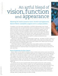
Dr. Douglas Anderson
When swinging a golf club,<br />
driving a car, or cooking dinner<br />
we need to judge <strong>the</strong> distance<br />
of objects near and far.<br />
In our everyday activities, we take depth perception <strong>for</strong> granted, not realizing that<br />
this is a complex brain process involving two functional eyes as well as many visual<br />
clues. For example, if someone in <strong>the</strong> distance obscures part of a hedge, your brain<br />
can determine that <strong>the</strong> hedge is fur<strong>the</strong>r away than <strong>the</strong> person.<br />
Through familiarity, <strong>the</strong> brain knows <strong>the</strong> typical<br />
size of people and common objects. When driving<br />
on <strong>the</strong> highway, we know that a tiny image of a car<br />
in <strong>the</strong> rear-view mirror means that vehicle is far<br />
away. When we look at a flat painting, our brain<br />
uses size and position in<strong>for</strong>mation to interpret<br />
“distances” created by <strong>the</strong> artist’s use of<br />
perspective.<br />
“When a friend starts walking toward you, you<br />
don’t perceive that person as getting bigger, but<br />
your brain interprets <strong>the</strong> enlarging image to mean<br />
<strong>the</strong> person is coming toward you,” explains Douglas<br />
R. Anderson, M.D.,FARVO, professor of<br />
ophthalmology and holder of <strong>the</strong> Douglas R.<br />
Anderson Chair in Ophthalmology. “We see in three<br />
dimensions (3-D): height, width, and depth, which<br />
can be ei<strong>the</strong>r distance or thickness.”<br />
Because our two eyes see objects from slightly<br />
different angles, <strong>the</strong> images <strong>the</strong>y send to <strong>the</strong> brain<br />
are slightly different. Through a process called<br />
“stereopsis,” <strong>the</strong> brain interprets those differences<br />
to help determine <strong>the</strong> distances to objects in our<br />
fields of vision. When objects are very far away,<br />
<strong>the</strong>re is very little distinction between <strong>the</strong> eye’s two<br />
images. There<strong>for</strong>e, we can’t perceive <strong>the</strong> varying<br />
distances of <strong>the</strong> stars and planets. At moderate<br />
distances, such as walking through a room or<br />
(Photo at left) The development of a mechanized remotelyoperated<br />
slit lamp biomicroscope (fondly called “Don’s<br />
Drone”) may greatly improve access to specialists <strong>for</strong> remote<br />
patients providing <strong>the</strong>m with better and more af<strong>for</strong>dable<br />
eyecare. The system, conceived by Dr. Donald Budenz, and<br />
fabricated by Dr. Jean-Marie Parel (shown), permits remote<br />
3-D stereo-viewing of <strong>the</strong> patient’s eye with <strong>the</strong> full<br />
functionality of a standard slip lamp. Made possible<br />
through <strong>the</strong> support of <strong>the</strong> Department of Defense, this<br />
project is part of a growing paradigm of teleophthalmolgy<br />
and Defense-sponsored projects <strong>for</strong> <strong>Bascom</strong> <strong>Palmer</strong>.<br />
parking a car, stereopsis supplements o<strong>the</strong>r visual<br />
cues. In general, stereopsis provides <strong>the</strong> greatest help<br />
when viewing nearby items, such as when sewing or<br />
cooking. Such tasks are more difficult without<br />
normal binocular (two-eye) vision because monocular<br />
(one-eye) vision does not provide those two slightly<br />
contrasting images to our brains.<br />
“Depth perception is thus a ra<strong>the</strong>r complex<br />
process,” adds Anderson. To see <strong>the</strong> importance of<br />
stereopsis, try to per<strong>for</strong>m this simple task: first, cover<br />
or close one eye. Then try to bring toge<strong>the</strong>r <strong>the</strong><br />
index fingers of your two hands with your elbows<br />
moderately bent. It’s very difficult to get those fingers<br />
to meet without binocular vision.<br />
Many animals, like fish, horses, cattle or deer, have<br />
depth perception only from non-stereoscopic clues.<br />
Their eyes are located toward <strong>the</strong> side of <strong>the</strong> face,<br />
giving <strong>the</strong>m a broader field of vision to detect<br />
predators. However, <strong>the</strong> two eyes do not see <strong>the</strong><br />
same objects at differing angles to provide stereopsis.<br />
That difference underscores <strong>the</strong> importance of<br />
stereopsis in humans.<br />
At <strong>Bascom</strong> <strong>Palmer</strong> <strong>Eye</strong> <strong>Institute</strong>, our clinicians<br />
strive to preserve binocular vision whenever possible,<br />
allowing stereoscopic depth perception of near<br />
objects. Our physicians also use a variety of<br />
instruments to measure depth and distance. For<br />
example, highly sophisticated diagnostic tools can<br />
measure how long it takes <strong>for</strong> an ultrasound pulse or<br />
focused beam of light to be reflected back to <strong>the</strong><br />
instrument. This in<strong>for</strong>mation-ga<strong>the</strong>ring process –<br />
which is made possible by powerful computing<br />
technology – allows our clinicians to create 3-D<br />
images of <strong>the</strong> eye and identify changes caused by<br />
diseases. In addition, our biomedical <strong>research</strong>ers have<br />
been leaders in <strong>the</strong> development of innovative 3-D<br />
“<br />
Seeing in three<br />
dimensions –<br />
or depth<br />
perception –<br />
is a ra<strong>the</strong>r<br />
complex<br />
process. Most<br />
people take<br />
<strong>the</strong>ir binocular<br />
vision <strong>for</strong><br />
granted –<br />
until <strong>the</strong>y try<br />
to per<strong>for</strong>m a<br />
simple task<br />
that involves<br />
depth perception,<br />
like trying<br />
to touch <strong>the</strong><br />
index fingers<br />
on each hand<br />
with one<br />
eye closed.”<br />
— Douglas R. Anderson, M.D.<br />
3-D <strong>Vision</strong><br />
13<br />
BASCOM PALMER EYE INSTITUTE
3-D <strong>Vision</strong><br />
BASCOM PALMER EYE INSTITUTE<br />
14<br />
imaging technologies and techniques designed to<br />
improve patient care around <strong>the</strong> world.<br />
This issue of Images takes an in-depth look at 3-D<br />
vision of <strong>the</strong> eyes. It also illustrates how <strong>Bascom</strong> <strong>Palmer</strong>’s<br />
clinical and <strong>research</strong> teams continue to advance<br />
<strong>the</strong> frontiers of medicine by improving 3-D<br />
methods to observe <strong>the</strong> eye’s structure and<br />
examine diseased regions.<br />
An In-Depth Look at 3-D <strong>Vision</strong><br />
Stereopsis is <strong>the</strong> special effect used to create<br />
<strong>the</strong> 3-D movie “Avatar” and o<strong>the</strong>r blockbuster<br />
films. It also accounts <strong>for</strong> <strong>the</strong> arrival of<br />
3-D television, <strong>the</strong> creation of 3-D<br />
commercials <strong>for</strong> special events like <strong>the</strong><br />
World Cup, and 3-D photographs that<br />
literally jump off <strong>the</strong> magazine page. Today, “3-D”<br />
is a big part of <strong>the</strong> multimedia entertainment<br />
industry. By wearing an inexpensive set of red and<br />
blue filters, such as those included in this issue of<br />
Images, your eyes can convert a fuzzy-looking<br />
multicolor photograph or video into a sharp image with<br />
depth. The filters allow each eye to see only one of <strong>the</strong><br />
two images, as <strong>the</strong> stereopsis enhances and emphasizes<br />
<strong>the</strong> o<strong>the</strong>r depth-related visual clues.<br />
Creating Stereoscopic 3-D Images<br />
In 1838, British scientist Sir Charles Wheatstone<br />
explained stereopsis in detail and constructed <strong>the</strong> first<br />
stereoscope. Lenses and mirrors combined two different<br />
photographs into one “solid”<br />
image. A few decades<br />
later, ano<strong>the</strong>r British<br />
inventor, David<br />
Brewster, developed<br />
<strong>the</strong> prism stereoscope and<br />
touched off a craze <strong>for</strong><br />
“stereogram” photos. In<br />
Stereoscope courtesy of<br />
Dr. Louis Goldszer, optometrist,<br />
retired, Pittsburgh, PA and<br />
Dr. Robert Goldszer, chief<br />
medical officer, Mount Sinai<br />
Medical Center, Miami Beach<br />
<strong>the</strong> 20th century, American<br />
children enjoyed 3-D images<br />
of scenes around <strong>the</strong> world<br />
using <strong>the</strong>ir ViewMaster<br />
stereoscopes.<br />
Meanwhile, Hollywood made 3-D movies using glasses<br />
with red and blue filters to create a sense of depth.<br />
Although <strong>the</strong> first three-dimension motion picture<br />
feature, “The Power of Love,” was made in 1922, only<br />
in <strong>the</strong> 1950s did 3-D movies catch on with <strong>the</strong> public, as<br />
fire-breathing monsters, rocket ships and speeding<br />
bullets appeared to fly right at <strong>the</strong> audience, pulling<br />
patrons right into <strong>the</strong> action.<br />
In a major advance, filmmakers in <strong>the</strong> ’50s began using<br />
polarization, ra<strong>the</strong>r than red-blue filters, to produce movies<br />
with more vivid, natural color. Today’s 3-D movies are filmed<br />
with two synchronized cameras taking slightly different views<br />
of <strong>the</strong> same scene, each seen with a polarized<br />
filter at a separate orientation. Moviegoers wear<br />
glasses with polarized filters at orientations that<br />
permit each eye to see only one image. The result<br />
is a more com<strong>for</strong>table “big-screen” experience,<br />
as shown by attendance of large audiences<br />
at 3-D IMAX <strong>the</strong>aters. A fur<strong>the</strong>r advance<br />
is emerging with 3-D television. The<br />
screen shows alternating images<br />
very rapidly, and <strong>the</strong> filters are<br />
synchronized so that one<br />
eye sees every o<strong>the</strong>r<br />
image. These special<br />
effects may be particularly<br />
striking with large images<br />
that move <strong>for</strong>ward on <strong>the</strong><br />
screen toward <strong>the</strong> viewer.<br />
Helping Children Develop 3-D <strong>Vision</strong><br />
Newborn babies see <strong>the</strong> world as a confusing place, filled<br />
with blurry, indistinct shapes and washed-out colors. It takes<br />
time <strong>for</strong> an infant’s eyes to learn how to focus on objects near<br />
and far, and <strong>for</strong> <strong>the</strong> brain to interpret <strong>the</strong> visual in<strong>for</strong>mation it<br />
receives through <strong>the</strong> optic nerves.<br />
However, in 2 to 4 percent of infants, <strong>the</strong> two eyes are<br />
misaligned – a disorder known as strabismus (called “squint”<br />
in <strong>the</strong> United Kingdom). Without early treatment, a child with<br />
strabismus may never develop stereopsis. They depend on<br />
o<strong>the</strong>r clues to judge distance. “Many individuals are not aware<br />
<strong>the</strong>y lack stereopsis, and assume that <strong>the</strong>ir vision is <strong>the</strong> same as<br />
in o<strong>the</strong>r people,” says Craig A. McKeown, M.D., professor of<br />
clinical ophthalmology and a specialist in pediatric disorders of<br />
<strong>the</strong> eye.<br />
“Newborn babies move from poor vision to nearly adult<br />
levels of vision by age 3,” he adds. “To develop good vision,<br />
children have to use <strong>the</strong>ir eyes. For <strong>the</strong> brain to have <strong>the</strong> ability<br />
of seeing with stereopsis, <strong>the</strong> eyes need to be well aligned and<br />
used toge<strong>the</strong>r during <strong>the</strong> first few years of life when humans<br />
develop <strong>the</strong>ir binocular vision.”<br />
Misaligned eyes have different images. If <strong>the</strong> brain of an<br />
infant is unable to merge <strong>the</strong>m to create stereoscopic<br />
appreciation of depth, <strong>the</strong> brain’s attention is turned away<br />
from one image to avoid double vision. If <strong>the</strong> brain occasionally<br />
switches attention from one eye to <strong>the</strong> o<strong>the</strong>r, each eye will<br />
(Photo at right)<br />
Dr. Craig McKeown and <strong>the</strong> team of <strong>Bascom</strong> <strong>Palmer</strong> pediatric<br />
ophthalmologists treat more than 7,000 children each year.
BASCOM PALMER EYE INSTITUTE<br />
16<br />
The Four<br />
Dimensions<br />
of Life<br />
In ma<strong>the</strong>matics<br />
and science,<br />
<strong>the</strong> dimensions<br />
of life have very<br />
specific meanings:<br />
1-D<br />
Length:<br />
Picture a dot or a<br />
long line stretching<br />
out to infinity.<br />
2-D<br />
Height and width:<br />
Like a photo or<br />
painting, a twodimensional<br />
image<br />
covers a flat area.<br />
However, visual cues<br />
within <strong>the</strong> image –<br />
such as smaller<br />
figures in <strong>the</strong><br />
background – can<br />
create <strong>the</strong> illusion<br />
of depth.<br />
Height, width<br />
and depth:<br />
Using various<br />
stereopsis techniques,<br />
two slightly different<br />
images can be<br />
brought into<br />
alignment so <strong>the</strong> eyes<br />
perceive a 3-D image.<br />
Height, width, depth<br />
and time:<br />
A series of 3-D<br />
images taken at<br />
different times can<br />
provide a physician<br />
with important<br />
in<strong>for</strong>mation about <strong>the</strong><br />
progress of an eye<br />
disease or o<strong>the</strong>r<br />
condition.<br />
have good vision, but both eyes will not be used<br />
simultaneously. If <strong>the</strong> brain habitually ignores one<br />
eye in favor of <strong>the</strong> o<strong>the</strong>r, it never develops full visual<br />
potential, a condition called “amblyopia.”<br />
“On any given day, <strong>Bascom</strong> <strong>Palmer</strong>’s William and<br />
Norma Horvitz Children’s Center is filled with<br />
children who have ei<strong>the</strong>r limited or no stereoscopic<br />
capabilities,” says McKeown. “We see many children<br />
with strabismus, amblyopia, and o<strong>the</strong>r problems so<br />
that only one eye can see well. We also see adults<br />
with similar conditions.”<br />
While <strong>the</strong>re can be many causes <strong>for</strong> strabismus,<br />
this disorder is often related to <strong>the</strong> brain’s signaling to<br />
<strong>the</strong> tiny sets of muscles that control <strong>the</strong> positioning of<br />
<strong>the</strong> eyeballs. As a result, one eye may be turned in or<br />
out, up or down, or be rotated in one direction or<br />
ano<strong>the</strong>r. For decades, <strong>Bascom</strong> <strong>Palmer</strong>’s<br />
ophthalmologists have been per<strong>for</strong>ming corrective<br />
surgery to equalize those tiny muscle groups.<br />
From one cause or ano<strong>the</strong>r, it is estimated that<br />
between 3 million to 9 million people in <strong>the</strong> United<br />
States have vision problems that keep <strong>the</strong>m from<br />
enjoying 3-D television or movies. Not all come from<br />
childhood conditions. “Many of our patients will be<br />
unable to see <strong>the</strong> three-dimensional photographs<br />
appearing in this issue of Images,” says Eduardo C.<br />
Alfonso, M.D., <strong>Bascom</strong> <strong>Palmer</strong>’s chairman. “The lack<br />
of depth perception in some adults results from<br />
glaucoma, age-related macular degeneration,<br />
cataracts, or various o<strong>the</strong>r eye disorders. To someone<br />
unable to see in 3-D, <strong>the</strong> photographs will look<br />
blurry, discolored, and out of focus without <strong>the</strong> red<br />
and blue filters. But even wearing <strong>the</strong> filters, that<br />
person will see only one of <strong>the</strong> two images flat on<br />
<strong>the</strong> page like any o<strong>the</strong>r picture.”<br />
Diagnosis and Treatment<br />
In some cases, parents may be able to see a<br />
misalignment, unsteady gaze in a child’s eyes, or an<br />
unusual way a child looks around. However, not all<br />
children with a slight misalignment have obvious<br />
symptoms or signs. That’s why physicians<br />
recommend vision assessments of ocular alignment<br />
and visual development early in an infant’s life.<br />
The first eye evaluation should be per<strong>for</strong>med by<br />
<strong>the</strong> pediatrician during <strong>the</strong> initial newborn screening<br />
examination, and ideally again as part of well-baby<br />
examinations and regular pediatric visits. This is<br />
particularly important during visual development,<br />
which continues through <strong>the</strong> first decade of life.<br />
“The earlier a problem is detected and treatment<br />
initiated,” McKeown adds, “<strong>the</strong> more likely we are<br />
to have a favorable outcome.”<br />
The <strong>Bascom</strong> <strong>Palmer</strong> team also works with many<br />
teenagers and adults who have double vision, which<br />
can result from a recurrence of childhood strabismus<br />
or from a new problem. In all <strong>the</strong>se cases, strabismus<br />
results in a loss of stereoscopic vision.<br />
An indication that a child may not see in 3-D<br />
might be a lack of reaction during a 3-D movie or TV<br />
show when o<strong>the</strong>rs are reacting with “ohs” and<br />
“ahs” and reaching <strong>for</strong> objects that appear to come<br />
out of <strong>the</strong> screen. “If we are certain that a child<br />
doesn't have stereopsis, I often tell <strong>the</strong> parents to<br />
avoid 3-D movies, since <strong>the</strong>y may be frustrating to<br />
<strong>the</strong> child,” McKeown says. “Recently, I have begun<br />
to tell <strong>the</strong>m that it may not be wise to spend <strong>the</strong><br />
extra money to buy one of <strong>the</strong> new 3-D television<br />
sets unless o<strong>the</strong>rs in <strong>the</strong> family will enjoy it.”<br />
Through <strong>the</strong> years, McKeown has seen many<br />
patients who have learned to compensate <strong>for</strong> <strong>the</strong><br />
loss of one eye – and, as a consequence, <strong>the</strong> loss of<br />
stereopsis – by using o<strong>the</strong>r visual cues. Such patients<br />
include a football quarterback blinded in one eye as<br />
a result of a childhood accident, but who needed<br />
surgery when his remaining eye drifted off center.<br />
“3-D vision is necessary in football, but this young<br />
man was able to succeed using clues o<strong>the</strong>r than<br />
stereopsis,” McKeown says. “However, having<br />
stereoscopic ability is essential to some types of<br />
careers, such as per<strong>for</strong>ming certain types of surgery<br />
or passing a commercial or military pilot’s<br />
examination,” he adds.<br />
Although <strong>Bascom</strong> <strong>Palmer</strong>’s Pediatric Service sees<br />
several hundred patients a week with strabismus,<br />
most are treated successfully without surgery.<br />
“Because a child’s body is developing and changing<br />
in many ways, <strong>the</strong> character and treatment of<br />
strabismus may fluctuate through <strong>the</strong> years,” says<br />
McKeown. “We tell parents that <strong>the</strong> right type of<br />
treatment today may be different in <strong>the</strong> <strong>future</strong>. Our<br />
goals are <strong>for</strong> <strong>the</strong> child to grow up with good vision<br />
in each eye, to maintain 3-D binocular vision and to<br />
have eyes with a normal ‘straight’ appearance.”<br />
Studying <strong>the</strong> <strong>Eye</strong> in 3-D<br />
<strong>Bascom</strong> <strong>Palmer</strong> has a long history of studying <strong>the</strong><br />
eye in three dimensions. Beginning in <strong>the</strong> 1970s, all<br />
photography at <strong>Bascom</strong> <strong>Palmer</strong> was done with film<br />
(Photo at right)<br />
Glaucoma specialist, Dr. Donald Budenz, has helped<br />
develop <strong>the</strong> parameters <strong>for</strong> advanced, three-dimensional<br />
imaging <strong>for</strong> glaucoma at <strong>Bascom</strong> <strong>Palmer</strong>.
in three-dimension. Dr. Edward W. D. Norton,<br />
founding chairman of <strong>Bascom</strong> <strong>Palmer</strong>, and fellowfounder,<br />
Dr. J. Donald M. Gass, believed that details<br />
of a disease or disorder were better seen when<br />
looking at it in three-dimensions. Gass’s Stereoscopic<br />
Atlas of Macular Diseases – Diagnosis and<br />
Treatment, first published in 1970, and now in its<br />
fourth edition, continues to set <strong>the</strong> standard <strong>for</strong><br />
stereoscopic imaging of medical conditions. Threedimensional<br />
imaging enabled Gass and Norton to<br />
discover and describe hundreds of ophthalmic<br />
clinical conditions.<br />
Today, sophisticated 3-D scanning technology –<br />
such as magnetic resonance imaging, computed<br />
tomography, ultrasound and optical coherence<br />
tomography (OCT) – provides <strong>Bascom</strong> <strong>Palmer</strong><br />
physicians with a better understanding of <strong>the</strong> eye,<br />
from <strong>the</strong> thin tear film in front, to <strong>the</strong> retina and<br />
optic nerve in back. For instance, <strong>Bascom</strong> <strong>Palmer</strong>’s<br />
physicians use 3-D technology in <strong>the</strong> treatment of<br />
glaucoma, a leading cause of blindness that affects<br />
more than two million Americans. Glaucoma is a<br />
family of ocular diseases characterized by progressive<br />
damage to <strong>the</strong> optic nerve, which is <strong>the</strong> part of <strong>the</strong><br />
eye that carries <strong>the</strong> images we see to <strong>the</strong> brain.<br />
“<strong>Bascom</strong> <strong>Palmer</strong>’s 3-D technology allows us to<br />
diagnose glaucoma sooner and chart its progression<br />
with better certainty than older techniques, such as<br />
visual fields and optic nerve photographs,” says<br />
Donald L. Budenz, M.D., M.P.H., professor of<br />
ophthalmology and glaucoma specialist.<br />
Unlike stereopsis, <strong>the</strong>se powerful 3-D technologies<br />
create in-depth “models” of <strong>the</strong> eye. First, <strong>the</strong>y<br />
generate two-dimensional images, effectively<br />
“slicing” <strong>the</strong> eye into many very thin sections,<br />
sometimes from different directions. During <strong>the</strong><br />
scanning process, some types of tissue, such as bone<br />
or nerves, absorb or reflect more of <strong>the</strong> light, sound<br />
or x-rays than o<strong>the</strong>r tissues, creating 2-D images of<br />
<strong>the</strong> eye’s structure. Powerful computer software<br />
applications <strong>the</strong>n combine <strong>the</strong> two (or more) sets of<br />
images into a detailed 3-D representation of <strong>the</strong> eye.<br />
These representations can show a tiny embedded<br />
object, a retinal tear, an enlarged blood vessel or<br />
many o<strong>the</strong>r conditions affecting a patient’s sight and<br />
(Photo at left) Biomedical engineer, Derek Nankivel (left)<br />
and <strong>research</strong> assistant, Marco Ruggeri, are part of <strong>Bascom</strong><br />
<strong>Palmer</strong>’s strong Ophthalmic Biophysics Center. Working<br />
with <strong>the</strong> clinical faculty, our engineers and scientists use<br />
state-of-<strong>the</strong>-art technology to invent, fabricate and develop<br />
instruments used <strong>for</strong> <strong>the</strong> diagnosis and treatment of<br />
eye disease. .<br />
allow measurements not previously possible. As a<br />
result, 3-D imaging in ophthalmology is critical to<br />
capturing <strong>the</strong> unique relationships among anatomic<br />
structures of <strong>the</strong> eye that traditional 2-D imaging<br />
with clinical photographs, ultrasound and OCT does<br />
not document reliably.<br />
“Currently, <strong>the</strong> greatest utility of 3-D imaging is<br />
<strong>the</strong> ability to document <strong>the</strong> structural alterations in<br />
<strong>the</strong> eye to allow physicians to better understand<br />
tissue relationships,” says Timothy G. Murray, M.D.,<br />
M.B.A., professor of ophthalmology, vitreoretinal<br />
specialist and expert on retinoblastoma, <strong>the</strong> most<br />
common <strong>for</strong>m of eye cancer in children. “This 3-D<br />
imaging has played a particularly large role in<br />
evaluation of <strong>the</strong> pediatric patient with complex<br />
retinal disease, evaluation of intraocular tumors, and<br />
in complex vitreoretinal disease in <strong>the</strong> adult, such as<br />
diabetic retinopathy, vitreomacular traction, epiretinal<br />
membrane and macular hole,” he adds.<br />
In addition, 3-D imaging allows <strong>the</strong> physician-intraining<br />
to experience <strong>the</strong> unique complexities of<br />
ocular disease, Murray says. “It is a major advance in<br />
teaching and has great potential <strong>for</strong> telemedicine<br />
evaluation. As technology advances, 3-D imaging<br />
will become <strong>the</strong> standard approach to imaging of<br />
<strong>the</strong> eye <strong>for</strong> clinical and surgical management.”<br />
Advancing <strong>the</strong> Frontiers<br />
In <strong>the</strong> past decade, <strong>Bascom</strong> <strong>Palmer</strong>’s clinicians and<br />
<strong>research</strong>ers have made fur<strong>the</strong>r advances in 3-D<br />
imaging. Today, spectral domain optical coherence<br />
tomography (SDOCT) has become <strong>the</strong> best clinical<br />
method <strong>for</strong> high-speed imaging of many eye<br />
conditions. In fact, <strong>Bascom</strong> <strong>Palmer</strong>’s SDOCT<br />
equipment can conduct 256 scans in half a second –<br />
less than <strong>the</strong> blink of an eye – using infrared light,<br />
which causes no discom<strong>for</strong>t <strong>for</strong> patients.<br />
Just as a computer screen presents 2-D images<br />
using pixels – <strong>the</strong> more pixels, <strong>the</strong> sharper <strong>the</strong><br />
resolution – SDOCT technology can create tiny 3-D<br />
cubes called “voxels” (<strong>for</strong> volume). Sophisticated<br />
modeling software <strong>the</strong>n allows <strong>the</strong> physician to<br />
change <strong>the</strong> viewing angle, providing different<br />
perspectives on <strong>the</strong> optic nerve, cornea, lens or o<strong>the</strong>r<br />
parts of <strong>the</strong> eye.<br />
Today’s SDOCT imaging technology allows <strong>Bascom</strong><br />
<strong>Palmer</strong> ophthalmologists to visualize <strong>the</strong> eye in 3-D,<br />
helping <strong>the</strong>m detect disease, diagnose o<strong>the</strong>r<br />
problems and follow changes in <strong>the</strong> eye. Some<br />
SDOCT instruments can automatically track <strong>the</strong><br />
movements of <strong>the</strong> eye, making it an effective tool<br />
To schedule an<br />
appointment with<br />
a <strong>Bascom</strong> <strong>Palmer</strong><br />
specialist,<br />
please call<br />
1-888-845-0002<br />
or visit<br />
bascompalmer.org<br />
3-D <strong>Vision</strong><br />
19<br />
BASCOM PALMER EYE INSTITUTE
Lens material in <strong>the</strong> anterior chamber<br />
secondary to trauma<br />
Coats’ disease with a total retinal detachment<br />
Familial exudative vitreoretinopathy with<br />
retinal traction and laser treatment<br />
<strong>for</strong> patients with low levels of vision and those who cannot keep <strong>the</strong>ir eyes focused in a<br />
steady position.<br />
Under <strong>the</strong> guidance of Jean-Marie Parel, Ph.D., Ing., ETS-G, director of <strong>the</strong> Ophthalmic<br />
Biophysics Center (OBC) and <strong>the</strong> Henri and Flore Lesieur Professor of Ophthalmology,<br />
<strong>Bascom</strong> <strong>Palmer</strong> biomedical engineers are pressing ahead with key enhancements to<br />
SDOCT imaging. Currently, SDOCT scans typically have limited depth – enough to provide<br />
a 3-D picture of ei<strong>the</strong>r <strong>the</strong> retina or <strong>the</strong> cornea but not o<strong>the</strong>r structures, such as <strong>the</strong> entire<br />
anterior segment or <strong>the</strong> crystalline lens. This system is also used by scientists to understand<br />
glaucoma and o<strong>the</strong>r eye diseases, ultimately leading to better diagnosis and treatment.<br />
<strong>Bascom</strong> <strong>Palmer</strong>’s <strong>research</strong> team uses ultrasound biomicroscopy (UBM) to produce 3-D<br />
images of <strong>the</strong> crystalline lens. With UBM, high-frequency sound waves are bounced off<br />
<strong>the</strong> inside of <strong>the</strong> eye and <strong>the</strong> “echo” patterns are captured in an image called a sonogram<br />
or echogram. “We can correlate changes in <strong>the</strong> sound with changes in <strong>the</strong> crystalline<br />
lens,” says biomedical engineer Derek Nankivil. “The optical and mechanical lens<br />
properties correlate with <strong>the</strong> speed of sound.” The novel UBM scanning system designed,<br />
and built by Parel’s team, provides a 3-D set of data <strong>for</strong> a computer display. The doctor can<br />
rotate <strong>the</strong> image and “fly” through <strong>the</strong> tissue <strong>for</strong> a close-up at different angles.<br />
Benefits <strong>for</strong> <strong>the</strong> Patient<br />
Taking a series of 3-D images allows clinicians to make precise measurements of<br />
changes in <strong>the</strong> eye over time – an important factor in treating glaucoma, age-related<br />
macular degeneration, cataracts and o<strong>the</strong>r chronic conditions. In glaucoma, <strong>for</strong> instance, a<br />
patient gradually loses nerve fibers. SDOCT scans help <strong>the</strong> clinician diagnose glaucoma<br />
since <strong>the</strong>y reveal thinning of <strong>the</strong> layer of a patient’s nerve fibers and can determine<br />
treatment effectiveness by measuring any fur<strong>the</strong>r loss of such fibers.<br />
“If <strong>the</strong>re is a fur<strong>the</strong>r loss, we can adjust <strong>the</strong> treatment right away,” says Anderson. “We<br />
can also plot <strong>the</strong> rate at which <strong>the</strong> nerve fibers are declining to help decide how<br />
aggressive treatment should be.”<br />
In <strong>the</strong> <strong>future</strong>, <strong>Bascom</strong> <strong>Palmer</strong>’s <strong>research</strong> on 3-D imaging may lead to fur<strong>the</strong>r<br />
breakthroughs in <strong>the</strong> treatment of many eye conditions. “Being able to see real-time<br />
images of <strong>the</strong> cornea, lens and iris all at once in 3-D would be a fantastic benefit during<br />
corneal transplants or o<strong>the</strong>r eye surgeries,” says Parel.<br />
Finally, Parel believes that readily available 3-D imaging could provide an accurate and<br />
objective life-long record of any changes to an individual’s eye. “If we took 3-D images at<br />
a young age or at <strong>the</strong> time of a first eye examination, we would have a reliable<br />
benchmark <strong>for</strong> <strong>the</strong> <strong>future</strong>,” he says. “If that person comes back later and is not seeing<br />
well, we could take a new 3-D image and compare <strong>the</strong> differences. That would be a huge<br />
benefit to <strong>the</strong> patient and a major advancement in ophthalmic medicine.”n<br />
BASCOM PALMER EYE INSTITUTE<br />
Retinoblastoma, <strong>the</strong> most common <strong>for</strong>m of<br />
eye cancer in children. This photograph was<br />
awarded “Best of Show” Stereo Division,<br />
Ophthalmic Photographers’ Society/ American<br />
Academy of Ophthalmology 2010<br />
All photos on this page by Ditte J. Hess, CRA,<br />
Director of Photographic Educational and Research<br />
Training Programs, <strong>Bascom</strong> <strong>Palmer</strong> <strong>Eye</strong> <strong>Institute</strong>.<br />
20
Faculty named nation’s best<br />
Twenty-seven <strong>Bascom</strong> <strong>Palmer</strong> <strong>Eye</strong> <strong>Institute</strong> physicians are<br />
included in America’s Top Doctors. Published each year by Castle<br />
Connolly, physicians are selected by <strong>the</strong>ir peers throughout <strong>the</strong><br />
country <strong>for</strong> <strong>the</strong>ir medical experience and skill. The inclusion of<br />
our faculty members on this notable list is ano<strong>the</strong>r indicator of<br />
<strong>the</strong> extraordinary quality of medical care at <strong>Bascom</strong> <strong>Palmer</strong>.<br />
The physicians are named below with <strong>the</strong>ir practice locations.<br />
Top Doctors<br />
Eduardo C. Alfonso, M.D. Hilda Capo, M.D. Janet L. Davis, M.D., M.A. Thomas E. Johnson, M.D.<br />
Vitreoretinal Diseases<br />
Janet L. Davis, M.D., M.A. (M, P)<br />
Yale L. Fisher, M.D. (PBG)<br />
Harry W. Flynn, Jr., M.D. (M)<br />
Timothy Murray, M.D., M.B.A. (M)<br />
Carmen A. Puliafito, M.D., M.B.A. (PBG)<br />
Philip J. Rosenfeld, M.D., Ph.D. (M, PBG)<br />
Stephen G. Schwartz, M.D., M.B.A. (N, PBG)<br />
Corneal and External Diseases<br />
Eduardo C. Alfonso, M.D. (M)<br />
George F. Corrent, M.D., Ph.D. (N, PBG)<br />
William W Culbertson, M.D. (M)<br />
Richard K. Forster, M.D. (M)<br />
Carol L. Karp, M.D. (M)<br />
Terrence P. O’Brien, M.D. (PBG)<br />
Victor L. Perez, M.D. (M, PBG)<br />
Glaucoma<br />
Donald L. Budenz, M.D., M.P. H. (M)<br />
David S. Greenfield, M.D. (PBG)<br />
Paul F. Palmberg, M.D., Ph.D. (M)<br />
Richard K. Parrish II, M.D. (M, P)<br />
America’s Top Doctors ® 2010/11<br />
Neuro-Ophthalmology<br />
Joel S. Glaser, M.D. (M)<br />
John R. Guy, M.D. (M, PBG)<br />
Norman J. Schatz, M.D. (M)<br />
Ophthalmic Plastic and<br />
Reconstructive Surgery<br />
Thomas E. Johnson, M.D. (M)<br />
David T. Tse, M.D. (M, PBG, N)<br />
Pediatric Glaucoma<br />
Elizabeth Hodapp, M.D. (M)<br />
Pediatric Ophthalmology<br />
Hilda Capo, M.D. (M)<br />
Craig A. McKeown, M.D. (M, PBG, N)<br />
Comprehensive Ophthalmology<br />
Lori M. Ventura, M.D. (M)<br />
Key: M Miami<br />
PBG Palm Beach Gardens<br />
P Plantation<br />
N Naples<br />
To schedule an<br />
appointment with<br />
a <strong>Bascom</strong> <strong>Palmer</strong><br />
specialist,<br />
please call<br />
1-888-845-0002<br />
or visit<br />
bascompalmer.org<br />
21<br />
BASCOM PALMER EYE INSTITUTE
Announcements<br />
BASCOM PALMER EYE INSTITUTE<br />
22<br />
Faculty continues expansion<br />
CHRIS R. ALABIAD, M.D., an<br />
ophthalmic plastic and orbital surgery<br />
specialist, joins <strong>the</strong> faculty as an<br />
assistant professor of clinical<br />
ophthalmology. Alabiad received a<br />
bachelor of science degree and a<br />
doctor of medicine degree from <strong>the</strong><br />
University of Miami. He completed an<br />
internal medicine residency at Massachusetts General<br />
Hospital at Harvard University followed by an<br />
ophthalmology residency and an American Society of<br />
Ophthalmic Plastic and Reconstructive Surgery-approved<br />
fellowship in oculofacial plastic and reconstructive surgery<br />
at <strong>Bascom</strong> <strong>Palmer</strong>. He is certified by <strong>the</strong> American Board<br />
of Internal Medicine and <strong>the</strong> American Board of<br />
Ophthalmology. Alabiad is available <strong>for</strong> consultation on<br />
oculofacial plastic, reconstructive and orbital surgeries, as<br />
well as ophthalmic and orbital oncology at <strong>Bascom</strong><br />
<strong>Palmer</strong>’s locations in Miami and Naples. He will also see<br />
patients at <strong>the</strong> Miami Veterans Hospital. His <strong>research</strong><br />
interests include orbital inflammatory disease, orbital<br />
infectious disease, and orbital and adnexal oncology.<br />
JORGE A. FORTUN, M.D., joins<br />
<strong>Bascom</strong> <strong>Palmer</strong> as an assistant<br />
professor of clinical ophthalmology. A<br />
vitreoretinal specialist, Fortun received<br />
a bachelor of science degree from<br />
Vanderbilt University and a doctor of<br />
medicine degree from <strong>the</strong> University<br />
of Michigan Medical School. He <strong>the</strong>n<br />
completed an ophthalmology residency, serving as chief<br />
resident at Baylor College of Medicine Affiliated<br />
Hospitals, and a fellowship in vitreoretinal diseases and<br />
surgery at Emory <strong>Eye</strong> Center of <strong>the</strong> Emory University<br />
School of Medicine. He is certified by <strong>the</strong> American Board<br />
of Ophthalmology. Fortun is available <strong>for</strong> consultation at<br />
<strong>Bascom</strong> <strong>Palmer</strong> <strong>Eye</strong> <strong>Institute</strong> at Palm Beach Gardens on<br />
vitreoretinal diseases and surgery, including macular<br />
degeneration, retinal vascular disease, diabetic<br />
retinopathy, macular surgery and retinal detachment<br />
repair. His <strong>research</strong> interests include macular degeneration,<br />
retinal vascular disease, and innovation of surgical<br />
instrumentation and techniques.<br />
AMY C. SCHEFLER, M.D., joins<br />
<strong>the</strong> faculty as assistant professor of<br />
clinical ophthalmology. Schefler, a<br />
vitreoretinal specialist, received a<br />
bachelor of arts degree from Yale<br />
University and a doctor of medicine<br />
degree from Weill Medical College of<br />
Cornell University. She completed an<br />
ophthalmology residency, serving as chief resident, and<br />
fellowships in vitreoretinal surgery and ocular oncology at<br />
<strong>Bascom</strong> <strong>Palmer</strong>. Additionally, she underwent training in<br />
ocular oncology at Wills <strong>Eye</strong> <strong>Institute</strong> and Memorial<br />
Sloan-Kettering Cancer Center. Certified by <strong>the</strong> American<br />
Board of Ophthalmology, Schefler is available <strong>for</strong><br />
consultation on pediatric and adult ocular tumors, and<br />
vitreoretinal surgery at <strong>Bascom</strong> <strong>Palmer</strong> <strong>Eye</strong> <strong>Institute</strong> at<br />
Miami and Palm Beach Gardens. Her <strong>research</strong> interests<br />
include pediatric and adult ocular tumors.<br />
SARA TULLIS WESTER, M.D.,<br />
joins <strong>Bascom</strong> <strong>Palmer</strong> as assistant<br />
professor of clinical ophthalmology.<br />
Wester received a bachelor of arts<br />
degree from Dartmouth College prior<br />
to receiving a doctor of medicine<br />
degree from Harvard Medical School.<br />
She <strong>the</strong>n completed both an<br />
ophthalmology residency and an American Society of<br />
Ophthalmic Plastic and Reconstructive Surgery-approved<br />
fellowship in oculofacial plastic and reconstructive surgery<br />
at <strong>Bascom</strong> <strong>Palmer</strong>. She is certified by <strong>the</strong> American Board<br />
of Ophthalmology. Wester is available <strong>for</strong> consultation on<br />
oculofacial plastic, reconstruc tive and orbital surgery, and<br />
cosmetic facial rejuvenation at <strong>Bascom</strong> <strong>Palmer</strong>’s locations<br />
in Miami, Plantation and Palm Beach Gardens. Her<br />
<strong>research</strong> interests include orbital and adnexal oncology<br />
and orbital inflammatory disease.<br />
To schedule an appoint ment with a<br />
<strong>Bascom</strong> <strong>Palmer</strong> specialist, please call<br />
1-888-845-0002, or visit bascompalmer.org
<strong>Bascom</strong> <strong>Palmer</strong><br />
ranked #1 in ophthalmology<br />
<strong>for</strong> seventh consecutive year<br />
For <strong>the</strong> seventh consecutive year, <strong>Bascom</strong><br />
<strong>Palmer</strong> <strong>Eye</strong> <strong>Institute</strong> was ranked <strong>the</strong><br />
nation’s best in ophthalmology in<br />
U.S.News & World Report. <strong>Bascom</strong> <strong>Palmer</strong><br />
has received <strong>the</strong> #1 ranking nine times<br />
since <strong>the</strong> rankings began 20 years ago.<br />
The magazine’s Best Hospitals guide<br />
ranks America’s top hospitals in 16<br />
medical specialties. The annual issue is a<br />
resource <strong>for</strong> consumers who seek optimal<br />
care in <strong>the</strong> diagnosis, treatment and<br />
management of difficult medical problems.<br />
“We are honored to receive this prestigious recognition, especially<br />
since our patients share in this special accolade,” says Eduardo C.<br />
Alfonso, M.D., professor and chairman of <strong>Bascom</strong> <strong>Palmer</strong> <strong>Eye</strong> <strong>Institute</strong>.<br />
“At <strong>Bascom</strong> <strong>Palmer</strong>, we provide <strong>the</strong> best clinical care possible. We do<br />
so through <strong>the</strong> expertise and compassion of our stellar team of 1,200<br />
ophthalmologists, vision <strong>research</strong>ers, nurses, ophthalmic technicians<br />
and outstanding support staff. Ensuring personalized, exceptional care<br />
<strong>for</strong> each of our patients is always our priority and we deeply<br />
appreciate being highlighted <strong>for</strong> that ongoing ef<strong>for</strong>t.”<br />
For close to five decades, <strong>Bascom</strong> <strong>Palmer</strong> has been at <strong>the</strong> <strong>for</strong>efront<br />
of innovation in ophthalmology. This past year, a <strong>Bascom</strong> <strong>Palmer</strong><br />
physician and his surgical team per<strong>for</strong>med an extraordinary surgery<br />
in which a blind patient’s sight was restored by removing her<br />
eyetooth, fitting it with an optical lens and implanting it into her eye.<br />
<strong>Bascom</strong> <strong>Palmer</strong> physicians also pioneered <strong>the</strong> off-label use of <strong>the</strong><br />
colorectal cancer drug Avastin, to treat <strong>the</strong> wet <strong>for</strong>m of age-related<br />
macular degeneration and o<strong>the</strong>r eye diseases.<br />
OPHTHALMOLOGY<br />
Rank Hospital Reputation (%)<br />
1 <strong>Bascom</strong> <strong>Palmer</strong> <strong>Eye</strong> <strong>Institute</strong> at <strong>the</strong> University of Miami 71.8<br />
2 Wilmer <strong>Eye</strong> <strong>Institute</strong>, Johns Hopkins Hospital, Baltimore 67.6<br />
3 Wills Hospital, Philadelphia 58.4<br />
4 Mass. <strong>Eye</strong> and Ear Infirmary, Massachusetts Gen. Hosp., Boston 30.2<br />
5 Jules Stein <strong>Eye</strong> <strong>Institute</strong>, UCLA Medical Center, Los Angeles 30.1<br />
6 University of Iowa Hospitals and Clinics, Iowa City 17.7<br />
7 Duke University Medical Center, Durham, N.C. 14.3<br />
8 Doheny <strong>Eye</strong> <strong>Institute</strong>, USC University Hospital, Los Angeles 10.2<br />
9 University of Cali<strong>for</strong>nia, San Francisco Medical Center 8.8<br />
10 Cleveland Clinic 7.6<br />
11 Mayo Clinic, Rochester, Minn. 7.5<br />
12 Methodist Hospital, Houston 6.9<br />
13 New York <strong>Eye</strong> and Ear Infirmary 6.8<br />
14 Emory University Hospital, Atlanta 6.7<br />
15 Barnes-Jewish Hospital /Washington University, St. Louis 6.6<br />
16 Hospital of <strong>the</strong> University of Pennsylvania, Philadelphia<br />
17 W.K. Kellogg <strong>Eye</strong> Center, University of Michigan, Ann Arbor 4.1<br />
“<strong>Bascom</strong> <strong>Palmer</strong> will continue to<br />
provide unparalleled care <strong>for</strong> our<br />
patients. Our mission to enhance <strong>the</strong><br />
quality of life by improving sight,<br />
preventing blindness and advancing<br />
ophthalmic knowledge through<br />
compassionate patient care and<br />
innovative vision <strong>research</strong> is <strong>the</strong><br />
engine that drives us every day.”<br />
Announcements<br />
Ophthalmology Times names <strong>Bascom</strong> <strong>Palmer</strong> best program in country<br />
For <strong>the</strong> third year in a row, <strong>Bascom</strong> <strong>Palmer</strong> <strong>Eye</strong> <strong>Institute</strong> was named <strong>the</strong><br />
Best Overall Ophthalmology Department in <strong>the</strong> United States by<br />
Ophthalmology Times. At a presentation during <strong>the</strong> American Academy of<br />
Ophthalmology’s annual meeting in Chicago, Illinois, Mark Dlugoss and Sheryl<br />
Stevenson of Ophthalmology Times presented Drs. Eduardo Alfonso and<br />
Steven Gedde with <strong>the</strong> award. <strong>Bascom</strong> <strong>Palmer</strong> also ranked first <strong>for</strong> having <strong>the</strong><br />
Best Residency Program and Best Clinical (Patient Care) Program.<br />
Patient care has always been <strong>the</strong> prime focus <strong>for</strong> <strong>Bascom</strong> <strong>Palmer</strong>, which has<br />
retained its number one ranking in this area <strong>for</strong> all 15 years of <strong>the</strong> survey.<br />
23<br />
BASCOM PALMER EYE INSTITUTE
Awards and Honors<br />
DR. STEPHEN G. SCHWARTZ (right)<br />
has assumed <strong>the</strong> presidency of<br />
<strong>the</strong> 500-member Florida Society<br />
of Ophthalmology. Schwartz is<br />
<strong>the</strong> first <strong>Bascom</strong> <strong>Palmer</strong> full-time<br />
faculty member to be elected<br />
president of <strong>the</strong> Society, although<br />
Dr. <strong>Bascom</strong> H. <strong>Palmer</strong>, <strong>for</strong> whom<br />
<strong>the</strong> <strong>Institute</strong> is named, served as<br />
president in 1948. Dr. Bradley<br />
Fouraker, outgoing president, is<br />
shown on <strong>the</strong> left.<br />
VICTOR L. PEREZ, M.D. was selected as <strong>the</strong> recipient of <strong>the</strong><br />
INNOVATOR AWARD at <strong>the</strong> 2010 South Florida Business Journal’s<br />
annual Excellence in Health Care Awards.<br />
Dr. Perez received <strong>the</strong> award <strong>for</strong> per<strong>for</strong>ming <strong>the</strong> first modified<br />
osteo-odonto keratopros<strong>the</strong>sis, or “eye-tooth” surgery in <strong>the</strong> United<br />
States — a revolutionary procedure done last year that restored<br />
patient Kay Thornton’s vision after nearly a decade of blindness. A<br />
cornea specialist, Dr. Perez led <strong>the</strong> multidisciplinary surgical team that<br />
brilliantly combined medicine, dentistry and ophthalmology to remove<br />
Thornton’s tooth, fit it with an optical lens<br />
and implant it into her eye.<br />
“<strong>Bascom</strong> <strong>Palmer</strong> <strong>Eye</strong> <strong>Institute</strong><br />
congratulates Dr. Perez and his entire team<br />
on having received this prestigious<br />
recognition,” said Eduardo C. Alfonso,<br />
M.D., chairman of <strong>Bascom</strong> <strong>Palmer</strong>. “The<br />
innovation used to create this revolutionary<br />
procedure is remarkable. We are all so<br />
pleased it was such a success and that our<br />
patient has been given back <strong>the</strong> precious<br />
gift of sight.”<br />
BASCOM PALMER EYE INSTITUTE<br />
24<br />
RICHARD K. LEE, M.D., PH.D., was selected as one of 10 inaugural winners of <strong>the</strong> University of Miami Miller<br />
School of Medicine FACULTY CITIZENSHIP AWARD. This award recognizes faculty members who exemplify <strong>the</strong><br />
highest standard of service to <strong>the</strong> School and display good citizenship, integrity and an inspirational positive<br />
attitude. “I want to take <strong>the</strong> opportunity to recognize<br />
remarkable individuals who exemplify <strong>the</strong> highest principles of<br />
service to our patients, our community and our Miller School of<br />
Medicine,” said Pascal J. Goldschmidt, M.D., senior vice<br />
president <strong>for</strong> medical affairs and dean of <strong>the</strong> University of<br />
Miami Miller School of Medicine. “These people have given<br />
selflessly of <strong>the</strong>ir time, talent and energy to help fulfill our goals<br />
in clinical care, <strong>research</strong>, education and community service.”<br />
Lee is a notable advocate <strong>for</strong> <strong>the</strong> prevention of blindness in<br />
<strong>the</strong> United States. He assumed an immediate leadership role in<br />
<strong>the</strong> Haiti earthquake relief ef<strong>for</strong>ts, providing emergent surgical<br />
care and setting up a makeshift eye clinic. Lee’s continued<br />
A <strong>Bascom</strong> <strong>Palmer</strong> team prepares <strong>for</strong> travel to Haiti:<br />
Dr. Thomas Shane; Ashlee Vainisi, RN; Dr. Thomas<br />
Johnson; Emmanuel Paz, CRNA; and Dr. Richard Lee.<br />
dedication to <strong>the</strong> Haitian people is demonstrated by his<br />
ongoing ef<strong>for</strong>ts to organize donations of supplies, equipment,<br />
eye glasses and ophthalmic medical books. Lee continues to<br />
oversee teams from <strong>Bascom</strong> <strong>Palmer</strong> <strong>Eye</strong> <strong>Institute</strong>, who are sent to Haiti to work with, train and support local<br />
ophthalmologists and deliver medications and o<strong>the</strong>r eye-care supplies. He also has a long-term commitment to<br />
combat glaucoma as one of <strong>the</strong> leading causes of irreversible but preventable blindness in <strong>the</strong> United States<br />
and works in local communities to provide vision screenings through <strong>Bascom</strong> <strong>Palmer</strong>’s <strong>Vision</strong> Van.
Scientist of <strong>the</strong> Year<br />
“Scientists are our angels,” said Betti Lidsky, co-founder of Hope <strong>for</strong> <strong>Vision</strong>, when Jeffrey L. Goldberg, M.D, Ph.D.<br />
was named Scientist of <strong>the</strong> Year at Hope <strong>for</strong> <strong>Vision</strong>’s annual Party with a Purpose. “We want to recognize Dr.<br />
Goldberg <strong>for</strong> his <strong>research</strong> in retinal disease, stem cells and nanotechnology. This is a celebration of scientists and<br />
all <strong>the</strong> work <strong>the</strong>y do. They are our unsung heroes.”<br />
Hope <strong>for</strong> <strong>Vision</strong>, a nonprofit organization dedicated to raising awareness of retinal<br />
degenerative and o<strong>the</strong>r blinding disease, has been instrumental in funding critical<br />
ophthalmic <strong>research</strong> initiatives, including <strong>Bascom</strong> <strong>Palmer</strong>’s Adrienne Arsht Hope <strong>for</strong><br />
<strong>Vision</strong> Retinal Degeneration Research Laboratory.<br />
In his laboratory <strong>research</strong>, Goldberg, an ophthalmologist and neurosocientist, is<br />
studying why neurons in <strong>the</strong> retina, especially retinal ganglion cells, fail to regenerate<br />
<strong>the</strong>mselves in degenerative diseases like glaucoma or after injury. Retinal ganglion cells<br />
transmit visual in<strong>for</strong>mation from <strong>the</strong> retina’s photoreceptors through <strong>the</strong> optic nerve to<br />
<strong>the</strong> brain.<br />
Goldberg was recently inducted into <strong>the</strong> American Society of Clinical Investigation<br />
(ASCI). One of <strong>the</strong> most prestigious organizations in <strong>the</strong> country, <strong>the</strong> ASCI is an honor<br />
society of physician-scientists, those who translate findings in <strong>the</strong> laboratory to <strong>the</strong><br />
advancement of clinical practice. The Society’s members are in <strong>the</strong> upper ranks of<br />
academic medicine and industrial healthcare. “Membership in <strong>the</strong> ASCI is a testament to<br />
<strong>the</strong> exceptional <strong>research</strong> that Dr. Goldberg is conducting at <strong>Bascom</strong> <strong>Palmer</strong>,” says Eduardo<br />
Alfonso, M.D., <strong>Bascom</strong> <strong>Palmer</strong>’s chairman. “Dr. Goldberg’s work on retinal and optic nerve<br />
regeneration and his innovation in cell <strong>the</strong>rapies will propel stem cell <strong>research</strong> into<br />
Drs. Eduardo Alfonso and Jeffrey Goldberg<br />
clinical use.”<br />
Goldberg’s lab recently joined with <strong>the</strong> University of Miami’s Interdisciplinary Stem Cell <strong>Institute</strong>, which will<br />
allow Goldberg to more rapidly translate cell <strong>the</strong>rapies into clinical trials. “We’re excited about Dr. Goldberg’s<br />
<strong>research</strong> leading to new treatments and cures <strong>for</strong> blindness,” Alfonso adds.<br />
DAVID S. GREENFIELD,<br />
M.D., professor of<br />
ophthalmology, was<br />
presented with <strong>the</strong><br />
AMERICAN GLAUCOMA<br />
SOCIETY’S CLINICIAN–<br />
SCIENTIST OF THE YEAR<br />
AWARD, one of <strong>the</strong><br />
Society’s highest<br />
honors. Greenfield, a clinician who takes care of<br />
patients with glaucoma, sees an essential role <strong>for</strong><br />
<strong>research</strong> to advance not only <strong>the</strong> science, but<br />
patient care as well. Greenfield received this<br />
prestigious honor <strong>for</strong> his work in glaucoma<br />
imaging and diagnosis.<br />
Congratulations to corneal specialists EDUARDO C. ALFONSO, M.D.,<br />
RICHARD AWDEH, M.D., WILLIAM CULBERTSON, M.D., DAVID<br />
GOLDMAN, M.D., TERRENCE O’BRIEN, M.D., and SONIA YOO, M.D.,<br />
<strong>for</strong> being selected by Premier Surgeon <strong>for</strong> being among <strong>the</strong> 250 top<br />
innovators in premium intraocular lens (IOL) implant surgery.<br />
When <strong>the</strong> eye’s natural lens is removed during cataract surgery, it<br />
is replaced with an artificial IOL. A traditional IOL is monofocal,<br />
which means that it offers vision at one distance only, and <strong>the</strong><br />
patient may still require eyeglasses to see at near or far distances.<br />
Premium IOLs are multifocal lenses that allow <strong>the</strong> patient to see well<br />
at more than one distance without <strong>the</strong> use of eyeglasses.<br />
The editors of Premier Surgeon selected surgeons who are<br />
educating <strong>the</strong>ir colleagues about premium IOLs, conducting <strong>research</strong><br />
to optimize patient clinical outcomes, and looking into <strong>the</strong> diagnostic<br />
technology to assist in selecting <strong>the</strong> best lens <strong>for</strong> <strong>the</strong> patient.<br />
25<br />
BASCOM PALMER EYE INSTITUTE
Faculty Profile<br />
Seeing a different<br />
BASCOM PALMER EYE INSTITUTE<br />
26<br />
To say <strong>the</strong> competition is steep is an understatement. Each<br />
year, following medical school training and internship, nearly<br />
450 young physicians apply to <strong>Bascom</strong> <strong>Palmer</strong>’s<br />
ophthalmology residency program. Only seven are accepted.<br />
A three-year program, limited to 21 residents, it is ranked by<br />
Ophthalmology Times as <strong>the</strong> nation’s best. After <strong>the</strong>ir first year,<br />
residents can participate in a medical service elective—an<br />
optional two-week trip, often to a developing country,<br />
conducted under <strong>the</strong> supervision of a senior physician. The<br />
residents travel abroad to learn about novel approaches and<br />
techniques used to treat serious eye conditions. Over <strong>the</strong><br />
past two years, eight <strong>Bascom</strong> <strong>Palmer</strong> residents have taken<br />
such an elective and collectively visited locations scattered<br />
across four continents. Following are <strong>the</strong> travel experiences<br />
of three of those residents, <strong>the</strong> impact it had on <strong>the</strong>m, and<br />
why <strong>the</strong>y are certain it will make <strong>the</strong>m better doctors.<br />
Where Blindness is an Everyday Challenge<br />
“Cataracts are <strong>the</strong> leading cause of blindness in <strong>the</strong> world,<br />
affecting tens of millions of people,” states Charles Wykoff,<br />
<strong>Bascom</strong> <strong>Palmer</strong> 2010-2011 chief residents Lisa C. Olmos, M.D., M.B.A.<br />
and Charles C. Wykoff, M.D., D. Phil.<br />
M.D., D.Phil., chief resident at <strong>Bascom</strong> <strong>Palmer</strong>. “Yet this<br />
condition is often curable. There is a new approach to dealing<br />
with this global health issue,” he explains, “and it is faster,<br />
RESIDENCY TRAINING AT BASCOM PALMER<br />
cheaper and less dependent on technology than <strong>the</strong><br />
1st Year: Residents learn <strong>the</strong> basic techniques of diagnosis techniques we typically use in developed parts of <strong>the</strong> world.”<br />
and medical management of a variety of eye diseases.<br />
The approach Wykoff refers to is small incision cataract surgery<br />
(SICS). Over <strong>the</strong> past several years it has proven dramatically<br />
2nd Year: Dedicated to in-depth exposure to ophthalmic<br />
effective in developing countries—primarily India, which has<br />
subspecialties: retina, cornea and external diseases, glaucoma,<br />
one of <strong>the</strong> planet’s largest blind populations. Wykoff traveled<br />
neuro-ophthalmology, pediatric ophthalmology, and oculo -<br />
to Aravind <strong>Eye</strong> Hospital in India last year to see SICS in<br />
plastics, as well as an introduction to intraocular surgery.<br />
action. Upon returning to Miami, he taught <strong>the</strong> procedure to<br />
3rd Year: Residents assume full responsibility <strong>for</strong> <strong>the</strong> medical<br />
several <strong>Bascom</strong> <strong>Palmer</strong> colleagues. <strong>Bascom</strong> <strong>Palmer</strong> has<br />
and surgical care of patients, learning about all types of<br />
per<strong>for</strong>med SICS on only about 12 patients, as <strong>the</strong> number of<br />
eye diseases as <strong>the</strong>y receive fur<strong>the</strong>r subspecialty training.<br />
candidates in <strong>the</strong> United States is very low. “That’s because<br />
<strong>the</strong> majority of our patients have access to quality eye care<br />
and receive treatment long be<strong>for</strong>e <strong>the</strong>ir cataracts become exceedingly dense,” states Wykoff.<br />
“Ideally,” he says, “<strong>the</strong> physicians who learn this procedure will volunteer in developing<br />
countries to help make a dent in <strong>the</strong> tremendous need <strong>for</strong> this vital service.”
perspective<br />
The main advantage of SICS is that an efficient surgical team—using a<br />
single operating table—can per<strong>for</strong>m up to 10 cases per hour. They do so<br />
by consistently completing five-minute procedures on <strong>the</strong> densest of<br />
cataracts with minimal turnover time and no sutures. In comparison, a<br />
cataract procedure in <strong>the</strong> United States takes an average of 30 to 60<br />
minutes. “Being able to help ten times <strong>the</strong> number of people in <strong>the</strong> same<br />
time span is impressive,” he says, “but <strong>the</strong> vast backlog of cases in India,<br />
Africa, Asia and South America remains staggering.”<br />
Compared to cataract removal by phacoemulsification (phaco), <strong>the</strong><br />
technique commonly used <strong>for</strong> cataract surgery in <strong>the</strong> United States, SICS<br />
creates a bigger wound, requires longer healing time, and may yield less<br />
than ideal vision outcomes of about 20/30 versus 20/20. However,<br />
considering <strong>the</strong> alarmingly high rate of blinding cataracts in some regions,<br />
it is an effective, low-cost way to address <strong>the</strong> epidemic, cure blindness and<br />
help people return to productive lives. With a severe shortage of<br />
ophthalmologists in developing countries, this procedure has huge<br />
potential. “Teaching this technique is exciting,” summarizes Wykoff. “It is a<br />
building block <strong>for</strong> giving <strong>the</strong> gift of sight back to millions of people. My<br />
experiences in India and Nepal serve as daily reminders of <strong>the</strong> vast need<br />
<strong>for</strong> quality eye care worldwide. Next year, I would like to go back as a<br />
volunteer <strong>for</strong> <strong>the</strong> Himalayan Cataract Project and plan to do so annually.”<br />
Although <strong>Bascom</strong> <strong>Palmer</strong> has always had a residency training<br />
program, <strong>the</strong> medical service elective was added to <strong>the</strong> curriculum in 2005 under <strong>the</strong><br />
supervision of Steven J. Gedde, M.D., <strong>Bascom</strong> <strong>Palmer</strong>’s residency program director <strong>for</strong> <strong>the</strong> past<br />
11 years. “Dr. Gedde’s contribution to ophthalmic education is outstanding,” says Eduardo C.<br />
Alfonso, M.D., <strong>Bascom</strong> <strong>Palmer</strong>’s chairman. “He is recognized as a superb educator and through<br />
his leadership, our residency program is second to none.”<br />
“There is no better teacher than firsthand experience,” states Gedde. “These trips allow<br />
residents to provide ophthalmic care in underserved areas of <strong>the</strong> world. As a result, <strong>the</strong>y<br />
return with an even stronger understanding of <strong>the</strong> complexities involved, including <strong>the</strong> overwhelming<br />
challenges o<strong>the</strong>r populations face every day in trying to alleviate vision loss and<br />
prevent blindness. It’s a learning experience <strong>the</strong>y will draw upon throughout <strong>the</strong>ir careers.”<br />
In 2006, Dr. Wykoff teamed up with ophthalmologists Geoffrey Tabin, M.D. of Utah and Sanduk Ruit,<br />
M.D. of Nepal to co-author “Fighting Global Blindness – Improving World <strong>Vision</strong> Through Cataract<br />
Elimination.” This step-by-step guide illustrates how low-cost and efficiently run programs can restore<br />
sight to patients blind from cataracts.<br />
“Teaching this technique<br />
[SICS] is exciting. It is a<br />
building block <strong>for</strong> giving<br />
<strong>the</strong> gift of sight back to<br />
millions of people.<br />
My experiences in India<br />
and Nepal serve as daily<br />
reminders of <strong>the</strong> vast<br />
need <strong>for</strong> quality eye care<br />
worldwide.”<br />
— Charles Wykoff, M.D., D. Phil.<br />
27Residency Training<br />
BASCOM PALMER EYE INSTITUTE
Faculty Expands<br />
“There is no better teacher<br />
than firsthand experience.”<br />
—Steven J. Gedde, M.D.; director, <strong>Bascom</strong> <strong>Palmer</strong> Residency Program<br />
A Unique Cure <strong>for</strong> a Rare Condition<br />
Having established an<br />
excellent educational<br />
connection in <strong>the</strong><br />
Middle East, <strong>Bascom</strong><br />
<strong>Palmer</strong> <strong>Eye</strong> <strong>Institute</strong> has<br />
trained more than 40<br />
fellows in <strong>the</strong> region.<br />
“It was day and night,” states Andrea Lora Kossler, M.D., a <strong>Bascom</strong> <strong>Palmer</strong> oculoplastics fellow.<br />
“She is a darling little girl and this procedure will make a dramatic difference in her life as<br />
she grows up. I plan to per<strong>for</strong>m this operation in <strong>the</strong> <strong>future</strong> and hope to have a similar impact<br />
upon many more children’s lives.”<br />
“Day and night” was <strong>the</strong> “be<strong>for</strong>e and after” appearance of a 22-<br />
month-old girl, who Kossler met at <strong>the</strong> Magrabi <strong>Eye</strong> Hospital at Cairo,<br />
Egypt while on a medical service elective as a third-year resident. The<br />
largest sub-specialized medical-care network in <strong>the</strong> region, Magrabi<br />
Hospitals and Centers consists of 32 hospitals and centers in nine<br />
countries throughout <strong>the</strong> Middle East and Africa. The child was at <strong>the</strong><br />
Cairo facility <strong>for</strong> a follow-up exam after implantation of an orbital<br />
tissue expander (OTE), designed and patented by David T. Tse, M.D., of<br />
<strong>Bascom</strong> <strong>Palmer</strong> <strong>Eye</strong> <strong>Institute</strong>. Tse, in conjunction with <strong>Bascom</strong><br />
<strong>Palmer</strong>’s Jean-Marie Parel, Ph.D., created <strong>the</strong> device to treat children<br />
born without one eye, a congenital defect that typically results in<br />
lifelong facial disfiguration. The OTE is an expandable balloon<br />
anchored to <strong>the</strong> socket bone that is gradually inflated with varying<br />
volumes of saline. The fluid exerts pressure against <strong>the</strong> eye socket to<br />
stimulate bone growth. A successful OTE implant reduces <strong>the</strong> need <strong>for</strong><br />
additional surgeries, significantly lessening trauma <strong>for</strong> <strong>the</strong> child.<br />
“Fortunately, Dr. Tse was visiting Magrabi while I was <strong>the</strong>re,” shares<br />
Kossler, “As a result, I participated in <strong>the</strong> follow-up care of eight babies who have had OTE<br />
implants. It was an incredible opportunity.” This experience came full circle when Kossler<br />
assisted Tse this past August with <strong>the</strong> second OTE implant done at <strong>Bascom</strong> <strong>Palmer</strong>.<br />
Kossler also studied under oculoplastic surgeon Dr. Mohammed Hafez while in Cairo. The<br />
scope of congenital abnormalities Kossler saw was extensive, including babies born without<br />
eyelids. “<strong>Bascom</strong> <strong>Palmer</strong>’s relationship with Magrabi expands <strong>the</strong> boundaries of medicine and<br />
surgery,” states Kossler. “It’s a merging of exceptional ideas and talent that helps residents like<br />
me to learn. Experiencing a different culture and its approach to patient care enhanced my<br />
patient-communication skills and ability to better understand what patients are going<br />
through. Such lessons are often just as important as learning a specific surgical skill.”<br />
BASCOM PALMER EYE INSTITUTE<br />
28
Enhanced Outcomes via Innovation<br />
Specializing in cataract surgery, Daniel B. Driscoll, M.D., a cornea fellow at <strong>Bascom</strong> <strong>Palmer</strong>,<br />
spent his medical elective earlier this year at The Lions <strong>Eye</strong> <strong>Institute</strong> in Australia—<strong>the</strong> largest<br />
eye <strong>research</strong> institute in <strong>the</strong> sou<strong>the</strong>rn hemisphere. While <strong>the</strong>re, he gained experience on a<br />
newly enhanced cataract-removal device. He also left with a deeper appreciation <strong>for</strong> <strong>the</strong><br />
importance of doctor/patient interaction.<br />
The standard procedure to remove a cataract, which is a clouding of <strong>the</strong> eye’s naturally<br />
clear lens, is to make a circular hole in <strong>the</strong> lens capsule. A phaco machine, equipped with a<br />
metal probe running back and <strong>for</strong>th at a high frequency, breaks <strong>the</strong> lens into tiny pieces and<br />
gently aspirates those elements out of <strong>the</strong> eye using ultrasound waves. In most cases, <strong>the</strong><br />
focusing power of <strong>the</strong> lens is restored by replacing it with a permanent<br />
intraocular lens implant. In Australia, <strong>the</strong> instrument Driscoll was<br />
trained on is called <strong>the</strong> Barrett Axe, used in conjunction with a phaco<br />
machine. The Barrett Axe cuts cataracts in half vertically and <strong>the</strong>n<br />
removes <strong>the</strong> pieces.<br />
“Its main advantage,” states Driscoll, “is that it produces a quick<br />
vertical break of <strong>the</strong> nucleus, allowing <strong>the</strong> cataract to be removed with<br />
less ultrasound. That feature aids post-operative recovery because<br />
certain nerves in <strong>the</strong> cornea can be damaged by high levels of<br />
ultrasound energy.” Driscoll was trained in Australia by Dr. Graham<br />
Barrett who created <strong>the</strong> device. Upon returning to South Florida,<br />
Driscoll requested that a Barrett Axe be sent to <strong>Bascom</strong> <strong>Palmer</strong> <strong>for</strong> trial<br />
use and possible <strong>future</strong> purchase.<br />
“It’s important to me to be knowledgeable of <strong>the</strong> latest developments<br />
in ophthalmic equipment,” he states. “The more tools we have at our<br />
disposal, <strong>the</strong> more tailored and effective <strong>the</strong> treatment options are <strong>for</strong><br />
our patients.”<br />
Driscoll also shared what he learned on a more personal level.<br />
“Watching doctors in a different setting interact with patients rein<strong>for</strong>ced <strong>for</strong> me that even<br />
though eye problems are universal, people are individuals,” he says. “They appreciate you<br />
taking a little more time to answer questions and listen to <strong>the</strong>ir concerns. Doing so also<br />
provides <strong>the</strong> doctor with more detailed in<strong>for</strong>mation about symptoms and outcomes. I hope to<br />
always incorporate that approach when treating patients.”<br />
Describing <strong>the</strong> importance of medical education at <strong>Bascom</strong> <strong>Palmer</strong>, Gedde states, “Our<br />
residency program standards are <strong>the</strong> highest. What residents learn here is invaluable;<br />
however, education has a ripple effect,” he continues. “That’s why <strong>the</strong> impact of a medical<br />
service elective cannot be measured on any one level. Such trips can pique residents’ interests<br />
in a clinical subspecialty, excite <strong>the</strong>m about a new area of vision <strong>research</strong>, or deepen <strong>the</strong>ir<br />
compassion <strong>for</strong> patients. The ‘bigger picture’ of how ophthalmology changes lives stays with<br />
<strong>the</strong> doctors who take <strong>the</strong>se trips. It provides a different perspective about patient care that<br />
benefits <strong>the</strong>m well into <strong>the</strong> <strong>future</strong>.” n<br />
Over <strong>the</strong> past two<br />
years, <strong>Bascom</strong> <strong>Palmer</strong><br />
residents have<br />
particpated in medical<br />
service electives in<br />
Australia, Asia, Africa<br />
and South America.<br />
Residency Training<br />
29<br />
BASCOM PALMER EYE INSTITUTE
Events<br />
Richard and Bernis Rosenbloom<br />
Herme’ de Wyman Miro, Lois Pope and Ari Rifkin<br />
Weonlyhave eyes <strong>for</strong> you<br />
This spring, <strong>Bascom</strong> <strong>Palmer</strong>’s supporters celebrated <strong>the</strong> 29th annual<br />
Evening of <strong>Vision</strong> Gala at The Beach Club in Palm Beach. All eyes were<br />
on Herme’de Wyman Miro and Ari Rifkin, <strong>the</strong> event’s co-chairs, as well<br />
as Lois Pope, <strong>the</strong> evening’s honorary chair.<br />
The highlight of <strong>the</strong> “eye-deal” evening was a mesmerizing musical<br />
interlude provided by internationally acclaimed violinist virtuoso,<br />
Ferenc Illenyi, and his guitarist, Barry Roberts. Originally from Budapest,<br />
Illenyi currently holds a position in <strong>the</strong> first violin section of <strong>the</strong><br />
Houston Philharmonic. Graciously underwritten by Co-Chairman Ari<br />
Rifkin, Illenyi’s per<strong>for</strong>mance brought <strong>the</strong> audience to its feet with a<br />
rousing standing ovation.<br />
“While we greatly value <strong>the</strong> national and international recognition<br />
that <strong>Bascom</strong> <strong>Palmer</strong> receives,” Eduardo C. Alfonso, M.D., <strong>Bascom</strong> <strong>Palmer</strong>’s<br />
chairman, told <strong>the</strong> guests, “we are even more grateful to all of you here<br />
tonight who make our good work possible.” Proceeds from <strong>the</strong> gala are<br />
used to support patient care and vision <strong>research</strong> at <strong>Bascom</strong> <strong>Palmer</strong>.<br />
“We are grateful to all of you here tonight<br />
who make our good work possible.”<br />
BASCOM PALMER EYE INSTITUTE<br />
30<br />
— Eduardo C. Alfonso, M.D.<br />
David and Rhoda Chase
Lois Pope, Dr. and Mrs. Terrence O’Brien<br />
Mercedes Cassidy, Gordon and Trudy Brekus<br />
Profiles in Philanthropy<br />
Dr. Alfonso with gala chairs Ari Rifkin and<br />
Herme’ de Wyman Miro<br />
Doris Hastings and Nick Kindred<br />
Dr. and Mrs. Philip Rosenfeld<br />
31<br />
BASCOM PALMER EYE INSTITUTE
BASCOM PALMER EYE INSTITUTE<br />
32Profile in Philanthropy<br />
Benefactor Saundra Dreikorn Creates Meaningful Legacy<br />
<strong>Bascom</strong> <strong>Palmer</strong> <strong>Eye</strong> <strong>Institute</strong> gratefully acknowledges <strong>the</strong> friendship, benevolence and caring spirit of<br />
Saundra Dreikorn who first experienced <strong>Bascom</strong> <strong>Palmer</strong>’s dedication to vision care, <strong>research</strong>, and<br />
education in 2002, and has been an avid volunteer and supporter since that time. Her exemplary<br />
generosity will touch <strong>the</strong> lives of many <strong>for</strong> years to come.<br />
Sandra invites you to join her as a member of <strong>Bascom</strong> <strong>Palmer</strong>’s Society of Encouraging Endowment<br />
(SEE). This Society includes compassionate individuals who ensure <strong>Bascom</strong> <strong>Palmer</strong>’s <strong>future</strong> by making <strong>the</strong><br />
<strong>Institute</strong> a beneficiary of <strong>the</strong>ir estate or financial plans.<br />
Sandra shares with you why she became a member of SEE and what <strong>Bascom</strong> <strong>Palmer</strong> means to her:<br />
In 1968, my mo<strong>the</strong>r experienced a serious eye condition. We were told that nothing<br />
could be done—until we learned of <strong>Bascom</strong> <strong>Palmer</strong>. They gave us <strong>the</strong> hope-filled<br />
words: “I think we can help.” And <strong>the</strong>y did.<br />
In 2002, I moved to Florida and attended <strong>the</strong> <strong>Institute</strong>’s annual Palm Beach Friends<br />
Luncheon. Dr. Carmen Puliafito, <strong>the</strong> chairman of <strong>Bascom</strong> <strong>Palmer</strong> at that time, spoke<br />
at <strong>the</strong> event. Impressed by his expertise, I scheduled an appointment. The<br />
outstanding treatment I received greatly enhanced my strong regard <strong>for</strong> <strong>the</strong><br />
<strong>Institute</strong>. In fact, I was so impressed I began volunteering weekly at <strong>the</strong> Palm Beach<br />
Gardens location. It was rewarding to meet patients whose vision had not only<br />
improved, but in various cases, been saved by <strong>the</strong> dedicated physicians. We are so<br />
<strong>for</strong>tunate to have this amazing facility in our community.<br />
An annual donor and member of <strong>the</strong> <strong>Institute</strong>’s “Evening of <strong>Vision</strong>” Gala Committee, I also support <strong>Bascom</strong><br />
<strong>Palmer</strong> by being a planned giving donor. By investing in <strong>Bascom</strong> <strong>Palmer</strong>’s worthwhile mission, I have created a<br />
meaningful legacy. Planned gifts can be directed to a specific disease, <strong>research</strong>, or a chosen physician’s work. My<br />
gift is directed to age-related macular degeneration <strong>research</strong>, as a result of <strong>the</strong> excellent care my late mo<strong>the</strong>r<br />
received more than 40 years ago.<br />
Please join me and become a member of <strong>Bascom</strong> <strong>Palmer</strong>’s “Society <strong>for</strong> Encouraging Endowment.” For<br />
additional in<strong>for</strong>mation, contact <strong>the</strong> <strong>Institute</strong>’s Development Office at 305-326-6190. Your gift will be an important<br />
part of <strong>Bascom</strong> <strong>Palmer</strong>’s <strong>future</strong>. Thank you.<br />
Dates to Remember:<br />
PALM BEACH FRIENDS LUNCHEON & MEDICAL FORUM<br />
JANUARY 21, 2011<br />
The Mar-a-Lago Club, Palm Beach, Florida<br />
Herme’ de Wyman Miro, Honorary Luncheon Chairman<br />
Presentation:<br />
New Horizons <strong>for</strong> Macular Degeneration Treatment<br />
Philip J. Rosenfeld, M.D., Ph.D.<br />
Ask <strong>the</strong> Doctors:<br />
Eduardo Alfonso, M.D., Yale Fisher, M.D., Jorge Fortun, M.D.<br />
30TH ANNUAL EVENING OF VISION GALA, VISIONS OF PALM BEACH<br />
MARCH 12, 2011<br />
The Breakers, Palm Beach, Florida<br />
Mr. and Mrs. Dale McNulty, Gala Chairmen
Non-Profit Org.<br />
U.S. Postage<br />
PAID<br />
Miami, Florida<br />
Permit No. 438<br />
<strong>Bascom</strong> <strong>Palmer</strong> <strong>Eye</strong> <strong>Institute</strong> / Anne Bates Leach <strong>Eye</strong> Hospital<br />
Evelyn F. and William L. McKnight <strong>Vision</strong> Research Center<br />
P.O. Box 016880 (D-880), Miami, Florida 33101<br />
Special 3-D issue.<br />
glasses inside!<br />
Images is produced by <strong>Bascom</strong><br />
<strong>Palmer</strong> <strong>Eye</strong> <strong>Institute</strong> of <strong>the</strong><br />
University of Miami Miller<br />
School of Medicine with <strong>the</strong><br />
support of <strong>the</strong> George C.<br />
Brosius Endowment Fund.<br />
Images is published biannually<br />
and is available free upon<br />
request.<br />
If you do not wish to receive<br />
Images in <strong>the</strong> <strong>future</strong>, please<br />
send your request in writing to<br />
<strong>the</strong> University of Miami Privacy<br />
Office, P.O. Box 019132 (M-879),<br />
Miami, Florida 33101.<br />
Editor<br />
Marla Bercuson<br />
Director of Marketing &<br />
Communications<br />
Eduardo C. Alfonso, M.D.<br />
Kathleen and Stanley J. Glaser<br />
Chair in Ophthalmology<br />
Chairman, <strong>Bascom</strong> <strong>Palmer</strong><br />
<strong>Eye</strong> <strong>Institute</strong><br />
Stanley H. Arkin<br />
Chairman, Board of Governors,<br />
Anne Bates Leach <strong>Eye</strong> Hospital<br />
<strong>Bascom</strong> <strong>Palmer</strong> <strong>Eye</strong> <strong>Institute</strong><br />
Anne Bates Leach <strong>Eye</strong> Hospital<br />
900 NW 17 Street<br />
Miami, Florida 33136<br />
Telephone 305-326-6000<br />
Toll free in USA 800-329-7000<br />
Palm Beach Gardens<br />
7101 Fairway Drive<br />
Palm Beach Gardens,<br />
Florida 33418<br />
Telephone 561-515-1500<br />
Naples<br />
311 9th Street North<br />
Naples, Florida 34102<br />
Telephone 239-659-3937<br />
Plantation<br />
1000 South Pine Island Road<br />
Plantation, Florida 33324<br />
Telephone 954-465-2700<br />
24-Hour Emergency<br />
305-326-6170<br />
Patient Appointments<br />
305-243-2020<br />
Toll free 888-845-0002<br />
bascompalmer.org<br />
Some See 3-D, Some Don’t<br />
See story on page 11