Tooth in a Bag: Same-Day Monolithic Zirconia Crown - Eureka Smile ...
Tooth in a Bag: Same-Day Monolithic Zirconia Crown - Eureka Smile ...
Tooth in a Bag: Same-Day Monolithic Zirconia Crown - Eureka Smile ...

Create successful ePaper yourself
Turn your PDF publications into a flip-book with our unique Google optimized e-Paper software.
2<br />
TECHNOLOGY<br />
<strong>Tooth</strong> <strong>in</strong> a <strong>Bag</strong>: <strong>Same</strong>-<strong>Day</strong><br />
<strong>Monolithic</strong> <strong>Zirconia</strong> <strong>Crown</strong><br />
Jack D. Griff<strong>in</strong> Jr,<br />
DMD<br />
INTRODUCTION<br />
The goal of every efficient practice is to provide<br />
the most durable restorations possible, while<br />
exceed<strong>in</strong>g the m<strong>in</strong>imum aesthetic needs of<br />
the patient. Newer materials and techniques<br />
make this easier than ever before. 1,2<br />
Full-contour (monolithic) zirconia<br />
crowns have become popular the last few<br />
years because of their flexural strength<br />
(1,000+ MPa), tooth color, m<strong>in</strong>imal wear on<br />
oppos<strong>in</strong>g teeth, conservative tooth preparation,<br />
and potential for excellent long-term<br />
cl<strong>in</strong>ical success. 3,4<br />
<strong>Monolithic</strong> yttria-stabilized tetragonal<br />
polycrystall<strong>in</strong>e zirconia has become very<br />
widely used the last few years because of its<br />
durability, excellent fit, and improved aesthetics<br />
(Table). Without a layer<strong>in</strong>g porcela<strong>in</strong>,<br />
the cl<strong>in</strong>ical performance has been excellent,<br />
as long as tooth preparation is adequate and<br />
the dental laboratory and cl<strong>in</strong>ical materials<br />
are handled <strong>in</strong> the correct mannner. 5,6<br />
Porcela<strong>in</strong> can be pressed or stacked to the<br />
surface of zirconia to improve aesthetics but<br />
this add-on material has had a history of<br />
chipp<strong>in</strong>g, breakage, and delam<strong>in</strong>ation,<br />
result<strong>in</strong>g <strong>in</strong> compromised restorations.<br />
Whether layer<strong>in</strong>g porcela<strong>in</strong> to metal, lithium<br />
disilicate, or zirconia; the weak l<strong>in</strong>k of<br />
the restoration is the relative weak bond of<br />
the layer<strong>in</strong>g porcela<strong>in</strong> to the underly<strong>in</strong>g substrate<br />
(cop<strong>in</strong>g). It is at this junction is where<br />
the potential for failure is highest. 7-10 In<br />
those cases where the cosmetic demand can<br />
be met without layer<strong>in</strong>g porcela<strong>in</strong>, the<br />
potential exists for monolithic restorations<br />
to outperform those with aesthetic enhanc<strong>in</strong>g<br />
porcela<strong>in</strong> additions. 11,12 The mantra by<br />
this cl<strong>in</strong>ician is to use the strongest material<br />
available that meets the m<strong>in</strong>imum cosmetic<br />
need of the patient.<br />
Because there is no layer<strong>in</strong>g porcela<strong>in</strong>,<br />
monolithic zirconia posterior crowns have<br />
the potential to outlast layered restorations<br />
such as the PFM because there is no porcela<strong>in</strong><br />
to delam<strong>in</strong>ate, chip, or fracture. In very aesthetically<br />
demand<strong>in</strong>g situations, the practitioner<br />
must choose a material to meet the<br />
cosmetic desires of the patient. If the patient<br />
is unhappy with the appearance, it often<br />
matters not how durable the material is.<br />
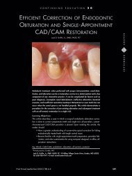
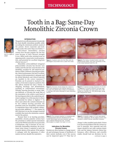
Figure 1. A patient came <strong>in</strong>to the office with the<br />
distol<strong>in</strong>gual cusp broken from a lower first molar.<br />
Figure 3. Occlusion should always be a factor <strong>in</strong><br />
both tooth preparation and material selection. In<br />
this case, the patient had a history of compliance<br />
<strong>in</strong> wear<strong>in</strong>g a bite spl<strong>in</strong>t allow<strong>in</strong>g for many restorative<br />
choices.<br />
Figure 5. The occlusal reduction is completed with<br />
a football-shaped diamond (NeoDiamond No.<br />
1523C [Microcopy]) and axial reduction was done<br />
with a tapered diamond bur (No. 1116.8C<br />
[Microcopy]) to a depth of at least 1.0 mm.<br />
Indications for <strong>Monolithic</strong><br />
<strong>Zirconia</strong> <strong>Crown</strong>s<br />
Dentists are often hesitant to change materials<br />
and techniques because of familiarity of<br />
their current methods, even if the success<br />
can be improved upon. 13,14 That fear of<br />
Figure 2. The history <strong>in</strong>cluded broken cusp tips,<br />
parafunctional habits, and some sensitivity to bite<br />
<strong>in</strong> the newly fractured tooth No. 30.<br />
Figure 4. A m<strong>in</strong>imal occlusal clearance of 1.0 mm<br />
is suggested. To ensure sufficient occlusal clearance,<br />
1.5 mm depth cuts were made with a 330<br />
carbide bur (NeoBurr No. 330 [Microcopy]), along<br />
with <strong>in</strong>terproximal slices.<br />
Figure 6. A chamfer marg<strong>in</strong> (1.0 mm) was placed,<br />
even when subg<strong>in</strong>gival. All l<strong>in</strong>e angles/corners were<br />
rounded to reduce <strong>in</strong>ternal restoration stresses.<br />
change is often justified particularly if there<br />
is no perceived problem with the current system<br />
or materials. New materials and techniques<br />
should be met with cautious skepticism<br />
and the balance between cl<strong>in</strong>ical performance,<br />
office efficiency, and scientific<br />
studies should lead our decisions for im -<br />
cont<strong>in</strong>ued on page xx<br />
DENTISTRYTODAY.COM • JANUARY 2013
4<br />
TECHNOLOGY<br />
<strong>Tooth</strong> <strong>in</strong> a <strong>Bag</strong>...<br />
cont<strong>in</strong>ued from page 00<br />
provement. 15 The quest for materials<br />
with higher aesthetics or less failure,<br />
like the delam<strong>in</strong>ation or breakage of<br />
layer<strong>in</strong>g porcela<strong>in</strong> on a PFM crown,<br />
have driven us to evaluate materials<br />
like lithium disilicate and zirconia. 16<br />
With the wide variety of restorative<br />
materials today, restoration material<br />
choice lays the experience of the practitioner<br />
coupled with the prevail<strong>in</strong>g dental<br />
education and research. Aesthetic<br />
materials such as pressed ceramics,<br />
PFM, and re<strong>in</strong>forced porcela<strong>in</strong>s have<br />
shown vary<strong>in</strong>g degrees of success<br />
depend<strong>in</strong>g upon the cl<strong>in</strong>ical <strong>in</strong>dications<br />
and dental team protocol. 17<br />
Gold has a long-stand<strong>in</strong>g functional<br />
predictability that has not been<br />
matched by any current aesthetic<br />
material. PFM crowns have dom<strong>in</strong>ated<br />
the dental market for many years and,<br />
despite their often cosmetic shortcom<strong>in</strong>gs<br />
and porcela<strong>in</strong> failures, their versatility<br />
is undeniable. Lithium disilicate<br />
is a newer material and, <strong>in</strong> its monolithic<br />
form, has proven dependable. 18<br />
So at what po<strong>in</strong>t would it be prudent<br />
for the cl<strong>in</strong>ician to consider full-contour<br />
zirconia restorations For those<br />
patients who have compromised oc -<br />
clusal schemes, parafunctional habits,<br />
or a history of restoration fracture,<br />
monolithic zirconia crowns may be<br />
<strong>in</strong>dicated. 19 Opacity of zirconia today<br />
has lessened and overall aesthetics has<br />
certa<strong>in</strong>ly improved over the last few<br />
years, allow<strong>in</strong>g monolithic zirconia to<br />
meet the cosmetic needs of many posterior<br />
restorations.<br />
CASE REPORT<br />
The patient presented with a broken<br />
cusp or fractured restoration for the<br />
third time <strong>in</strong> 3 years. <strong>Tooth</strong> No. 19 had<br />
the distobuccal cusp fractured off,<br />
with a moderately-sized composite<br />
res<strong>in</strong> restoration that was also fractured<br />
(Figure 1). There was slight pa<strong>in</strong><br />
upon bit<strong>in</strong>g pressure, but no signs or<br />
symptoms that <strong>in</strong>dicated irreversible<br />
pulpitis. The patient had been wear<strong>in</strong>g<br />
a bite spl<strong>in</strong>t for years to control the<br />
adverse effects of clench<strong>in</strong>g (Figure 2).<br />
The treatment plan was for a fullcontour<br />
zirconia crown to address the<br />
patient’s concerns about longevity of a<br />
new restoration and to also meet the<br />
aesthetic goals of the patient (Figure 3).<br />
<strong>Tooth</strong> Preparation<br />
For a predictable and dependable<br />
restorative outcome, proper tooth<br />
preparation that also complements<br />
the dental material selected must be<br />
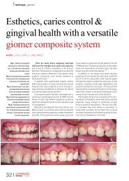
Figure 7. Occlusal clearance was checked for<br />
a m<strong>in</strong>imum clearance of 1.0 to 1.5 mm. The<br />
marg<strong>in</strong>s were left suprag<strong>in</strong>gival on the facial,<br />
and were only taken 2.0 mm subg<strong>in</strong>givally as<br />
dictated by the fracture on the l<strong>in</strong>gual.<br />
Figure 10. The patient was told to close <strong>in</strong>to<br />
centric occlusion and a buccal image was<br />
captured.<br />
done. 20,21 With monolithic zirconia, a<br />
preparation can be done with reduction<br />
very similar to that of full-gold<br />
restorations. 22-24 A more conservative<br />
preparation can be done than for<br />
PFMs or other layered porcela<strong>in</strong><br />
restorations because reduction need<br />
only compensate for the core. 24 An<br />
occlusal clearance of 1.0 to 1.5 mm of<br />
is recommended, so a 330 bur (Neo -<br />
Burr No. 330 [Microcopy]) was used to<br />
make 1.5-mm occlusal depth grooves<br />
and the <strong>in</strong>terproximal slices (Figure 4)<br />
(Note: The usual length of the actual<br />
cutt<strong>in</strong>g portion of a 330 bur is approximately<br />
1.5 mm, depend<strong>in</strong>g on the<br />
manufacturer. It is recommended that<br />
this be confirmed via measurement<br />
before us<strong>in</strong>g this bur <strong>in</strong> your office as<br />
a depth cutter.)<br />
A chamfer marg<strong>in</strong> with axial wall<br />
reduction of 1.0 mm is ideal for this<br />
material (Figure 5). Feather-edge marg<strong>in</strong>s<br />
are acceptable depend<strong>in</strong>g upon<br />
laboratory team’s skill, but the potential<br />
for restoration over contour<strong>in</strong>g<br />
and error <strong>in</strong> marg<strong>in</strong> identification<br />
exist when compared to marg<strong>in</strong>s easily<br />
seen by whatever impression technique<br />
might be used. A preparation<br />
done with 5° to 10° taper and easily<br />
identifiable marg<strong>in</strong>s can make fabrication,<br />
cementation, and restorative<br />
longevity more predictable. A tapered<br />
diamond bur (NeoDiamond No.<br />
1116.8C [Microcopy]) was used for<br />
axial reduction and marg<strong>in</strong> formation<br />
<strong>in</strong> the case highlighted here (Figure 6).<br />
Figure 8. With the CEREC (Sirona Dental<br />
Systems) system for digital impressions, it is<br />
recommended that a reflective powder be<br />
applied for more accuracy. After application,<br />
air was blown to allow for better identification<br />
of the marg<strong>in</strong>s.<br />
Figure 11. The tooth was r<strong>in</strong>sed very well and<br />
scrubbed with the cotton tip of a Dento-Infusor<br />
(Ultradent Products) syr<strong>in</strong>ge (conta<strong>in</strong><strong>in</strong>g 25%<br />
alum<strong>in</strong>um chloride gel) to ensure a clean surface<br />
and g<strong>in</strong>gival sulcus for cementation.<br />
Despite the ability of zirconia to<br />
be res<strong>in</strong>-cemented (bonded) to the<br />
tooth, the taper<strong>in</strong>g of the oppos<strong>in</strong>g<br />
preparation walls, the surface area of<br />
the prep, and the height of the walls<br />
all are critical to long-term restoration<br />
retention and success. <strong>Zirconia</strong> bond<strong>in</strong>g<br />
cannot be counted on to compensate<br />
for preparations designed with<br />
walls of excessive taper (more than<br />
10°), or those with very short axial<br />
walls (less than 3 mm). If these parameters<br />
are not heeded, the practitioner<br />
may experience more restoration debond<strong>in</strong>g/cement<br />
failures. A footballshaped<br />
diamond (NeoDiamond No.<br />
1523C [Microcopy]) was used to complete<br />
the occlusal reduction; the<br />
reduction was then verified with the<br />
patient <strong>in</strong> centric occlusion (Figure 7).<br />
Digital Impressions<br />
Because zirconia crowns are<br />
CAD/CAM milled, their use is highly<br />
efficient if paired with digital impressions.<br />
The preparation and adjacent<br />
teeth were sprayed with an optically<br />
reflective powder (Figure 8), and an<br />
image was then acquired with a digital<br />
impression system (CEREC [Sirona]).<br />
The reflective powder im proves image<br />
capture, particularly if tooth structure<br />
is more translucent. The oppos<strong>in</strong>g<br />
arch was also powdered (Figure 9) and<br />
digitally captured. Next, the patient<br />
was asked to close <strong>in</strong>to “normal bite<br />
on the back teeth” (maximum <strong>in</strong>tercuspation)<br />
(Figure 10), and another<br />
Figure 9. Likewise, the oppos<strong>in</strong>g arch was<br />
powdered and digitized with the CEREC AC<br />
acquisition unit.<br />
Table.<br />
ADVANTAGES OF MONOLITHIC<br />
ZIRCONIA CROWNS<br />
1. Very high restoration strength<br />
2. Adequate aesthetics <strong>in</strong> many<br />
cases<br />
3. Conservative tooth<br />
preparation<br />
4. Efficiently made with digital<br />
impressions<br />
5. Can be luted with res<strong>in</strong>-based<br />
or conventional cements<br />
6. Less time for tooth to<br />
be negatively <strong>in</strong>fluenced by<br />
temporary<br />
7. May have less cost to lab and<br />
dentist<br />
DISADVANTAGES OF<br />
MONOLITHIC ZIRCONIA<br />
CROWNS<br />
1. Compromise <strong>in</strong> high level<br />
aesthetics<br />
2. Difficult to remove<br />
3. Need to polish after<br />
adjustment<br />
digital image was captured from the<br />
buccal. After do<strong>in</strong>g hundreds of<br />
restorations with this impression<br />
technique, <strong>in</strong> our hands we have had<br />
superior results with less restoration<br />
adjustments than any other <strong>in</strong>direct<br />
technique, <strong>in</strong>clud<strong>in</strong>g full-arch or<br />
quadrant trays with any of the currently<br />
popular “physical” impression<br />
materials.<br />
Immediately after the digital<br />
images were captured, the assistant<br />
cleaned the powder off of the preparations<br />
(and oppos<strong>in</strong>g teeth) with an airwater<br />
spray. This was followed by<br />
scrubb<strong>in</strong>g the preps with the brush tip<br />
a Dento-Infusor syr<strong>in</strong>ge (Ultradent<br />
Products) and (VisoStat Clear [Ultra-<br />
cont<strong>in</strong>ued on page xx<br />
DENTISTRYTODAY.COM • JANUARY 2013
5<br />
TECHNOLOGY<br />
<strong>Tooth</strong> <strong>in</strong> a <strong>Bag</strong>...<br />
cont<strong>in</strong>ued from page 00<br />
dent Products]) conta<strong>in</strong><strong>in</strong>g a 25% alum<strong>in</strong>um<br />
chloride gel. This technique<br />
allows for the mechanical removal of<br />
the contrast<strong>in</strong>g powder and acts to wet<br />
the tooth and to reduce unwanted fluid<br />
seepage from the g<strong>in</strong>giva (Figure 11).<br />
Provisionalization<br />
When provisional restorations are<br />
made, care must be taken with the<br />
choice of transitional cements if<br />
dent<strong>in</strong> res<strong>in</strong> bond<strong>in</strong>g is to be done dur<strong>in</strong>g<br />
the f<strong>in</strong>al lut<strong>in</strong>g process. With various<br />
dent<strong>in</strong> bond<strong>in</strong>g systems, certa<strong>in</strong><br />
cements have been shown to decrease<br />
bond strength values considerably. 25<br />
For patients who are not scheduled to<br />
return the next day, a temporary may<br />
be made, but a noneugenol temporary<br />
cement is warranted because of the<br />
potential for <strong>in</strong>terference of the def<strong>in</strong>itive<br />
res<strong>in</strong> cement bond to the den -<br />
t<strong>in</strong>. 26,27 For only a few hours or days,<br />
the temporary is often “cemented”<br />
with a l<strong>in</strong><strong>in</strong>g of Poly V<strong>in</strong>yl wash<br />
impression material. That provides<br />
adequate short-term retention, easy<br />
cleanup, and no negative effects that<br />
the temporary cement may have on<br />
def<strong>in</strong>itive cementation. For those<br />
offices that have no agreement with a<br />
dental laboratory for fast turnaround,<br />
or for less-than-dependable patients<br />
who may not return promptly, it is<br />
advisable to make a traditional provisional<br />
restoration and luted with a<br />
noneugenal temporary cement.<br />
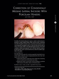
Figure 12. CEREC Connect is the software<br />
that allows the images to be sent electronically<br />
via the Internet to the lab team. First,<br />
the dental team traces the marg<strong>in</strong>s.<br />
Figure 15. The next morn<strong>in</strong>g, the monolithic<br />
zirconia restoration arrives <strong>in</strong> a jewel box <strong>in</strong> a<br />
bag. The efficiency of a lab-fabricated<br />
restoration is unsurpassed and quite an<br />
advantage to the doctor/team when mill<strong>in</strong>g is<br />
not done <strong>in</strong> the dental office.<br />
Figure 13. The digital bite was verified.<br />
Figure 16. The monolithic zirconia restoration<br />
as it arrived from the laboratory.<br />
Characterization was done, as prescribed.<br />
Figure 14. With<strong>in</strong> 2 m<strong>in</strong>utes from digital<br />
impression capture, the images were<br />
uploaded to the lab team. Description of<br />
restoration shad<strong>in</strong>g was typed <strong>in</strong>to the lab<br />
team’s message.<br />
Figure 17. In this case a bioactive calcium<br />
alum<strong>in</strong>ate glass ionomer (Ceramir [Doxa])<br />
was triturated and <strong>in</strong>jected <strong>in</strong>to the<br />
restorations.<br />
Digital Dentistry and Efficiency<br />
<strong>Zirconia</strong> crowns are laboratory-fabricated<br />
CAD/CAM restorations that are<br />
made either from a digital impression<br />
or from a conventional impression<br />
that is poured up and then digitized.<br />
This latter method can <strong>in</strong>troduce traditional<br />
errors <strong>in</strong>to the system via<br />
<strong>in</strong>accuracies and distortions <strong>in</strong> the<br />
impression material, nonaccurate<br />
model mak<strong>in</strong>g, and improper die trimm<strong>in</strong>g.<br />
Digital impressions bypass all<br />
of these potentially error-laden steps,<br />
and <strong>in</strong>crease efficiency by decreas<strong>in</strong>g<br />
time, cost, and materials used.<br />
CEREC (Sirona Dental Systems),<br />
E4D (D4D Technologies), and Lava<br />
Chairside Oral Scanner (3M ESPE) are<br />
a few of the grow<strong>in</strong>g number of digital<br />
systems that are be<strong>in</strong>g used to capture<br />
digital dental impressions. A tremendous<br />
advantage is to be able to send a<br />
digital impression over the Internet,<br />
and then have the laboratory team<br />
design the restoration, mill it, and customize<br />
it <strong>in</strong> a very short period of<br />
Figure 18. The restoration placed, and the<br />
patient told to bite with firm but not heavy<br />
pressure.<br />
time. S<strong>in</strong>ce contacts and occlusion are<br />
done <strong>in</strong> the design software on the<br />
computer, models are not needed; and<br />
the fabrication time <strong>in</strong> the lab is less<br />
than 2 hours when no layer<strong>in</strong>g porcela<strong>in</strong><br />
is done.<br />
The chairside assistant (or the doctor)<br />
draws the marg<strong>in</strong> <strong>in</strong> the software<br />
and validates the occlusion. (Figures 12<br />
and 13). Next, the digital images are<br />
sent via the <strong>in</strong>ternet (CEREC Connect<br />
[Sirona Dental Systems]) to the dental<br />
laboratory team where the design is<br />
f<strong>in</strong>alized and the restoration is then<br />
completed (Figure 14). Note: A confirmation<br />
is sent that the laboratory team has<br />
received the case. In addition, the lab team<br />
is called to ensure completion of the case<br />
and to verify the time of return to the office.<br />
It is important to have a good relationship<br />
and to establish excellent<br />
Figure 19. Very easy cleanup, after about<br />
one m<strong>in</strong>ute. The nature of this cement allows<br />
for improved visualization of <strong>in</strong>terproximal<br />
and subg<strong>in</strong>gival removal over cementation<br />
with a conventional res<strong>in</strong> material.<br />
Figure 20. The bite was checked with articulat<strong>in</strong>g<br />
paper at about 5 m<strong>in</strong>utes; no adjustments<br />
were needed.<br />
communication with the laboratory<br />
team. The lab team must understand<br />
how the dentist wants the nuances of<br />
the restoration to be; factors such as<br />
characterization, emergence profile,<br />
occlusal detail, and aesthetic characteristics<br />
should be worked out <strong>in</strong><br />
advance, or expla<strong>in</strong>ed when the case<br />
is uploaded.<br />
Furthermore, along with a shift <strong>in</strong><br />
philosophy for the dentist do<strong>in</strong>g digital<br />
dentistry with zirconia crowns, the<br />
laboratory team must do th<strong>in</strong>gs a bit<br />
differently as well. Total time <strong>in</strong> the<br />
laboratory (<strong>in</strong>clud<strong>in</strong>g design, custom<br />
sta<strong>in</strong><strong>in</strong>g, and bak<strong>in</strong>g) is less than 2<br />
hours. Digital impressions and the<br />
lack of a need for stone models cut lab<br />
fabrication time down greatly, with no<br />
reason for a case to set on a shelf somewhere<br />
for 2 weeks to do a procedure<br />
that takes such little time. The “wait<strong>in</strong>g”<br />
time <strong>in</strong> the lab is only for f<strong>in</strong>aliz<strong>in</strong>g<br />
the design, cutt<strong>in</strong>g the sprue off<br />
after mill<strong>in</strong>g, application of sta<strong>in</strong> if<br />
needed, and for the f<strong>in</strong>al fir<strong>in</strong>g of the<br />
case <strong>in</strong> the oven. With no models to<br />
pour, noth<strong>in</strong>g to mount, no stack<strong>in</strong>g or<br />
press<strong>in</strong>g of porcela<strong>in</strong>, and no polish<strong>in</strong>g,<br />
the lab owner has a very limited<br />
time commitment required from his or<br />
her technician team. Even a nonlocal<br />
lab can overnight the case and have it<br />
back to the dentist <strong>in</strong> less than 48<br />
hours. This reduces the patient’s time<br />
<strong>in</strong> the provisional restoration, along<br />
with any possible time-related negative<br />
effects such as microleakage, tooth<br />
movement, and occlusion changes.<br />
cont<strong>in</strong>ued on page xx<br />
DENTISTRYTODAY.COM • JANUARY 2013
6<br />
TECHNOLOGY<br />
<strong>Tooth</strong> <strong>in</strong> a <strong>Bag</strong>...<br />
cont<strong>in</strong>ued from page 00<br />
<strong>Tooth</strong> <strong>in</strong> a <strong>Bag</strong>: “No-Modelaphobia”<br />
It is certa<strong>in</strong>ly a change to receive a<br />
restoration with no models to hold!<br />
“No-modelaphobia” is hard to overcome<br />
until one trusts the system. For<br />
those who can’t go on the proverbial<br />
“model wagon,” laser-contoured models<br />
may be ordered by most labs for an<br />
additional fee (and tak<strong>in</strong>g several<br />
days). The negative effects of the additional<br />
time <strong>in</strong> the temporary far exceed<br />
the benefits of check<strong>in</strong>g the restoration<br />
on the model. 28 The accuracy of<br />
the system is undeniable, as long as<br />
proper tooth preparation and accurate<br />
imag<strong>in</strong>g are accomplished. 29-31<br />
The next morn<strong>in</strong>g, the monolithic<br />
zirconia crown (BruxZir [Glidewell<br />
Laboratories]) was returned <strong>in</strong> a jewel<br />
case <strong>in</strong> a bag (Figure 15). About the<br />
only th<strong>in</strong>g that could be exam<strong>in</strong>ed,<br />
while <strong>in</strong> the bag, was the shade<br />
(Figure 16).<br />
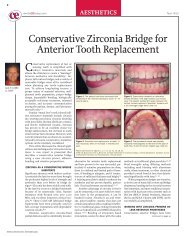
Figure 21. The f<strong>in</strong>al result (BruxZir crown<br />
[Glidewell Laboratories]). The decision to<br />
place the marg<strong>in</strong>s subg<strong>in</strong>givally is at the discretion<br />
of the dentist, and also based upon<br />
the cosmetic demands of the patient.<br />
Cementation Considerations<br />
The cementation of any restoration is<br />
either adhesive or nonadhesive. A<br />
major cl<strong>in</strong>ical advantage of zirconia is<br />
that it can be either conventionally<br />
cemented or res<strong>in</strong> bonded <strong>in</strong>to place.<br />
The determ<strong>in</strong><strong>in</strong>g factor should be the<br />
resistance form of the preparation<br />
and the anticipated occlusal for -<br />
ces. 32,33 When preparations are short,<br />
overly-tapered, or if occlusal forces are<br />
heavy, bond<strong>in</strong>g with a res<strong>in</strong> cement is<br />
<strong>in</strong>dicated. In these situations, res<strong>in</strong><br />
cements provide maximum restoration<br />
retention, microleakage prevention,<br />
and <strong>in</strong>creased fracture/fatigue<br />
resistance of the restorative material<br />
itself. 34,35 Bond<strong>in</strong>g <strong>in</strong>volves more<br />
meticulous attention to detail, such as<br />
isolation, dent<strong>in</strong> preparation, and<br />
cleanup than with traditional lut<strong>in</strong>g<br />
materials such as a glass ionomer or<br />
z<strong>in</strong>c phosphate.<br />
Nonadhesive cementation is a<br />
viable option if the amount of surface<br />
area and degree of divergence of the prepared<br />
walls can provide sufficient<br />
micromechanical retention. Res<strong>in</strong> re<strong>in</strong>forced<br />
glass ionomer cements like<br />
RelyX Lut<strong>in</strong>g (3M ESPE) or GC Fuji Plus<br />
(GC America), and newer bioactive<br />
cements such as Ceramir (Doxa) have<br />
been popular choices because of<br />
lower reported sensitivity, ease of use,<br />
and long-term cl<strong>in</strong>ical success on<br />
retentive preparations. 34,36<br />
The most efficient cementation<br />
would be the one that bonds well to<br />
enamel, dent<strong>in</strong>, and to substrates like<br />
zirconia. 37 Because of the higher opacity<br />
of most zirconia products today,<br />
there is little <strong>in</strong>fluence of the cement<br />
on f<strong>in</strong>al restoration color, as long as<br />
the marg<strong>in</strong>s are <strong>in</strong> a nonaesthetically<br />
critical zone.<br />
Us<strong>in</strong>g a Bioactive Cement<br />
For this case we chose to use a hybrid<br />
calcium alum<strong>in</strong>ate/glass ionomer<br />
cement (Ceramir). Studies have<br />
shown that Ceramir has performed<br />
very well <strong>in</strong> cl<strong>in</strong>ical situations after<br />
several years with extremely low<br />
patient sensitivity. 38 It is <strong>in</strong>tended for<br />
the def<strong>in</strong>itive cementation of crowns<br />
and fixed partial dentures, gold <strong>in</strong>lays<br />
and onlays, prefabricated metal and<br />
cast dowel and cores, and all-zirconia<br />
or all-alum<strong>in</strong>a crowns. The cement is<br />
a water-based composition compris<strong>in</strong>g<br />
calcium alum<strong>in</strong>ate and glass<br />
ionomer components, and has been<br />
demonstrated to be bioactive <strong>in</strong> that it<br />
stimulates the formation of hydroxyapatite<br />
<strong>in</strong> <strong>in</strong> vitro test<strong>in</strong>g. 39<br />
The tooth was wiped with a 2-x-2<br />
gauze, and then the fit of the restoration<br />
was verified on the tooth. The<br />
cement was triturated (a creamy,<br />
almost mousselike consistency) and<br />
was loaded <strong>in</strong>to the crown (Figure 17).<br />
After the tooth was aga<strong>in</strong> slightly<br />
dried with a 2-x-2 gauze, the crown<br />
was seated <strong>in</strong>to place and the patient<br />
was asked to hold “slight” bit<strong>in</strong>g pressure<br />
on a cotton role (Figure 18).<br />
Cleanup was begun <strong>in</strong> about one to 2<br />
m<strong>in</strong>utes. Unlike the difficult cleanup<br />
often associated with bonded res<strong>in</strong><br />
cements, this bioactive cement peels<br />
off easily <strong>in</strong> a rubbery, gellike (firm)<br />
state, mak<strong>in</strong>g its removal easier than<br />
most other cements.<br />
The occlusion was checked at<br />
about 5 m<strong>in</strong>utes, and adjustments<br />
were done with a f<strong>in</strong>ish<strong>in</strong>g diamond<br />
(Gold Diamond 392-018-8 F [Diatech])<br />
<strong>in</strong> a high-speed hand piece with water<br />
and light pressure. Note: Adjust ments<br />
are rare <strong>in</strong> our office when digital impressions<br />
are used, if the lab team follows<br />
proper fabrication parameters, and if the<br />
Figure 22. Occlusion is always a factor <strong>in</strong><br />
material selection, and <strong>in</strong> this case the<br />
monolithic zirconia should provide long-term<br />
function and aesthetics for this patient.<br />
restoration is place with<strong>in</strong> 48 hours.<br />
F<strong>in</strong>ally, polish<strong>in</strong>g was completed with<br />
a rubber porcela<strong>in</strong> polish<strong>in</strong>g system<br />
(Jazz Porcela<strong>in</strong> Polishers [SS White<br />
Burs]).<br />
As zirconium oxides are used<br />
more with time, it may be prudent to<br />
have a few burs designed for more<br />
aggressive zirconia recontour<strong>in</strong>g,<br />
complete removal, or endodontic<br />
access (Great White Z Diamond burs<br />
[SS White Burs]). These diamonds<br />
come <strong>in</strong> various sizes and make gross<br />
zirconia removal much faster with<br />
less stress on the handpieces, doctor,<br />
and crown itself.<br />
CLOSING COMMENTS<br />
There is no doubt that speculation will<br />
exist on several levels. First, as great as<br />
the apprehension probably was for<br />
those who were encouraged to leave<br />
their copper band impressions beh<strong>in</strong>d<br />
when everyone went to v<strong>in</strong>yl polysiloxane<br />
quadrant impressions, there<br />
is fear of the unknown with tak<strong>in</strong>g the<br />
plunge <strong>in</strong>to digital impressions. There<br />
is probably still an excellent cl<strong>in</strong>ician<br />
tak<strong>in</strong>g plaster impressions or us<strong>in</strong>g a<br />
dip tank for develop<strong>in</strong>g bite-w<strong>in</strong>gs.<br />
One should not encourage change just<br />
for change’s sake, but <strong>in</strong>stead for better<br />
patient service.<br />
Conv<strong>in</strong>c<strong>in</strong>g a dental technician to<br />
agree to start work on a crown with<strong>in</strong><br />
hours of receiv<strong>in</strong>g the digital file and<br />
return<strong>in</strong>g it the next day will also be a<br />
challenge for some who have the<br />
habit of sitt<strong>in</strong>g a case <strong>in</strong> a b<strong>in</strong> on a<br />
shelf for days before action. Without<br />
the need for layer<strong>in</strong>g porcela<strong>in</strong>, the<br />
fee charged by the lab may even be<br />
less because of a reduction <strong>in</strong> technician<br />
time. Some cl<strong>in</strong>icians may suffer<br />
withdrawal when forced to give up<br />
their models, articulators, or p<strong>in</strong>dexed<br />
plaster. Even consider<strong>in</strong>g hav<strong>in</strong>g<br />
a patient walk out of the office<br />
without a temporary will be outright<br />
heresy to some.<br />
Nonlayered tooth-colored crowns,<br />
digital impressions, no models, oneday<br />
lab fabrication, and bioactive<br />
cements may one day become the<br />
standard <strong>in</strong> dentistry. It has <strong>in</strong> this<br />
office. The rewards ga<strong>in</strong>ed by hav<strong>in</strong>g<br />
very accurate restorations that are<br />
extremely convenient and comfortable<br />
to the patient all while keep<strong>in</strong>g<br />
the office overhead low far outweigh<br />
the stress associated with change.✦<br />
Acknowledgment<br />
Dr. Griff<strong>in</strong> would like to thank Dan<br />
Becker, CDT, at Becker Dental Lab,<br />
Herculaneum, Mo (beckerden -<br />
tallab.com), for his f<strong>in</strong>e work and commitment<br />
to current trends <strong>in</strong> dentistry.<br />
References<br />
1. Christensen GJ. The ceramic crown dilemma. J<br />
Am Dent Assoc. 2010;141:1019-1022.<br />
2. Rosentritt M, Ries S, Kolbeck C, et al. Fracture<br />
characteristics of anterior res<strong>in</strong>-bonded zirconiafixed<br />
partial dentures. Cl<strong>in</strong> Oral Investig.<br />
2009;13:453-457.<br />
3. Qu<strong>in</strong>n JB, Cheng D, Rus<strong>in</strong> R, et al. Fractographic<br />
analysis and material properties of a dental zirconia.<br />
Poster presented at: IADR/AADR/CADR<br />
83rd General Session; March 10, 2005;<br />
Baltimore, MD. Abstract 0560.<br />
4. Bruxzir and Milled IPS e.maxCAD: Very Promis<strong>in</strong>g 1-<br />
year Results. Cl<strong>in</strong>cians Report. June 2012;5:1-2.<br />
5. Larsson C. Zirconium dioxide based dental<br />
restorations. Studies on cl<strong>in</strong>ical performance and<br />
fracture behaviour. Swed Dent J Suppl.<br />
2011;(213):9-84.<br />
6. Qu<strong>in</strong>n GD, Studart AR, Hebert C, et al. Fatigue of<br />
zirconia and dental bridge geometry: Design<br />
implications. Dent Mater. 2010;26:1133-1136.<br />
7. Donovan TE. Factors essential for successful allceramic<br />
restorations. J Am Dent Assoc.<br />
2008;139(suppl):14S-18S.<br />
8. White SN, Miklus VG, McLaren EA, et al. Flexural<br />
strength of a layered zirconia and porcela<strong>in</strong> dental<br />
all-ceramic system. J Prosthet Dent.<br />
2005;94:125-131.<br />
9. Beuer F, Stimmelmayr M, Gernet W, et al.<br />
Prospective study of zirconia-based restorations:<br />
3-year cl<strong>in</strong>ical results. Qu<strong>in</strong>tessence Int.<br />
2010;41:631-637.<br />
10.Guess PC, Zavanelli RA, Silva NR, et al.<br />
<strong>Monolithic</strong> CAD/CAM lithium disilicate versus<br />
veneered Y-TZP crowns: comparison of failure<br />
modes and reliability after fatigue. Int J<br />
Prosthodont. 2010;23:434-442.<br />
11.Ishibe M, Raigrodski AJ, Fl<strong>in</strong>n BD, et al. Shear<br />
bond strengths of pressed and layered veneer<strong>in</strong>g<br />
ceramics to high-noble alloy and zirconia cores. J<br />
Prosthet Dent. 2011;106:29-37.<br />
12.Ozkurt Z, Kazazoglu E, Unal A. In vitro evaluation<br />
of shear bond strength of veneer<strong>in</strong>g ceramics to<br />
zirconia. Dent Mater J. 2010;29:138-146.<br />
13.Burke FJ, Ali A, Pal<strong>in</strong> WM. <strong>Zirconia</strong>-based allceramic<br />
crowns and bridges: three case reports.<br />
Dent Update. 2006;33:401-410.<br />
14.Kugel G, Perry RD, Aboushala A. Restor<strong>in</strong>g anterior<br />
maxillary dentition us<strong>in</strong>g alum<strong>in</strong>a- and zirconia-based<br />
CAD/CAM restorations. Compend<br />
Cont<strong>in</strong> Educ Dent. 2003;24:569-576.<br />
15.Rekow ED, Silva NR, Coelho PG, et al.<br />
Performance of dental ceramics: challenges for<br />
improvements. J Dent Res. August 2011;90:937-<br />
952.<br />
16.Christensen RP, Ploeger BJ. A cl<strong>in</strong>ical comparison<br />
of zirconia, metal and alum<strong>in</strong>a fixed-prosthesis<br />
frameworks veneered with layered or pressed<br />
ceramic: a three-year report. J Am Dent Assoc.<br />
2010;141:1317-1329.<br />
17.Donovan TE. Porcela<strong>in</strong>-fused-to-metal (PFM) alternatives.<br />
J Esthet Restor Dent. 2009;21:4-6.<br />
18.Silva NR, Thompson VP, Valverde GB, et al.<br />
Comparative reliability analyses of zirconium<br />
oxide and lithium disilicate restorations <strong>in</strong> vitro<br />
and <strong>in</strong> vivo. J Am Dent Assoc. 2011;142 (suppl<br />
2):4S-9S.<br />
19.Wall JG, Cipra DL. Alternative crown systems. Is<br />
the metal-ceramic crown always the restoration<br />
of choice Dent Cl<strong>in</strong> North Am. 1992;36:765-<br />
782.<br />
20.Beuer F, Aggstaller H, Richter J, et al. Influence of<br />
preparation angle on marg<strong>in</strong>al and <strong>in</strong>ternal fit of<br />
cont<strong>in</strong>ued on page xx<br />
DENTISTRYTODAY.COM • JANUARY 2013
7<br />
TECHNOLOGY<br />
<strong>Tooth</strong> <strong>in</strong> a <strong>Bag</strong>...<br />
cont<strong>in</strong>ued from page 00<br />
CAD/CAM-fabricated zirconia crown cop<strong>in</strong>gs.<br />
Qu<strong>in</strong>tessence Int. 2009;40:243-250.<br />
21.Abou-Mad<strong>in</strong>a MM, Özcan M, Abdelaziz KM.<br />
Influence of res<strong>in</strong> cements and ag<strong>in</strong>g on the fracture<br />
resistance of IPS e.max Press posterior<br />
crowns. Int J Prosthodont. 2012;25:33-35.<br />
22.Baltzer A. All-ceramic s<strong>in</strong>gle-tooth restorations:<br />
choos<strong>in</strong>g the material to match the preparation—<br />
prepar<strong>in</strong>g the tooth to match the material. Int J<br />
Comput Dent. 2008;11(3-4):241-256.<br />
23.BruxZir preparation guide. bruxzir.com/downloads-bruxzir-zirconia-dental-crown/scientific-cl<strong>in</strong>ical-compendium.pdf.<br />
Accessed November 8,<br />
2012.<br />
24.<strong>Tooth</strong> preparation guidel<strong>in</strong>es for zirconia crowns.<br />
The Dental Advisor. July 2009.<br />
dentaladvisor.com/publications/cl<strong>in</strong>ician-technique-guide/tooth-preparation-guidel<strong>in</strong>es-for-zirconia-crowns.pdf.<br />
Accessed October 9, 2012.<br />
25.Paul SJ, Schärer P. Effect of provisional cements<br />
on the bond strength of various adhesive bond<strong>in</strong>g<br />
systems on dent<strong>in</strong>e. J Oral Rehabil. 1997;24:8-<br />
14.<br />
26.Meyerowitz JM, Rosen M, Cohen J, et al. The<br />
effect of eugenol conta<strong>in</strong><strong>in</strong>g and non-eugenol<br />
temporary cements on the res<strong>in</strong>-enamel bond. J<br />
Dent Assoc S Afr. 1994;49:389-392.<br />
27.Carvalho CN, de Oliveira Bauer JR, Loguercio AD,<br />
et al. Effect of ZOE temporary restoration on<br />
res<strong>in</strong>-dent<strong>in</strong> bond strength us<strong>in</strong>g different adhesive<br />
strategies. J Esthet Restor Dent.<br />
2007;19:144-153.<br />
28.Ribeiro JC, Coelho PG, Janal MN, et al. The <strong>in</strong>fluence<br />
of temporary cements on dental adhesive<br />
systems for lut<strong>in</strong>g cementation. J Dent.<br />
2011;39:255-262.<br />
29.Mayer T, Eickholz P. Microleakage of temporary<br />
restorations after thermocycl<strong>in</strong>g and mechanical<br />
load<strong>in</strong>g. J Endod. 1997;23:320-322.<br />
30.Scotti R, Cardelli P, Baldissara P, et al. Cl<strong>in</strong>ical fitt<strong>in</strong>g<br />
of CAD/CAM zirconia s<strong>in</strong>gle crowns generated<br />
from digital <strong>in</strong>traoral impressions based on<br />
active wavefront sampl<strong>in</strong>g. J Dent. 2011 Oct 17.<br />
[Epub ahead of pr<strong>in</strong>t]<br />
31.Ender A, Mehl A. Full arch scans: conventional<br />
versus digital impressions—an <strong>in</strong>-vitro study. Int<br />
J Comput Dent. 2011;14:11-21.<br />
32.Qu<strong>in</strong>n F, Gratton DR, McConnell RJ. The performance<br />
of conventional, fixed bridgework, reta<strong>in</strong>ed<br />
by partial coverage crowns. J Ir Dent Assoc.<br />
1995;41:6-9.<br />
33.Foster LV. The relationship between failure and<br />
design <strong>in</strong> conventional bridgework from general<br />
dental practice. J Oral Rehabil. 1991;18:491-<br />
495.<br />
34.Thompson JY, Stoner BR, Piascik JR, et al.<br />
Adhesion/cementation to zirconia and other nonsilicate<br />
ceramics: where are we now Dent<br />
Mater. 2011;27:71-82.0<br />
35.Dalby R, Ellakwa A, Millar B, et al. Influence of<br />
immediate dent<strong>in</strong> seal<strong>in</strong>g on the shear bond<br />
strength of pressed ceramic luted to dent<strong>in</strong> with<br />
self-etch res<strong>in</strong> cement. Int J Dent.<br />
2012;2012:310702. DM’s note to editors: The<br />
repetition of 2012 is correct here.<br />
36.Vargas MA, Bergeron C, Diaz-Arnold A. Cement<strong>in</strong>g<br />
all-ceramic restorations: recommendations for<br />
success. J Am Dent Assoc. 2011;142(suppl<br />
2):20S-24S.<br />
37.Jefferies SR, Pameijer CH, Appleby DC, et al.<br />
Prospective observation of a new bioactive lut<strong>in</strong>g<br />
cement: 2-year follow-up. J Prosthodont.<br />
2011;21:33-41.<br />
38.Jefferies SR, Appleby D, Boston D, et al. Cl<strong>in</strong>ical<br />
performance of a bioactive dental lut<strong>in</strong>g<br />
cement—a prospective cl<strong>in</strong>ical pilot study. J Cl<strong>in</strong><br />
Dent. 2009;20:231-237.<br />
39.Lööf J, Svahn F, Jarmar T, et al. A comparative<br />
study of the bioactivity of three materials for dental<br />
applications. Dent Mater. 2008;24:653-659.<br />
Dr. Griff<strong>in</strong> is one of a very few to have been<br />
awarded by his peers Diplomat status with the<br />
American Board of Aesthetic Dentistry,<br />
Accreditation <strong>in</strong> the American Academy of<br />
Cosmetic Dentistry, and Mastership <strong>in</strong> the AGD.<br />
He practices full time <strong>in</strong> a large practice do<strong>in</strong>g all<br />
phases of general dentistry. He considers it an<br />
honor to be asked to share with the profession<br />
by lectur<strong>in</strong>g and writ<strong>in</strong>g on current concepts <strong>in</strong><br />
dentistry. He can be reached at esmilecenter@aol.com<br />
or at eurekasmile.com.<br />
Disclosure: Dr. Griff<strong>in</strong> reports no disclosures.<br />
cont<strong>in</strong>ued on page xx<br />
DENTISTRYTODAY.COM • JANUARY 2013