Primary Human Osteoclast Differentiation & Function on Dentine
Primary Human Osteoclast Differentiation & Function on Dentine
Primary Human Osteoclast Differentiation & Function on Dentine

Create successful ePaper yourself
Turn your PDF publications into a flip-book with our unique Google optimized e-Paper software.
<str<strong>on</strong>g>Primary</str<strong>on</strong>g> <str<strong>on</strong>g>Human</str<strong>on</strong>g> <str<strong>on</strong>g>Osteoclast</str<strong>on</strong>g> <str<strong>on</strong>g>Differentiati<strong>on</strong></str<strong>on</strong>g><br />
and <str<strong>on</strong>g>Functi<strong>on</strong></str<strong>on</strong>g> <strong>on</strong> <strong>Dentine</strong> Discs and<br />
Corning ® Osteo Assay Surface<br />
SnAPPShots<br />
A brief technical report<br />
from the Corning<br />
Development Group<br />
A. Fawad Faruqi and<br />
James Beltzer<br />
Corning Incorporated<br />
Sullivan Park, NY 14831<br />
Introducti<strong>on</strong><br />
In vitro assays that can measure both the formati<strong>on</strong> and<br />
functi<strong>on</strong> of osteoclasts are critical for furthering our understanding<br />
of the biology of normal b<strong>on</strong>e remodeling and diseases.<br />
Some of the more notable disease processes caused by<br />
increased b<strong>on</strong>e resorpti<strong>on</strong> include osteoporosis, metastatic<br />
cancer, and Paget’s disease. The gold standard system for<br />
assessing osteoclast activity is both time c<strong>on</strong>suming and<br />
difficult to quantify. The typical assay involves primary<br />
osteoclasts and utilizes b<strong>on</strong>e or dentine slices to evaluate<br />
osteoclast differentiati<strong>on</strong> and functi<strong>on</strong>. An inorganic coating<br />
has been developed <strong>on</strong> standard polystyrene multiple well<br />
plates by Corning that can be used for in vitro assays and<br />
begins to address the challenges typically associated with<br />
osteoclast activity experiments. A comparis<strong>on</strong> was performed<br />
with the Corning Osteo Assay Surface multiple plates and<br />
dentine discs using cultures of primary human osteoclast<br />
precursors cultured with macrophage col<strong>on</strong>y stimulating<br />
factor (MCSF), and receptor activator of nuclear factor-χB<br />
ligand (RANKL) which stimulates osteoclast differentiati<strong>on</strong><br />
and activity. The results dem<strong>on</strong>strate that the osteoclast differentiati<strong>on</strong><br />
can be assessed rapidly and accurately <strong>on</strong> the<br />
Corning Osteo Assay surface using tartrate-resistant acid<br />
phosphatase (TRAP) staining. Visualizati<strong>on</strong> of TRAP positive<br />
multinucleated cells <strong>on</strong> dentine discs with normal light<br />
microscopy is difficult. <str<strong>on</strong>g>Osteoclast</str<strong>on</strong>g> activity can be assessed<br />
by measuring the area of the mineral coating removal as a<br />
model of pit formati<strong>on</strong> <strong>on</strong> Corning Osteo Assay Surface,<br />
but this activity is extremely difficult to visualize <strong>on</strong> dentine<br />
discs. In summary, both osteoclast differentiati<strong>on</strong> and activity<br />
can be readily measured with the Corning Osteo Assay<br />
Surface coated multiple well polystyrene plates, and the<br />
results are c<strong>on</strong>sistent and reproducible for assays using<br />
osteoclast precursors whereas use of dentine discs for similar<br />
experiments can be problematic.<br />
Materials and Methods:<br />
<str<strong>on</strong>g>Human</str<strong>on</strong>g> <str<strong>on</strong>g>Osteoclast</str<strong>on</strong>g> Precursor Cells<br />
<str<strong>on</strong>g>Human</str<strong>on</strong>g> osteoclast precursor cells (L<strong>on</strong>za Cat. No. 2T-110)<br />
were rapidly thawed and washed with <str<strong>on</strong>g>Osteoclast</str<strong>on</strong>g> Precursor<br />
Basal Medium (L<strong>on</strong>za Cat. No. PT-8201) which had been<br />
supplemented with 10% fetal bovine serum (FBS), 2 mM<br />
L-glutamine and 100 units/mL penicillin and streptomycin<br />
(Pen/Strep). Cells were counted and viability determined by<br />
using trypan blue exclusi<strong>on</strong>. <str<strong>on</strong>g>Primary</str<strong>on</strong>g> human osteoclast precursors<br />
cannot be passaged however the cells can be differentiated<br />
into osteoclasts in the presence of RANK ligand<br />
(66 ng/mL) and M-CSF (33 ng/mL). Precursor cells grown<br />
in the presence of M-CSF but lacking soluble RANK ligand<br />
will expand in number but will not differentiate into functi<strong>on</strong>al<br />
osteoclasts. All cells, including a n<strong>on</strong>-differentiati<strong>on</strong><br />
c<strong>on</strong>trol, were resuspended to a final c<strong>on</strong>centrati<strong>on</strong> of 50,000<br />
cells/mL. 100 µL of cell suspensi<strong>on</strong> was plated per well in a<br />
96 well Corning Osteo Assay Surface plate (Corning Cat. No.<br />
3988XX1) or polystyrene 96 well plate to which had been<br />
added a single dentine disc (Immunodiagnostic Systems Cat.<br />
No. AE-8050). Cells were cultured at 37°C in a humidified<br />
atmosphere of 5% CO 2 . After 5 to 7 days of culture, the<br />
osteoclasts could be identified as large multinucleate cells<br />
when visualized by phase microscopy. Media was changed<br />
<strong>on</strong> day 3 of culture.
IDS <strong>Dentine</strong> Discs<br />
The dentine discs were supplied sterile and handled in a<br />
biosafety cabinet to maintain sterility. The c<strong>on</strong>tents of the<br />
sterile c<strong>on</strong>tainer were emptied <strong>on</strong> a Petri dish to facilitate<br />
handling. Forceps were used to carefully transfer the discs<br />
to a 96 well multiple well plate. The discs were pre-wetted<br />
for at least <strong>on</strong>e hour with phosphate buffered saline and the<br />
soluti<strong>on</strong> was removed prior to adding cells.<br />
Tartrate-Resistant Acid Phosphatase Quantitati<strong>on</strong><br />
and Cell Staining<br />
In order to analyze for the presence of the osteoclast specific<br />
marker TRAP, a staining kit from B-Bridge Internati<strong>on</strong>al<br />
(Cat. No. AK04) was used. Briefly, 30 µL of culture supernatant<br />
was added to 170 µL of the chromogenic substrate<br />
which had been diluted in Tartrate-c<strong>on</strong>taining buffer<br />
(3 mg/5 mL as described in the kit). Plates were incubated<br />
for 3 hours at 37°C and absorbance read at 540 nm in a<br />
microplate reader (PerkinElmer ® Victor3). For TRAP<br />
staining, the cells were washed with 100 µL of PBS and<br />
fixed for 5 minutes with the fixative reagent at room temperature.<br />
The cells were washed three times with distilled<br />
water and then stained for 20 to 60 minutes at 37°C with<br />
the chromogenic substrate and finally washed with distilled<br />
water to stop the reacti<strong>on</strong> when optimum color was achieved.<br />
L<strong>on</strong>ger incubati<strong>on</strong>s with the stain caused precipitati<strong>on</strong> and<br />
were avoided.<br />
Resorpti<strong>on</strong> Pit Assay<br />
To analyze the surface for pit formati<strong>on</strong> the medium was<br />
aspirated from the wells <strong>on</strong> day 5 and 100 µL of 10% bleach<br />
soluti<strong>on</strong> was added. Cells were incubated with the bleach<br />
soluti<strong>on</strong> for 5 minutes at room temperature. The wells were<br />
washed twice with distilled water and allowed to dry at room<br />
temperature for 3 to 5 hours. Individual pits or multiple pit<br />
clusters were observed using a microscope at 25 to 100x<br />
magnificati<strong>on</strong>. In order to observe the resorpti<strong>on</strong> pits <strong>on</strong><br />
dentine, the discs were first stained with 1% toluidine blue<br />
for 5 minutes (1).<br />
Imaging <strong>on</strong> <strong>Dentine</strong><br />
The TRAP stained cells and resorpti<strong>on</strong> pits <strong>on</strong> dentine discs<br />
were visualized and images captured using an upright microscope<br />
(Nik<strong>on</strong> ® Eclipse 50iPOL) equipped with a ColorView<br />
soft imaging system.<br />
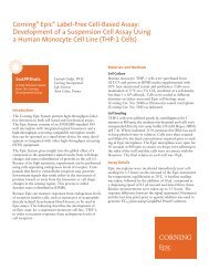
OD @ 540 nm<br />
1.6<br />
1.4<br />
1.2<br />
1.0<br />
0.8<br />
0.6<br />
0.4<br />
0.2<br />
0<br />
<strong>Dentine</strong><br />
Surface<br />
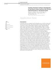
Figure 1. Soluble TRAP activity found in the medium of human osteoclast<br />
precursor cells which had been cultured for 5 days <strong>on</strong> the Corning Osteo<br />
Assay Surface or dentine surface (*p < 0.05 relative to dentine).<br />
Results and Discussi<strong>on</strong><br />
The b<strong>on</strong>e slice assay, in which cultured osteoclasts form pits<br />
<strong>on</strong> a mineralized substrate, has been widely used to investigate<br />
the effects of many factors <strong>on</strong> b<strong>on</strong>e resorpti<strong>on</strong> and is<br />
c<strong>on</strong>sidered to be the “gold standard” method (2,3). It has<br />
been widely used to study resorpti<strong>on</strong> by human osteoclasts<br />
either isolated directly from b<strong>on</strong>e or cultured from marrow.<br />
One recent advance in the field has been the development<br />
of a technique to induce osteoclast formati<strong>on</strong> from peripheral<br />
blood cells by the additi<strong>on</strong> of RANK ligand (4). Using<br />
this technique large numbers of functi<strong>on</strong>al osteoclasts can<br />
be generated which will form resorpti<strong>on</strong> pits in mineralized<br />
substrates.<br />
Poietics ® human osteoclast precursors, which had been<br />
expanded using media c<strong>on</strong>taining M-CSF and plated <strong>on</strong>to<br />
Corning ® Osteo Assay Surface 96 well plates or IDS dentine<br />
discs were differentiated for 5 to 7 days in the presence<br />
of RANK ligand, then tested for the producti<strong>on</strong> of soluble<br />
TRAP using the B-Bridge kit (described above). Figure 1<br />
shows a comparis<strong>on</strong> of TRAP activity in the culture medium<br />
derived from human osteoclast precursors cultured <strong>on</strong><br />
Corning Osteo Assay Surface or <strong>on</strong> dentine discs. Cells<br />
grown <strong>on</strong> the Osteo Assay Surface express more than twice<br />
the TRAP activity in the medium compared with dentine<br />
discs. <strong>Dentine</strong> discs present several challenges for tissue<br />
culture, as the discs are easily damaged during handling and<br />
culturing of cells. Additi<strong>on</strong>ally, the discs are pr<strong>on</strong>e to floating<br />
and do not completely fill the bottom of an individual<br />
well (using 96 multiple well plates). These factors could<br />
c<strong>on</strong>tribute to cell loss <strong>on</strong> the dentine surface as the cells<br />
attach to the plastic well instead, and ultimately a lower<br />
TRAP value.<br />
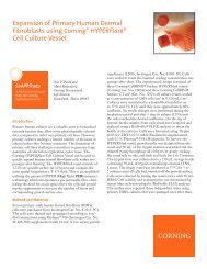
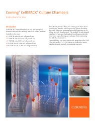
Active and fully differentiated osteoclasts are characterized<br />
as large multinucleate cells which display positive TRAP<br />
staining. Figure 2A shows human osteoclast precursors<br />
which had been cultured for 5 days <strong>on</strong> Corning Osteo Assay<br />
Surface fixed and stained for TRAP as described above.<br />
The cells were uniformly stained for TRAP and had greater<br />
than 3 nuclei per cell, the standard metric for differentiated<br />
osteoclasts. Cells prepared in exactly the same manner can<br />
not be visualized directly <strong>on</strong> dentine disks, as the dentine<br />
*<br />
Corning Osteo Assay<br />
Surface
discs are not transparent. To visualize fixed and TRAP<br />
stained cells <strong>on</strong> dentine discs required the use of an upright<br />
microscope and brightfield illuminati<strong>on</strong>. Figure 2B shows<br />
TRAP positively stained cells <strong>on</strong> dentine discs. The size<br />
and number of TRAP positive staining cells <strong>on</strong> the dentine<br />
surface is much lower than that observed <strong>on</strong> the Corning ®<br />
Osteo Assay Surface. The dentine surface is quite uneven<br />
and displays numerous scratches and knife marks. While<br />
it is possible to visualize TRAP stained cells <strong>on</strong> the discs,<br />
it is very difficult – if not impossible – to distinguish individual<br />
nuclei, making the quantificati<strong>on</strong> of cells with 3 or<br />
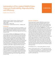
more. Resorpti<strong>on</strong> of a mineralized substrate such as b<strong>on</strong>e<br />
slices and dentine discs are c<strong>on</strong>sidered the “gold standard”<br />
for measuring osteoclast functi<strong>on</strong>. Figure 3A shows resorpti<strong>on</strong><br />
pits produced by human osteoclast precursors cultured<br />
<strong>on</strong> Corning Osteo Assay Surface for 5 days and then<br />
removed by bleaching the well. The pits are numerous and<br />
readily visible. As has been noted above, it is not possible to<br />
visualize resorpti<strong>on</strong> pits in dentine discs using a standard<br />
cell culture inverted microscope. In fact, using an upright<br />
microscope and brightfield illuminati<strong>on</strong> was not sufficient<br />
to visualize resorpti<strong>on</strong> pits <strong>on</strong> dentine discs. Figure 3B<br />
shows resorpti<strong>on</strong> pits <strong>on</strong> dentine discs which had been<br />
stained with toluidine blue. The staining resulted in dark<br />
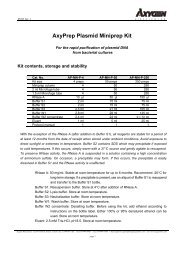
Corning Osteo Assay Surface<br />
<strong>Dentine</strong> Surface<br />
100 µm<br />
Figure 2. <str<strong>on</strong>g>Human</str<strong>on</strong>g> osteoclast precursors were fixed after 5 days of growth <strong>on</strong> the Corning Osteo Assay Surface or dentine surface and<br />
stained for the osteoclast marker TRAP.<br />
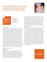
Corning Osteo Assay Surface<br />
<strong>Dentine</strong> Surface<br />
400 µm<br />
Figure 3. Resorpti<strong>on</strong> pits observed <strong>on</strong> the Corning Osteo Assay Surface and dentine surface after 5 days of culture of human osteoclast<br />
precursor cells. The dentine surface was stained with toluidine blue to visualize the pits.
lue pits which were visible in spite of a high level of background<br />
staining which also brought out the uneven surface<br />
and numerous scratches and knife marks. The high background<br />
and numerous imperfecti<strong>on</strong>s of the dentine discs<br />
make the quantificati<strong>on</strong> of resorpti<strong>on</strong> pit area all but impossible.<br />
It can be argued that Corning ® Osteo Assay Surface<br />
represents a robust, c<strong>on</strong>venient, and reproducible substitute<br />
for b<strong>on</strong>e slices or dentine discs.<br />
C<strong>on</strong>clusi<strong>on</strong>s<br />
It was dem<strong>on</strong>strated that the Corning Osteo Assay Surface<br />
can be used to easily measure soluble TRAP activity, to visualize<br />
and quantify multinucleated TRAP positive stained<br />
differentiating osteoclasts, and to visualize resorpti<strong>on</strong> pits<br />
created by differentiated osteoclasts. In c<strong>on</strong>trast, dentine<br />
discs were difficult to handle, easily damaged and do not<br />
cover the entire bottom of the well. We speculate that these<br />
handling issues result in fewer cells <strong>on</strong> the dentine discs and<br />
therefore a much lower soluble TRAP value. In fact, the soluble<br />
TRAP value <strong>on</strong> Corning Osteo Assay Surface was more<br />
than twice that of what was observed for the dentine discs.<br />
Corning Osteo Assay Surface is a c<strong>on</strong>venient tool for the<br />
measure of the number of differentiated osteoclasts, because<br />
TRAP staining cells can be easily visualized using an inverted<br />
microscope. The uniformity and clarity of the surface in the<br />
Corning multiple well plates makes the counting of nuclei<br />
quite straightforward. On the other hand, fixed and stained<br />
cells can not be visualized <strong>on</strong> dentine discs using the standard<br />
cell culture inverted microscope. When TRAP stained cells<br />
<strong>on</strong> the dentine discs are examined using an upright microscope<br />
and brightfield illuminati<strong>on</strong>, the cells are much smaller<br />
and fewer in number compared to what was observed <strong>on</strong><br />
Corning Osteo Assay Surface. Additi<strong>on</strong>ally, the unevenness<br />
of the background and numerous imperfecti<strong>on</strong>s make<br />
counting the number of nuclei for the individual cells<br />
all but impossible.<br />
The visualizati<strong>on</strong> and quantificati<strong>on</strong> of resorpti<strong>on</strong> pits formed<br />
by differentiated osteoclasts is also easily accomplished<br />
<strong>on</strong> Corning Osteo Assay Surface after the cells have been<br />
removed. The pits are numerous and quite large when<br />
viewed under an inverted microscope without the need for<br />
additi<strong>on</strong>al staining. As discussed above, visualizati<strong>on</strong> of pits<br />
<strong>on</strong> dentine discs required the use of an upright microscope<br />
and brightfield illuminati<strong>on</strong>. It was also necessary to stain<br />
the dentine discs with toluidine blue in order to visualize<br />
the pits. There were not as many pits <strong>on</strong> the dentine discs<br />
and they appeared smaller in size. Because of the high<br />
background of the stained discs, it would be very difficult<br />
to quantify the area of resorpti<strong>on</strong> pits <strong>on</strong> the discs.<br />
References<br />
1. Saftig, P. et. al. (1998). PNAS 95:13453-13458.<br />
2. Boyde, A., Ali, N.N. and J<strong>on</strong>es, S.J. (1984). Br. Dent. J.<br />
156:216-220.<br />
3. Chambers, T.J., Revell, P.A., Fuller, K., and Athanasou, N.<br />
(1984). J. Cell Sci. 55:383-399.<br />
4. It<strong>on</strong>aga, I., Sabokbar, A., Neale, S.D., and Athanasou, N.A.<br />
(1999). Biochem. Biophys. Res. Comm. 264:590-595.<br />
For additi<strong>on</strong>al product or technical informati<strong>on</strong>, please visit www.corning.com/lifesciences<br />
or call 1.800.492.1110. Outside the United States, please call 978.442.2200 or c<strong>on</strong>tact your<br />
local Corning sales office listed below..<br />
Corning Incorporated<br />
Life Sciences<br />
Tower 2, 4th Floor<br />
900 Chelmsford St.<br />
Lowell, MA 01851<br />
t 800.492.1110<br />
t 978.442.2200<br />
f 978.442.2476<br />
www.corning.com/lifesciences<br />
Worldwide<br />
Support Offices<br />
A S I A / P A C I F I C<br />
Australia/New Zealand<br />
t 0402-794-347<br />
China<br />
t 86-21-5467-4666<br />
f 86-21-5407-5899<br />
India<br />
t 91 124 4604000<br />
f 91 124 4604099<br />
Corning is a registered trademarks of Corning Incorporated, Corning, NY.<br />
All other trademarks in this document are the property of their respective owners.<br />
Corning Incorporated, One Riverfr<strong>on</strong>t Plaza, Corning, NY 14831-0001<br />
Japan<br />
t 81 3-3586 1996<br />
f 81 3-3586 1291<br />
Korea<br />
t 82 2-796-9500<br />
f 82 2-796-9300<br />
Singapore<br />
t 65 6733-6511<br />
f 65 6861-2913<br />
Taiwan<br />
t 886 2-2716-0338<br />
f 886 2-2716-0339<br />
E U R O P E<br />
France<br />
t 0800 916 882<br />
f 0800 918 636<br />
Germany<br />
t 0800 101 1153<br />
f 0800 101 2427<br />
The Netherlands<br />
t 31 20 655 79 28<br />
f 31 20 659 76 73<br />
United Kingdom<br />
t 0800 376 8660<br />
f 0800 279 1117<br />
All Other European<br />
Countries<br />
t 31 (0) 20 659 60 51<br />
f 31 (0) 20 659 76 73<br />
L AT I N A M E R I C A<br />
Brasil<br />
t (55-11) 3089-7419<br />
f (55-11) 3167-0700<br />
Mexico<br />
t (52-81) 8158-8400<br />
f (52-81) 8313-8589<br />
© 2010 Corning Incorporated Printed in U.S.A. 11/10 POD CLS-AN-162