AXIOM Innovations November 2011 3.96MB - Siemens Healthcare
AXIOM Innovations November 2011 3.96MB - Siemens Healthcare AXIOM Innovations November 2011 3.96MB - Siemens Healthcare
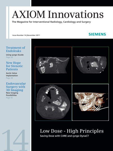
AXIOM Innovations The Magazine for Interventional Radiology, Cardiology and Surgery Issue Number 14 | November 2011 Treatment of Endoleaks Using syngo iGuide Page 26 New Hope for Stenotic Patients Aortic Valve Implantation Page 38 Endovascular Surgery with 3D Imaging New Imaging Possibilities Page 42 Low Dose - High Principles Saving Dose with CARE and syngo DynaCT
- Page 2 and 3: Editorial “Keeping image quality
- Page 4 and 5: Content Content 16 Radiation Reduct
- Page 6 and 7: News Less Radiation During Catheter
- Page 8 and 9: News A New Era: Hybrid Operating Ro
- Page 10 and 11: Cover Story CARE Low Dose, High Pri
- Page 12 and 13: Cover Story CARE “The Siemens eng
- Page 14 and 15: Cover Story CARE Combined Applicati
- Page 16 and 17: Cover Story CARE A Pediatric Radiol
- Page 18 and 19: Cover Story CARE doses are constant
- Page 20 and 21: Cover Story CARE Treatment of Compl
- Page 22 and 23: Cover Story CARE Evaluation of CARE
- Page 24 and 25: Cover Story CARE Table 2: Screening
- Page 26 and 27: Angiography syngo iGuide Endoleak T
- Page 28 and 29: Angiography syngo iGuide “In the
- Page 30 and 31: Angiography syngo iFlow Quantitativ
- Page 32 and 33: Angiography syngo DynaCT Revascular
- Page 34 and 35: Cardiology Artis zee in EP Overlay
- Page 36 and 37: Cardiology syngo DynaCT Cardiac Acc
- Page 38 and 39: Cardiology Hybrid OR Minimally Inva
- Page 40 and 41: Cardiology Hybrid OR of his teenage
- Page 42 and 43: Surgery Hybrid Surgery 3D Imaging B
- Page 44 and 45: Surgery Hybrid Surgery A look into
- Page 46 and 47: Surgery Thoracic Surgery / Pneumono
- Page 48 and 49: Surgery Thoracic Surgery / Pneumono
- Page 50 and 51: Surgery Heart Valve Implantations H
<strong>AXIOM</strong> <strong>Innovations</strong><br />
The Magazine for Interventional Radiology, Cardiology and Surgery<br />
Issue Number 14 | <strong>November</strong> <strong>2011</strong><br />
Treatment of<br />
Endoleaks<br />
Using syngo iGuide<br />
Page 26<br />
New Hope<br />
for Stenotic<br />
Patients<br />
Aortic Valve<br />
Implantation<br />
Page 38<br />
Endovascular<br />
Surgery with<br />
3D Imaging<br />
New Imaging<br />
Possibilities<br />
Page 42<br />
Low Dose - High Principles<br />
Saving Dose with CARE and syngo DynaCT
Editorial<br />
“Keeping image quality<br />
high and radiation<br />
dose low is one of the<br />
key drivers in the<br />
development of our<br />
imaging portfolio.”<br />
Dr. Heinrich Kolem,<br />
CEO of the Angiography & Interventional X-Ray Business Unit (AX)<br />
at <strong>Siemens</strong> <strong>Healthcare</strong><br />
2 <strong>AXIOM</strong> <strong>Innovations</strong> · <strong>November</strong> <strong>2011</strong> · www.siemens.com/healthcare-magazine
Editorial<br />
Dear Reader,<br />
Dr. Heinrich Kolem<br />
CEO AX Division<br />
Earlier this year, I had the opportunity to<br />
attend the Cardiovascular and Interventional<br />
Radiological Society of Europe’s<br />
(CIRSE) annual conference in Munich.<br />
The CIRSE is one of the most renowned<br />
conferences for interventional imaging.<br />
There were approximately 6,000<br />
attendees eager to learn about the latest<br />
technologies and advances in their<br />
clinical field. I was very pleased to see<br />
how many visitors were interested in<br />
our products and solutions. For the first<br />
time, we showcased how “The Future is<br />
Flexible” with the Artis zeego, our multiaxis<br />
C-arm system based on robotic<br />
technology. We introduced the Hybrid IR<br />
concept which offers new opportunities<br />
for interventional radiologists looking<br />
to expand to more complex cases. In<br />
order to meet current and future imaging<br />
needs, Artis zeego allows a variety<br />
of working heights and delivers large<br />
volume image results.<br />
Improving clinical workflow and<br />
radiation dose reduction are of great<br />
importance to our customers and their<br />
patients. They are also key drivers for<br />
our development. We know that our<br />
customers carry a lot of responsibility<br />
for each individual patient they treat.<br />
In return, it is our responsibility to offer<br />
reliable systems to support our customers<br />
worldwide. We are constantly<br />
exploring new possibilities to reduce<br />
dose without compromising image quality.<br />
The title of this issue reflects that<br />
line of thought, “Low Dose - High Principles.”<br />
Our dose saving initiative with<br />
CARE (Combined Applications to Reduce<br />
Exposure) and CLEAR (an imaging technology<br />
to enhance visualization with<br />
X-ray) not only reduces radiation dose<br />
for patients and staff, but also boosts<br />
image quality to an outstanding level.<br />
A growing trend in the market for interventional<br />
cardiology and hybrid surgery<br />
is the minimally invasive replacement<br />
of valves. With demographic changes in<br />
our society and a growing percentage of<br />
elderly patients, new treatment methods<br />
will evolve. Minimally invasive procedures<br />
are an exciting new treatment<br />
possibility especially for high-risk<br />
patients who cannot undergo open surgery.<br />
New innovative applications such<br />
as syngo Aortic ValveGuide* help surgeons<br />
and cardiologists replace calcified<br />
aortic heart valves with more guidance<br />
and a new automated workflow.<br />
Looking back on a very exciting year<br />
for <strong>Siemens</strong> <strong>Healthcare</strong>, I am proud<br />
to present to you this issue of <strong>AXIOM</strong><br />
<strong>Innovations</strong>. You can read more about<br />
CARE technology, new functionalities in<br />
endovascular and thoracic surgery, and<br />
many other innovative topics. Enjoy!<br />
Dr. Heinrich Kolem<br />
* The syngo Aortic ValveGuide is pending 510(k) clearance, and is not yet commercially available in the United States.<br />
<strong>AXIOM</strong> <strong>Innovations</strong> · <strong>November</strong> <strong>2011</strong> · www.siemens.com/healthcare-magazine 3
Content<br />
Content<br />
16<br />
Radiation Reduction<br />
is Key in Pediatrics<br />
26<br />
Enhanced Endoleak<br />
Treatment Through<br />
Improved Navigation<br />
10 A long-term commitment<br />
to reduce radiation dose<br />
<strong>Siemens</strong> has long been a leader in<br />
addressing the need to provide optimal<br />
images at the lowest possible<br />
radiation dose following the ALARA<br />
principle. Many institutions worldwide<br />
working with <strong>Siemens</strong> angiography<br />
systems use the CARE<br />
applications in their daily routine<br />
when imaging patients of all ages.<br />
It is most importatnt to them to<br />
keep radiation dose low, especially<br />
for children and also their own staff.<br />
3 Editorial<br />
6 News<br />
10 Cover Story<br />
Low Dose, High Principles:<br />
Cardiac CARE for All<br />
A Report from Fundación<br />
Cardioinfantil in Colombia<br />
A Pediatric Radiologist's Approach<br />
to Radiation Reduction<br />
Interview with Dr. Anne Marie<br />
Cahill, Children's Hospital of<br />
Philadelphia<br />
Cover<br />
Imaging of temporal<br />
mandibular joint with<br />
reduced radiation dose.<br />
Courtesy of Anne<br />
Marie Cahill, M.D.,<br />
Department of<br />
Interventional<br />
Radiology,<br />
Children´s Hospital<br />
of Philadelphia,<br />
Pennsylvania, USA<br />
Treatment of Complex Head and<br />
Neck Malformation in an Infant<br />
Clinical Case<br />
CAREposition<br />
An Evaluation of Radiation-Free<br />
Positioning<br />
61 Imprint<br />
4 <strong>AXIOM</strong> <strong>Innovations</strong> · <strong>November</strong> <strong>2011</strong> · www.siemens.com/healthcare-magazine
Content<br />
32<br />
syngo Neuro PBV IR Helps During<br />
Revascularization Treatment<br />
42<br />
Advanced Technology Revolutionizes<br />
Endovascular Surgery<br />
Angiography<br />
26 Endoleak Treatment:<br />
Enhanced Visualization Meets<br />
Enhanced Navigation<br />
syngo iGuide facilitates better<br />
Workflow<br />
30 Quantitative Evidences of<br />
Hemodynamic Improvement of<br />
Brain Arteriovenous Malformation<br />
after Surgical Intervention<br />
Clinical case<br />
32 Revascularization Treatment<br />
of Acute Cerebral Stenosis<br />
Clinical case<br />
Cardiology<br />
34 Overlay of MRI Images<br />
in Pulmonary Vein Isolation<br />
A clinical evaluation<br />
by Prof. Rukshen Weerasooriya<br />
36 Accurate Placement<br />
of Aortic Valves<br />
Clinical case<br />
38 Minimally Invasive Aortic Valve<br />
Implantation Offers New Hope to<br />
Stenotic Patients<br />
Interview with Dr. Antonio Dager<br />
at Angiographia de Occidente in<br />
Colombia<br />
Surgery<br />
42 3D Imaging Brings a New Vision<br />
to Endovascular Surgery<br />
Interview with Dr. Alan Lumsden,<br />
Director of the Methodist Hospital<br />
DeBakey Heart and Vascular Center<br />
in Houston<br />
48 Accuracy Far Beyond Conventional<br />
Pneumological Practice<br />
Dr. Hohenforst-Schmidt pioneers<br />
new navigation techniques in<br />
pneumology<br />
48 Navigation in the Lung<br />
Clinical Case<br />
50 Heart Valve Replacement –<br />
Significant Changes Thanks to<br />
Hybrid Surgery<br />
Dr. Tatsuhiro Komiya talks about<br />
new surgical procedures<br />
54 An Innovative Setting –<br />
Hybrid Room 3D Imaging Delivers<br />
Groundbreaking Care<br />
St. Joseph Hospital in California<br />
chooses an interdisciplinary<br />
approach to maximize patient care<br />
Customer Care<br />
58 Upcoming Congresses &<br />
Workshops<br />
60 Subscription & Information<br />
<strong>AXIOM</strong> <strong>Innovations</strong> · <strong>November</strong> <strong>2011</strong> · www.siemens.com/healthcare-magazine 5
News<br />
Less Radiation During<br />
Catheter Interventions<br />
<strong>Siemens</strong>’ Artis zee® angiography system has<br />
recently integrated the MediGuide Technology<br />
from St. Jude Medical, a medical positioning<br />
system that visualizes the position of a catheter<br />
without radiation.<br />
Like a GPS system, the MediGuide Technology<br />
locates the catheter during cardiac interventions<br />
and projects its precise position in real time on<br />
a previously acquired fluoroscopy image of the<br />
patient. During electrophysiological interventions,<br />
a miniaturized sensor integrated into the catheter<br />
can be located by receiving electromagnetic positioning<br />
signals from the MediGuide transmitters,<br />
which are incorporated into the detector housing<br />
of the Artis zee system. The MediGuide Technology<br />
then calculates the respective position and orientation<br />
of the catheter and displays it in real time<br />
on fluoroscopic images of the patient that were<br />
recorded earlier. The technology also compensates<br />
for patient movement caused by respiration and<br />
heart motion.<br />
The MediGuide Technology provides significant<br />
benefits especially during complex electrophysiological<br />
procedures. The electrophysiologist no<br />
longer has to take fluoroscopic images of the<br />
patient each time the catheter is re-positioned.<br />
As a result, less radiation and less contrast agent<br />
use is expected.<br />
The Heart Center Leipzig, Germany, has already<br />
performed the first interventions with Artis<br />
zee and the MediGuide Technology. “The lowradiation,<br />
precise localization of the catheter tip<br />
onto the pre-recorded fluoroscopy image is a most<br />
impressive function, because the system is able to<br />
compensate the motion from the heart beat and<br />
breathing,” said Professor Dr. Gerhard Hindricks,<br />
Director of the Rhythmology Department. “For my<br />
team and me, this is clearly the future in electrophysiology.”<br />
Localization of the catheter supported by MediGuide Technology<br />
and EnSite Velocity Mapping System.<br />
6 <strong>AXIOM</strong> <strong>Innovations</strong> · <strong>November</strong> <strong>2011</strong> · www.siemens.com/healthcare-magazine
News<br />
Integrated FFR Calculation Saves<br />
Space and Time<br />
The measurement of fractional<br />
flow reserve (FFR) helps to determine<br />
the hemodynamic significance<br />
of a coronary stenosis and<br />
to guide balloon and stent placement.<br />
FFR is being measured by<br />
comparing the blood pressures<br />
distal (behind) and proximal (in<br />
front) to a lesion. Once this ratio<br />
reaches 0.75 or below, a stenosis<br />
is considered hemodynamically<br />
significant and a stent placement<br />
is recommended.<br />
The FAME study published<br />
in 2009 (N Engl J Med 2009;<br />
360:213-224) evaluated FFRguided<br />
CAD treatment and<br />
clearly found an increase in<br />
patient survival of 30 % while at<br />
the same time treatment costs<br />
decreased by 30 %.<br />
St Jude Medical and Volcano<br />
offer FFR measurement solutions<br />
in the cath lab that now<br />
have been fully integrated with<br />
the <strong>Siemens</strong> <strong>AXIOM</strong> Sensis XP<br />
recording system, which offers<br />
simple plug&play connectivity<br />
and makes using an external<br />
workstation obsolete.<br />
For getting the new FFR measurement<br />
capabilities, <strong>AXIOM</strong><br />
Sensis XP software must be<br />
upgraded to the latest software<br />
version due for release at the<br />
end of <strong>2011</strong>. The FFR option can<br />
already be ordered today and will<br />
become available with the new<br />
software version.<br />
All systems in the field currently<br />
running software version VC10<br />
will be upgraded free of charge to<br />
VC11 starting in spring 2012.<br />
<strong>AXIOM</strong> Sensis XP running VC11 software and FFR option.<br />
<strong>AXIOM</strong> <strong>Innovations</strong> · <strong>November</strong> <strong>2011</strong> · www.siemens.com/healthcare-magazine 7
News<br />
A New Era: Hybrid Operating Rooms<br />
in the American Hospital<br />
Dr. Genco Yücel, Department of Cardiology,<br />
in front of the department’s new Artis zeego.<br />
As one of the most modern hospitals<br />
not only in Turkey but the entire region,<br />
the American Hospital in Istanbul<br />
continues its leading role in hybrid<br />
operating room practices with the<br />
recently installed <strong>Siemens</strong> Artis zeego<br />
multi-axis robotic angiography system.<br />
Thanks to the Artis zeego system,<br />
both advanced hybrid practices and<br />
standardized cardiologic procedures can<br />
be performed. In the new procedure<br />
room that has an angiography<br />
operating table that tilts in four<br />
different directions, a laminar flow<br />
unit and anesthesia device pendants,<br />
the latest technological innovations,<br />
such as a 56" Artis zee Large Display,<br />
an integrated Intravascular Ultrasound<br />
(IVUS), an integrated hemodynamic<br />
recording system (<strong>Siemens</strong> <strong>AXIOM</strong><br />
Sensis XP), etc., are used. At the center<br />
where TAVI (Transcatheter Aortic Valve<br />
Implantation) procedures have recently<br />
begun to be performed with the use<br />
of the Artis zeego system and <strong>Siemens</strong><br />
syngo DynaCT Cardiac software,<br />
procedures can be completed more<br />
rapidly and reliably with the comfort<br />
introduced by Artis zeego and the wide<br />
movement capability of the C-arm.<br />
The American Hospital is currently able<br />
to perform interventional procedures,<br />
thanks to the hybrid system it has<br />
recently acquired, as well as ongoing<br />
medical, technical and technological<br />
developments. Operating in Istanbul,<br />
Turkey since 1920, the American<br />
Hospital offers the latest technologies<br />
to aid the hundreds of millions of<br />
people in the region as a hospital held<br />
in high regard not only in Turkey but in<br />
neighboring countries as well.<br />
Hybrid OR With Trumpf Table Opened<br />
at Clinical Center Ludwigsburg<br />
Since 2010, <strong>Siemens</strong> optionally delivers Artis zeego or Artis<br />
zee ceiling with the TruSystem 7500 OR table by Trumpf.<br />
In May <strong>2011</strong> another hybrid operating room with Trumpf´s<br />
OR table and <strong>Siemens</strong> Artis zeego went live at the clinical<br />
center Ludwigsburg, Germany – meanwhile this is the sixth<br />
installation worldwide that combines an Artis system with a<br />
Trumpf OR table. The room at the clinical center Ludwigsburg<br />
is located in a brand new building as part of a 35 million euro<br />
project to build one of the most modern surgical centers in<br />
Germany with altogether 9 fully digital connected operating<br />
rooms. With this new hybrid OR, patients will benefit from<br />
fewer shifts during the procedures, less radiation because of<br />
faster and fewer acquisitions, and fewer complications since<br />
the success of the intervention can be verified right in the OR<br />
at the end of the procedure.<br />
<strong>Siemens</strong> is also going to offer a similar integration of the<br />
Artis zeego and Artis zee ceiling with Maquet OR tables in<br />
early 2013.<br />
View of the new hybrid operating room in Ludwigsburg with the Trumpf<br />
TruSystem 7500 OR table and <strong>Siemens</strong> Artis zeego.<br />
8 <strong>AXIOM</strong> <strong>Innovations</strong> · <strong>November</strong> <strong>2011</strong> · www.siemens.com/healthcare-magazine
News<br />
<strong>Siemens</strong> Integrates<br />
Maquet’s Magnus OR Table<br />
for Hybrid ORs<br />
1 2<br />
Fig. 1: <strong>Siemens</strong> Artis zeego with Maquet’s Magnus table with breakable<br />
top supports complex procedures in advanced positions.<br />
Fig. 2: Maquet also offers a radiolucent table top for artifact-free imaging<br />
in endovascular procedures.<br />
The versatile Magnus OR table* from<br />
Maquet will be available together with<br />
Artis zeego and Artis zee ceiling. With the<br />
existing family of Artis tables, Trumpf’s<br />
TruSystem 7500 tables and the Magnus<br />
table to follow, <strong>Siemens</strong> can provide<br />
three fully integrated table systems. This<br />
means synchronized C-arm and OR table<br />
movements, 2D and 3D imaging, all with<br />
a single integrated operation module.<br />
And the right table for surgical requirements<br />
in the hybrid OR.<br />
The key benefit of the new solution is<br />
the multidisciplinary use of the hybrid<br />
OR – which means it will allow for both<br />
interventional imaging and for open<br />
procedures where extremely flexible<br />
patient positioning may be required.<br />
The Magnus OR table is fitted with<br />
exchangeable table tops – a fully radiolucent<br />
carbon top (Fig. 2) as well as a<br />
highly flexible, segmented table top<br />
(Fig. 1). The latter allows surgeons the<br />
most appropriate patient positioning,<br />
including complicated positions,<br />
occurring especially in orthopedics<br />
and neurosurgery. The Artis zeego is<br />
the only robotic C-arm-system on the<br />
market that can adapt to the complete<br />
height adjustment of the table. The<br />
multi-axis robotic system also follows<br />
the movements of the operating table.<br />
It flexibly adjusts to even complicated<br />
positions of the segmented table top.<br />
This flexibility also allows free access<br />
from any side, with a comfortable space<br />
for the anesthetist and his equipment.<br />
If open procedures without imaging<br />
equipment are performed, Artis zeego’s<br />
multiple park positions allow for maximum<br />
space in the OR field.<br />
Equipped with an additional floating<br />
carbon table top, the integrated solution<br />
enables full cath lab functionality.<br />
The table top provides 360° radiotranslucency<br />
for superb X-ray images,<br />
in cluding 3D. A transport system<br />
en sures the easy exchange of the table<br />
tops. The patient can be prepared and<br />
positioned on the table top in the<br />
induction area ensuring ahead of time<br />
maximum efficiency for the OR.<br />
First clinical user tests of the integrated<br />
solution with Artis zeego are anticipated<br />
to start in May 2012; general availability<br />
is planned for December 2012. The integration<br />
with Artis zee ceiling is scheduled<br />
to be available in spring 2013.<br />
Of course <strong>Siemens</strong> will continue to<br />
offer the tried-and-true family of fully<br />
integrated Artis zee tables. They are<br />
available in three different versions: the<br />
standard table, table with tilt, and the<br />
OR table with tilt and cradle. The carbon<br />
table tops allow for high-end imaging,<br />
including 3D. The Artis table family is<br />
the solution of choice for cardiac and<br />
vascular procedures providing an extensive<br />
longitudinal travel range, full body<br />
coverage and a free floating radiolucent<br />
table top.<br />
* WIP. The information about this product is preliminary.<br />
The product is under development and not commercially<br />
available in the U.S., and its future availability cannot<br />
be ensured.<br />
<strong>AXIOM</strong> <strong>Innovations</strong> · <strong>November</strong> <strong>2011</strong> · www.siemens.com/healthcare-magazine 9
Cover Story CARE<br />
Low Dose,<br />
High Principles:<br />
Cardiac CARE for All<br />
10 <strong>AXIOM</strong> <strong>Innovations</strong> · <strong>November</strong> <strong>2011</strong> · www.siemens.com/healthcare-magazine
CARE Cover Story<br />
At Fundación Cardioinfantil in<br />
Bogotá, Colombia, no one is<br />
turned away for lack of ability<br />
to pay. Using the most<br />
advanced equipment from<br />
<strong>Siemens</strong>, the hospital’s<br />
dedicated staff is making<br />
interventional imaging safer<br />
for everyone, especially those<br />
who need it most: their<br />
youngest, weakest patients.<br />
By Chris Kraul<br />
<strong>AXIOM</strong> <strong>Innovations</strong> · <strong>November</strong> <strong>2011</strong> · www.siemens.com/healthcare-magazine 11
Cover Story CARE<br />
“The <strong>Siemens</strong><br />
engineers have<br />
helped a lot in<br />
giving individual<br />
training to our<br />
personnel with<br />
the goal of<br />
minimizing the<br />
radiation used in<br />
a given exam<br />
and to devise<br />
additional<br />
measures of<br />
radiological<br />
protection.”<br />
Miguel Ronderos, M.D.,<br />
Department of Cardiology, Fundación<br />
Cardioinfantil, Bogotá, Colombia<br />
Any day now, Nelsy, a five-year-old<br />
Colombian girl, will undergo an angioplasty<br />
at Bogotá’s Fundación Cardioinfantil<br />
hospital that will save her life.<br />
Pale, underweight and short of breath,<br />
Nelsy was screened in late May by the<br />
clinic’s outreach team in the southwestern<br />
city of Pasto and found to have persistent<br />
ductus arteriosis, a congenital<br />
condition that, without a procedure to<br />
close the ductus, would have proved<br />
fatal by her mid-teens.<br />
Nelsy, whose mother and ten-year old<br />
brother both died of heart disease, will<br />
be brought to Bogotá free of charge<br />
from her home 300 miles away, a poor<br />
fishing village on Colombia’s southwestern<br />
Pacific coast. She and her father will<br />
be put up in the clinic’s housing and,<br />
despite the fact that they have no way<br />
to pay the hospital bills, Nelsy will be<br />
given the kind of care that has made<br />
Fundación Cardioinfantil a private,<br />
nonprofit institution, a beacon of cardiovascular<br />
treatment in Latin America.<br />
The hospital draws paying patients from<br />
throughout the Caribbean and Central<br />
America, and from as far away as<br />
Florida and Peru. It also takes on scores<br />
of pro bono “social” cases per year, like<br />
Nelsy’s, mainly from Colombia but it<br />
accepts desperately poor patients from<br />
Ecuador and Venezuela as well.<br />
Making the Safety Factor a<br />
High Priority<br />
The procedure in the hospital’s heart<br />
catheterization lab to close the ductus<br />
will only take 45 minutes. But it will<br />
save Nelsy from probable death by<br />
pneumonia in the coming years and<br />
give her a good chance of leading a<br />
long, healthy life, says Miguel Ronderos,<br />
MD, a cardiologist who trained at<br />
University of Alabama-Birmingham and<br />
who was part of the outreach team that<br />
diagnosed Nelsy. He will also perform<br />
the angioplasty.<br />
“We immediately classified Nelsy as<br />
a medical emergency, because she is<br />
in heart failure right now. But she is<br />
also what we call a social emergency,<br />
because her mother and brother have<br />
died and her father is worried that he<br />
will lose her as well,” says Ronderos.<br />
By coming to Fundación Cardioinfantil<br />
in Bogotá for treatment, Nelsy will<br />
receive another benefit that could be<br />
just as consequential to her long-term<br />
health. Her procedure will be performed<br />
using <strong>Siemens</strong>’ state-of-the-art “low<br />
dose” interventional imaging technology.<br />
The hospital has four <strong>Siemens</strong> Artis<br />
systems that – with their advanced<br />
software and imaging innovations – can<br />
dramatically cut the radiation exposure<br />
of patients like Nelsy, compared with<br />
what they would have received a few<br />
years ago.<br />
Each system is bundled with <strong>Siemens</strong>’<br />
proprietary CARE (Combined Applications<br />
to Reduce Exposure) applications<br />
that can help to decrease typical<br />
exposure in catheterization labs. It does<br />
that by providing a broad range of dose<br />
saving applications, enhanced monitoring<br />
and reporting of the radiation being<br />
generated and a flexibility of use that<br />
allows doctors at Fundacion Cardioinfantil<br />
to tailor treatment to a patient’s<br />
specific age, weight and diagnosis.<br />
“The advances that we are seeing in<br />
<strong>Siemens</strong>’ imaging are allowing us to be<br />
more precise with our procedures with<br />
less risk of complications and failures,”<br />
said Dr. Ronderos. Additionally he states<br />
the better the imaging, the less time he<br />
spends diagnosing the condition and<br />
placing coils, stents and heart valves.<br />
That reduces the patient’s and the medical<br />
staff’s exposure to radiation, he said.<br />
That lessened exposure is important<br />
for the operator as well as the patient.<br />
Medical staff who administer the procedures<br />
are concerned about their own<br />
risk as well.<br />
Making angiography safer has been a<br />
<strong>Siemens</strong> objective since the company<br />
entered the medical equipment field<br />
in the early 1960s, and especially<br />
since a low dose application initiative<br />
was launched in 1994. Since then,<br />
the company has achieved a series of<br />
breakthroughs, all with the goal of<br />
12 <strong>AXIOM</strong> <strong>Innovations</strong> · <strong>November</strong> <strong>2011</strong> · www.siemens.com/healthcare-magazine
CARE Cover Story<br />
A Vision of Improved <strong>Healthcare</strong> Access<br />
Fundación Cardioinfantil’s history began in<br />
1971 when two Colombian doctors, brothers<br />
Reinaldo and Camilo Cabrera, returned home<br />
from completing their medical training at the<br />
Texas Heart Institute in Houston determined<br />
to address the inequalities of their country’s<br />
healthcare. High on their list of concerns was<br />
the incidence of congenital heart defects,<br />
affecting one in every 100 babies. In developed<br />
countries, such defects are easily<br />
diagnosed and treated, but they too often<br />
proved to be death sentences in Colombia.<br />
Poverty and difficult geography make access<br />
to quality medical care an impossible dream<br />
for much of the population.<br />
With the help of ex-president Carlos Lleras<br />
Restrepo, the new physicians opened the predecessor<br />
of today’s foundation in a corner of<br />
Bogotá’s Misericordia Hospital, a charity clinic<br />
for children, specifically to offer cardiovascular<br />
care to poor youth in the urban area. They<br />
relied on donations of services, equipment,<br />
cash and eventually land from the city of<br />
Bogotá, where the current 300-bed hospital<br />
is situated.<br />
As it grew, the clinic broadened its mission to<br />
include children who could pay for treatment<br />
as part of a scheme by which paying patients<br />
helped subsidize care for the poor. Later, as<br />
the clinic’s reputation for quality care spread,<br />
adult heart patients were admitted, also to<br />
help subsidize treatment for poor children.<br />
Adults now comprise about 70 percent of the<br />
caseload.<br />
In 1986, the Cabrera brothers decided to provide<br />
care to young patients for whom a trip to<br />
Bogotá for life-saving treatment was impossible.<br />
The hospital began organizing outreach<br />
brigades – teams of a half-dozen doctors and<br />
nurses – to seek out sick children in remote,<br />
poverty-stricken areas of the country. Now,<br />
about eight times a year, the brigades hold<br />
two-day cardiovascular clinics at partner<br />
hospitals from one end of the country to the<br />
other where young patients in need gather.<br />
Two months prior to each visit, the foundation’s<br />
community relations department starts<br />
getting the word out via radio announcements<br />
and posters that any family who has<br />
a child with possible heart problems will be<br />
examined for free. It was in one such brigade<br />
visit held at Pasto’s Hospital Los Angeles in<br />
May that Ronderos diagnosed Nelsy, one<br />
of 400 young people who lined up for an<br />
examination, and put her on the list of kids<br />
to be brought to Bogotá. She and her father<br />
traveled eight hours by boat and four hours<br />
by bus to get to the examination site.<br />
Fundación Cardioinfantil’s staff examine<br />
about 4,000 poor young people per year<br />
during these outreach clinics. Of those,<br />
250 children and adolescents, or about<br />
20 percent of all the children examined,<br />
are subsequently brought to the hospital<br />
for heart surgery or angiography at the<br />
bustling Bogotá hospital, free of charge.<br />
For their service to Colombia, the Cabrera<br />
brothers were decorated in July 2010 by<br />
then-president Alvaro Uribe with the Order<br />
of Boyaca, the highest national honor<br />
attainable by a Colombian civilian. One of the<br />
doctors, Reinaldo Cabrera, died in <strong>November</strong><br />
2010, but his brother Camilo continues to<br />
be active and currently is the director of the<br />
foundation.<br />
<strong>AXIOM</strong> <strong>Innovations</strong> · <strong>November</strong> <strong>2011</strong> · www.siemens.com/healthcare-magazine 13
Cover Story CARE<br />
Combined Applications to Reduce Exposure:<br />
An Integrated Answer to Dose Reduction<br />
CAREvision: pulsed fluoroscopy application that provides extremely<br />
low frequencies to meet individual dose-saving targets. Pulses can<br />
drop from a range of 30 p/s to only 0.5 p/s. At 7.5 p/s, a 75 % dose<br />
reduction is achieved.<br />
CAREfilter: a variable CU-filter (0.1 mm – 0.9 mm) is automatically set<br />
according to the current transparency of the object/C-arm angulation,<br />
without any necessary interaction from the user. Dose reduction: up<br />
to 50 %.<br />
CAREposition: positioning without repeated fluoroscopy. The feature<br />
is especially needed during long-lasting neurointerventions that can<br />
take several hours as the provider can control patient positioning<br />
without the need for additional fluoroscopy.<br />
CAREprofile: radiation-free adjustment of collimators as well<br />
as radiation-free semitransparent filter parameter setting. Dose<br />
reduction: Up to 9 %.<br />
CAREguard: a new real-time application that monitors skin dose<br />
exposure and allows for effective skin dose control. Three separate<br />
thresholds can be defined with warning indicators that alert<br />
for length of exposure time. The feature reduces exposure for<br />
radiologists, technicians, and patients.<br />
Low Dose Acquisition: additional low dose protocols that can be<br />
accessed hands free, directly from a footswitch. These tools can<br />
reduce radiation dose by 67 %.<br />
Low Dose syngo DynaCT: an optional feature, offers CT-like 3D<br />
imaging for radiosensitive patients and others. As an example, 5 sec<br />
protocol can be done at 0.1 µGy/frame instead of 0.36 µGy/frame,<br />
which results in a 72 % reduction.<br />
CAREreport: a structured dose report that contains all patient<br />
demographics, procedure, and dose information. Using commercially<br />
available programs or in-house software, this information can be<br />
filtered for further processing, such as dose analysis.<br />
limiting dosage while enhancing image<br />
quality. Improved safety is doubly critical<br />
because of the expanding range of<br />
diagnoses and treatments for which<br />
angiography is now used, going far<br />
beyond its original cardiovascular application<br />
to include neuro and abdominal<br />
interventions and surgeries.<br />
The low dose equipment is achieving<br />
that goal with a variety of innovations.<br />
Proprietary technology pulses radiation<br />
instead of emitting continuous<br />
fluoroscopy. With CAREvision, high<br />
quality images are now captured at<br />
7.5 frames per second, down from 30<br />
frames previously, making possible a<br />
75 percent reduction in radiation during<br />
the procedure. CAREreport offers a<br />
running account of radiation dispersed<br />
along with the patient’s demographics,<br />
procedure and dose information.<br />
A Two-Way Flow of Information<br />
But there is more than that to low<br />
dose. Efficiency gains from powerful<br />
new <strong>Siemens</strong> software have improved<br />
the imaging and visibility of devices.<br />
Over the last five years, <strong>Siemens</strong> syngo<br />
DynaCT has made soft tissue imaging<br />
possible, enabling physicians to get<br />
additional soft tissue information in<br />
the interventional suite and thus get a<br />
better understanding of the structure of<br />
the vessels and organs they are dealing<br />
with.<br />
Dr. Dario Echeverri, who heads Fundacion<br />
Cardioinfantil’s catheterization lab<br />
and who is also the current president<br />
of the Latin American Society of Interventionist<br />
Cardiology, said the CARE<br />
application “CAREguard” is especially<br />
valuable because it tracks radiation skin<br />
dose thresholds of patients and medical<br />
staff and sounds a warning if they are<br />
exceeded.<br />
“It allows us to do a continuous followup<br />
of the radiation dispersed by the<br />
machines and helps us reduce exposure<br />
to patients and MDs, nurses and technicians<br />
working in the lab,” Echeverri said.<br />
Cognizant of the importance of low<br />
dose imaging, <strong>Siemens</strong> sends a free<br />
update of the latest software VC14<br />
including all CARE (Combined Applications<br />
to Reduce Exposure) and CLEAR<br />
(image quality) features to all customers<br />
of its Artis zee angiography systems<br />
around the world.<br />
<strong>Siemens</strong>’ advances now enable doctors<br />
to reduce the number of times a typical<br />
heart patient has to undergo imaging<br />
exams. A decade ago, Nelsy might have<br />
had to undergo three different procedures<br />
during her stay at Cardioinfantil:<br />
one to diagnose, a second to treat her<br />
disease, and a third to confirm success.<br />
But the likelihood now is that a single<br />
procedure will suffice.<br />
“Nowadays, we must be much more<br />
careful with our exams in arriving at a<br />
diagnosis,” Dr. Ronderos said. “Each day<br />
we are more aware of the importance to<br />
health of the cumulative dose of radiation<br />
over the course of a lifetime and,<br />
with the <strong>Siemens</strong> technology we have,<br />
combined with our understanding and<br />
quality of the imaging, we can minimize<br />
exposure in a given exam.”<br />
14 <strong>AXIOM</strong> <strong>Innovations</strong> · <strong>November</strong> <strong>2011</strong> · www.siemens.com/healthcare-magazine
CARE Cover Story<br />
Fewer procedures also mean a patient<br />
receives fewer injections of contrast<br />
agent, which if overused can damage<br />
kidneys.<br />
As one of a dozen <strong>Siemens</strong> “reference<br />
sites” at leading hospitals in Latin<br />
America and the Caribbean, Fundación<br />
Cardioinfantil has become a key proving<br />
ground for <strong>Siemens</strong>’ low dose<br />
imaging initiative in the region. The<br />
clinic also has become an incubator for<br />
more intensive relationships between<br />
<strong>Siemens</strong> product managers and the<br />
medical professionals who use the<br />
company’s equipment. The company<br />
and its clinical customers see such close<br />
relationships as a necessity, given the<br />
high stakes in cardiovascular care: the<br />
life-saving benefits of cutting-edge therapies<br />
as well as the potential hazards of<br />
the imaging devices.<br />
“Over the last year, a real two-way flow<br />
of information with <strong>Siemens</strong> has begun,<br />
so that we can receive not just the<br />
low dose technology but training and<br />
advice, and we want that to continue,”<br />
says cardiologist Echeverri. “By the same<br />
token, we offer suggestions on design<br />
improvements to better protect the<br />
patients and people who work in the<br />
room.” Those suggestions have included<br />
smaller tables to accommodate child<br />
patients and design changes for protective<br />
clothing.<br />
The advances of the Artis zee system<br />
give Dr. Echeverri added flexibility in<br />
devising different protocols “according<br />
to the patient and his or her condition,<br />
instead of using the same program for<br />
all patients. This is new with the latest<br />
software of our Artis zee.” Adds Dr.<br />
Ronderos: “The <strong>Siemens</strong> engineers have<br />
helped a lot in giving individual training<br />
to our personnel with the goal of<br />
minimizing the radiation used in a given<br />
exam and to devise additional measures<br />
of radiological protection.”<br />
In February, <strong>Siemens</strong> brought in experts<br />
to the clinic from Germany to lead a<br />
dose and radiation workshop on safe<br />
operation of the angiography systems.<br />
That was an important gesture,<br />
says Echeverri, because he, Ronderos<br />
and other medical staff operate the<br />
machines themselves without specially<br />
trained technicians.<br />
“We want the relationship with <strong>Siemens</strong><br />
to go far beyond buying and selling<br />
equipment. We want <strong>Siemens</strong> to help us<br />
in an integral way with training, marketing<br />
and advice, a friend who will help<br />
distinguish the foundation. The Low<br />
Dose Workshop with <strong>Siemens</strong> was very<br />
encouraging, and it produced concrete<br />
results,” says Echeverri.<br />
In exchange for going the extra mile,<br />
<strong>Siemens</strong> is allowed to bring prospective<br />
clients to Cardioinfantil to see<br />
the systems in action, and even bring<br />
in doctors and nurses from foreign<br />
countries to receive training on the<br />
machines. <strong>Siemens</strong> also gets important<br />
feedback that is incorporated into the<br />
systems’ software and design. A bonus<br />
for <strong>Siemens</strong> is its association with one<br />
of Colombia’s most admired institutions<br />
for its mission of delivering lifesaving<br />
cardiovascular care to poor children and<br />
young people who otherwise might die<br />
prematurely.<br />
Attesting to Fundación Cardioinfantil’s<br />
high standards is its cooperation<br />
arrangement with the Cleveland Clinic,<br />
which sends medical staff to Bogotá<br />
once a year to conduct professional<br />
seminars.<br />
“Most companies just want to sell<br />
machines and there is usually a disconnect<br />
between the sales and technical<br />
staff of these companies. <strong>Siemens</strong> is<br />
changing that trend. Together we are<br />
constantly refining the proper use of<br />
the machines,” Dr. Ronderos concluded.<br />
“We have come to a nice arrangement.”<br />
Chris Kraul is a freelance writer based in Bogotá,<br />
Colombia, covering stories a wide range of topics.<br />
A reporter for the Los Angeles Times for 22<br />
years, he was most recently the Latin American<br />
Bureau Chief at the newspaper’s Bogotá office.<br />
Contact<br />
vera.juennemann@siemens.com<br />
“CAREguard<br />
allows us to do<br />
continuous<br />
follow-up of the<br />
radiation<br />
dispersed by the<br />
machines and<br />
helps us reduce<br />
exposure to<br />
patients and<br />
MDs, nurses and<br />
technicians<br />
working in the<br />
lab.”<br />
Dario Echeverri, M.D.,<br />
Department of Cardiology, Fundación<br />
Cardioinfantil, Bogotá, Colombia<br />
<strong>AXIOM</strong> <strong>Innovations</strong> · <strong>November</strong> <strong>2011</strong> · www.siemens.com/healthcare-magazine 15
Cover Story CARE<br />
A Pediatric Radiologist’s<br />
Approach to Radiation<br />
Reduction<br />
<strong>Siemens</strong> has long been a leader in addressing the need<br />
to provide optimal images at the lowest possible radiation<br />
dose. <strong>AXIOM</strong> <strong>Innovations</strong>’ Robert L. Bard discussed<br />
radiation dose savings and their clinical application in<br />
pediatric patients with Anne Marie Cahill, MD, of the<br />
Children’s Hospital of Philadelphia.<br />
By Robert L. Bard<br />
There is considerable concern among<br />
both medical professionals and the public<br />
these days about radiation exposure,<br />
in part due to the growing use of imaging<br />
equipment in both diagnostic and<br />
interventional medicine. Anne Marie<br />
Cahill, MD, Director of Interventional<br />
Radiology at the Children’s Hospital<br />
of Philadelphia, has worked with the<br />
<strong>Siemens</strong>’ product development group<br />
to find new ways of imaging with lower<br />
radiation doses and at the same time<br />
achieving the best possible images. This<br />
principle is commonly referred to as the<br />
“ALARA” principle: as low as reasonably<br />
achievable.<br />
While clinicians must be aware of acute<br />
radiation exposure from any individual<br />
procedure, they must be even more<br />
diligent in managing chronic radiation<br />
doses for their patients who receive<br />
either multiple procedures acutely or<br />
chronic procedures in a long-term evaluation<br />
of their condition. These concerns<br />
are paramount for Cahill because she<br />
frequently deals with complex cases.<br />
Furthermore, radiation exposure is<br />
especially important for her because she<br />
is treating children.<br />
<strong>Siemens</strong> products have long been<br />
designed to help clinicians obtain quality<br />
images while following the ALARA<br />
principle. <strong>Siemens</strong> addresses the ALARA<br />
principle with its Combined Applications<br />
to Reduce Exposure, or “CARE”<br />
package. CARE is available throughout<br />
the <strong>Siemens</strong> line of imaging products<br />
and addresses several aspects of the<br />
imaging process, including the documentation<br />
and monitoring of radiation<br />
exposure of both patients and clinicians.<br />
A primary feature of CARE is the ability<br />
to standardize radiation doses at the<br />
lowest possible level for each and every<br />
procedure or image. CARE permits the<br />
interventional radiologist to choose<br />
programs in advance so that ALARA can<br />
be achieved. Study-specific imaging programs<br />
automatically using the lowest<br />
possible radiation dose while providing<br />
the best possible images, but clinicians<br />
still have the ability to increase the<br />
dose if there is an area of interest that<br />
requires further investigation. For example,<br />
Cahill commonly evaluates patients<br />
with vascular anomalies where she first<br />
selects a low dose 3D program, syngo<br />
DynaCT, to deliver the lowest necessary<br />
dose of radiation to image the vasculature.<br />
However, if she notices a section<br />
of blood vessel that is not imaged well<br />
or appears to be abnormal during the<br />
initial scan, she has the freedom and<br />
ability to interrupt the standardized<br />
program and she can concentrate the<br />
imaging on that particular area and<br />
investigate any potential abnormalities.<br />
<strong>Siemens</strong> CAREguard is available with<br />
all Artis zee angiography systems,<br />
providing skin dose control software<br />
that helps limit and document radiation<br />
levels. With CAREguard, radiation<br />
16 <strong>AXIOM</strong> <strong>Innovations</strong> · <strong>November</strong> <strong>2011</strong> · www.siemens.com/healthcare-magazine
CARE Cover Story<br />
<strong>AXIOM</strong> <strong>Innovations</strong> · <strong>November</strong> <strong>2011</strong> · www.siemens.com/healthcare-magazine 17
Cover Story CARE<br />
doses are constantly measured and<br />
the user can set the parameters to sitespecific<br />
values. If pre-defined skin dose<br />
levels are exceeded, an audible warning<br />
sounds, and a skin dose indicator<br />
flashes on the live display. A warning<br />
pop-up is also prominently displayed on<br />
the touchscreen of the system’s tableside<br />
control, which is located within the<br />
interventional radiologist’s view during<br />
the procedure. Radiation exposure levels<br />
can be generated with CAREreport,<br />
and they can also be archived in the<br />
PACS system along with the procedural<br />
images. Cahill and her colleagues regularly<br />
take advantage of this feature by<br />
providing patients with radiation information<br />
when reviewing clinical results.<br />
Clinical Applications<br />
for Low-Dose Imaging<br />
Cahill has worked closely with <strong>Siemens</strong><br />
in developing C-arm CT protocols that<br />
provide 3D information in the interventional<br />
radiation suite at very low<br />
radiation doses. She reports that she<br />
has experienced three major benefits of<br />
the <strong>Siemens</strong> products in their ability to<br />
image at the lowest possible radiation<br />
doses: Store Fluoro, Overlay Reference,<br />
and Low Dose syngo DynaCT.<br />
Store Fluoro enables the user to store a<br />
regular fluoro scene during a procedure<br />
for documentation instead of doing a<br />
new acquisition. Cahill explains that this<br />
functionality allows her to decrease the<br />
number of acquisitions she requires as<br />
compared to previous clinical methods.<br />
She can save images on the display<br />
monitor instead of obtaining further<br />
images and exposing the patient – and<br />
staff – to additional levels of radiation.<br />
Prior to having this capability, it would<br />
have been necessary for her to acquire<br />
additional images to assure she had all<br />
of the information she needed before<br />
the patient left the interventional<br />
radiology suite.<br />
Similar to Store Fluoro, Overlay Reference<br />
can decrease the number of<br />
images acquired as compared to previous<br />
clinical imaging methods. Not<br />
only are fewer images necessary using<br />
Overlay Reference, but the images are<br />
obtained at a lower radiation level.<br />
Therefore, radiation is decreased as<br />
compared to traditional applications that<br />
relied upon traditional fluoroscopy.<br />
During common vascular imaging, radiation<br />
dose is decreased because, as Cahill<br />
states, “We can do an angiogram and<br />
save a specific image and then overlay<br />
that image onto the live fluoro. Later<br />
that image can be used to track smaller<br />
arteries. This process saves radiation<br />
because we do not perform roadmaps<br />
(comprehensive tracking of blood flow<br />
through the vasculature) as much as we<br />
used to.”<br />
C-arm CT and Joint Injections<br />
The Children’s Hospital of Philadelphia<br />
also uses syngo DynaCT to reduce radiation<br />
exposure in patients who require<br />
imaging where the radiation dose is a<br />
particular concern. C-arm computed<br />
tomography permits joint injections<br />
with the use of CT and fluoroscopy and<br />
AVM nidus extending from T6 to L1 supplied by a left intercostal artery<br />
1<br />
2 3<br />
1 Conventional intercostal artery angiogram 2 VRT (Volume Rendering Technique) 3 Embedded MPR Image of AVM nidus<br />
18 <strong>AXIOM</strong> <strong>Innovations</strong> · <strong>November</strong> <strong>2011</strong> · www.siemens.com/healthcare-magazine
CARE Cover Story<br />
omits an additional dose of radiation<br />
from traditional CT. Cahill references her<br />
research using C-arm CT, which shows<br />
that radiation doses can be significantly<br />
decreased when treating and evaluating<br />
patients with temporal mandibular<br />
joint (TMJ) disorder. She explains that<br />
radiation savings are largely obtained by<br />
reducing the need to acquire traditional<br />
computed tomography examinations<br />
following interventional treatments.<br />
TMJ disorder commonly requires<br />
patients to receive repeated injections<br />
once or twice a year, and the radiation<br />
dose savings adds up during the course<br />
of treatment. Pediatric TMJ patients are<br />
one specific group where imaging with<br />
the ALARA principle is crucial because<br />
of the repeated radiation exposure of a<br />
vulnerable area, the jaw, because of its<br />
proximity to the eyes and thyroid.<br />
Joint injections are a common application<br />
of C-arm CT because of the ability<br />
to image at very low dose. Dr. Cahill<br />
estimates based on phantom studies<br />
about a 50 percent reduction when<br />
compared to conventional CT and a<br />
further radiation dose reduction of 6%<br />
from a manufacturer setting. This dose<br />
reduction is possible due to the inherent<br />
high contrast of bone and needle<br />
in CT imaging. The benefit of such a<br />
significant dose savings also applies to<br />
the treatment of children with juvenile<br />
arthritis, who are sometimes as young<br />
as two or three years old and, as in TMJ<br />
cases, require multiple injections during<br />
the course of their treatment. The<br />
importance of reducing the amount of<br />
acute and chronic radiation is quite evident<br />
in these delicate patients.<br />
Other areas where significant radiation<br />
savings are achieved include procedures<br />
where syngo DynaCT is used to guide<br />
the needle during bone biopsies and<br />
to assess the airway and lesion during<br />
vascular malformation therapy, whereby<br />
the intermittent imaging used in the<br />
latter is used to determine the relationship<br />
of the airway to the treated lesion<br />
to help decide if the patient's breathing<br />
tube can be safely removed post procedure.<br />
In vascular malformation therapy<br />
this helps clinicians determine whether<br />
the breathing tube can be safely<br />
removed from patients who are asleep<br />
under general anesthesia.<br />
In addition, vascular anomalies are<br />
treated using Low Dose syngo DynaCT<br />
as well, and the radiation savings – also<br />
about 50 percent of conventional CT – is<br />
largely obtained once again by omitting<br />
a post-procedural traditional CT. One of<br />
the major advantages of treating vascular<br />
anomalies in this fashion is that the<br />
success of the sclerotherapy procedure<br />
can be evaluated immediately after the<br />
procedure by the same interventional<br />
radiologist at the same location, which<br />
is an efficient use of hospital resources<br />
and an excellent means to improve<br />
patient satisfaction to their clinical care.<br />
The importance of dose savings –<br />
especially in young patients – will<br />
only increase with the growth of<br />
non-invasive imaging and radiologic<br />
interventions. <strong>Siemens</strong> has addressed<br />
the need to provide high-quality images<br />
at the lowest possible radiation dose<br />
with its CARE package, which provides<br />
a clear indication of the radiation levels<br />
used for every image or procedure. This<br />
ALARA approach improves the accountability<br />
of the interventional radiologist,<br />
interventional cardiologist, and administrators<br />
while promoting a healthy<br />
environment for patients and staff.<br />
Robert L. Bard is a freelance medical writer certified<br />
by the American Medical Writer’s Association<br />
who also conducts clinical research at the<br />
University of Michigan’s Division of Cardiovascular<br />
Medicine.<br />
Contact<br />
heike.theessen@siemens.com<br />
“Store Fluoro allows<br />
us to decrease the<br />
number of acquired<br />
images. With the<br />
Store Fluoro<br />
functionality we can<br />
save images on the<br />
display monitor<br />
instead of obtaining<br />
further images and<br />
exposing the patient,<br />
and staff, to<br />
additional levels of<br />
radiation.”<br />
Anne Marie Cahill, M.D., Department<br />
of Interventional Radiology, Children’s<br />
Hospital of Philadelphia, PA, USA<br />
<strong>AXIOM</strong> <strong>Innovations</strong> · <strong>November</strong> <strong>2011</strong> · www.siemens.com/healthcare-magazine 19
Cover Story CARE<br />
Treatment of Complex Head and Neck<br />
Lymphatic Malformation in an Infant<br />
Supported by syngo DynaCT<br />
Courtesy of Anne Marie Cahill, M.D., Deddeh Ballah, B.A.<br />
Department of Interventional Radiology, Children’s Hospital of Philadelphia, PA, USA<br />
Children's Hospital of Philadelphia<br />
– Founded in 1855 as the nation's<br />
first hospital devoted exclusively<br />
to caring for children, it has been<br />
the birthplace of many firsts in pediatric<br />
medicine. In interventional<br />
radiology employees are working<br />
hand in hand to provide best<br />
possible care for their patients.<br />
Patient history<br />
At 40 weeks in utero the male fetus<br />
was diagnosed with a large neck mass<br />
on prenatal ultrasound. Prenatal MR<br />
imaging was performed demonstrating<br />
a 5.7 cm by 2.7 cm left neck mass.<br />
The patient was delivered at CHOP<br />
via Caesarean section for concern of<br />
airway compromise. The baby boy<br />
required routine resuscitation and was<br />
transferred uneventfully to the neonatal<br />
intensive care unit.<br />
Diagnosis<br />
On the third day of life, a neck MRI with<br />
and without gadolinium contrast was<br />
performed on the neonate, showing a<br />
T2 hyperintense, multicystic lesion with<br />
evidence of hemorrhage that extended<br />
to the posterior neck and deeper<br />
structures of the neck, including the<br />
prevertebral and retropharyngeal space,<br />
confirming a macrocystic lymphatic<br />
malformation.<br />
Treatment<br />
This patient underwent three doxycycline<br />
sclerotherapy procedures over the<br />
course of 24 months. Doxycycline is an<br />
antibiotic that causes inflammation of<br />
the lymphatic malformation epithelium<br />
that eventually leads to scarring and<br />
regression of the lesion. The second<br />
and third sclerotherapy procedures used<br />
Low Dose syngo DynaCT imaging to<br />
confirm sclerotherapy distribution in the<br />
lesion. The syngo DynaCT was acquired<br />
with a dose value of 0.7 mGy.<br />
Comments<br />
syngo DynaCT is used to confirm sclerotherapy<br />
agent distribution and determine<br />
proximity to the airway in patients<br />
with complex head and neck lymphatic<br />
malformations. Intraprocedural syngo<br />
DynaCT images can provide information<br />
regarding the volume of lesion treated<br />
and the necessity for further treatment<br />
sessions. It enables us to create image<br />
reconstructions in three planes on a<br />
separate workstation allowing us to<br />
compare the extent of the lesion treated<br />
to the pre-treatment lesion in those<br />
three planes on MRI.<br />
Since this is essentially a CT scan it is<br />
imperative to achieve the lowest possible<br />
dose while preserving diagnostic<br />
information. With the inherent contrast<br />
provided by the sclerotherapy agent we<br />
are able to reduce the dose provided<br />
by our typical syngo DynaCT to 6 % of<br />
the manufacturer setting and 50 % of<br />
conventional CT.<br />
In the future, syngo DynaCT can be<br />
used to direct procedures. By identifying<br />
areas of the lesions that lack sclerotherapy<br />
agent after injection, additional<br />
injections may be performed during<br />
treatment potentially reducing the number<br />
of future sessions.<br />
Pediatr Radiol. <strong>2011</strong> Apr;41(4):<br />
476-82. Epub 2010 Nov 16.<br />
Results:<br />
The manufacturer's default setting gave an equivalent<br />
CTDI of 4.8 mGy. Optimizing the dose settings and<br />
adding copper filtration reduced the radiation dose by<br />
94 %. This represents a 50 % reduction from conventional<br />
CT.<br />
Contact<br />
heike.theessen@siemens.com<br />
20 <strong>AXIOM</strong> <strong>Innovations</strong> · <strong>November</strong> <strong>2011</strong> · www.siemens.com/healthcare-magazine
CARE Cover Story<br />
1<br />
1 Axial T2 weighted MR image demonstrating<br />
the macrocystic lymphatic malformation left neck.<br />
2<br />
2 Correlating axial syngo DynaCT image<br />
demonstrating the distribution of the<br />
sclerotherapy agent throughout the lesion<br />
when compared to the MRI. Note the close<br />
proximity of the sclerotherapy agent and<br />
lesion to the airway guiding post-procedure<br />
therapy, resulting in the patient remaining<br />
intubated post-procedure.<br />
<strong>AXIOM</strong> <strong>Innovations</strong> · <strong>November</strong> <strong>2011</strong> · www.siemens.com/healthcare-magazine 21
Cover Story CARE<br />
Evaluation of CAREposition:<br />
Radiation-Free Positioning with<br />
CAREposition Results in a Decrease<br />
in Screening Times<br />
Brendan Erskine, and Elissa Marshall from the Department of<br />
Radiology, The Alfred, Melbourne, Australia have conducted a<br />
study on the potential of CAREposition to reduce the fluoroscopic<br />
screening time during various diagnostic procedures. The study<br />
was presented as a paper: “Evaluation of <strong>Siemens</strong> CAREposition,<br />
Radiation-Free Positioning For Angiography and Fluoroscopy” at<br />
the ASMIRRT <strong>2011</strong> conference.<br />
by Brendan Erskine and Elissa Marshall<br />
Introduction<br />
CAREposition is a <strong>Siemens</strong> functionality<br />
that enables accurate repositioning of<br />
the patient under visual control without<br />
radiation exposure. Guidance is provided<br />
by a centrally positioned crosshair<br />
marking the central beam while the<br />
collimated field of view (FOV) is visually<br />
displayed as a white outlined rectangular<br />
box. This is displayed on the last<br />
image hold and adjusts automatically<br />
to changes in position of the table and<br />
C-arm as well as changes in FOV.<br />
Background<br />
CAREposition functionality is aimed<br />
purely at reducing fluoroscopic screening<br />
time and therefore the primary<br />
focus of this study was to quantify these<br />
savings, not to investigate dose minimization.<br />
While a reduction in screening<br />
time will result in lower patient dose,<br />
ultimately dose is a product of many<br />
factors, beyond the scope of this study,<br />
such as:<br />
• Patient habitus<br />
• Object-to-image distance<br />
• Source-to-object distance<br />
• Field of view<br />
• Exposure factors (kV, mA)<br />
• Exposure time<br />
• Detector quantum efficiency (DQE)<br />
• Pulsed fluoroscopy<br />
• Adaptive dose filtration by the<br />
application of copper/aluminum<br />
prefiltration<br />
Other techniques for reducing screening<br />
time,<br />
• Last Image Hold (LIH) holds the last<br />
fluoroscopic image on the viewing<br />
monitor and incorporates radiationfree<br />
collimation and wedge filter<br />
placement<br />
• ‘Fluoro Store’ or ‘Fluoro Loop’ permit<br />
the operator to review and<br />
save the last fluoroscopic imaging<br />
sequence. While not directly reducing<br />
the screening time, this software<br />
functionality may indirectly reduce<br />
screening time and patient dose by<br />
allowing the storage of fluoroscopic<br />
sequences instead of formal (higher<br />
dose) angiographic sequences<br />
22 <strong>AXIOM</strong> <strong>Innovations</strong> · <strong>November</strong> <strong>2011</strong> · www.siemens.com/healthcare-magazine
CARE Cover Story<br />
Table 1: Screening Times Pre & Post CAREposition<br />
30:00<br />
Mean Screening Time (min)<br />
25:00<br />
20:00<br />
15:00<br />
10:00<br />
05:00<br />
pre CAREposition<br />
post CAREposition<br />
00:00<br />
Aorto-femoral Angiography<br />
Femoral Angiography<br />
AV Fistulography<br />
AV Fistula – Interventional<br />
Tunneled Line Insertion<br />
Jugular Vonoplasty<br />
ERCP<br />
PICC Line Insertion<br />
Gastrografin Swallow<br />
Defecating Proctography<br />
Pain Management<br />
Video Swallow<br />
Erskine Brendan and Marshall Elissa, Dept. of Radiology, The Alfred, Melbourne, Australia<br />
• Some fluoroscopic systems especially<br />
mobile image intensifiers (I.I.) incorporate<br />
the use of I.I. mounted laser<br />
guidance to assist positioning without<br />
the need for radiation. This relatively<br />
cheap method of image guidance<br />
suffers from many drawbacks such<br />
as the difficulty of laser visualization<br />
in brightly lit rooms and refraction<br />
of the laser through plastic I.I. covers.<br />
Also, accurate positioning of the<br />
laser is made more difficult on heavily<br />
draped patients.<br />
Methods<br />
Screening times from procedures performed<br />
on all angiographic and fluoroscopic<br />
machines were obtained over a<br />
six month period. At the trial midpoint,<br />
three multipurpose fluoroscopic/angiographic<br />
machines were enabled with<br />
<strong>AXIOM</strong> <strong>Innovations</strong> · <strong>November</strong> <strong>2011</strong> · www.siemens.com/healthcare-magazine 23
Cover Story CARE<br />
Table 2: Screening time measurements during diagnostic and interventional procedures<br />
Procedure Case Numbers PRE CAREposition POST CAREposition<br />
Endoscopic retrograde<br />
pancreato-cholangiography (ERCP)<br />
94 04:52 03:42<br />
Gastrografin Swallow 65 01:18 01:24<br />
Video Swallow 50 03:24 03:20<br />
Defecating proctography 39 01:46 01:27<br />
Peripherally Inserted Central Catheter (PICC) 656 00:45 00:45<br />
Tunnelled catheter insertion<br />
(Hickmans, Permcath, Portocath)<br />
Pain management injections<br />
(Epidural, Facet joint, Medial branch block)<br />
103 01:28 01:00<br />
126 00:47 00:47<br />
Aorto-femoral angiography – Diagnostic 27 03:26 03:11<br />
Femoral angiography – Diagnostic 25 01:30 01:11<br />
Arterio-venous (AV) fistulography –<br />
Diagnostic<br />
Arterio-venous (AV) fistulography –<br />
Interventional<br />
19 01:06 00:49<br />
22 14:50 14:02<br />
Jugular venoplasty – Interventional 47 27:09 20:11<br />
Erskine Brendan and Marshall Elissa, Dept. of Radiology, The Alfred, Melbourne, Australia<br />
CAREposition software licensing:<br />
• <strong>Siemens</strong> <strong>AXIOM</strong> Artis MP<br />
(fluoroscopy)<br />
• <strong>Siemens</strong> <strong>AXIOM</strong> Artis MP<br />
(angiography)<br />
• <strong>Siemens</strong> <strong>AXIOM</strong> Artis dMP<br />
(fluoroscopy/angiography)<br />
All machines employed remote (pedestal)<br />
C-arm/table controls ran exclusively<br />
by the radiographer. Our main angiographic<br />
machine, a <strong>Siemens</strong> Artis zee<br />
biplane, had CAREposition licensing<br />
from installation and was therefore<br />
excluded from the trial.<br />
Although data from approximately<br />
2,200 procedures were obtained,<br />
the variability of fluoroscopic and<br />
angiographic procedures (particularly<br />
interventional) will always lead to a<br />
wide spread of results. To obtain the<br />
most accurate data, only procedures<br />
(diagnostic or interventional) with<br />
standardized imaging and relatively<br />
high case numbers were chosen. These<br />
procedures along with results are demonstrated<br />
in Table 1.<br />
Study limitations<br />
Since we are primarily a teaching institution,<br />
it is likely that even without the<br />
use of CAREposition we might have<br />
observed a small reduction in screening<br />
times given increasing experience<br />
of both medical and allied staff. Also,<br />
despite the six month trial encompassing<br />
approximately 2,200 fluoroscopic/<br />
angiographic procedures, a greater<br />
period of data collection would have<br />
certainly provided more accurate<br />
results.<br />
Discussion<br />
[…] CAREposition appeared most<br />
beneficial in the scenario of sequential<br />
diagnostic imaging of peripheral<br />
vasculature, whereby accurate overlap<br />
between acquisitions could be achieved<br />
with minimal fluoroscopy. Aorto-femoral<br />
diagnostic angiographic studies saw a<br />
7 % reduction in screening time (3:26<br />
to 3:11 min), with single leg diagnostic<br />
studies demonstrating a 21 % reduction<br />
(1:30 to 1:11 min). A similar result was<br />
demonstrated for diagnostic AV fistulography,<br />
where a 26 % reduction (1:06 to<br />
0:49 min) was observed. Several examples<br />
for procedures are listed in Table 2.<br />
When operators gain confidence in the<br />
24 <strong>AXIOM</strong> <strong>Innovations</strong> · <strong>November</strong> <strong>2011</strong> · www.siemens.com/healthcare-magazine
CARE Cover Story<br />
1a 1b 1c<br />
1 Utilization of CAREposition during three angiographic procedures:<br />
a Trans-jugular liver biopsy<br />
b Cerebral angiography<br />
c Femoral angiography<br />
accuracy of CAREposition functionality,<br />
huge reductions in screening time could<br />
be achieved. Such was highlighted when<br />
reliance exclusively on CAREposition for<br />
guidance of angiographic positioning in<br />
three peripheral diagnostic studies comprising<br />
19 separate acquisitions, were<br />
achieved with totals of only 4, 6 and 8<br />
seconds fluoroscopy time […]<br />
[…] screening times in interventional<br />
procedures are heavily dependent on<br />
both the patient pathology and the<br />
skill of the clinician. Although difficult<br />
to quantify, CAREposition appeared<br />
extremely useful in complex and challenging<br />
interventional procedures<br />
especially when multiple working<br />
projections were employed. In the<br />
angiographic intervention of AV fistulas<br />
only a small reduction (5 %) in screening<br />
time was demonstrated, however,<br />
noting our case numbers were small.<br />
A much larger result was demonstrated<br />
in jugular venoplasty procedures. This<br />
interventional procedure was chosen<br />
because it provided the largest subject<br />
group of all the interventional procedures<br />
and involved standardized imaging<br />
of both internal jugular veins and<br />
azygos system with subsequent angioplasty<br />
of stenosed vessels, providing us<br />
with a more accurate reflection of the<br />
benefits of CAREposition.<br />
Conclusion<br />
CAREposition provides radiation-free fluoroscopic<br />
and angiographic positioning<br />
with the use of graphic markers overlaid<br />
on the fluoroscopic last image hold.<br />
Our study has demonstrated that the uti-<br />
lization of CAREposition has resulted in a<br />
modest decrease in screening times for<br />
a range of diagnostic procedures with<br />
greatest benefit being demonstrated<br />
when used in sequential diagnostic<br />
imaging of peripheral vasculature.<br />
Thanks to the following angiographers for their<br />
assistance in compiling procedural data: Jane<br />
Chow, Karen Patel, Alix Fell, Wayne Chan, Neal<br />
Russell, Lisa Broadley.<br />
Contact<br />
vera.juennemann@siemens.com<br />
<strong>AXIOM</strong> <strong>Innovations</strong> · <strong>November</strong> <strong>2011</strong> · www.siemens.com/healthcare-magazine 25
Angiography syngo iGuide<br />
Endoleak Treatment:<br />
Enhanced Visualization<br />
Meets Enhanced Navigation<br />
Diagnostic CT scan showes an Endoleak Type II.<br />
The CT images are fused with the<br />
syngo DynaCT dataset.<br />
syngo iGuide allows you to place target and<br />
skin entry point on the CT image, which is<br />
overlaid onto the syngo DynaCT.<br />
1 2 3<br />
syngo iGuide integrated needle guidance<br />
enables vascular surgeons and<br />
interventional radiologists to efficiently,<br />
precisely, and effectively treat type II<br />
endoleaks, one of the most vexing<br />
complications following endovascular<br />
abdominal aortic aneurysm repair.<br />
By Sameh Fahmy, MS<br />
The endovascular repair of abdominal<br />
aortic aneurysms (EVAR) with stent<br />
grafts has surged in popularity over<br />
the past decade, dramatically reducing<br />
the amount of recovery time for<br />
patients in comparison with open repair.<br />
However, approximately one-third 1 of<br />
patients who undergo EVAR will have<br />
an endoleak, a condition in which<br />
blood flows outside the lumen of the<br />
endograft but within the aneurysm sac,<br />
potentially increasing the risk of rupture.<br />
In type II endoleaks, aortic branches that<br />
26 <strong>AXIOM</strong> <strong>Innovations</strong> · <strong>November</strong> <strong>2011</strong> · www.siemens.com/healthcare-magazine
syngo iGuide Angiography<br />
syngo iGuide<br />
provides a<br />
bull's eye<br />
view and progression<br />
views for<br />
optimal needle<br />
tracking.<br />
After reaching the target a catheter will be<br />
placed in the endoleak.<br />
A DSA scene shows clearly all endoleak related<br />
vessels. The feeding vessels will be embolized<br />
with various coils and Cordis Trufill n-Butyl<br />
Cyanoacrylate glue.<br />
4 5 6<br />
have been excluded by the endograft,<br />
such as the lumbar arteries and the<br />
inferior mesenteric artery, perfuse the<br />
aneurysm sac via retrograde blood flow.<br />
Type II endoleaks are the most common<br />
endoleak and can have multiple feeding<br />
and draining vessels that make their<br />
treatment challenging. “Sometimes<br />
when we get referred to these patients,<br />
they may have had three or four transfemoral<br />
attempts at blocking the en -<br />
do leak off,” says Alan Lumsden, MD,<br />
Medical Director of the Methodist Hospital<br />
DeBakey Heart and Vascular Center<br />
in Houston, Texas, USA, and Professor of<br />
Cardiovascular Surgery at Weill Cornell<br />
Medical College of Cornell University.<br />
In July 2010, Lumsden and his colleagues<br />
began using syngo iGuide integrated<br />
needle guidance, a software<br />
solution that enables physicians to perform<br />
needle procedures more precisely<br />
and efficiently using the Artis zee family<br />
of C-arm systems. Because the planning<br />
of the procedure is accomplished using<br />
cross-sectional images acquired with<br />
syngo DynaCT, the solution frees up<br />
the hospital’s computed tomography<br />
(CT) scanners for routine diagnostic<br />
imaging and enhances workflow by<br />
eliminating the need to transfer the<br />
patient during the procedure. “The most<br />
important thing for us is that the aneurysm<br />
stops growing,” says Lumsden,<br />
who uses the multi-axis Artis zeego.<br />
“And in the majority of patients that<br />
we have treated with syngo iGuide, the<br />
aneurysm has stopped growing and the<br />
endoleak has gone away.”<br />
<strong>AXIOM</strong> <strong>Innovations</strong> · <strong>November</strong> <strong>2011</strong> · www.siemens.com/healthcare-magazine 27
Angiography syngo iGuide<br />
“In the majority of<br />
patients that we<br />
have treated with<br />
syngo iGuide, the<br />
aneurysm has<br />
stopped growing<br />
and the endoleak<br />
has gone away.”<br />
Alan B. Lumsden, M.D., Medical Director<br />
Methodist DeBakey Heart and Vascular<br />
Center, The Methodist Hospital,<br />
Houston, TX, USA<br />
Interventional radiologists such as<br />
David Lacey, MD, at Iowa Methodist<br />
Medical Center in Des Moines, Iowa<br />
report similar promising results. “I’m<br />
more apt to be able to select small vessels<br />
and embolize them, and our results<br />
have been very good,” says Lacey, who<br />
in 2008 became one of the first users of<br />
syngo iGuide in the USA.<br />
Saving Time<br />
and Streamlining Workflow<br />
The two most common approaches<br />
for treating type II endoleaks are the<br />
transarterial approach, often via transfemoral<br />
access, and direct translumbar<br />
puncture. The success rate of the transarterial<br />
approach is low, with up to 80<br />
percent recurrence rate due to the complexity<br />
of endoleaks and the potentially<br />
circuitous route. 2<br />
“The transfemoral approach has been<br />
tried and largely has failed,” Lumsden<br />
says. “The idea behind the translumbar<br />
procedure is that instead of going<br />
through multiple feeding branches, you<br />
go right where they all come together<br />
and work into those feeding branches<br />
from inside that. The access in trying to<br />
go transfemoral or transmesenteric is<br />
very long and torturous; some catheters<br />
can’t even get to them.”<br />
Lacey explains that before Iowa Methodist<br />
Medical Center began using syngo<br />
iGuide with its ceiling-mounted Artis<br />
zee, he and his colleagues would use a<br />
translumbar approach and advance the<br />
needle into the endoleak cavity using CT<br />
fluoroscopy. After confirming that the<br />
needle was in position by the pulsatile<br />
return of blood, the physicians would<br />
exchange the needle over a guidewire,<br />
secure the wire with tape, and then<br />
transfer the patient to the interventional<br />
radiology angio suite. The patient<br />
would then be re-prepped before physicians<br />
used the guidewire to advance a<br />
catheter under fluoroscopic guidance<br />
into the endoleak cavity, where coils<br />
and glue are used for the embolization.<br />
With the installation of syngo iGuide,<br />
however, the entire procedure is performed<br />
in the angio suite – and in a<br />
much shorter amount of time. Detailed<br />
anatomical information is obtained with<br />
syngo DynaCT, and the images are fused<br />
with a diagnostic CT that was obtained<br />
earlier. To define the needle path, physicians<br />
mark the needle target point in<br />
the CT dataset and then mark the location<br />
of the skin entry point. By scrolling<br />
through the multiplanar reformations,<br />
physicians can follow the needle path<br />
easily and verify that there are no vital<br />
structures, such as the inferior vena<br />
cava, in the way of the planned path.<br />
syngo iGuide automatically proposes<br />
two needle progression views, in which<br />
the planned path is graphically overlaid<br />
onto the fluoroscopic image. The needle<br />
is advanced under fluoro guidance, and<br />
an additional syngo DynaCT run is performed<br />
in slab mode to check the final<br />
position of the needle. Once the needle<br />
position is verified, the physicians perform<br />
the embolization.<br />
Lacey estimates that the use of syngo<br />
iGuide reduces the time it takes to perform<br />
the procedure by an hour, while<br />
Lumsden notes that it may save even<br />
more time in particularly complicated<br />
cases. “The last one we did probably<br />
took an hour,” Lumsden says. “Before, it<br />
probably took two to three hours.”<br />
In addition to the measurable reduction<br />
in time associated with the use of<br />
syngo iGuide, the elimination of patient<br />
transfer from the CT has the theoretical<br />
advantage of maintaining sterility, since<br />
physicians no longer have to transfer a<br />
patient with a wire extending from the<br />
aneurysm sac, out the patient’s back,<br />
and to the outside world.<br />
A CT scanner can be used for many<br />
diagnostic scans in the time it takes to<br />
complete a single needle procedure, so<br />
the elimination of the CT scanner for<br />
needle guidance also has clear financial<br />
benefits. Lacey points out that the<br />
increasing use of interventions such as<br />
CT biopsies and CT ablations has put<br />
additional scheduling demands on the<br />
scanners at Iowa Methodist Medical<br />
Center, making any technology that<br />
reduces demand for the CT scanners<br />
especially welcome.<br />
28 <strong>AXIOM</strong> <strong>Innovations</strong> · <strong>November</strong> <strong>2011</strong> · www.siemens.com/healthcare-magazine
syngo iGuide Angiography<br />
“The 3D overlay lets<br />
you look at any angle<br />
to figure out<br />
how to best navigate<br />
the otherwise unpredictable<br />
pathway<br />
into this lesion.”<br />
David L. Lacey, M.D., Department of Radiology,<br />
Iowa Methodist Medical Center,<br />
Des Moines, Iowa, USA<br />
Lacey points out several components of<br />
syngo iGuide that he says reflect a level<br />
of refinement not seen in similar needle<br />
guidance systems. The ability to obtain<br />
a control scan after the needle has been<br />
placed helps ensure accuracy by automatically<br />
correcting for patient motion<br />
or even the deformation of tissues<br />
caused by the pressure of the needle on<br />
the skin. The Artis zee ceiling-mounted<br />
system he uses is equipped with an<br />
integrated laser crosshair that precisely<br />
marks the skin entry point. This reduces<br />
radiation dose by eliminating the need<br />
for fluoroscopy to match the needle tip<br />
with the path in the bulls-eye view and<br />
improves ease of use. After acquiring<br />
the initial syngo DynaCT scan to define<br />
small structures, Lacey uses syngo<br />
DynaCT in low-dose mode to further<br />
minimize radiation to the patient.<br />
Once the endoleak site has been<br />
accessed, Lacey uses an additional tool<br />
known as syngo iPilot to overlay a 3D<br />
syngo DynaCT angiography image on<br />
the live fluoro as he navigates the catheter.<br />
“The 3D overlay lets you look at any<br />
angle to figure out how to best navigate<br />
the otherwise unpredictable pathway<br />
into this lesion,” Lacey says. “It allows<br />
you to very quickly and easily pick off<br />
the supplying vessels and really helps<br />
you address the full extent of the leak.”<br />
Handling Challenging Cases<br />
with Ease<br />
Lumsden says that using conventional<br />
techniques, most physicians are quite<br />
happy just to get the needle into the<br />
aneurysm sac. The precision that syngo<br />
iGuide enables, however, allows physicians<br />
to enter the aneurysm sac at a<br />
location that maximizes their likelihood<br />
of embolizing the vessels that are feeding<br />
the endoleak.<br />
“If you have two lumbar arteries that are<br />
feeding into the aneurysm and we enter<br />
right alongside one of them, we can’t<br />
bend the catheters around to get to it,”<br />
Lumsden explains. “So one of the things<br />
we’re looking at is trying to choose<br />
the trajectory that will give us the best<br />
opportunity to catheterize and block off<br />
these feeding vessels.”<br />
The physicians say the power of the<br />
technology is evident by the ease with<br />
which it handles challenging cases.<br />
Lacey points out that he often encounters<br />
aneurysm sacs that measure less<br />
than a centimeter. The difficulty of<br />
accessing them is compounded in larger<br />
patients, who in many cases require a<br />
20-centimeter needle. “That’s a long<br />
way to go to get an accurate placement<br />
with a needle, but it turns out to be<br />
pretty easy to do with this system,” he<br />
says. “I’m always impressed that I can hit<br />
that target, often in just one pass, using<br />
this system.”<br />
The physicians say that long-term<br />
follow-up and several additional cases<br />
will be required to quantify improved<br />
patient outcomes using syngo iGuide,<br />
but the early results are certainly promising.<br />
Lacey says the success in treating<br />
type II endoleaks at Iowa Methodist<br />
Medical Center has already led to referrals.<br />
In one recent case, the referring<br />
physician tried and failed to treat the<br />
patient using conventional tools and<br />
sent the patient to Iowa Methodist,<br />
nearly four hours away.<br />
Lacey adds that syngo iGuide is so easy<br />
to use that it can give physicians who<br />
lack significant experience or confidence<br />
with needle procedures the ability<br />
to successfully perform procedures,<br />
including type II endoleak repairs, that<br />
they might otherwise find challenging.<br />
Using syngo iGuide is so simple, he<br />
says, “It’s kind of like cheating.”<br />
Sameh Fahmy, MS, is an award-winning freelance<br />
medical and technology journalist based<br />
in Athens, Georgia, U.S.<br />
1 Baum RA, et. al. Endoleaks after Endovascular<br />
Repair of Abdominal Aortic Aneurysms J Vasc Inerv<br />
Radiol 2003; 14:1111-1117<br />
2 Baum et al. Treatment of type 2 endoleaks after<br />
endovascular repair of abdominal aortic aneurysms:<br />
comparison of transarterial and translumbar<br />
techniques. J VascSurg 2002; 35:23–29.<br />
Contact<br />
heike.theessen@siemens.com<br />
<strong>AXIOM</strong> <strong>Innovations</strong> · <strong>November</strong> <strong>2011</strong> · www.siemens.com/healthcare-magazine 29
Angiography syngo iFlow<br />
Quantitative Evidences of Hemodynamic<br />
Improvement of Brain Arteriovenous<br />
Malformation after Surgical Intervention<br />
Supported by Artis zee and syngo iFlow<br />
Courtesy of Wan-Yuo Guo, M.D., Ph.D.<br />
Department of Radiology, Taipei Veterans General Hospital, Taipei, Taiwan<br />
Wan-Yuo Guo, M.D., practices<br />
in the Department of Radiology<br />
in the Taipei Veterans General<br />
Hospital.<br />
Patient history<br />
32-year-old female suffered from intermittent<br />
partial seizures with secondary<br />
tonic and clonic generalization for<br />
5 years under regular medical treatment.<br />
The seizure frequency increased<br />
recently. The deteriorated seizure control<br />
brought the patient back to hospital<br />
to seek further treatment.<br />
Diagnosis<br />
Left cuneus region S-M grade II arteriovenous<br />
malformation (AVM).<br />
Treatment<br />
Craniotomy with surgical removal of<br />
the AVM was undertaken. The surgical<br />
course was uneventful. After surgery,<br />
no more seizures were noted during<br />
medical treatment.<br />
Comments<br />
DSA with syngo iFlow post-processing<br />
quantitatively illustrates the peri-surgical<br />
hemodynamic changes that related<br />
to the AVM. Before surgery (Fig. 1), TTP<br />
(time to peak opacification on DSA) of<br />
left PCA, the main feeding artery of<br />
AVM, is 2.83 seconds, which is shorter<br />
than the right PCA (3.83 seconds). The<br />
difference mainly results from the AV<br />
shunts that occur in the AVM, where<br />
a pressure gradient of blood flow is<br />
present. The gradient decreases the<br />
resistance in the artery and “steals”<br />
blood flow from the right side to the<br />
left PCA and makes the TTP of left PCA<br />
shorter. After surgical removal of the<br />
AVM (Fig. 2), the AV shunts are deleted<br />
and the pressure gradient is no longer<br />
present. No more blood flow is needed<br />
to compensate the pressure gradient.<br />
Equalization of TTP (3.13 seconds) is<br />
illustrated on left PCA and right PCA.<br />
Contact<br />
janina.beilner@siemens.com<br />
30 <strong>AXIOM</strong> <strong>Innovations</strong> · <strong>November</strong> <strong>2011</strong> · www.siemens.com/healthcare-magazine
syngo iFlow Angiography<br />
1<br />
LPCA<br />
RPCA<br />
1 Before surgery, the TTP of the left PCA (the main feeding artery of AVM) is 2.83 seconds, which<br />
is shorter than the right PCA (3.83 seconds).<br />
2<br />
2 After surgical removal of the AVM, the AV shunt is deleted and the pressure gradient is no longer<br />
present. No more blood flow is needed to compensate the pressure gradient. The TTP of the left PCA<br />
and right PCA are equalized (3.13 seconds).<br />
<strong>AXIOM</strong> <strong>Innovations</strong> · <strong>November</strong> <strong>2011</strong> · www.siemens.com/healthcare-magazine 31
Angiography syngo DynaCT<br />
Revascularization Treatment<br />
of Acute Cerebral Stenosis<br />
Supported by intra-arterial syngo Neuro PBV IR<br />
Courtesy of Qiang Zhang, M.D.<br />
Department of Diagnostic Radiology, Military General Hospital of Beijing PLA, China<br />
Qiang Zhang, M.D.<br />
is working with an<br />
Artis zeego system<br />
in his lab.<br />
Patient history<br />
A 47-year-old male patient with a<br />
16-year history of hypertension and<br />
onset of diabetes mellitus 5 years ago<br />
presented with symptoms of paroxysmal<br />
numbness and weakness in his left<br />
extremities.<br />
Diagnosis<br />
Digital subtraction angiography (DSA)<br />
demonstrates a severe stenosis (80 %)<br />
in M1 segment of the right middle cerebral<br />
artery (MCA). A 3D intra-arterial<br />
syngo Neuro PBV IR examination was<br />
performed to examine the viability of<br />
the right brain parenchyma.<br />
Treatment<br />
A 2.5 x 9 mm intracranial stent (Apollo,<br />
MicroPort, China) was implanted<br />
at the position of stenosis through<br />
balloon dilatation. The procedure was<br />
performed under general anesthesia.<br />
DSA examination showed the morphological<br />
improvement of the M1 segment<br />
of the right MCA. The stenosis was<br />
reduced to less than 10 %.<br />
After stenting, a syngo Neuro PBV IR<br />
run was performed by using the same<br />
acquisition and injection protocol as<br />
in diagnosis in order to quantitatively<br />
assess the quality of treatment. A significant<br />
improvement of the CBV value of<br />
the right brain hemisphere could be<br />
observed by the comparison pre- and<br />
post-stenting PBV imaging.<br />
Comments<br />
syngo Neuro PBV IR provides a mean to<br />
assess cerebral blood volume (CBV) in<br />
the catheter suite. It is a powerful and<br />
convenient tool to visualize the revascularization<br />
with cerebral ischemia directly<br />
before and after intervention. Different<br />
from the common syngo Neuro PBV IR<br />
examinations, in which an intra-venous<br />
injection protocol is used, in this case,<br />
a selective intra-arterial injection protocol<br />
was adopted. The pre- and posttreatment<br />
blood volume measurements<br />
were restricted to the right brain, the<br />
area that is supplied by the right common<br />
carotid artery. This way, the effect<br />
of revascularization on the right MCA<br />
could be best presented. More importantly,<br />
the usage of contrast agent in<br />
the intra-arterial injection was only 10 %<br />
of the amount used in intra-venous<br />
injection. Although this protocol has<br />
not been approved systematically, we<br />
believe that intra-arterial syngo Neuro<br />
PBV IR measurement could have a large<br />
application potential in conjunction<br />
with interventional treatment of cerebrovascular<br />
diseases. Ongoing studies<br />
are necessary to further approve the<br />
measurement with selective intra-arterial<br />
injection and to explore its clinical<br />
benefits.<br />
Intra-arterial PBV<br />
Examination protocol<br />
Imaging protocol<br />
8s DSA<br />
Contrast quantity 16 cc (350 mg/cc)<br />
diluted to 50 %<br />
Injection rate<br />
1 cc/s<br />
Injection duration<br />
16 s<br />
X-ray delay<br />
8 s<br />
Injection site<br />
right common<br />
carotid artery<br />
Reconstruction preset Neuro PBV<br />
Contact<br />
jingfeng.han@siemens.com<br />
32 <strong>AXIOM</strong> <strong>Innovations</strong> · <strong>November</strong> <strong>2011</strong> · www.siemens.com/healthcare-magazine
syngo DynaCT Angiography<br />
1 2<br />
1 Pre-treatment: A severe stenosis in the M1 segment<br />
of the right MCA.<br />
2 Post-treatment: Right intracranial vasculature<br />
was improved by stenting.<br />
3 4<br />
3 Pre-treatment syngo Neuro PBV imaging of the right<br />
brain hemisphere.<br />
4 Post-treatment syngo Neuro PBV imaging of the right<br />
brain hemisphere.<br />
<strong>AXIOM</strong> <strong>Innovations</strong> · <strong>November</strong> <strong>2011</strong> · www.siemens.com/healthcare-magazine 33
Cardiology Artis zee in EP<br />
Overlay of MRI Images<br />
in Pulmonary Vein Isolation<br />
MRI images help the electrophysiologist to understand the anatomy and<br />
plan the right ablation strategy for pulmonary vein isolations. The 3D image<br />
is superimposed onto the live fluoroscopy und improves the guidance of the<br />
catheter during ablation therapy in the EP lab.<br />
By Professor Rukshen Weerasooriya<br />
Radio-frequency (RF) catheter ablation<br />
of atrial fibrillation (AF) is highly<br />
dependent on an understanding of the<br />
complex and variable anatomy of the<br />
pulmonary vein (PV) – left atrial (LA)<br />
junction. While CT 3D fluoroscopic<br />
overlay technique has been previously<br />
described, we have investigated the<br />
use of magnetic resonance (MRI) image<br />
fluoroscopic overlay (with syngo iPilot)<br />
which has the distinct advantage of<br />
lower exposure to ionizing radiation.<br />
Figure 1 demonstrates the anteroposterior<br />
view of the position of the<br />
circumferential mapping catheter and<br />
the ablation catheter in the left superior<br />
pulmonary vein. The fluoro overlay<br />
involves a surface model generated<br />
using syngo InSpace EP and syngo iPilot.<br />
Figure 2, on the other hand, offers a<br />
view onto the right pulmonary veins.<br />
A decapolar catheter has been placed<br />
within the coronary sinus via a long<br />
curved sheath and an esophageal<br />
temperature probe is also present.<br />
Using this technology, major aspects<br />
of anatomy such as the intervenous<br />
ridges, venous calibre, early branching,<br />
accessory veins, roof pouches and<br />
appendage position can be appreciated.<br />
The syngo InSpace EP software enables<br />
cross-sectional representation of the<br />
left atrial anatomy (Fig. 3); a type of<br />
“endoscopic” fluoro overlay view, as<br />
well as tagging of ablation points,<br />
shown as pink tags at the ostium of the<br />
1 2<br />
1 Automated coregistration<br />
of 3D MRI of LA and<br />
pulmonary veins.<br />
Intra-procedural overlay<br />
during PV RF ablation.<br />
2 Position marker of<br />
ablated region on MRIbased<br />
overlay during<br />
ablation of right inferior<br />
pulmonary vein.<br />
34 <strong>AXIOM</strong> <strong>Innovations</strong> · <strong>November</strong> <strong>2011</strong> · www.siemens.com/healthcare-magazine
Artis zee in EP Cardiology<br />
“The ability to leave tags<br />
at ablation sites gives us<br />
confidence.<br />
The clear demonstration<br />
of anatomical variants of<br />
the pulmonary veins is<br />
really important as there<br />
is no such thing as normal<br />
left atrial anatomy –<br />
variation is the norm.”<br />
Prof. Weerasooriya, M.D.<br />
surrounded by his team.<br />
R. Weerasooriya M.D.,<br />
Department of Cardiology,<br />
Hollywood Private Hospital, Perth, Australia<br />
pulmonary veins (Fig. 1 and 2).<br />
The MR images were obtained using<br />
a <strong>Siemens</strong> 1.5T Avanto scanner. All<br />
sequences were acquired during a<br />
breath-hold of 15-20 seconds. An initial<br />
test bolus is given to assess transit time<br />
to the left atrium. Left atrial morphology<br />
was acquired after injection of<br />
0.2 mmol/kg of gadolinium-diethylene<br />
triamine penta-acetic acid ( injection<br />
rate 3-4 cc/sec) and appropriate time<br />
delay using a Flash 3D sequence<br />
3<br />
3 Automated 1-click segmentation of the<br />
left atrium during ablation preparation<br />
with axial imaging and voxel size<br />
1.0 x 0.8 x 1.0 mm; TR/TE 2.91/1.1 msec;<br />
flip angle 25 degrees; FoV 350 mm;<br />
matrix 224 x 448.<br />
Prior to the ablation procedure, a low<br />
dose 5-second 3D rotational fluoroscopic<br />
imaging (syngo DynaCT) without<br />
contrast is performed with T6 to T9 in<br />
the field of image. Using syngo InSpace<br />
3D/3D Fusion, the syngo DynaCT and<br />
MR data are displayed fused together.<br />
The bony landmarks of the vertebral<br />
column are used for alignment and<br />
registration of the MR data set to enable<br />
accurate image overlay. During the ablation,<br />
the segmented left atrium from<br />
the MRI is displayed superimposed on<br />
the live fluoro image using syngo iPilot.<br />
The MR-fluoro overlay image, derived<br />
from the MR data set, is updated in<br />
real time to match the position of the<br />
angiography system maintaining an<br />
accurate overlay. An internal view of the<br />
left atrium is obtained using clip planes.<br />
Ablation points can be recorded by<br />
placing a tag at the tip of the ablation<br />
catheter. The tags are shown as part of<br />
the MR fluoro overlay.<br />
syngo InSpace 3D/3D Fusion, syngo<br />
InSpace EP, and syngo iPilot software<br />
have enabled rapid and efficient fusion<br />
of the MR and fluoroscopic images at<br />
Hollywood Private Hospital in Perth,<br />
Australia. Using this software, pulmonary<br />
vein isolation can be undertaken<br />
in a safe and effective manner. Studies<br />
are currently underway to carefully<br />
evaluate the success and complication<br />
rates as well as radiation dose using<br />
this novel technology further. Initial<br />
results of applying this new approach<br />
to MR-guided PV isolation on a group of<br />
43 consecutive patients with symptomatic<br />
AF have allowed us to perform the<br />
entire procedure without an EP mapping<br />
system. Despite removing the dedicated<br />
electroanatomical mapping system,<br />
cases are performed with similar fluoro<br />
time, radiation dose to the patient,<br />
and procedure time, while the preparation<br />
time has been reduced. A further<br />
improvement is the marked dose reduction<br />
to the operator performing the case<br />
as the low dose 3D acquisition can be<br />
performed with clinical staff out of the<br />
room.<br />
Contact<br />
anne-eloise.cournut@siemens.com<br />
<strong>AXIOM</strong> <strong>Innovations</strong> · <strong>November</strong> <strong>2011</strong> · www.siemens.com/healthcare-magazine 35
Cardiology syngo DynaCT Cardiac<br />
Accurate Placement of Aortic Valves<br />
Supported by syngo DynaCT Cardiac<br />
Walters DL, M.D. 1 , Crowhurst J., Chief Radiographer 1 , and Aroney C., M.D. 1,2<br />
1<br />
Cardiology Department, Prince Charles Hospital, Brisbane, Qld., Australia<br />
2<br />
Holy Spirit Northside Private Hospital, Chermside Qld., Australia<br />
“We found that the use of syngo DynaCT<br />
Cardiac significantly reduces the overall<br />
contrast volume and radiation dose required<br />
for the TAVR procedures. It accurately<br />
determines the choice of valve deployment<br />
position.”<br />
J. Crowhurst,Chief Radiographer, Department of Cardiology,<br />
Prince Charles Hospital Brisbane, Australia<br />
The emerging technique of Transcatheter<br />
Aortic Valve Replacement (TAVR),<br />
is offering treatment options to many<br />
patients who are ineligible for open<br />
heart surgery due to frailty and other<br />
co-morbidities.<br />
The success of this procedure is greatly<br />
dependent on the alignment of the<br />
valve prosthesis in the aortic root.<br />
The valve must be profiled perfectly<br />
perpendicular to the X-ray beam before<br />
deployment. This requires multiple<br />
aortograms being performed which is<br />
time-consuming, inaccurate and uses<br />
significant contrast media volumes.<br />
Performing a pre-operative or perioperative<br />
CT scan can demonstrate the<br />
correct C-arm angle for placement of<br />
the valve. The cardiology team at the<br />
Prince Charles Hospital has compared<br />
the effectiveness of pre-operative CT<br />
against peri-operative C-arm CT for<br />
these procedures.<br />
Method<br />
Patients enrolled in the TAVI program<br />
were separated into two groups. One<br />
group was studied with pre-operative<br />
CT scans and the other underwent<br />
peri-operative C-arm CT (syngo DynaCT<br />
Cardiac). Predicted C-arm angulations<br />
from both groups were decided using<br />
<strong>Siemens</strong> syngo InSpace 3D software.<br />
In all cases, the Artis zee system was<br />
positioned using the CT scan’s predicted<br />
angle. An aortogram was performed to<br />
confirm the accuracy of the predicted<br />
profile. Total contrast volume was noted<br />
for the re-valving procedure including<br />
contrast required for the syngo DynaCT<br />
Cardiac.<br />
Comments<br />
In the pre-operative CT group, the<br />
correct C-arm angulation was demonstrated<br />
in less than 30 % of cases,<br />
versus 93 % of cases where syngo<br />
DynaCT Cardiac was used to determine<br />
the angle. Initial findings show the<br />
average re-valving procedural contrast<br />
volume for the pre-operative CT group<br />
was 285 cc, versus 210 cc for the syngo<br />
DynaCT group. As a comparison over<br />
the combined CT + re-valving procedure,<br />
the total contrast volume for the<br />
pre-operative CT group was 335 cc, versus<br />
210 cc for the syngo DynaCT group.<br />
Not only does the use of syngo DynaCT<br />
Cardiac result in the reduction of contrast<br />
required, it importantly also allows<br />
us to achieve a significant reduction<br />
in radiation dose. Indeed, through the<br />
entire assessment and intervention<br />
the amount of dose per patient was<br />
reduced on average from 255.5 CGy<br />
with a pre-procedural CT to 214.2 CGy<br />
when syngo DynaCT Cardiac is used<br />
instead.<br />
Conclusion<br />
The treatment with peri-operative<br />
C-arm CT with syngo DynaCT Cardiac<br />
significantly demonstrates a more precise<br />
angle of the C-arm when compared<br />
to traditional pre-operative CT. As the<br />
use of contrast media is critical for<br />
patients suffering from renal impairment,<br />
the use of syngo DynaCT Cardiac<br />
indicated to be the system of choice as<br />
less contrast media and radiation dose<br />
was delivered in comparison to preoperative<br />
CT.<br />
Contact<br />
anne-eloise.cournut@siemens.com<br />
36 <strong>AXIOM</strong> <strong>Innovations</strong> · <strong>November</strong> <strong>2011</strong> · www.siemens.com/healthcare-magazine
syngo DynaCT Cardiac Cardiology<br />
“Due to the increased<br />
accuracy of valve placement<br />
with syngo DynaCT Cardiac,<br />
this technique has become<br />
an integral part of all of the<br />
TAVI cases within our<br />
hospital.”<br />
Darren Walters, M.D., Director of Cardiology,<br />
Prince Charles Hospital Brisbane, Australia<br />
1 2<br />
1 Aortic root segmentation result based<br />
on syngo DynaCT Cardiac 3D Volume.<br />
2 Overlay of anatomical information and landmarks onto live fluoro<br />
for image guidance during valve positioning.<br />
<strong>AXIOM</strong> <strong>Innovations</strong> · <strong>November</strong> <strong>2011</strong> · www.siemens.com/healthcare-magazine 37
Cardiology Hybrid OR<br />
Minimally Invasive Aortic<br />
Valve Implantation Offers<br />
New Hope to Stenotic Patients<br />
Since 2008, the Angiografia de Occidente<br />
cardiology group has performed 70<br />
percutaneous aortic valve implantations.<br />
<strong>Siemens</strong> Artis zee imaging equipment is<br />
considered “indispensable” for the implants.<br />
In Europe, the procedure is rapidly<br />
becoming standard practice for elderly<br />
patients unsuitable for open-heart surgery.<br />
by Chris Kraul<br />
The city of Cali in southwestern Colombia<br />
might not leap to mind as a hotbed<br />
of cutting-edge medical technology, but<br />
Dr. Antonio Dager and his Angiografia<br />
de Occidente clinic are in the vanguard<br />
of nothing less than a revolution in<br />
minimally invasive cardiovascular procedures<br />
that is being helped along by<br />
<strong>Siemens</strong> Artis zee imaging systems.<br />
Since March 2008, Dager and his associates<br />
have performed 70 transfemoral<br />
aortic valve implants (TAVIs), more than<br />
any other clinic in South America. In all<br />
cases, Artis zee equipment was used<br />
to help diagnose the disease, usually<br />
aortic stenosis, and place the implants.<br />
Although not yet approved by the U.S.<br />
Food and Drug Administration, the relatively<br />
new procedure is rapidly becoming<br />
standard care in Europe, Canada,<br />
and Latin America for elderly stenotic<br />
patients with comorbidities who are not<br />
eligible for open-heart surgery because<br />
of their high-risk status.<br />
Due to the relatively high volume of<br />
implants and Dager’s success rate of 96<br />
percent, his clinic was named in <strong>November</strong><br />
2010 to participate in a Medtronicsponsored<br />
study of angioplasty centers<br />
worldwide that tracks the effectiveness<br />
of aortic valve implants. Medtronic is<br />
one of the principal manufacturers of<br />
the valves. The nine <strong>Siemens</strong> imaging<br />
systems that Dager’s clinic uses are<br />
considered very helpful by the hospital<br />
for performing the 8,000 procedures –<br />
divided about evenly between diagnostic<br />
procedures and angioplasties – that<br />
Dager and three other interventional<br />
cardiologists at Angiografia de Occidente<br />
perform annually at seven locations<br />
in Cali, Popayan, and Pereira.<br />
Essential for the 45-minute TAVI procedure<br />
is his Artis zee system, which is<br />
equipped with syngo DynaCT Cardiac for<br />
rotational angiography. It generates precise<br />
three-dimensional images that can<br />
be rotated, enabling him to see the aortic<br />
root from all angles. “It’s better than life.<br />
It helps me pinpoint the exact place in<br />
the inferior portion of the aorta to align<br />
38 <strong>AXIOM</strong> <strong>Innovations</strong> · <strong>November</strong> <strong>2011</strong> · www.siemens.com/healthcare-magazine
CARE Cover Story<br />
the TAVI device. At first there was a lot of<br />
mispositioning, but with syngo DynaCT,<br />
results have improved significantly over<br />
the last three years,” Dager said.<br />
For pre-procedural TAVI planning Dager<br />
also makes use of computed tomography<br />
(CT). With the <strong>Siemens</strong> SOMATOM®<br />
Definition CT scanner he is able to precisely<br />
determine size, morphology and<br />
position of the diseased aortic valve.<br />
Furthermore the distance of the coronary<br />
ostia to the aortic annulus can be<br />
accurately assessed.<br />
The way we have to see it<br />
“Without this equipment, our work<br />
would be impossible. It can show<br />
with great definition the anatomy the<br />
way we have to see it,” said Bernardo<br />
Caicedo, MD, Dager’s partner at Angiografia<br />
de Occidente.<br />
Adds Dager: “These systems are an<br />
extension of your senses, your mind,<br />
and your thoughts about the patient’s<br />
condition. You can think out a case as<br />
you do the procedure, because the feedback<br />
and capture are instantaneous.”<br />
The systems also feature <strong>Siemens</strong> lowdose<br />
radiation technology that, for<br />
several reasons, makes the procedures<br />
significantly safer for patients and<br />
medical staff: Better images let Dager<br />
see organs and the progress of the<br />
guidewire more clearly, which reduces<br />
the time – and radiation – needed to<br />
make a diagnosis or place the implant.<br />
Advanced features like automated selection<br />
of filters, dose free repositioning<br />
of collimator blades and table as well as<br />
low dose acquisition protocols reduce<br />
the exposure. The upshot, says Angiografia<br />
de Occidente radiology technician<br />
Francisco Corredor, is that a typical<br />
angioplasty now requires significantly<br />
less radiation than a typical procedure a<br />
few years ago.<br />
“Angiography systems used to disperse<br />
radiation indiscriminately in a conelike<br />
path. Now, it is very targeted,”<br />
says Dager. He speaks English with a<br />
faint Cajun accent, having spent some<br />
<strong>AXIOM</strong> <strong>Innovations</strong> · <strong>November</strong> <strong>2011</strong> · www.siemens.com/healthcare-magazine 39
Cardiology Hybrid OR<br />
of his teenage years in New Orleans,<br />
where his father was Colombian consul.<br />
Dager’s day starts at 7:30 a.m. and<br />
sometimes doesn’t end until 10 p.m. if<br />
he has a “papa caliente” – Spanish for<br />
“hot potato”, or emergency. On average,<br />
Dager performs ten diagnostic and<br />
interventional procedures per day, some<br />
lasting a few minutes, some up to three<br />
hours.<br />
Among the 70 TAVIs he and his partners<br />
have done were eight U.S. patients<br />
who came to Dr. Dager’s clinic in Cali<br />
at the insistence of cardiologists at the<br />
University of Miami Miller School of<br />
Medicine, with which Dager maintains<br />
close ties. The referrals usually come, he<br />
says, because the patients don’t qualify<br />
for insurance reimbursement, and having<br />
the implant done at Angiografia de<br />
Occidente costs half as much as the cost<br />
of the procedure at a U.S. clinic.<br />
A dream come true<br />
Dager’s relations with the University of<br />
Miami medical school date from 1985,<br />
when he was accepted as a four-year<br />
cardiology and hemodynamics fellow<br />
under the school’s William J Harrington<br />
Program for Latin American medical<br />
students and physicians. He cites the<br />
program as the source of much of his<br />
professional success and of his enduring<br />
passion for following the state-of-theart<br />
in medical technology. By the time<br />
Dager began his fellowship in Miami,<br />
the Cartagena native had already spent<br />
a decade in general and intensive care<br />
practice in Cali after attending the Universidad<br />
del Valle medical school there.<br />
But he had always hoped to specialize in<br />
cardiology, particularly after a beloved<br />
uncle died of aortic stenosis in 1974,<br />
when the diagnosis was a virtual death<br />
sentence. The UM Harrington fellowship<br />
helped him realize his dream. Upon<br />
his return to Cali in 1989, he founded<br />
Angiografia de Occidente with Dr.<br />
“Without this equipment, our<br />
work would be impossible.<br />
It can show with great<br />
definition the anatomy the<br />
way we have to see it.”<br />
Bernardo Caicedo, M.D., Department of Cardiology,<br />
Angiographia de Occidente Clinic, Colombia<br />
Dr. Dager plans the next steps during the treatment by<br />
checking the fluoroscopic image in the control room.<br />
Caicedo, a close friend who was his chief<br />
resident during his internship in 1974.<br />
Better survival rates<br />
Dager also gets referrals because the<br />
results of the TAVIs are so compelling.<br />
Patients in their 70s diagnosed with aortic<br />
stenosis who receive the implanted<br />
valve have a 77-percent likelihood of<br />
surviving the first year and a 69-percent<br />
chance of surviving two years, results<br />
that roughly track a Canadian study,<br />
Dager says. Those who do not receive<br />
implants have only a 50-percent survival<br />
rate one year after diagnosis and<br />
only 30-percent chances of survival<br />
after two years.<br />
In their nearly four decades in medicine<br />
together, Drs. Dager and Caicedo<br />
say, they have witnessed a demographic<br />
shift in patient population<br />
due to Colombia’s modernizing and<br />
urbanizing population. “We’re in the<br />
midst of an epidemic in diet-related<br />
40 <strong>AXIOM</strong> <strong>Innovations</strong> · <strong>November</strong> <strong>2011</strong> · www.siemens.com/healthcare-magazine
Hybrid OR Cardiology<br />
“It’s better than life. It helps me pinpoint the exact place in<br />
the inferior portion of the aorta to align the TAVI device.<br />
At first there was a lot of mispositioning, but with syngo<br />
DynaCT, results have improved significantly over the last<br />
three years.”<br />
Antonio E. Dager, M.D., Department of Cardiology, Angiographia de Occidente Clinic, Cali, Colombia<br />
heart diseases. Lifestyles have changed<br />
from home cooking and daily siestas to<br />
more junk food and stress. As a result,<br />
we see lots more atherosclerotic and<br />
peripheral artery disease than we did 20<br />
or 30 years ago. Stenoses get detected<br />
in patients’ 40s and 50s instead of their<br />
60s and 70s,” says Dager.<br />
Acceleration<br />
in procedures foreseen<br />
That shift (which is in progress in varying<br />
degrees the world over), together<br />
with the growing success rate in TAVI<br />
procedures, is why experts are projecting<br />
a rapid acceleration in percutaneous<br />
aortic valve replacements in coming<br />
years. The procedures have already<br />
taken off in Europe, where more than<br />
20,000 TAVIs have been performed up<br />
to now, up from only 1,000 in 2007,<br />
Dager says. He believes the need for<br />
aortic valve replacements will further<br />
increase, and possibly also among<br />
younger patients, who are more likely<br />
these days to receive implants via openheart<br />
surgery.<br />
Chris Kraul, a former foreign correspondent<br />
with the Los Angeles Times, is now a freelance<br />
writer based in Bogota, Colombia.<br />
Contact<br />
hanno.herrmann@siemens.com<br />
<strong>AXIOM</strong> <strong>Innovations</strong> · <strong>November</strong> <strong>2011</strong> · www.siemens.com/healthcare-magazine 41
Surgery Hybrid Surgery<br />
3D Imaging<br />
Brings a New<br />
Vision to<br />
Endovascular<br />
Surgery<br />
Alan Lumsden, MD, a leader in the<br />
field of endovascular surgery, is<br />
Medical Director of the Methodist<br />
Hospital DeBakey Heart and Vascular<br />
Center Houston, Texas and Professor<br />
and Chairman of the Department of<br />
Cardiovascular Surgery at the<br />
Methodist Hospital, Houston, Texas.<br />
Lumsden, who has received more<br />
than $1.8 million in research funding<br />
and has contributed more than 300<br />
papers to the medical literature, says<br />
3D imaging in the operating room,<br />
robotic catheter guidance, and other<br />
advanced technologies have the<br />
potential to revolutionize endovascular<br />
surgery by improving<br />
accuracy as well as safety.<br />
By Sameh Fahmy, MS<br />
42 <strong>AXIOM</strong> <strong>Innovations</strong> · <strong>November</strong> <strong>2011</strong> · www.siemens.com/healthcare-magazine
Titel Kategorie<br />
“We had angio<br />
suites, but we<br />
didn’t have CT<br />
scanners in the<br />
angio suite<br />
until syngo<br />
DynaCT.<br />
Now we have a<br />
whole new set<br />
of imaging<br />
modalities that<br />
we can work<br />
from.”<br />
Alan B. Lumsden, M.D.,<br />
Medical Director Methodist Hospital<br />
DeBakey Heart and Vascular Center,<br />
Houston, Texas, U.S.<br />
<strong>AXIOM</strong> <strong>Innovations</strong> · <strong>November</strong> <strong>2011</strong> · www.siemens.com/healthcare-magazine 43
Surgery Hybrid Surgery<br />
A look into the new hybrid OR<br />
featuring Artis zeego.<br />
Which early advances in imaging<br />
have been most consequential for<br />
surgeons<br />
The single most important piece of<br />
equipment was the portable C-arm. That<br />
has, in my opinion, transformed what we<br />
could do. No hospital is going to invest<br />
three or four million dollars in building a<br />
hybrid room before you had significant<br />
patient volume and re imbursement to<br />
justify building that. The next level really<br />
was fixed imaging suites in the operating<br />
rooms. Or I should also say, access to<br />
fixed imaging, because not all of them<br />
are in the operating room.<br />
Tell me about some of the more<br />
recent milestones in imaging.<br />
I think the next one is syngo DynaCT<br />
image fusion; that’s the next revolution<br />
that’s about to take place. We had angio<br />
suites, but we didn’t have CT scanners<br />
in the angio suite until syngo DynaCT.<br />
Now we have a whole new set of imaging<br />
modalities that we can work from.<br />
Can you give me an example in<br />
which syngo DynaCT is particularly<br />
beneficial<br />
Type II endoleak management is one<br />
example in vascular surgery. Another<br />
example would be for the thoracic<br />
surgeon. Rather than having a patient<br />
undergo a needle biopsy, and possibly<br />
bringing the patient back for a scope if<br />
the needle biopsy is non-diagnostic and<br />
then having a third procedure to resect<br />
the tumor, I think it could all be done in<br />
one shot. You could import the initial CT<br />
scan, fuse it on top of the patient, use<br />
something like syngo iGuide (integrated<br />
needle guidance), stick a needle in it<br />
for a biopsy, send it to the pathologist,<br />
keep the patient asleep, look at it. If it’s<br />
non-diagnostic, we’re going to stick a<br />
scope in there, take a biopsy, and send<br />
it to the pathologist. And if it’s a lung<br />
cancer, it needs to be resected and it’s<br />
going to happen right there. I think the<br />
technology is transformational.<br />
How can 3D imaging aid procedural<br />
planning<br />
Let me give you a vision, although the<br />
pieces aren’t all necessarily meshing<br />
together yet. One of the advantages<br />
of syngo DynaCT is 3D reconstruction,<br />
but we don’t look at it in 3D. We have<br />
a 3D rendering on a 2D model. We<br />
would really like to be able to look at<br />
that 3D image and interact with it in<br />
3D with tools that allow us to plan the<br />
operation. We would then push it to the<br />
simulators and practice on Mrs. Smith’s<br />
renal artery angioplasty today. We could<br />
then take that same data set and push it<br />
to the Artis zeego tomorrow, fuse it on<br />
top of the patient and do the procedure<br />
having planned it three-dimensionally<br />
in a cave environment and having practiced<br />
on our simulator. Now you may<br />
not have to do this in every case, but it’s<br />
the opportunity to do this on the more<br />
sophisticated cases that’s significant.<br />
And the final part of this, and this is<br />
several years away, is using flexible<br />
44 <strong>AXIOM</strong> <strong>Innovations</strong> · <strong>November</strong> <strong>2011</strong> · www.siemens.com/healthcare-magazine
Hybrid Surgery Surgery<br />
catheter robotics. Can we automate<br />
parts of this Because that same<br />
centerline that we’ve used down the<br />
middle of the aorta to measure a length<br />
for an endograft could be a guide path<br />
for a robot. And that’s not that far<br />
away. We have the catheter robots and<br />
are really interested in this interface<br />
between 3D navigation, which you<br />
can pinpoint with a robot, and the 3D<br />
imaging that <strong>Siemens</strong> provides.<br />
What are some benefits of the robotic<br />
movement of the Artis zeego<br />
Right now, I think it’s the ease of positioning<br />
the patient. But in the future it’s<br />
going to be in the speed of the rotation<br />
of the image intensifier and detector,<br />
although I think that’s going to have to<br />
be done under an IDE (investigational<br />
device exemption) with the FDA (U.S.<br />
Food and Drug Administration). The<br />
faster we can acquire the images, the<br />
fewer motion artifacts. It also may allow<br />
you to start measuring flow in vascular<br />
territories by looking at the speed in<br />
which they are being filled. So that’s<br />
why the robotic component of the Artis<br />
zeego is pretty exciting. Right now we’re<br />
really not using it to its maximal capability.<br />
A lot of these things are still under<br />
development.<br />
How do the 3D imaging capabilities of<br />
syngo DynaCT improve your ability to<br />
treat patients in the hybrid suite<br />
I think it can improve accuracy. I’ll give<br />
you an example of splenic and renal<br />
artery aneurysms. There was a patient<br />
recently whose CT scan I looked at in<br />
the office and it looked like this was a<br />
saccular aneurysm that was fairly easy<br />
to treat. When we did the 3D reconstruction<br />
and looked at it, there were<br />
actually several branches coming out of<br />
this aneurysm. It was marginal whether<br />
we should be treating it or not and<br />
we backed out of treating that patient<br />
because we thought the risk/benefit<br />
ratio was in favor of leaving it alone.<br />
Tell me about the use of syngo<br />
DynaCT as a navigational aid.<br />
With the catheter robotics that are currently<br />
available, we have highly accurate<br />
control and can move millimeter<br />
by millimeter when we need to. The<br />
first human cases have been done in<br />
relatively straightforward situations, the<br />
femoral artery, but the benefit really<br />
comes in some of the complex catheterization<br />
capabilities, so for example<br />
branched aortic endografts. One of the<br />
advantages perhaps of these robots<br />
is that when we’ve done the syngo<br />
DynaCT and we know the location of<br />
the renal artery is behind an endograft,<br />
where we can’t inject it with dye<br />
directly, we can potentially with a robot<br />
puncture that endograft over the renal<br />
artery and then gain access to the renal<br />
artery. It adds a whole new conceptual<br />
capability in how we’re going to manage<br />
those patients.<br />
What are some advantages of<br />
combining 3D imaging with robotic<br />
catheter guidance<br />
Currently, when you take a catheter and<br />
navigate to the left coronary artery, you<br />
really don’t navigate anything. You have<br />
a series of wall interactions between the<br />
catheter and the wall of the aorta all the<br />
way up – and that’s where the complications<br />
come from. So centerline navigation<br />
is highly appealing. Can we get<br />
a catheter up there and never touch the<br />
wall or minimize wall contact We may<br />
not be able to do it along the entire<br />
length, but in that critical terminal<br />
10, 15 centimeters, can we build that<br />
capability We can draw a centerline on<br />
syngo DynaCT, we can lay those lines on<br />
the patient so we can see a virtual line,<br />
and we’re starting to have the ability to<br />
have that degree of accuracy in steering<br />
catheters. And those centerlines are just<br />
a series of points in space with coordinates<br />
– and robots follow directions<br />
pretty well.<br />
What kind of training needs to be<br />
done for vascular surgeons to maximize<br />
the potential of 3D imaging<br />
When you see surgeons interacting with<br />
imaging equipment, it’s not optimal.<br />
The radiation safety aspects basically<br />
are not there; the image optimization<br />
is often not where it should be. Those<br />
are gaps that we need to be teaching.<br />
I think there is an enormous imaging<br />
training opportunity, need actually, in<br />
the surgical world.<br />
What do you see on the horizon for<br />
the future of imaging in surgery<br />
Right now, I think that you’re beginning<br />
to see vascular surgeons using these<br />
tools and beginning to present and talk<br />
about them. But orthopedics, urology,<br />
general surgery, and thoracic surgery all<br />
have potential applications.<br />
Sameh Fahmy, MS, is an award-winning freelance<br />
medical and technology journalist based<br />
in Athens, Georgia, U.S.<br />
Contact<br />
thomas.hartkens@siemens.com<br />
<strong>AXIOM</strong> <strong>Innovations</strong> · <strong>November</strong> <strong>2011</strong> · www.siemens.com/healthcare-magazine 45
Surgery Thoracic Surgery / Pneumonology<br />
Accuracy Far Beyond<br />
Conventional Pneumological<br />
Practice<br />
Worldwide, cancer is a leading cause of death,<br />
with lung cancer causing more deaths than<br />
stomach, liver, colon and breast cancer. Taking<br />
biopsies from small lung nodules to diagnose<br />
cancer at an early stage poses an opportunity<br />
and a challenge at the same time. The smaller<br />
the nodule, the higher the survival rate of the<br />
patient, but the more difficult the biopsy.<br />
An efficient solution lies in guided navigation.<br />
With no artificial contrast medium but air the<br />
tumor is clearly visible with syngo DynaCT.<br />
by Lena Schnabl<br />
According to the WHO the lung tumor<br />
group is often diagnosed in the<br />
advanced stages, resulting in poor<br />
prognosis. However, a study from the<br />
American Cancer Society shows that if<br />
discovered early enough the overall survival<br />
rate is 88 %, making precautious,<br />
precise and reliable diagnosis essential.<br />
In June <strong>2011</strong> the National Lung Screening<br />
Trial (NLST) publicized its primary<br />
results. The trial was launched in 2002<br />
and compared two ways of detecting<br />
lung cancer: low-dose helical computed<br />
tomography (CT) and standard chest<br />
X-ray. The primary results show a 20 %<br />
decrease in mortality from lung cancer<br />
in the low-dose CT group as compared<br />
to the radiography group. The rate of<br />
positive results was more than three<br />
times higher with low-dose CT screening<br />
than with radiographic screening.<br />
A number of trials of low-dose CT<br />
screening are currently underway in<br />
Europe. More lung screenings will most<br />
likely result in more positive findings,<br />
such as the detection of a high number<br />
of small nodules that make clarification<br />
through biopsies necessary.<br />
Taking biopsies from small lung nodules<br />
to diagnose cancer at an early stage<br />
poses an opportunity and a challenge<br />
at the same time. The smaller the<br />
nodule, the higher the survival rate of<br />
the patient, but the more difficult the<br />
biopsy. The likelihood of a lesion being<br />
malignant varies depending on age and<br />
smoking behavior as well as its size:<br />
As published in the ‘Guidelines for Management<br />
of Small Pulmonary Nodules<br />
Detected on CT Scans’, lesions measuring<br />
less than 4 millimeters found in lowrisk<br />
individuals prove malignant in only<br />
one percent of cases. Those measuring<br />
8 to 10 millimeters have a 10 to 20<br />
percent chance of being malignant and<br />
50 percent of lesions over 20 millimeters<br />
are cancerous, making clarification<br />
essential.<br />
Challenge: Increase Accuracy<br />
of Tissue Samples<br />
However, Schreiber found out that the<br />
accuracy of tissue samples taken from<br />
lesions of this size is relatively low:<br />
33 percent in the case of lesions less<br />
than 20 mm as opposed to 62 percent<br />
for lesions over 20 millimeters. Small<br />
pulmonary nodules (SPN) tend to be<br />
benign rather than malignant, but<br />
they require more invasive treatment<br />
because conventional bronchoscopies<br />
on lesions of this size result in a low<br />
accuracy rate. Thus tissue samples can<br />
be false-negative. Dr. Wolfgang Hohenforst-Schmidt,<br />
Senior Physician at the<br />
Department of Cardiology, Angiology<br />
and Pneumology (Head of the Department<br />
Prof. Dr. Johannes Brachmann) at<br />
the Coburg Hospital in Germany says:<br />
“This poses a real problem. If the tissue<br />
sample proves to be malignant, I hit the<br />
lesion without doubt. But in the case<br />
of benign results the main question<br />
is, if I ought to believe these results.<br />
We urgently need an enhanced solution<br />
for the diagnostic clarification of small,<br />
peripherally located lung lesions in<br />
order to prevent false-negative results.<br />
This is only feasible via navigation.”<br />
According to Hohenforst-Schmidt, the<br />
solution lies in guided navigation with<br />
syngo DynaCT. As the pulmonary medicine<br />
unit is located in the Department<br />
of Cardiology, Angiology and Pneumology<br />
the lung specialist has access to<br />
an angiography suite equipped with a<br />
46 <strong>AXIOM</strong> <strong>Innovations</strong> · <strong>November</strong> <strong>2011</strong> · www.siemens.com/healthcare-magazine
Thoracic Surgery / Pneumonology Surgery<br />
“I believe hybrid rooms are the future of<br />
medicine. In such multi-level rooms, where<br />
angiography and surgery take place on the<br />
same table, cardiologists as well as pulmonologists,<br />
hepatologists and surgeons can<br />
gain from the benefits of intraprocedural<br />
imaging.”<br />
Wolfgang Hohenforst-Schmidt, M.D., Department of Cardiology-Angiology-<br />
Pneumology, Klinikum Coburg, Coburg, Germany<br />
ceiling-mounted angiography system.<br />
The syngo DynaCT application generates<br />
a CT-like dataset within a few seconds.<br />
Real-time fluoroscopic images are<br />
superimposed on this during the bronchoscopy,<br />
facilitating navigation within<br />
the bronchial tree. Hohenforst-Schmidt<br />
ensures that the diaphragm remains in<br />
a fixed position via jet ventilation during<br />
deep sedation. He uses no artificial<br />
contrast medium but air to make the<br />
tumor clearly visible with syngo DynaCT.<br />
According to the lung specialist, the<br />
major benefit of this approach is that it is<br />
a real-time procedure. Both stages – the<br />
acquisition of the syngo DynaCT dataset<br />
and the bronchoscopy under fluoroscopic<br />
guidance – are performed at the<br />
same time, in the same place and with<br />
the diaphragm in the same position.<br />
“The increase in the hit rate to<br />
around two thirds poses a<br />
seismic shift.”<br />
To date, the lung specialist has taken<br />
biopsies from over forty patients via<br />
syngo DynaCT-guided bronchoscopies<br />
– with impressive results: During the<br />
proof-of-concept period the hit rate<br />
was 80 % in lesions with an average<br />
diameter of 24x23x23 mm, and 58 %<br />
in very small nodules with an average<br />
of 15x14x16 mm – only by forceps<br />
biopsies. “The increase in the hit rate<br />
to around two thirds poses a seismic<br />
shift”, says the lung specialist, “But the<br />
problem of false-negative results is still<br />
conspicuous. To solve this, we want<br />
to bring additional instruments, such<br />
as needles, brushes, suction-cytology<br />
and 20-Mhz-miniprobes to the lesion in<br />
the near future. The expectation from<br />
other studies is that the additional use<br />
of these instruments will bring another<br />
10 - 15 % of detection rate.”<br />
Hohenforst-Schmidt has also done several<br />
transthoracic biopsies using syngo<br />
DynaCT and syngo iGuide, the live and<br />
integrated needle guidance software.<br />
Tissue samples taken during transthoracic<br />
biopsies are usually bigger and<br />
therefore more accurate diagnosis is<br />
possible. syngo DynaCT enables the verification<br />
of the correct needle position<br />
and thus makes transthoracic needle<br />
procedures more effective.<br />
Furthermore, the lung specialist has<br />
demonstrated that it is possible to dye<br />
small lesions so that they are more<br />
visible and easier to remove during<br />
subsequent surgical interventions. Additionally,<br />
Hohenforst-Schmidt believes<br />
syngo DynaCT can become a powerful<br />
tool to make progress in local ablative<br />
therapy modalities like radiofrequency<br />
ablation, microwave ablation, laser<br />
induced thermotherapy or intratumoral<br />
chemotherapy.: “Intratumoral chemotherapy<br />
can considerably extend the<br />
lifespan of patients and I am convinced<br />
that syngo DynaCT can contribute to<br />
doing so more precisely.”<br />
Other types of ablation, such as RFA,<br />
microwave and LITT or endoluminal<br />
brachytherapy also depend on exact<br />
navigation. Depending on the tumor<br />
size, these “total” local ablations are<br />
expedient in the context of a therapy<br />
using intratumoral chemotherapy,<br />
since affected lymph tracts and lymph<br />
nodes can absorb the outflowing chemotherapeutic<br />
agent in significantly<br />
higher concentrations than is the case<br />
with systemic administration. Undiscovered<br />
residual tumors in lymph tracts<br />
and lymph nodes are not reached by<br />
the local ablation, however, they are<br />
reached by intratumoral chemotherapy.<br />
syngo DynaCT<br />
in the Hybrid Room<br />
When asked which departments could<br />
benefit from angiographic imaging, the<br />
lung specialist answers that it is mainly<br />
suited to hospitals with a high patient<br />
circulation, in which at least 800-1,000<br />
bronchoscopies are performed annually.<br />
“I believe hybrid rooms are the future<br />
of medicine. In such multi-level rooms,<br />
where angiography and surgery take<br />
place on the same table, cardiologists<br />
as well as pulmonologists, hepatologists<br />
and surgeons can gain from the benefits<br />
of intra-procedural imaging.”<br />
Lena Schnabl is a social scientist and works in<br />
the editorial team of <strong>Siemens</strong> healthcare communications.<br />
Among other topics, she writes<br />
about early detection and diagnostics of cancer<br />
on a regular basis<br />
References<br />
• American Cancer Society, Cancer Facts & Figures<br />
<strong>2011</strong>; Intl. Early Lung Cancer Action Program<br />
Investigators, 2006;<br />
• The National Lung Screening Trial Research Team,<br />
The New England Journal of Medicine; Reduced<br />
Lung-Cancer Mortality with Low-Dose Computed<br />
Tomographic Screening, June 29, <strong>2011</strong><br />
• MacMahon H. et al., Radiology 2005; 237:395-400;<br />
Guidelines for Management of Small Pulmonary<br />
Nodules Detected on CT Scans<br />
• Schreiber G. et al., Chest 2003<br />
Contact<br />
anne.figel@siemens.com<br />
<strong>AXIOM</strong> <strong>Innovations</strong> · <strong>November</strong> <strong>2011</strong> · www.siemens.com/healthcare-magazine 47
Surgery Thoracic Surgery / Pneumonology<br />
Navigation in the Lung<br />
Supported by syngo DynaCT Cardiac<br />
Courtesy of Wolfgang Hohenforst-Schmidt , M.D.<br />
Institute of Internal Medicine, Klinikum Coburg, Germany<br />
“Local ablative therapies like intratumoral<br />
chemotherapy may considerably extend<br />
the lifespan of patients and I am convinced<br />
that syngo DynaCT can contribute to doing<br />
so more precisely in the future.”<br />
Wolfgang Hohenforst-Schmidt, M.D., Department of Cardiology-Angiology-<br />
Pneumology, Klinikum Coburg, Coburg, Germany<br />
Patient History<br />
A 70-year-old female.<br />
Diagnosis<br />
The patient was diagnosed with moderately<br />
differentiated lung adenomatous<br />
carcinoma four years ago and initially<br />
treated with chemotherapy. Currently<br />
the patient presents with multiple<br />
intrapulmonary nodules, metastases<br />
in the sternum and multiple osteoplastic<br />
metastases in the thoracic spine.<br />
Additionally the patient has cerebral<br />
metastases.<br />
The patient was admitted for an update<br />
to diagnose the response to chemotherapy.<br />
In general the patient suffers<br />
from moderate dyspnea.<br />
Treatment<br />
A rapid data acquisition was performed<br />
with syngo DynaCT Cardiac under deep<br />
sedation and jet ventilation. In this<br />
intra-procedural 3D data set the tumor<br />
is clearly visible. Navigation through<br />
the bronchi is possible in different<br />
ways:<br />
1 The 3D volume is overlaid with live<br />
fluoroscopy. This way, the physician<br />
can see live the movement of the<br />
biopsy forceps toward the tumor and<br />
make sure the biopsy captures the<br />
tissue (Fig. 1).<br />
2 In addition to option 1, it is possible<br />
to manually mark a path through the<br />
bronchi as well as the volume of the<br />
tumor on the workstation. This path<br />
can then again be overlaid and followed<br />
when advancing the forceps to<br />
the tumor (Fig. 2).<br />
3 Another option is to do a virtual<br />
bronchoscopy. The software currently<br />
allows navigation up to the<br />
9th debranching of the bronchial<br />
tree (Fig. 3).<br />
Comments<br />
Intra-procedural 3D imaging of the<br />
bronchial tree with syngo DynaCT<br />
Cardiac together with the other software<br />
applications (syngo iPilot, syngo<br />
iGuide Toolbox and syngo FlyThrough)<br />
supports biopsies of even early-stage<br />
lung cancer. The open structure of the<br />
C-arm is well suited for interventions<br />
and proves to be useful in the field of<br />
pneumology.<br />
The demonstrated case, shows that an<br />
overlay of the tumor in the 3D data set<br />
and the real-time fluoroscopy is very<br />
precise due to the fact that the whole<br />
procedure – 3D data acquisition and<br />
bronchoscopy – is done in the same<br />
suite at the same time and nearly in<br />
the same position of the diaphragm,<br />
guaranteed by the apnea under deep<br />
sedation (not general anesthesia) and<br />
jet ventilation.<br />
We conclude that with syngo DynaCT<br />
Cardiac rapid onsite navigation in the<br />
lung could soon be a routine application<br />
in the bronchoscopy suite. syngo<br />
DynaCT Cardiac has the power to be the<br />
central part of a hybrid interventional<br />
pneumology / thoracic surgery suite.<br />
Contact<br />
anne.figel@siemens.com<br />
48 <strong>AXIOM</strong> <strong>Innovations</strong> · <strong>November</strong> <strong>2011</strong> · www.siemens.com/healthcare-magazine
Thoracic Surgery / Pneumonology Surgery<br />
1<br />
1 3D volume of the lung (syngo DynaCT<br />
Cardiac) with live fluoroscopy overlay<br />
(syngo iPilot).<br />
Please note:<br />
the clinical images are<br />
from different cases.<br />
2 3<br />
2 Manually marked path (syngo iGuide Toolbox) through the bronchi and<br />
marked tumor volume.<br />
3 Virtual bronchoscopy based on syngo FlyThrough.<br />
<strong>AXIOM</strong> <strong>Innovations</strong> · <strong>November</strong> <strong>2011</strong> · www.siemens.com/healthcare-magazine 49
Surgery Heart Valve Implantations<br />
Heart Valve Replacement – Significant<br />
Changes Thanks to Hybrid Surgery<br />
Today, Japan’s population<br />
is living longer<br />
than ever, but that also<br />
means a higher national<br />
incidence of elderly<br />
patients needing heart<br />
valve replacements.<br />
<strong>Siemens</strong> is collaborating<br />
with surgeons at<br />
Kurashiki Central Hospital<br />
to introduce<br />
lower-risk catheterbased<br />
procedures that<br />
eliminate the need for<br />
open-heart surgery in<br />
many of these cases.<br />
By Clark Shimazu<br />
Kurashiki Central Hospital is among<br />
three Japanese medical institutions<br />
whose multidisciplinary surgery teams<br />
conducted clinical trials of catheterdelivered<br />
Edwards prosthetic valves<br />
over the past year. The aim is to gain<br />
approval from the National Health Ministry<br />
for the innovative procedures. All<br />
of these institutions are equipped with<br />
<strong>Siemens</strong> hybrid rooms – a conventional<br />
OR equipped with an angiography<br />
system to allow for both open and minimally<br />
invasive treatment in one room.<br />
At Kurashiki Central Hospital, the room<br />
is equipped with a ceiling-mounted Artis<br />
zee angiography system from <strong>Siemens</strong>.<br />
The machine enables intra-procedural<br />
imaging in 2D and 3D and helps multifunctional<br />
teams of cardiac surgeons<br />
and cardiologists to best perform their<br />
procedures.<br />
Doing heart valve replacements in a<br />
hybrid room is also recommended in a<br />
recent joint paper from the Society of<br />
Thoracic Surgeons (STS) and the American<br />
College of Cardiology (ACC), two of<br />
the most important cardiac surgical and<br />
cardiological societies in the world.<br />
A multi-disciplinary team in a<br />
multi-disciplinary room<br />
“The methods for implanting heart<br />
valves cannot be learned on one’s<br />
own because close teamwork is essential<br />
among the different specialists,<br />
including interventional cardiologists,<br />
echocardiographers, imaging specialists<br />
and heart surgeons like myself,”<br />
says Tatsuhiro Komiya, MD, Chief of the<br />
hospital’s cardiovascular surgery department.<br />
“Since the launch of our joint<br />
program with <strong>Siemens</strong> in May 2010,<br />
each specialist has not only had to learn<br />
his own specific skills but also gain<br />
some familiarity with other team members’<br />
tasks, because we have to work in<br />
concert on the operation in a very short<br />
period of time.”<br />
The first round of clinical trials is especially<br />
important in Japan, where many<br />
elderly people among the aging population<br />
are outliving the normal duration<br />
of their heart valves. The aorta is the<br />
biggest artery in the body; therefore, its<br />
valve handles a larger flow than other<br />
heart valves. Seniors with a lot of comorbidities,<br />
however, are not indicated<br />
to undergo open-heart surgery due to<br />
the risk that this procedure poses for<br />
them.<br />
“For more than 30 years, we’ve relied<br />
on open-heart surgery, and that<br />
requires splitting the sternum,” Komiya<br />
explained. “Many patients with defective<br />
heart valves are more than 70 years<br />
old. Among the national population,<br />
there are annually some 10,000 cases<br />
of degenerative disease in the aortic<br />
valve, but medical institutions must<br />
50 <strong>AXIOM</strong> <strong>Innovations</strong> · <strong>November</strong> <strong>2011</strong> · www.siemens.com/healthcare-magazine
Heart Valve Implantations Surgery<br />
turn away many prospective patients<br />
due to their advanced age and physical<br />
frailty. As a consequence, we can do<br />
only 70 open-heart surgeries a year at<br />
Kurashiki Central.”<br />
Catheter Delivery<br />
The Kurashiki heart team is focusing on<br />
two different options for catheter-delivered<br />
replacements of the heart valves,<br />
transapical and transfemoral. The aortic<br />
valve from Edwards is replaced through<br />
a transapical insertion on a valvemounted<br />
catheter, which is introduced<br />
through a surgical puncture into the<br />
apex of the left ventricle.<br />
Produced by Edwards LifeSciences,<br />
based in Irvine, California, the prosthetic<br />
valves are based on an ingenious,<br />
yet simple design concept. They are<br />
bovine in origin and similar in size<br />
and shape to the natural valves in the<br />
human heart. These bio-valves are<br />
expandable wire-mesh stents which,<br />
when opened, hold the valve firmly<br />
in place. The Edwards valve does not<br />
require prior removal of the patient’s<br />
own valve, but instead is slid inside the<br />
defective valve, permanently pushing<br />
back its leaflets and immediately taking<br />
over the function of maintaining oneway<br />
blood flow.<br />
Catheter delivery minimizes or eliminates<br />
the need for open-heart surgery<br />
along with the trauma of opening large<br />
parts of the thorax. The patient also<br />
Tatsuhiro Komiya M.D., Chief at the Cardiovascular Surgery Department of the Kurashiki Central Hospital, Kurashiki, Japan<br />
<strong>AXIOM</strong> <strong>Innovations</strong> · <strong>November</strong> <strong>2011</strong> · www.siemens.com/healthcare-magazine 51
Surgery Heart Valve Implantations<br />
Kurashiki Central Hospital is famous for its innovative procedures.<br />
spends far less time in surgery, and<br />
there is no longer a need for external<br />
blood circulation with a cardiopulmonary<br />
bypass machine.<br />
Navigation of an Edwards valve into the<br />
correct location demands orchestration<br />
of many components, including several<br />
high-tech imaging modalities, intensive<br />
planning and preparation, real-time<br />
monitoring, and multidisciplinary collaboration<br />
in ensemble.<br />
Taking a Balloon Ride<br />
Komiya points out: “The single most<br />
important step is to align the valve ring<br />
on the aortic annulus between the left<br />
ventricle and the proximal ascending<br />
aorta. The valve must be precisely positioned<br />
on this line, neither too far in nor<br />
too short of the mark.”<br />
Echocardiograms, pre-operative CT<br />
and intra-operative syngo DynaCT<br />
images provide the heart team with<br />
a three-dimensional schema of the<br />
target region. The valve must also be<br />
perpendicular to the X-ray projection to<br />
capture the best real-time images of the<br />
catheter’s progression.<br />
The Edwards Sapien valve is compressed<br />
around a balloon catheter by a<br />
crimper to a diameter narrow enough<br />
to fit inside the patient’s aortic valve.<br />
Meanwhile, the cardiac surgeon uses a<br />
scalpel to open an entry port between<br />
the two ribs directly above the apex.<br />
After completing the purse string<br />
sutures, the surgeon punctures the apical<br />
area for insertion of an introducer<br />
sheath. A temporary pacemaker lead is<br />
placed in the heart. The valve-bearing<br />
catheter is introduced through the<br />
sheath. Turning the control knob, the<br />
operator then maneuvers the flexible<br />
catheter, curving it toward and into<br />
the left ventricular outflow tract. After<br />
checking the alignment on screen the<br />
temporary pacemaker stimulates the<br />
left ventricle with up to 200 beats per<br />
minute. The heart contracts so rapidly<br />
that the cardiac output is minimized.<br />
The balloon swells to enlarge the stent,<br />
locking the bio-valve into place. The<br />
pacemaker is stopped; the balloon is<br />
deflated. The heart again pumps.<br />
The team watches to determine<br />
whether the Edwards valve is firm and<br />
functioning. The catheter and electrode<br />
wire are retracted. The surgeon then<br />
removes the introducer sheath to close<br />
the puncture in the heart.<br />
“This, for me, is the most stressful<br />
moment, since any slip of the fingers<br />
could have catastrophic consequences,”<br />
says Komiya. “The heart is beating while<br />
I close the thorax and skin incision.”<br />
The multiplicity of tasks was done<br />
within a total span of just 12 to 14<br />
minutes.<br />
Positioning matters<br />
“A vast amount of planning precedes<br />
the operation,” Komiya states. “Echocardiograms<br />
and fluoroscopic images of<br />
each patient are examined from different<br />
angles so that the team can set the<br />
valve precisely into position.”<br />
If an aortic valve is positioned just millimeters<br />
off-center of the aortic annulus,<br />
a stent-in-stent procedure must be<br />
done, by slipping in a second wire-mesh<br />
ring to hold the first one firmly in place.<br />
“In one of our earlier operations, a valve<br />
was positioned slightly incorrectly,” says<br />
Komiya. “Since we had doubts about<br />
its ability to maintain a grip, the team<br />
did a stent-in-stent insertion.” That<br />
has held ever since, with no ill effects<br />
for the patient. “If a valve were ever to<br />
dislodge, that could be disastrous,” he<br />
adds. “Our recourse then would be the<br />
immediate start of open-heart surgery.”<br />
Which does not pose any technical problems<br />
- due to the fact that the operating<br />
room is a hybrid OR. The combined<br />
heart-valve unit provides quick backup<br />
in any unlikely event.<br />
“Among our patients, in one case,<br />
a coronary was obstructed by the<br />
valve expansion, so we had to switch<br />
52 <strong>AXIOM</strong> <strong>Innovations</strong> · <strong>November</strong> <strong>2011</strong> · www.siemens.com/healthcare-magazine
Heart Valve Implantations Surgery<br />
Lorem ipsum Dolor Team OR<br />
immediately to open-heart surgery in<br />
our hybrid OR,” Komiya discloses.<br />
The aortic valve trials at Kurashiki<br />
Central have spurred development of<br />
valves of smaller diameter that are more<br />
suitable for Japanese patients who on<br />
average are smaller than European or<br />
American patients.<br />
More extensive research and development<br />
on valve design must, of course,<br />
await ministry approval for the innovative<br />
valve replacement technique.<br />
“Just like the American health system,<br />
the Japanese one demands comprehensive<br />
clinical trials on innovative procedures,”<br />
Komiya says. “Our reporting of<br />
data must be quite rigorous to satisfy<br />
the strict requirements on testing medical<br />
claims.”<br />
Kurashiki Central and two other hospitals<br />
– in Osaka and Sakakibara – are<br />
now tracking the recuperation phase<br />
of some 50 patients who volunteered<br />
for the heart valve trials. Among this<br />
cohort, some received Medtronic’s<br />
CoreValve, an aortic valve that is being<br />
implanted via the femoral arteries<br />
instead of the apex of the heart.<br />
“The technique is rapidly developing,<br />
and Japanese patients and physicians<br />
are expressing enthusiasm,” Komiya<br />
concludes. “This is a new frontier for<br />
medical science and surgical practice<br />
worldwide and here in Japan.”<br />
Clark Shimazu is an environmental and technology<br />
writer based in Chiang Mai, Thailand.<br />
Dr. Komiya explains the placement of the prosthetic heart valve.<br />
Contact<br />
anne.figel@siemens.com<br />
<strong>AXIOM</strong> <strong>Innovations</strong> · <strong>November</strong> <strong>2011</strong> · www.siemens.com/healthcare-magazine 53
Surgery Hybrid OR<br />
An Innovative<br />
Setting –<br />
Hybrid Room 3D<br />
Imaging Delivers<br />
Groundbreaking<br />
Care<br />
The hybrid operating room at St.<br />
Joseph Hospital in Orange County,<br />
California, USA, allows cardiologists<br />
and surgeons to transition seamlessly<br />
from minimally invasive to openchest<br />
surgical procedures in the same<br />
environment. The interdisciplinary,<br />
multi-functional room maximizes<br />
patient care, improves overall<br />
outcomes and shortens hospital<br />
stays.<br />
By Diana Smith<br />
More and more hybrid rooms are installed<br />
in surgery departments. St. Joseph Hospital<br />
decided to go with the Artis zee system<br />
based on robotic technology.<br />
54 <strong>AXIOM</strong> <strong>Innovations</strong> · <strong>November</strong> <strong>2011</strong> · www.siemens.com/healthcare-magazine
Hybrid OR Surgery<br />
In 2010, St. Joseph Hospital became<br />
one of the first facilities in the United<br />
States to unveil a hybrid operating<br />
room, used to treat heart and vascular<br />
disorders in adult and pediatric patients.<br />
The 525-bed hospital completes more<br />
than 7,000 cardiac and vascular procedures<br />
per year.<br />
Located in densely populated Orange<br />
County, the facility is located in a state<br />
that has a significant elderly population,<br />
which is continuing to experience<br />
explosive growth. The California<br />
Department of Aging estimates that<br />
in 2010, one in five residents was 60<br />
years of age or older. That number is<br />
expected to increase significantly – 38<br />
percent in the next ten years*. As the<br />
hospital contemplated build-out of its<br />
operating room, evaluating volume and<br />
demographics, it became imperative to<br />
design a universal room that would be<br />
highly utilized and provide faster, better<br />
and more cost-effective care.<br />
Today, in St. Joseph Hospital’s fully<br />
equipped hybrid surgical suite, physicians<br />
perform open, minimally invasive,<br />
image-guided and/or catheter-based<br />
procedures, or a combination of procedures<br />
at the same time, in the same<br />
<strong>AXIOM</strong> <strong>Innovations</strong> · <strong>November</strong> <strong>2011</strong> · www.siemens.com/healthcare-magazine 55
Surgery Hybrid OR<br />
operating room. The hybrid room<br />
incorporates groundbreaking threedimensional<br />
technology, and is staffed<br />
by a specially trained team of nurses,<br />
radiological technicians and cardiovascular<br />
technicians.<br />
For patients requiring complex procedures<br />
or for high-risk patients, such as<br />
those over age 80 with valve disease<br />
or those who have had multiple heart<br />
and vascular surgeries, the hybrid room<br />
is an optimal setting. Because of the<br />
high-resolution and highly accurate 3D<br />
imaging available in the hybrid room,<br />
many of these patients are able to<br />
undergo minimally invasive procedures,<br />
rather than major surgery, resulting in<br />
the highest degree of safety, improved<br />
recovery time, and less time in the<br />
hospital setting.<br />
A New Level of Imaging<br />
St. Joseph Hospital’s hybrid operating<br />
room is equipped with a robotic, stateof-the-art<br />
imaging system, Artis zeego,<br />
which provides excellent image quality<br />
for interventional cardiology and cardiac<br />
surgery as well as flexible system<br />
positioning for anesthesiologists. The<br />
system has an unrestricted positioning<br />
ratio and acquires even three-dimensional<br />
images. The exceptional imaging<br />
technology allows physicians to perform<br />
the most complex adult and pediatric<br />
open heart surgeries as well as minimally<br />
invasive procedures. It is one of<br />
the few hybrid operating rooms in the<br />
country with this capability.<br />
To enhance clinical decision-making in<br />
the hybrid OR, high-resolution, dynamic<br />
images of the heart and vascular system<br />
are created with Artis zeego and<br />
<strong>Siemens</strong> syngo DynaCT software, resulting<br />
in a highly accurate 3D model that<br />
can be rotated freely in space on the<br />
workstation. With the advanced technology,<br />
physicians can precisely assess<br />
and measure structural defects in the<br />
chambers, walls and valves of the heart<br />
as well as blockages in the major blood<br />
vessels and/or arteries.<br />
The extreme level of accuracy helps<br />
reduce duration of procedure by simplifying<br />
navigation when treating defects<br />
in the heart or aneurysms of major<br />
blood vessels. As a result, many patients<br />
can be treated using minimally invasive<br />
procedures instead of open surgeries<br />
that would have previously been<br />
required. Additionally, physicians report<br />
that they are able to use significantly<br />
less contrast and take fewer images<br />
because the quality and clarity is exceptional.<br />
According to St. Joseph Hospital<br />
staff, imaging in 3D has resulted in<br />
improved outcomes, faster recovery<br />
times and less physical and emotional<br />
stress for patients and their families.<br />
Multiple Capabilities,<br />
Increased Efficiency<br />
St. Joseph Hospital chose the Artis<br />
zeego system for its exceptional technology,<br />
but the physicians and staff also<br />
enjoy the benefits of other features.<br />
Inspired by advances in automated<br />
manufacturing, the Artis zeego system<br />
can be positioned exactly the way it is<br />
needed and controlled with far greater<br />
ease and precision than traditional<br />
floor- or ceiling-mounted systems.<br />
A major benefit of the system is its large<br />
volume cross-sectional imaging, with up<br />
to 47 cm in diameter offering broader<br />
coverage of the anatomy. Additionally,<br />
clinicians in the hybrid room can adjust<br />
the working height of the system, therefore<br />
reducing fatigue and backache<br />
associated with fixed heights, heavy<br />
lead aprons and lengthy procedure<br />
times, an especially important factor<br />
when repairs can stretch into multiple<br />
hours.<br />
While the hybrid room is the most technologically<br />
advanced operating environment,<br />
it has also been a model for<br />
maximum efficiency and clinical workflow.<br />
Because it is set up for all kinds<br />
of patients, the hybrid room has a high<br />
utilization rate, optimizing efficiency<br />
and workflow. The room is scheduled<br />
every day and used for all types of<br />
vascular and cardiac procedures, including<br />
percutaneous pulmonary artery<br />
valve (Melody valve) insertion, plus<br />
overflow from the catheterization labs,<br />
and even STEMI (ST-segment elevation<br />
myocardial infarction) patients.<br />
Diana Smith is a freelance writer specializing<br />
in medical topics. She is based in Liberty Hill,<br />
Texas, USA.<br />
* Source: California Dept. of Aging,<br />
www.aging.ca.gov/stats/<br />
oldest_old_population.asp<br />
Contact<br />
anne.figel@siemens.com<br />
56 <strong>AXIOM</strong> <strong>Innovations</strong> · <strong>November</strong> <strong>2011</strong> · www.siemens.com/healthcare-magazine
Hybrid OR Surgery<br />
<strong>AXIOM</strong> <strong>Innovations</strong> · <strong>November</strong> <strong>2011</strong> · www.siemens.com/healthcare-magazine 57
Customer Care Congresses and Events<br />
Upcoming Congresses <strong>2011</strong> / 2012<br />
We always would like to give you the<br />
opportunity to get in “touch” with the<br />
real system and learn more about system<br />
handling to keep you in step with<br />
the latest technological advances. You<br />
will have the chance to experience our<br />
technology at international congresses,<br />
trade fairs, and workshops. In the list<br />
below you will find information on<br />
various events where we offer you the<br />
opportunity to meet AX.<br />
Title Location Short Description Date Contact<br />
RSNA<br />
Leipzig – Dallas<br />
LINC Houston<br />
Boston AF<br />
AsiaPCR/SingLI-<br />
VE<br />
Arab Health<br />
LINC Leipzig<br />
STS<br />
DGHTG<br />
EAU<br />
ECR<br />
ASCVTS<br />
CIT<br />
SIR<br />
ACC<br />
DGK<br />
Chicago, USA<br />
Leipzig,<br />
Germany<br />
Houston, Texas,<br />
USA<br />
Boston, USA<br />
Assembly and Annual<br />
Meeting, Radiological Society<br />
of North America<br />
International Live Case<br />
Meeting<br />
Live Interventional Neurology<br />
Conference<br />
Annual International Boston<br />
Atrial Fibrillation Symposium<br />
Nov 27 - Dec 2<br />
Dec 1 - 3<br />
Dec 5 - 8<br />
www.rsna.org<br />
www.ltcs-leipzig.com/<br />
www.linchouston.com<br />
Jan 12 - 14 www.afsymposium.com/<br />
Singapore International Symposium Jan 12 - 14 www.asiapcr.com/<br />
Dubai, UAE<br />
Leipzig<br />
Fort Lauderdale,<br />
USA<br />
Freiburg,<br />
Germany<br />
Paris, France<br />
Vienna, Austria<br />
Bali, Indonesia<br />
Beijing, China<br />
San Francisco,<br />
USA<br />
Chicago, USA<br />
Mannheim,<br />
Germany<br />
<strong>Healthcare</strong> Exhibition<br />
& Congress<br />
Live Course, The Leipzig<br />
Interventional Course<br />
Annual Meeting,<br />
The Society of Thoracic<br />
Surgeons<br />
Annual Conference,<br />
German Society for Thoracic<br />
and Cardiovascular Surgery<br />
Annual Congress, European<br />
Association of Urology<br />
Annual Meeting, European<br />
Society of Radiology<br />
Annual Meeting, Asian<br />
Society for Cardiovascular and<br />
Thoracic Surgery<br />
China Interventional<br />
Therapeutics Conference<br />
Annual Scientific Meeting,<br />
Society of Interventional<br />
Radiology<br />
Annual Scientific Session &<br />
Expo, American College of<br />
Cardiology<br />
Annual Conference, Deutsche<br />
Gesellschaft für Kardiologie<br />
-Herz- und Kreislaufforschung<br />
e.V.<br />
Jan 23 - 26<br />
Jan 25 - 28<br />
Jan 30 - Feb 1<br />
Feb 12 - 15<br />
Feb 24 - 28<br />
Mar 1 - 5<br />
Mar 8 - 11<br />
Mar 15 - 18<br />
Mar 24 - 29<br />
Mar 24 - 27<br />
Apr 11 - 14<br />
www.arabhealthonline.com/<br />
www.leipzig-interventional-course.de<br />
www.sts.org/education-meetings/<br />
sts-annual-meeting<br />
www.dgthg.de<br />
www.eauparis2012.org<br />
www.myesr.org<br />
www.ascvtsbali2012.org<br />
www.citmd.com<br />
www.sirmeeting.org<br />
www.acc.org<br />
www.dgk.org<br />
58 <strong>AXIOM</strong> <strong>Innovations</strong> · <strong>November</strong> <strong>2011</strong> · www.siemens.com/healthcare-magazine
Congresses and Events Customer Care<br />
Title Location Short Description Date Contact<br />
Charing Cross<br />
AANS<br />
London, UK<br />
Miami, USA<br />
CMEF Shenzhen, China<br />
TCTAP<br />
ECIO<br />
AATS<br />
GEST<br />
HRS<br />
Africa Health<br />
Seoul,<br />
South Korea<br />
Florence, Italy<br />
San Francisco,<br />
USA<br />
New York, USA<br />
Boston, USA<br />
Johannesburg,<br />
South Africa<br />
Vascular & Endovascular<br />
Consensus Update<br />
Annual Meeting,<br />
American Association of<br />
Neurological Surgeons<br />
China International<br />
Medical Equipment Fair<br />
Angioplasty Summit,<br />
Transcatheter Cardiovascular<br />
Therapeutics Asia Pacific<br />
Conference on Interventional<br />
Oncology (Organized by<br />
CIRSE)<br />
Annual Meeting, American<br />
Association for Thoracic<br />
Surgery<br />
Global Embolization<br />
Symposium and Technologies<br />
Annual Scientific<br />
Sessions, Heart Rhythm<br />
Society<br />
<strong>Healthcare</strong> Exhibition<br />
& Congress<br />
Apr 14 - 17<br />
Apr 14 - 18<br />
Apr 16 - 20<br />
Apr 25 - 27<br />
Apr 25 - 28<br />
Apr 28 - May 2<br />
May 3 - 6<br />
May 9 - 12<br />
May 14 - 16<br />
www.cxsymposium.com<br />
www.aans.org<br />
en.cmef.com.cn<br />
www.summit-tctap.com<br />
www.ecio2012.org/<br />
www.aats.org/<br />
www.gestweb.org<br />
www.hrsonline.org<br />
www.africahealthexhibition.com<br />
EuroPCR Paris, France Cardiovascular Course May 15 - 18 www.europcr.com<br />
Deutscher Röntgenkongress<br />
AEPC<br />
SPINEWEEK<br />
SVS<br />
ESTS<br />
Cardiostim<br />
Hamburg,<br />
Germany<br />
Istanbul, Turkey<br />
Amsterdam,<br />
Netherlands<br />
Washington,<br />
USA<br />
Essen, Germany<br />
Nice, France<br />
Congress, Deutsche<br />
Röntgengesellschaft<br />
Annual Meeting, Association<br />
for European Paediatric and<br />
Congenital Cardiology<br />
International Congress,<br />
organized by several spine<br />
associations<br />
Vascular Annual Meeting,<br />
Society for Vascular Surgery<br />
European Conference,<br />
European Society of<br />
Thoracic Surgeons<br />
WORLD CONGRESS Cardiac<br />
Electrophysiology & Cardiac<br />
Techniques<br />
May 16 - 19<br />
May 23 - 26<br />
May 28 - June 1<br />
June 7 - 9<br />
June 10 - 13<br />
June 13 - 16<br />
www.roentgenkongress.de<br />
www.aepc2012.org/index.php<br />
spineweek2012.com/<br />
www.vascularweb.org<br />
www.ests.org<br />
www.cardiostim.com<br />
WCIO<br />
Chicago, USA<br />
World Conference on<br />
Interventional Oncology<br />
June 14 - 17 wcio2012.com<br />
UKRC Manchester, UK UK Radiological Congress Jun 25 - 27 www.ukrc.org.uk<br />
ESC<br />
Munich, Germany<br />
AOCR Sydney, Australia<br />
CIRSE<br />
Lisbon, Portugal<br />
Annual Congress, European<br />
Society of Cardiology<br />
Annual Congress, Asian Oceanian<br />
Society of Radiology<br />
Annual Congress, Cardiovascular<br />
and Interventional<br />
Radiological Society of Europe<br />
Aug 25 - 29<br />
Aug 30 - Sept 2<br />
Sept 15 - 19<br />
www.escardio.org<br />
www.aocr2012.com<br />
www.cirse.org<br />
<strong>AXIOM</strong> <strong>Innovations</strong> · <strong>November</strong> <strong>2011</strong> · www.siemens.com/healthcare-magazine 59
Subscription<br />
<strong>Siemens</strong> <strong>Healthcare</strong> – Customer Magazines<br />
Our customer magazine family offers the latest information and background for every<br />
healthcare field. From the hospital director to the radiological assistant – here, you can<br />
quickly find information relevant to your needs.<br />
Medical Solutions<br />
Innovation and trends<br />
in healthcare. The<br />
magazine, published<br />
three times a year, is<br />
designed especially<br />
for members of hospital<br />
management,<br />
administration personnel,<br />
and heads of<br />
medical departments.<br />
eNews<br />
Our latest topics<br />
such as product<br />
news, reference<br />
stories, reports,<br />
and general<br />
interest topics<br />
are always<br />
available at:<br />
www.<strong>Siemens</strong>.com/<br />
healthcare-news<br />
<strong>AXIOM</strong> <strong>Innovations</strong><br />
Everything from the worlds<br />
of interventional radiology,<br />
cardiology, and surgery.<br />
This semi-annual magazine<br />
is primarily designed for<br />
physicians, physicists,<br />
researchers, and technical<br />
personnel.<br />
MAGNETOM Flash<br />
Everything from the world<br />
of magnetic resonance<br />
imaging. The magazine<br />
presents case reports,<br />
technology, product news,<br />
and how-to’s. It is primarily<br />
designed for physicians,<br />
physicists, and technical<br />
personnel.<br />
SOMATOM Sessions<br />
Everything from the world<br />
of computed tomography.<br />
With its innovations, clinical<br />
applications, and vision,<br />
this semiannual magazine<br />
is primarily designed for<br />
physicians, physicists,<br />
researchers, and technical<br />
personnel.<br />
Imaging Life<br />
Everything from the world<br />
of molecular imaging<br />
innovations. This bi-annual<br />
magazine presents clinical<br />
case reports, customer<br />
experiences, and product<br />
news and is primarily<br />
designed for physicians,<br />
hospital management and<br />
researches.<br />
For current and older issues and to order the magazines, please visit www.<strong>Siemens</strong>.com/healthcare-magazine<br />
60 <strong>AXIOM</strong> <strong>Innovations</strong> · <strong>November</strong> <strong>2011</strong> · www.siemens.com/healthcare-magazine
Imprint<br />
<strong>AXIOM</strong> <strong>Innovations</strong><br />
© <strong>2011</strong> by <strong>Siemens</strong> AG, Berlin and Munich, All Rights Reserved<br />
Publisher<br />
<strong>Siemens</strong> AG<br />
Medical Solutions<br />
Angiography & Interventional<br />
X-Ray Systems<br />
<strong>Siemens</strong>str. 1,<br />
91301 Forchheim, Germany<br />
Responsible for contents<br />
Heinrich Kolem, Ph.D<br />
Chief editor<br />
Nadine Meru, Ph.D.<br />
nadine.meru@siemens.com<br />
Co-editor<br />
Sabine Wich<br />
sabine.wich@siemens.com<br />
Editorial board<br />
Anne Figel, Vera Jünnemann,<br />
Dirk Sunderbrink, Andrea Beulcke,<br />
Thomas Hartkens, Anne-Eloise Cournut<br />
Contributors to this issue<br />
Heike Theessen, Jingfeng Han,<br />
Janina Beilner.<br />
All at <strong>Siemens</strong> <strong>Healthcare</strong><br />
Division of Vascular and Interventional<br />
Radiology<br />
Anne Marie Cahill, M.D.,<br />
Department of Interventional Radiology,<br />
Children’s Hospital of Philadelphia,<br />
Phiadelphia, PA, USA<br />
Antonio E. Dager, M.D.,<br />
Bernardo Caicedo, M.D.,<br />
Department of Cardiology,<br />
Angiographia de Occidente Clinic,<br />
Cali, Colombia<br />
Dario Echeverri, M.D.,<br />
Miguel Ronderos, M.D.,<br />
Department of Cardiology,<br />
Fundación Cardioinfantil,<br />
Bogotá, Colombia<br />
Brendan Erskine,<br />
Elissa Marshall,<br />
Department of Radiology,<br />
The Alfred Hospital,<br />
Melbourne, Australia<br />
Wan-Yuo Guo, M.D., Ph.D.,<br />
Department of Radiology,<br />
Taipei Veterans General Hospital,<br />
Taipei, Taiwan<br />
David L. Lacey, M.D.,<br />
Department of Radiology,<br />
Iowa Methodist Medical Center,<br />
Des Moines, Iowa, USA<br />
Alan B. Lumsden, M.D.,<br />
Methodist DeBakey<br />
Heart and Vascular Center,<br />
The Methodist Hospital,<br />
Houston, TX, USA<br />
Wolfgang Hohenforst-Schmidt, M.D.,<br />
Institute of Internal Medicine,<br />
Klinikum Coburg, Coburg, Germany<br />
Tatsuhiro Komiya M.D.,<br />
Department of the Kurashiki Central<br />
Hospital, Kurashiki, Japan<br />
Darren Walters, M.D.,<br />
J. Crowhurst,<br />
Department of Cardiology,<br />
Prince Charles Hospital Brisbane,<br />
Brisbane, Australia<br />
Rukshen Weerasooriya M.D.,<br />
Department of Cardiology,<br />
Hollywood Private Hospital,<br />
Perth, Australia<br />
Qiang Zhang, M.D.,<br />
Department of Diagnostic Radiology,<br />
Military General Hospital of Beijing PLA,<br />
Beijing, China<br />
Production<br />
Michael Brumme<br />
<strong>Siemens</strong> <strong>Healthcare</strong><br />
Layout and editorial staff<br />
Satzwerker – Jäger & Tuppi<br />
Karlsruhe, Germany<br />
Printer<br />
Rotaplan Offset Kammann Druck GmbH<br />
Hofer Strasse 1<br />
93057 Regensburg, Germany<br />
<strong>AXIOM</strong> <strong>Innovations</strong> on the net:<br />
www.siemens.com/<br />
healthcare-magazine<br />
Note in accordance with § 33 Para.1 of the Federal<br />
Data Protection Law: Dispatch is made using an address<br />
file which is maintained with the aid of an automated<br />
data processing system. We remind our<br />
readers that, when printed, X-ray films never disclose<br />
all the information content of the original. Artifacts<br />
in X-ray, CT, MR and ultrasound images are<br />
recognizable by their typical features and are generally<br />
distinguishable from existing pathology. As<br />
referenced above, healthcare practition ers are expected<br />
to utilize their own learning, training and<br />
expertise in evaluating images.<br />
Partial reproduction in printed form of individual<br />
contributions is permitted, provided the customary<br />
bibliographical data such as author’s name and title<br />
of the contribution as well as date and pages of<br />
“<strong>AXIOM</strong> <strong>Innovations</strong>” are cited. The editors request<br />
that two copies be sent to their attention. The consent<br />
of the authors and editors is required for the<br />
complete reprint of an article. Manuscripts submitted<br />
without prior agreement as well as suggestions,<br />
proposals and information are always welcome;<br />
they will be carefully assessed and submitted to the<br />
editorial board for review.<br />
<strong>AXIOM</strong> <strong>Innovations</strong> · <strong>November</strong> <strong>2011</strong> · www.siemens.com/healthcare-magazine 61
Global <strong>Siemens</strong> Headquarters<br />
Global <strong>Siemens</strong><br />
<strong>Healthcare</strong> Headquarters<br />
Legal Manufacturer<br />
<strong>Siemens</strong> AG<br />
Wittelsbacherplatz 2<br />
80333 Muenchen<br />
Germany<br />
<strong>Siemens</strong> AG<br />
<strong>Healthcare</strong> Sector<br />
Henkestr. 127<br />
91052 Erlangen<br />
Germany<br />
Phone: +49 9131 84-0<br />
www.siemens.com/healthcare<br />
<strong>Siemens</strong> AG<br />
Wittelsbacherplatz 2<br />
DE-80333 Muenchen<br />
Germany<br />
www.siemens.com/healthcare-magazine<br />
Order No. A91AX-81101-14C1-7600 | Printed in Germany | CC AX ZS 111120. | © 11.<strong>2011</strong>, <strong>Siemens</strong> AG<br />
On account of certain regional limitations of<br />
sales rights and service availability, we cannot<br />
guarantee that all products included in this<br />
brochure are available through the <strong>Siemens</strong><br />
sales organization worldwide. Availability and<br />
packaging may vary by country and are subject<br />
to change without prior notice. Some / All of<br />
the features and products described herein may<br />
not be available in the United States or other<br />
countries.<br />
The information in this document contains<br />
general technical descriptions of specifications<br />
and options as well as standard and optional<br />
features that do not always have to be present<br />
in individual cases.<br />
<strong>Siemens</strong> reserves the right to modify the design,<br />
packaging, specifications and options<br />
described herein without prior notice. Please<br />
contact your local <strong>Siemens</strong> sales representative<br />
for the most current information.<br />
In the interest of complying with legal requirements<br />
concerning the environmental compatibility<br />
of our products (protection of natural<br />
resources and waste conservation), we recycle<br />
certain components. Using the same extensive<br />
quality assurance measures as for factory-new<br />
components, we guarantee the quality of these<br />
recycled components.<br />
Note: Any technical data contained in this<br />
document may vary within defined tolerances.<br />
Original images always lose a certain amount<br />
of detail when reproduced.<br />
For product accessories see:<br />
www.siemens.com/medical-accessories<br />
Global Business Unit<br />
<strong>Siemens</strong> AG, Medical Solutions<br />
Angiography & Interventional X-Ray Systems<br />
<strong>Siemens</strong>str. 1<br />
DE-91301 Forchheim<br />
Germany<br />
Phone: +49 9191 18-0<br />
www.siemens.com/healthcare<br />
Local Contact Information<br />
Asia/Pacific:<br />
<strong>Siemens</strong> Medical Solutions<br />
Asia Pacific Headquarters<br />
The <strong>Siemens</strong> Center<br />
60 MacPherson Road<br />
Singapore 348615<br />
Phone: +65 9622-2026<br />
www.siemens.com/healthcare<br />
Canada:<br />
<strong>Siemens</strong> Canada Limited<br />
Medical Solutions<br />
2185 Derry Road West<br />
Mississauga ON L5N 7A6<br />
Canada<br />
Phone: +1 905 819-5800<br />
www.siemens.com/healthcare<br />
Europe/Africa/Middle East:<br />
<strong>Siemens</strong> AG, Medical Solutions<br />
Henkestr. 127,<br />
DE-91052 Erlangen<br />
Germany<br />
Phone: +49 9131 84-0<br />
www.siemens.com/healthcare<br />
USA:<br />
<strong>Siemens</strong> Medical Solutions U.S.A., Inc.<br />
51 Valley Stream Parkway<br />
Malvern, PA 19355-1406<br />
USA<br />
Phone: +1-888-826-9702<br />
www.siemens.com/healthcare<br />
Latin America:<br />
<strong>Siemens</strong> S.A., Medical Solutions<br />
Avenida de Pte. Julio A. Roca No 516, Piso 7<br />
C1067ABN Buenos Aires<br />
Argentina<br />
Phone: +54 11 4340 8400<br />
www.siemens.com/healthcare


