Pathology Innovations - Cleveland Clinic Laboratories > Home
Pathology Innovations - Cleveland Clinic Laboratories > Home
Pathology Innovations - Cleveland Clinic Laboratories > Home
Create successful ePaper yourself
Turn your PDF publications into a flip-book with our unique Google optimized e-Paper software.
<strong>Pathology</strong> and Laboratory Medicine Institute<br />
<strong>Clinic</strong>al and Translational<br />
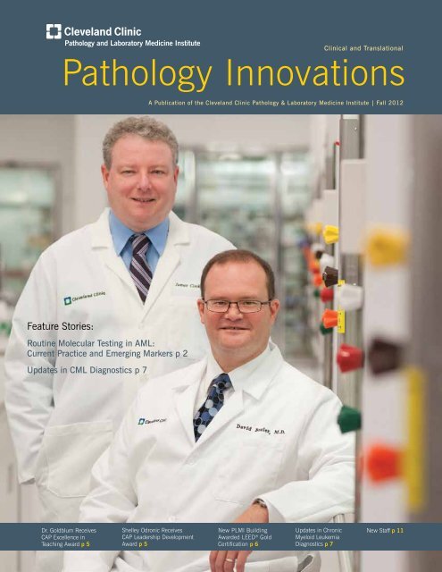
<strong>Pathology</strong> <strong>Innovations</strong><br />
A Publication of the <strong>Cleveland</strong> <strong>Clinic</strong> <strong>Pathology</strong> & Laboratory Medicine Institute | Fall 2012<br />
Feature Stories:<br />
Routine Molecular Testing in AML:<br />
Current Practice and Emerging Markers p 2<br />
Updates in CML Diagnostics p 7<br />
Dr. Goldblum Receives<br />
CAP Excellence in<br />
Teaching Award p 5<br />
Shelley Odronic Receives<br />
CAP Leadership Development<br />
Award p 5<br />
New PLMI Building<br />
Awarded LEED ® Gold<br />
Certification p 6<br />
Updates in Chronic<br />
Myeloid Leukemia<br />
Diagnostics p 7<br />
New Staff p 11
<strong>Pathology</strong> <strong>Innovations</strong> | <strong>Clinic</strong>al | Translational<br />
Routine Molecular Testing in AML:<br />
Current Practice and Emerging Markers<br />
By James R. Cook, MD, PhD<br />
The most common form of acute leukemia in adults, acute<br />
myeloid leukemia (AML) represents a group of clonal hematopoietic<br />
disorders in which both failure to differentiate and<br />
uncontrolled proliferation in the stem cell compartment result<br />
in the accumulation of myeloid blasts. 1,2 A diagnosis of AML<br />
is established through the identification of greater than 20%<br />
myeloid blasts in the bone marrow or peripheral blood. AML<br />
can be further subclassified into specific clinicopathologic<br />
entities that are characterized by distinct clinical features,<br />
differences in prognosis, and, in some cases, differences in<br />
terms of the optimal therapeutic management.<br />
Metaphase cytogenetic studies have been a critical part of<br />
AML diagnosis and classification for many years. Several distinct<br />
subtypes of AML are recognized on the basis of specific<br />
recurring cytogenetic abnormalities, such as balanced translocations<br />
leading to production of oncogenic fusion transcripts.<br />
For example, AML with t(8;21)(q22;q22) RUNX1-RUNX1T1<br />
or inv(16)(p13.1q22) CBFB-MYH11 (sometimes referred to<br />
collectively as “core binding factor leukemias”), each show<br />
characteristic morphologic findings and are associated with<br />
Table 1. AML with recurrent genetic abnormalities, as defined<br />
in the 2008 WHO classification<br />
AML with t(8;21)(q22;q22) RUNX1-RUNX1T1<br />
AML with inv(16)(p13.1q22) or t(16;16)(p13.1;q22) CBFB-MYH11<br />
APL with t(15;17)(q22;q12) PML-RARA<br />
AML with t(9;11)(p22;q23) MLLT3-MLL<br />
AML with t(6;9)(p23;q34) DEK-NUP214<br />
AML with inv(3)(q21q26.2) or t(3;3)(q21;q26.2) RPN1-EVI1<br />
AML with t(1;22)(p13;q13) RBM15-MKL1<br />
AML with mutated NPM1 *<br />
AML with mutated CEBPA *<br />
*provision entities in 2008 WHO edition<br />
a relatively good prognosis. In contrast, cases of AML with<br />
inv(3)(q21q26.2) RPN1-EVI1 are associated with a very<br />
aggressive clinical course and poor outcome. For this reason,<br />
metaphase cytogenetic analysis has long been considered<br />
part of the routine workup and classification of new AML.<br />
In more recent years, it has become clear that other distinct<br />
subtypes of AML can be identified on the basis of recurrent<br />
molecular abnormalities. The 2008 revision of the WHO<br />
classification (Table 1) of hematolymphoid tumors included<br />
two new provisional entities defined by the presence of specific<br />
mutations: AML with mutated NPM1 and AML with mutated<br />
CEBPA. 1,2 Additional studies since 2008 have confirmed that<br />
these disorders represent distinct clinicopathologic entities. In<br />
addition, other molecular abnormalities have been described<br />
that do not define specific subtypes, but are important prognostic<br />
markers in certain subtypes of AML. The most important of<br />
these prognostic markers to date are FLT3 and KIT. Molecular<br />
studies for mutations in NPM1, FLT3, CEBPA and KIT now<br />
represent part of the routine workup of AML, 3,4 and these<br />
genetic markers are included in current NCCN and European<br />
LeukemiaNet guidelines for diagnosis, prognostic<br />
assessment, and therapeutic decision-making<br />
(Table 2).<br />
The NPM1 gene at chromosome 5q35 encodes<br />
nucleophosmin, a nucleolar protein that appears<br />
to play a role as a histone chaperone and in<br />
ribosome biogenesis. Mutations in NPM1 are<br />
found in approximately one-third of AML, making<br />
this one of the most common abnormalities<br />
in AML. More than 40 different mutations in<br />
NPM1 have been described, but all represent<br />
small insertions (usually 4 nucleotides) in exon<br />
12. Mutations in NPM1 can be identified by<br />
PCR of the relevant region of exon 12 followed<br />
by capillary electrophoresis to assess for the<br />
characteristic 4bp insertion. Cases of AML<br />
with mutated NPM1 often show monocytic<br />
2
Fall | 2012<br />
Table 2. Risk status based on cytogenetic and molecular abnormalities in current NCCN guidelines. 3<br />
Risk Status Cytogenetic Abnormalities Molecular Abnormalities<br />
Better Risk inv(16) or t(16;16) Normal cytogenetics with NPM1 mutation or<br />
t(8;21)<br />
isolated CEBPA mutation without FLT3-ITD<br />
t(15;17)<br />
Intermediate Risk Normal cytogenetics t(8;21), t(16;16) or inv(16) with KIT mutation<br />
+8<br />
t(9;11)<br />
Other non-defined<br />
Poor Risk Complex (≥3 abnormalities) Normal cytogenetics with FLT3-ITD<br />
-5,5q-,-7,7q-<br />
11q23 - non t(9;11)<br />
inv(3), t(3;3)<br />
t(6;9)<br />
t(9;22)<br />
differentiation, and 85-95% of cases show a normal karyotype<br />
by metaphase cytogenetics. The prognosis of NPM1-mutated<br />
AML varies depending on whether or not a FLT3 mutation is<br />
also present, as discussed below.<br />
FLT3 is a receptor tyrosine kinase normally expressed at the<br />
cell surface of hematopoietic precursors. Several types of<br />
mutations in FLT3 occur in AML, including internal tandem<br />
duplications (ITD), tyrosine kinase domain (TKD) mutations,<br />
and rarely juxtamembrane region mutations. The most clinically<br />
important of these mutations is the ITD, a variably sized<br />
duplication that occurs in exons 14-15. PCR for this region<br />
and subsequent fragment length analysis by capillary electrophoresis<br />
allows for the detection of this abnormality. FLT3-ITD<br />
mutations do not define a specific subtype of AML, but rather<br />
can be found in many different forms of AML. The FLT3-ITD<br />
is associated with unfavorable prognosis in AML. FLT3-TKD<br />
mutations also appear to be associated with a less favorable<br />
prognosis in many studies, although this remains somewhat<br />
controversial.<br />
The combination of NPM1 and FLT3-ITD status can be used<br />
to stratify prognosis in AML, especially in cases with normal<br />
karyotype by metaphase cytogenetics. An NPM+/FLT3-ITDgenotype<br />
is associated with a favorable clinical course, while<br />
a NPM1-/FLT3-ITD+ genotype is associated with poor outcome<br />
and other permutations are associated with intermediate<br />
prognosis. This information can be used to guide the choice<br />
of therapy, as NPM1+/FLT3-ITD- cases may not benefit from<br />
undergoing allogeneic bone marrow transplant, while other<br />
cases with other genotypic profiles may.<br />
The CEBPA gene, located at chromosome 19q13.1, encodes<br />
a transcription factor that is involved in differentiation of<br />
granulocytes. Mutations in CEBPA occur in 5-15% of AML<br />
and are extremely diverse. More than 100 different CEBPA<br />
mutations have been described, including point mutations,<br />
insertions and deletions. Due to this marked diversity, mutations<br />
in CEBPA are best detected by Sanger sequencing of<br />
the entire coding region. The majority of mutated cases show<br />
biallelic mutations in CEBPA, usually with one located in the<br />
N-terminal region and one located in the C-terminal region.<br />
Cases with mutated CEBPA, especially those with biallelic<br />
mutations, show a favorable prognosis similar to cases with<br />
an NPM1+/FLT3-ITD- genotype, and often show monocytic<br />
differentiation. Approximately 70% of cases are associated<br />
with a normal karyotype by metaphase cytogenetics. Gene<br />
expression profiling studies have shown that CEBPA mutated<br />
AML has a distinct genetic signature, providing further evidence<br />
that this represents a distinct subtype of AML.<br />
Mutations in the KIT gene on chromosome 4q12 can be<br />
found in approximately 17% of AML cases, including a variety<br />
of cytogenetic subtypes. KIT encodes a receptor tyrosine kinase<br />
that signals through several cellular pathways, and mutations<br />
in this gene lead to increased proliferation. KIT mutations<br />
are very heterogeneous, but the vast majority of mutations in<br />
AML are located in exons 8 and 17. These diverse mutations<br />
3
<strong>Pathology</strong> <strong>Innovations</strong> | <strong>Clinic</strong>al | Translational<br />
are best identified by Sanger sequencing. KIT abnormalities<br />
appear to be especially important in AML with t(8;21) or<br />
inv(16). These core binding factor leukemias are normally<br />
considered to be associated with good prognosis. The presence<br />
of a concurrent KIT mutation, however, is associated with<br />
intermediate risk disease, at least in t(8;21) positive cases.<br />
The effect on prognosis in inv(16) AML is more controversial<br />
and may be weaker. The significance of KIT mutations in other<br />
forms of AML remains uncertain. The NCCN and European<br />
LeukemiaNet guidelines currently include KIT mutation analysis<br />
only for cases carrying t(8;21) and inv(16).<br />
While NPM1, FLT3, CEBPA and KIT mutation studies are<br />
now widely accepted as standard practice in AML diagnosis<br />
and prognostic assessment, studies over the last several years<br />
have identified a diverse array of other recurrently mutated<br />
genes that are also associated with prognostic differences in<br />
AML. Some of the more widely studied abnormalities in this<br />
category include duplications in MLL and mutations in DNMT3A,<br />
RUNX1, TET2, EZH2, ASXL1, IDH1, IDH2 and TP53. 5,6 AML<br />
researchers now face the challenge of determining which<br />
mutations, or combinations of these mutations, represent the<br />
most important abnormalities to identify, and how to use this<br />
information to guide selection of therapeutic regimens. For<br />
example, a recent landmark New England Journal of Medicine<br />
study by Patel et al 7 examined mutations in 17 genes, and<br />
identified an 11-gene panel of abnormalities that greatly refines<br />
prognostic evaluation of AML. It is likely that the next few years<br />
will see numerous similar studies in an effort to define an<br />
optimal consensus panel. The major challenge for molecular<br />
diagnostic laboratories in this period will be to identify techniques<br />
to evaluate such genetic panels in a timely, cost-effective<br />
manner. The Department of Molecular <strong>Pathology</strong> at PLMI is<br />
currently exploring options for the use of next-generation<br />
sequencing technologies for such applications.<br />
Cost-effective utilization of molecular testing is possible using<br />
an algorithmic approach. Samples for metaphase cytogenetics<br />
and DNA extraction should be obtained in any bone marrow<br />
biopsy for suspected AML. Extracted DNA may then simply be<br />
held in the laboratory, pending results of metaphase cytogenetic<br />
studies. In the case of normal karyotype, reflex testing for<br />
NPM1, FLT3, and CEBPA mutations is recommended. In the<br />
case of AML with t(8;21) or inv(16), KIT mutation studies<br />
may be performed. In contrast, in cases where metaphase<br />
cytogenetic results clearly identify a poor risk genotype, further<br />
molecular testing may not be necessary. This type of integrated,<br />
algorithmic approach is likely to become even more important<br />
as the number of mutational studies performed continues to<br />
grow in the future.<br />
References<br />
1. Swerdlow SH, Campo ES, Stein H, et al, eds. WHO<br />
Classification of Tumours of Haematopoietic and Lymphoid<br />
Tissues. Lyon, France: IARC;2008.<br />
2. Vardiman JW, Thiele J, Arber DA, et al. The 2008 revision<br />
of the World Health Organization (WHO) classification of<br />
myeloid neoplasms and acute leukemia: rationale and<br />
important changes. Blood 2009;114:937-951.<br />
3. O’Donnell MR, Abboud CN, Altman J, et al. Acute myeloid<br />
leukemia. J Natl Compr Nac Netw 2012;10:984-1021.<br />
4. Dohner H, Estey EH, Amadori S, et al. Diagnosis and<br />
management of acute myeloid leukemia in adults: recommendations<br />
from an international expert panel, on behalf<br />
of the European LeukemiaNet. Blood 2010;115:453-74.<br />
5. Dohner H and Gaidzik VI. Impact of genetic features on<br />
treatment decisions in AML. Hematology Am Soc Hematol<br />
Educ Program. 2011;36-42.<br />
6. Abdel-Wahab O. Molecular genetics of acute myeloid<br />
leukemia: clinical implications and opportunities for<br />
integrating genomics into clinical practice. Hematology<br />
2012;17 Suppl 1:S39-42.<br />
7. Patel JP, Gonen M, Figueroa ME, et al. Prognostic<br />
relevance of integrated genetic profiling in acute myeloid<br />
leukemia. N Engl J Med 2012;366:1079-89.<br />
4
Fall | 2012<br />
Dr. Goldblum Receives CAP Excellence in Teaching Award<br />
John R. Goldblum,<br />
MD, FACP, chair of<br />
the Department of<br />
Anatomic <strong>Pathology</strong>,<br />
received the CAP<br />
Excellence in Teaching<br />
Award from the College<br />
of American Pathologists<br />
(CAP) at the<br />
organization’s annual<br />
meeting in San Diego<br />
September 9.<br />
Dr. Goldblum was<br />
recognized for his<br />
expertise in anatomic<br />
and surgical pathology, particularly soft tissue pathology, and<br />
his effectiveness as a three-time faculty member at the CAP’s<br />
annual meetings. The College established the CAP Excellence<br />
in Teaching Award to individuals who contribute outstanding<br />
contributions as faculty for one or more education activities<br />
resulting in exceptional professional development opportunities<br />
for pathologists.<br />
“It is my sincere honor to receive this award,” says Dr. Goldblum.<br />
“Soft tissue pathology is among the more difficult areas of<br />
surgical pathology, and one of the most rewarding aspects<br />
of my professional career is serving as faculty at the CAP’s<br />
annual meetings.”<br />
Dr. Goldblum specializes in the interpretation of biopsy and<br />
resection specimens in the fields of soft tissue pathology and<br />
gastrointestinal pathology. Co-author of the world’s largest<br />
selling textbooks on soft tissue tumors and gastrointestinal<br />
pathology, he has lectured extensively and published more<br />
than 200 peer-reviewed articles.<br />
As an active member of the CAP, Dr. Goldblum served on the<br />
Surgical <strong>Pathology</strong> Committee. He also is a member of numerous<br />
other medical professional societies, including the Arthur Purdy<br />
Stout Society of Surgical Pathologists, the Gastrointestinal<br />
<strong>Pathology</strong> Society, the United States & Canadian Academy of<br />
<strong>Pathology</strong>, and the American Society for <strong>Clinic</strong>al <strong>Pathology</strong>.<br />
After growing up in Pittsburgh, Dr. Goldblum received his<br />
bachelor of science degree in biology and his medical degree<br />
from the University of Michigan. He went on to complete his<br />
residency in anatomic pathology serving as chief resident,<br />
and later he served as a fellow in surgical pathology under Dr.<br />
Sharon W. Weiss. He is certified in anatomic pathology by<br />
the American Board of <strong>Pathology</strong>.<br />
The College of American Pathologists is currently celebrating<br />
50 years as the gold standard in laboratory accreditation,<br />
serving more than 18,000 physician members and the global<br />
laboratory community. It is the world’s largest association<br />
composed exclusively of board-certified pathologists and is<br />
the worldwide leader in laboratory quality assurance.<br />
Shelley Odronic Receives CAP Leadership Development Award<br />
The College of American Pathologists Foundation presented Shelley Odronic,<br />
MD, a chief resident in Anatomic <strong>Pathology</strong>, with its 2012 CAP Foundation<br />
Leadership Development Award during the CAP House of Delegates/Residents<br />
Forum Joint Session on September 8. The award is a travel stipend to help<br />
defray travel expenses for attending two consecutive CAP meetings. The Joint<br />
Session preceded CAP ’12 – The Pathologists’ Meeting in San Diego.<br />
5
New PLMI Building Awarded LEED ® Gold Certification<br />
for Energy and Environmental Design<br />
The <strong>Cleveland</strong> <strong>Clinic</strong> <strong>Pathology</strong> and Laboratory Medicine Institute’s new<br />
LL Building was recently awarded LEED® Gold certification, established<br />
by the U.S. Green Building Council and verified by the Green Building<br />
Certification Institute (GBCI). LEED stands for “Leadership in Energy and<br />
Environmental Design” and is the nation’s preeminent program for the<br />
design, construction and operation of high performance green buildings.<br />
“From the very beginning,<br />
<strong>Cleveland</strong> <strong>Clinic</strong> and PLMI<br />
were dedicated to building a<br />
cutting-edge laboratory facility<br />
that also featured sustainable<br />
and environmentally friendly<br />
designs.”<br />
— Kandice Kottke-Marchant, MD, PhD,<br />
PLMI Chair<br />
“From the very beginning, <strong>Cleveland</strong> <strong>Clinic</strong> and PLMI were dedicated to<br />
building a cutting-edge laboratory facility that also featured sustainable<br />
and environmentally friendly designs,” says Kandice Kottke-Marchant,<br />
MD, PhD, PLMI Chair. “This achievement is certainly a testament to<br />
hours of hard work and dedication. A special thank you to Perspectus<br />
Architecture, Donley’s Inc. Construction, <strong>Cleveland</strong> <strong>Clinic</strong> Construction<br />
Management, and everyone else who had a hand in this accomplishment.”<br />
In order to obtain this certification, the team committed to green goals and<br />
enhanced sustainable performance in the following categories: sustainable<br />
sites, water efficiency, energy and atmosphere, materials and resources,<br />
indoor environmental quality, and innovation and design process.<br />
For example, energy efficiencies are achieved through natural lighting,<br />
daylight responsive lighting controls, LED lighting and exhaust air energy<br />
recovery, helping to achieve a 41.6% improvement in building energy<br />
performance compared to the baseline model. In addition, the use of<br />
water efficient irrigation systems, automated weather monitoring and<br />
drought-tolerant landscape materials reduces potable water use by 83%.<br />
Opened in January, the new building doubles the size of <strong>Cleveland</strong> <strong>Clinic</strong><br />
<strong>Laboratories</strong> and is designed to support testing needs in microbiology,<br />
special chemistry, immunopathology and molecular pathology.<br />
6
Fall | 2012<br />
Updates in Chronic Myeloid Leukemia Diagnostics<br />
By David Bosler, MD<br />
Molecular diagnostics testing plays an important role in many<br />
facets of diagnosis and management of chronic myelogenous<br />
leukemia (CML), including diagnosis, disease monitoring and<br />
prognosis, and response to therapy and resistance.<br />
The impact of molecular diagnostics on the care of patients<br />
with CML has only grown with time, with integral components<br />
of contemporary management including disease-defining diagnostic<br />
tests, guiding choice of therapy, monitoring of response<br />
to therapy through minimal residual disease testing, and testing<br />
for development of resistance to therapy. In addition, recent<br />
updates, such as the 2012 National Comprehensive Cancer<br />
Network clinical practice guidelines and International Scale<br />
for standardization of reporting between laboratories, continue<br />
to advance the appropriate use of molecular diagnostics to<br />
optimize patient care.<br />
The development of tyrosine kinase inhibitors (TKIs) such<br />
as imatinib revolutionized therapy for CML and has served<br />
as a model for other malignancies. After demonstration of its<br />
clinical utility, imatinib was approved for use in 2001 for the<br />
treatment of CML and was the first molecular-targeted therapy<br />
approved for use in human cancer. Other therapies, such as<br />
nilotinib, dasatinib and bosutinib have followed, with FDA<br />
approval of panotinib also anticipated. Since the introduction<br />
of imatinib, long-term survival rates for CML have increased<br />
to 95%, allowing better survival and quality of life when<br />
compared to previous therapeutic regimens.<br />
The transformation of CML treatment and course that was<br />
provided by tyrosine kinase inhibitors (TKIs) was largely made<br />
possible by early knowledge of the molecular genetic events<br />
that cause CML. CML has been known for decades as the<br />
myeloproliferative disease associated with the “Philadelphia<br />
chromosome,” an abnormally small chromosome 22 created<br />
by a reciprocal translocation of chromosomes 9 and 22. The<br />
BCR-ABL fusion gene created on this newly derived chromosome<br />
22 gives rise to a chimeric fusion protein with abnormally<br />
enhanced and unregulated tyrosine kinase activity. Unchecked,<br />
this tyrosine kinase activity sends signals downstream for<br />
myeloid precursors to proliferate that result in CML.<br />
Testing at Diagnosis<br />
Since CML is a disease defined by the presence of the BCR-ABL<br />
fusion, a sensitive method of detecting the fusion is critical to<br />
accurate diagnosis. Fluorescence in situ hybridization (FISH)<br />
for BCR-ABL is a sensitive and specific method that is most<br />
widely used for diagnosis. Dual color, dual fusion probe<br />
strategies employ fluorescently labeled probes that span and<br />
flank the full range of breakpoints for both BCR and ABL,<br />
allowing a fusion signal to be detected regardless of which<br />
break point is present. Since the probe sets flank the breakpoints,<br />
the reciprocal translocation also creates two fusion<br />
signals, significantly enhancing specificity over single fusion<br />
strategies. FISH can be performed on interphase cells, so<br />
results are not dependent on cell culture, resulting in the<br />
ability to perform testing on a wider range of samples as well<br />
as a more rapid turnaround time than can be achieved by the<br />
culture-dependent cytogenetic karyotyping. Additionally,<br />
BCR-ABL fusions can be detected by FISH in rare Philadelphia<br />
chromosome-negative cases of CML.<br />
Comprehensive multiplex PCR strategies have also been<br />
employed as a reliable and sensitive means of detecting BCR-<br />
ABL fusions. This method provides the flexibility and sensitivity<br />
of FISH testing because it can be designed to detect all possible<br />
break points. It also provides information at diagnosis about<br />
which break points are present, which cannot be determined<br />
by FISH.<br />
Although cytogenetic karyotyping detects t(9:22) and complex<br />
translocations involving these chromosomes in most cases,<br />
a small percentage of cases is not detectable by this method<br />
and would be missed if cytogenetics were used alone. Cytogenetic<br />
karyotyping is also time consuming and may result<br />
in delayed diagnosis compared to other methods. However,<br />
cytogenetic karyotyping maintains value as a diagnostic tool<br />
because it provides information about abnormalities that<br />
may be present elsewhere within the karyotype, such as a<br />
second Philadelphia chromosome, trisomy 8 or isochromosome<br />
17q. Since none of the other more targeted methods used in<br />
CML diagnosis detect these abnormalities, cytogenetic karyotyping<br />
provides information that is complementary to these<br />
other techniques.<br />
RT-PCR designed to detect the BCR-ABL transcript for the<br />
p210 fusion is a rapid and very analytically sensitive technique,<br />
but its use at initial diagnosis is limited since it detects only<br />
the p210 fusion. A negative result does not exclude CML since<br />
a small percentage of cases have alternate fusion transcripts<br />
that would be missed by a primer set designed to detect only<br />
7
<strong>Pathology</strong> <strong>Innovations</strong> | <strong>Clinic</strong>al | Translational<br />
the p210 fusion. Additionally, rare deletions occurring in or<br />
near the primers may cause false-negative results by PCR<br />
even when the correct breakpoints are targeted.<br />
In practice, molecular diagnostic testing for CML at diagnosis<br />
uses a combination of each of these methods. While FISH or<br />
multiplex PCR are best used to make the diagnosis, cytogenetic<br />
karyotyping provides complementary information regarding the<br />
presence of any other genetic abnormalities, and BCR-ABL<br />
RT-PCR is useful to provide a baseline level of fusion transcript<br />
for subsequent monitoring during therapy.<br />
Response to Therapy<br />
Although of limited use at initial diagnosis, RT-PCR for BCR-<br />
ABL p210 fusion is the gold standard for monitoring disease<br />
burden during therapy. BCR-ABL fusion transcripts are measured<br />
quantitatively over time to track response to therapy.<br />
The results are expressed in normalized copy number (NCN),<br />
the ratio of BCR-ABL copy number to the copy number of a<br />
control gene (often normal ABL), and compared to either the<br />
patient’s baseline NCN at diagnosis or a standardized baseline<br />
value. In tracking response to therapy, a major molecular<br />
response is defined as a 3 log reduction (or 0.1%) from<br />
this baseline value. One recent improvement to BCR-ABL<br />
quantitative testing is the use of standards and the International<br />
Scale (IS) to provide results that are comparable across<br />
different laboratories. Without the use of the International Scale,<br />
results can vary greatly between laboratories due to the many<br />
different methods used to obtain results. The International<br />
Scale uses calibrators of known concentrations derived from<br />
World Health Organization-certified NIBSC reference material<br />
for both BCR-ABL and ABL. By plotting patient results against<br />
the results of the calibration curves, the patient result can be<br />
converted to an International Scale value that is standardized<br />
across all different labs that use the IS. These standards have<br />
significantly advanced the efforts at inter-laboratory standardization<br />
and have improved the care of patients who are seen<br />
at more than one medical center.<br />
National Comprehensive Cancer Network (NCCN) Guidelines<br />
provide guidance for monitoring response to therapy in CML.<br />
Different levels and types of responses (hematologic, cytogenetic,<br />
molecular) are expected at different time points in order<br />
for a response to be considered optimal. For example, optimal<br />
responses based on the 2009 European LeukemiaNet (ELN)<br />
recommendations include complete hematologic response at<br />
3 months, complete cytogenetic response at 12 months, and<br />
major molecular response at 18 months. Patient responses<br />
are categorized as optimal, sub-optimal or failure by NCCN<br />
based on outcomes studies, and the criteria used for measuring<br />
response drive the specific methods used to monitor<br />
response at each time point. Although hematologic response<br />
and BCR-ABL fusion transcript levels can be determined<br />
from peripheral blood, cytogenetic karyotyping requires bone<br />
marrow sampling.<br />
2012 National Comprehensive Cancer Network<br />
(NCCN) Guidelines<br />
The 2012 NCCN CML Guidelines update the recommendations<br />
for disease monitoring and therapy based on newly available<br />
outcomes studies. The updated guidelines generally seek to<br />
provide greater clarity surrounding when to alter the current<br />
course of therapy by changing to a different TKI based on<br />
sub-optimal and/or failure level responses, providing clinicians<br />
with a proposed course of action using the best tools and<br />
knowledge available.<br />
One significant change in the 2012 guidelines compared to<br />
the 2009 European LeukemiaNet (ELN) Recommendations is<br />
the emphasis on early response. At the three-month evaluation,<br />
continuation of current therapy requires that the patient is<br />
demonstrating optimal response (ie. complete hematologic<br />
and partial cytogenetic responses), and also places increased<br />
emphasis on early molecular response by adding a one log<br />
reduction of BCR-ABL fusion transcripts to the criteria for<br />
optimal response. This added criterion is based on studies<br />
showing improved long-term overall survival in patients that<br />
achieve a one log reduction after 3 months of imatinib therapy.<br />
For anything less than optimal response at 3 months, the<br />
NCCN guidelines recommend evaluation of patient compliance<br />
and possible drug-drug interactions, as well as ABL kinase<br />
mutation analysis for the purposes of switching to the<br />
appropriate second generation TKI.<br />
Another significant change in the 2012 update of NCCN<br />
guidelines is the de- emphasized importance of achieving a<br />
major molecular response (MMR) at 18 months. MMR was<br />
previously the sole criterion for optimal response in the 2009<br />
ELN Recommendations. However many studies show that,<br />
among patients achieving complete cytogenetic response, the<br />
long-term survival does not vary significantly between those<br />
achieving MMR and those without MMR. The 2012 NCCN<br />
Guidelines therefore essentially lump optimal (MMR and<br />
complete cytogenetic response) and sub-optimal (complete<br />
cytogenetic response but no MMR) ELN responses together<br />
in order to make the more clinically important distinction<br />
between complete cytogenetic response and lack of complete<br />
cytogenetic response at 18 months. The lack of complete<br />
8
Fall | 2012<br />
cytogenetic response is what drives<br />
evaluation for mutations and switching<br />
to a second generation TKI at 18 months<br />
Mutation<br />
under the new guidelines. Failure to<br />
meet milestones at any time or loss of T3151<br />
response in therapy adherent patients<br />
should prompt ABL kinase mutation<br />
analysis. A complete list of the 2012<br />
NCCN guidelines for CML can be found<br />
at: National Comprehensive Cancer<br />
Network. NCCN clinical practice guidelines<br />
in oncology: chronic myelogenous<br />
leukemia. http://www.nccn.org/professionals/physician_gls/f_<br />
guidelines.asp.<br />
Therapy Resistance Testing<br />
Over 100 mutations at a variety of locations within the ABL<br />
kinase domain can confer resistance to various TKIs. Although<br />
the TKIs themselves are not mutagenic, use of a TKI creates<br />
an environment that can allow a mutated clone to emerge,<br />
somewhat analogous to the emergence of antibiotic resistant<br />
bacterial strains under the pressure of antibiotic use. These<br />
mutations may interfere with the binding or function of TKIs<br />
differentially, so that a given mutation may confer resistance<br />
to one TKI and not others. Failure to respond to TKI therapy<br />
in CML or loss of response is often due to the emergence of a<br />
clone bearing a resistance-conferring ABL kinase mutation,<br />
and given the differential effects of these various mutation,<br />
analysis of which mutation is present provides an important<br />
contribution to guiding choice of therapy. Gene sequencing is<br />
one of the most appropriate methods employed for ABL kinase<br />
mutation analysis given the number of possible mutations,<br />
although multiplex PCR approaches have also been used<br />
to effectively interrogate a more limited number of mutation<br />
hot spots.<br />
One of the challenges surrounding interpretation of ABL kinase<br />
mutation analysis is understanding the clinical implications of<br />
each particular mutation. A number of mutations that can be<br />
encountered in the ABL kinase domain have either no known<br />
effect or incompletely understood effect on TKI resistance.<br />
However, sufficient clarity has emerged for a subset of these<br />
mutations for them to be included into recommendations<br />
regarding therapy. In fact, for a few of these mutations, the<br />
differential effects on different second generation TKIs allow<br />
recommendation of one TKI over another (see Table 1 above).<br />
The T315I mutation is both the most common mutation<br />
detected after imatinib failure and the most TKI resistant of<br />
Table 1. Guideline Recommendations for Treating Patients with Mutations<br />
V299L, T315A, F317L/V/I/C<br />
Y253H, E255K/V, F359V/C/I<br />
Any other mutations<br />
ABL kinase mutations. Cases of CML that have the T315I<br />
mutation are resistant to all approved TKI therapies to date,<br />
making hematopoietic stem cell transplantation (HSCT) or<br />
clinical trial participation the current recommended options.<br />
A new drug called ponatinib, currently in clinical trials, has<br />
shown significant activity against CML with T3151 mutations,<br />
and may prove a viable new option for this currently refractory<br />
group. If ponatinib receives FDA approval and proves effective<br />
against T315I mutated cases, the role of testing for the<br />
T3151 will likely expand significantly.<br />
Summary<br />
Treatment<br />
HSCT or clinical trial (ponatinib)<br />
Consider nilotinib rather than dasatinib<br />
Consider dasatinib rather than nilotinib<br />
Consider high-dose imatinib or dasatinib<br />
or nilotinib<br />
At diagnosis, reliable and sensitive methods for detection of<br />
BCR-ABL fusion include fluorescence in situ hybridization and<br />
some comprehensive multiplex RT-PCR assays. Cytogenetic<br />
karyotyping and PCR for the p210 fusion also detect the vast<br />
majority of CML cases, but they miss cases that are cytogenetically<br />
cryptic or that involve variant breakpoints, respectively.<br />
Nonetheless, it remains important to perform these tests at<br />
diagnosis in order to get baseline information that will become<br />
important in disease monitoring. Assuming that a p210 BCR-<br />
ABL fusion is present at diagnosis, quantitative RT-PCR designed<br />
to detect the p210 fusion is the most sensitive method<br />
of monitoring residual disease. Obtaining a baseline result<br />
for the RT-PCR p210 assay is helpful in order to ensure that<br />
cases are subsequently negative for the BCR-ABL fusion during<br />
disease monitoring because they have achieved complete<br />
molecular response rather than because they have a variant<br />
rearrangement that is not detected by the assay. Additionally,<br />
cytogenetic karyotyping remains an important method of detecting<br />
additional cytogenetic abnormalities, such as a second<br />
Philadelphia chromosome, trisomy 8 or isochromosome 17q,<br />
which may be harbingers of disease progression and are not<br />
detected by RT-PCR or FISH. When molecular disease monitoring,<br />
hematologic findings or clinical course suggests loss of<br />
response to therapy, analysis of the ABL kinase for mutations<br />
9
<strong>Pathology</strong> <strong>Innovations</strong> | <strong>Clinic</strong>al | Translational<br />
Relevant CCL Tests:<br />
Test Name<br />
• Cytogenetic karyotyping<br />
• BCR-ABL FISH<br />
• RT-PCR for BCR-ABL p210 transcript<br />
• RT-PCR for BCR-ABL p190 transcript<br />
• ABL kinase mutation analysis<br />
• BCR-ABL multiplex PCR for diagnosis<br />
Ordering Code<br />
CHRBMH, CHRBLL<br />
BCRFSH<br />
BCRPCR<br />
190BCR<br />
KINASE<br />
(in development)<br />
8. Dessars B, El HH, Lambert F, Kentos A, Heimann P.<br />
Rational use of the EAC real-time quantitative PCR protocol<br />
in chronic myelogenous leukemia: report of three falsenegative<br />
cases at diagnosis. Leukemia. 2006;20:886-888.<br />
9. Gabert J, Beillard E, van der Velden VJH, et al. Standardization<br />
and quality control studies of ‘real-time’ quantitative<br />
reverse transcriptase polymerase chain reaction of fusion<br />
gene transcripts for residual disease detection in leukemia<br />
– a Europe Against Cancer Program. Leukemia.<br />
2003;17:2318-2357.<br />
that confer resistance to imatinib and/or other tyrosine kinase<br />
inhibitors helps to guide the choice of subsequent therapy.<br />
References<br />
1. Druker BJ, Lydon NB. Lessons learned from the development<br />
of an abl tyrosine kinase inhibitor for chronic<br />
myelogenous leukemia. J Clin Invest. 2000;105:3-7.<br />
2. Piccaluga PP, Rondoni M, Paolini S, Rosti G, Martinelli<br />
G, Baccarani M. Imatinib mesylate in the treatment<br />
of hematologic malignancies. Expert Opin Biol Ther.<br />
2007;7:1597-1611.<br />
3. Cortes JE, Tapaz M, O’Brien S, et al. Staging of chronic<br />
myeloid leukemia in the imatinib era: an evaluation<br />
of the World Health Organization proposal. Cancer.<br />
2006;106:1306-1315.<br />
4. Melo JV. The diversity of BCR-ABL fusion proteins<br />
and their relationship to leukemia phenotype. Blood.<br />
1996;88:2375-2384.<br />
5. Landstrom AP, Ketterling RP, Knudson RA, Tefferi A. Utility<br />
of peripheral blood dual color, double fusion fluorescent<br />
in situ hybridization for BCR/ABL fusion to assess cytogenetic<br />
remission status in chronic myeloid leukemia.<br />
Leuk Lymphoma. 2006;47:2055-2061.<br />
6. Costa D, Espinet B, Queralt R, et al. Chimeric BCR/ABL:<br />
gene detected by fluorescence in situ hybridization in<br />
three new cases of Philadelphia chromosome-negative<br />
chronic myelocytic leukemia. Cancer Genet Cytogenet.<br />
2003;141:114-119.<br />
7. Ou J, Vergilio JA, Bagg A. Molecular diagnosis and<br />
monitoring in the clinical management of patients with<br />
chronic myelogenous leukemia treated with tyrosine<br />
kinase inhibitors. Am J Hematol. 2008;83:296-302.<br />
10. Hughes T, Deininger M, Hochhaus A, et al. Monitoring<br />
CML patients responding to treatment with tyrosine kinase<br />
inhibitors: review and recommendations for harmonizing<br />
current methodology for detecting BCR-ABL transcripts<br />
and kinase domain mutations and for expressing results.<br />
Blood. 2006;108:28-37.<br />
11. Baccarani M, Saglio G, Goldman J, et al. Evolving concepts<br />
in the management of chronic myeloid leukemia:<br />
recommendations for an expert panel on behalf of the<br />
European LeukemiaNet. Blood. 2006;108:1809-1820.<br />
12. Marin D, Ibrahim AR, Lucas C, et al. Assessment of BCR-<br />
ABL1 transcript levels at 3 months is the only requirement<br />
for predicting outcome for patients with chronic myeloid<br />
leukemia treated with tyrosine kinase inhibitors. J Clin<br />
Oncol. 2012;30:232-238.<br />
13. National Comprehensive Cancer Network. NCCN clinical<br />
practice guidelines in oncology: chronic myelogenous<br />
leukemia. http://www.nccn.org/professionals/physician_<br />
gls/f_guidelines.asp. Revised July 5, 2012.<br />
14. Soverini S, Hochhaus A, Nicolini FE, et al. BCR-ABL kinase<br />
domain mutation analysis in chronic myeloid leukemia<br />
patients treated with tyrosine kinase inhibitors: recommendations<br />
from an expert panel on behalf of European<br />
LeukemiaNet. Blood. 2011; 118(5):1208-1215.<br />
15. Diamond JM, Melo JV. Mechanisms of resistance to BCR-<br />
ABL kinase inhibitors. Leuk Lymphoma. 2011 Feb;52<br />
Suppl 1:12-22.<br />
16. Redaelli S, Piazza R, Rostagno R, et al. Activity of Bosutinib,<br />
Dasatinib, and Nilotinib against 18 Imatinib-resistant<br />
BCR/ABL mutants. J Clin Oncol. 2009;27:469-471.<br />
10
Fall | 2012<br />
NEW STAFF<br />
Ilyssa Gordon, MD, PhD<br />
Anatomic <strong>Pathology</strong><br />
Board Certifications:<br />
Anatomic <strong>Pathology</strong>,<br />
<strong>Clinic</strong>al <strong>Pathology</strong><br />
Specialty Interests: Gastrointestinal<br />
and Pulmonary <strong>Pathology</strong><br />
Phone: 216.444.8245<br />
Email: gordon1@ccf.org<br />
James Lapinski, MD<br />
Regional <strong>Pathology</strong><br />
Board Certifications:<br />
Anatomic <strong>Pathology</strong>,<br />
<strong>Clinic</strong>al <strong>Pathology</strong><br />
Specialty Interests: Gastrointestinal<br />
<strong>Pathology</strong><br />
Phone: 440.312.4393<br />
Email: lapinsj@ccf.org<br />
Jinesh Patel, MD<br />
Anatomic <strong>Pathology</strong><br />
Board Certifications:<br />
Anatomic <strong>Pathology</strong>,<br />
<strong>Clinic</strong>al <strong>Pathology</strong><br />
Specialty Interests: Cytopathology and<br />
Head and Neck <strong>Pathology</strong><br />
Phone: 216.444.8143<br />
Email: patelj6@ccf.org<br />
Roger Klein, MD, JD<br />
Molecular <strong>Pathology</strong><br />
Board Certifications:<br />
<strong>Clinic</strong>al <strong>Pathology</strong>,<br />
Molecular <strong>Pathology</strong><br />
Specialty Interests: Molecular Oncology<br />
Phone: 216.445.0776<br />
Email: kleinr3@ccf.org<br />
Roy Lee, MD<br />
Associate Medical<br />
Director, CPI and<br />
Molecular <strong>Pathology</strong><br />
Board Certifications:<br />
Anatomic <strong>Pathology</strong>, <strong>Clinic</strong>al <strong>Pathology</strong><br />
Specialty Interests: Whole genome<br />
sequencing, bioinformatics, personalized<br />
medicine<br />
Phone: 216.444.8096<br />
Email: leer3@ccf.org<br />
Anthony Simonetti, MD<br />
Regional <strong>Pathology</strong> and<br />
CP Director, Preanalytic<br />
Board Certifications:<br />
Anatomic <strong>Pathology</strong>,<br />
<strong>Clinic</strong>al <strong>Pathology</strong><br />
Specialty Interests: Administration and<br />
Pre-Analytics<br />
Phone: 216.444.3587<br />
Email: simonea2@ccf.org<br />
Jesse McKenney, MD<br />
Anatomic <strong>Pathology</strong><br />
Board Certification:<br />
Anatomic <strong>Pathology</strong><br />
Specialty Interest:<br />
Genitourinary <strong>Pathology</strong><br />
Phone: 216.444.1058<br />
Email: mckennj@ccf.org<br />
11
<strong>Pathology</strong> <strong>Innovations</strong> Magazine<br />
offers information from the medical<br />
staff in the <strong>Cleveland</strong> <strong>Clinic</strong><br />
<strong>Pathology</strong> & Laboratory Medicine<br />
Institute about its research, services<br />
and laboratory technology.<br />
Raymond Tubbs, DO<br />
Medical Editor<br />
216.444.2844<br />
Editoral Board:<br />
Thomas Bauer, MD, PhD<br />
James Cook, MD, PhD<br />
John Goldblum, MD<br />
Eric Hsi, MD<br />
Lisa Yerian, MD<br />
Kathy Leonhardt, Director<br />
Marketing and Communications<br />
Gary Weiland, Editor<br />
Ruth Clark, Designer<br />
Don Gerda, Photographer<br />
© 2012 The <strong>Cleveland</strong> <strong>Clinic</strong> Foundation<br />
About the Authors<br />
James Cook, MD, PhD, is<br />
an associate professor of<br />
<strong>Pathology</strong> at the <strong>Cleveland</strong><br />
<strong>Clinic</strong> Lerner College of<br />
Medicine. He serves as<br />
Section Head of Molecular<br />
Hematopathology,<br />
Medical Director of the<br />
Manual Hematology<br />
Laboratory and Co-director<br />
of the Cell Culture Core Facility for the <strong>Pathology</strong><br />
and Laboratory Medicine Institute.<br />
Dr. Cook’s clinical interests focus on diagnostic<br />
hematopathology and molecular diagnostics.<br />
His research interests include diagnostic and<br />
prognostic markers in non-Hodgkin lymphoma<br />
and plasma cell disorders, and molecular<br />
diagnosis of leukemia and lymphoma. He is<br />
a member of the Lymphoma Translational<br />
Medicine Committee for the Southwest Oncology<br />
Group, and he regularly serves as faculty for<br />
post-graduate educational courses at national<br />
meetings.<br />
Dr. Cook received his undergraduate degree<br />
from Pennsylvania State University, and his<br />
medical and doctorate degrees from Washington<br />
University School of Medicine in St. Louis. He<br />
served his residency in anatomic pathology<br />
at Barnes-Jewish Hospital in St. Louis and<br />
completed a fellowship in hematopathology at<br />
the University of Pittsburgh School of Medicine.<br />
Dr. Cook is certified in anatomic pathology and<br />
hematopathology. He is a past recipient of the<br />
Pathologist in-Training Award from the Society<br />
for Hematopathology and the John Beach<br />
Hazard Distinguished Teaching Award in the<br />
<strong>Cleveland</strong> <strong>Clinic</strong> Institute of <strong>Pathology</strong> and<br />
Laboratory Medicine.<br />
Dr. Cook can be reached at cookj2@ccf.org.<br />
David Bosler, MD, is<br />
Head of <strong>Cleveland</strong> <strong>Clinic</strong><br />
<strong>Laboratories</strong> within the<br />
<strong>Pathology</strong> & Laboratory<br />
Medicine Institute at<br />
<strong>Cleveland</strong> <strong>Clinic</strong>. He is<br />
responsible for the<br />
strategic execution of<br />
<strong>Cleveland</strong> <strong>Clinic</strong><br />
<strong>Laboratories</strong>’ business plan for local, regional<br />
and national outreach of reference laboratory<br />
testing. Dr. Bosler also is Staff in Hematopathology<br />
in the Department of <strong>Clinic</strong>al <strong>Pathology</strong> and<br />
Assistant Professor, <strong>Cleveland</strong> <strong>Clinic</strong> Lerner<br />
College of Medicine. He is involved in bone<br />
marrow sign-out and assay development in<br />
molecular hematology, as well as serving as<br />
Co-Chair of the Point-of-Care Testing Compliance<br />
Council.<br />
Dr. Bosler received his undergraduate degree<br />
from Miami University and his medical degree<br />
from the University of Cincinnati. He completed<br />
fellowships in hematopathology and molecular<br />
genetic pathology at Mayo <strong>Clinic</strong>, served his<br />
residency at William Beaumont Hospital in Royal<br />
Oak, Mich., and obtained his medical degree<br />
from the University of Cincinnati College of<br />
Medicine. He has been on the <strong>Cleveland</strong> <strong>Clinic</strong><br />
staff since 2009.<br />
Dr. Bosler is a frequent speaker at national meetings<br />
and has authored numerous peer-reviewed<br />
journal articles and book chapters. He is certified<br />
as a diplomate of the American Board of <strong>Pathology</strong><br />
in Anatomic and <strong>Clinic</strong>al <strong>Pathology</strong>, Hematology<br />
and Molecular Genetic <strong>Pathology</strong>. He currently<br />
serves as a member of the Point-of-Care Testing<br />
Committee for the College of American<br />
Pathologists (CAP).<br />
Dr. Bosler can be reached at boslerd@ccf.org.<br />
The <strong>Cleveland</strong> <strong>Clinic</strong> Foundation<br />
<strong>Pathology</strong> <strong>Innovations</strong> Magazine<br />
9500 Euclid Avenue / LL2-1<br />
<strong>Cleveland</strong>, OH 44195<br />
clevelandcliniclabs.com


