SCREENING FOR CANCER OF THE CERVIX - BC Cancer Agency
SCREENING FOR CANCER OF THE CERVIX - BC Cancer Agency
SCREENING FOR CANCER OF THE CERVIX - BC Cancer Agency

Create successful ePaper yourself
Turn your PDF publications into a flip-book with our unique Google optimized e-Paper software.
<strong>SCREENING</strong> <strong>FOR</strong><br />
<strong>CANCER</strong> <strong>OF</strong> <strong>THE</strong> <strong>CERVIX</strong><br />
An Office Manual for Health Professionals<br />
This manual has been prepared by the Cervical <strong>Cancer</strong> Screening Program<br />
of the <strong>BC</strong> <strong>Cancer</strong> <strong>Agency</strong> to support effective use of the screening program.
Contact Information<br />
Cervical <strong>Cancer</strong> Screening<br />
Program (CCSP)<br />
Medical Leader<br />
Phone: 604-877-6000 Local 2068<br />
Fax: 604-629-2507<br />
E-mail: dvanniek@bccancer.bc.ca<br />
Chief Technologist<br />
Phone: 604-877-6000 Local 4907<br />
Fax: 604-629-2507<br />
E-mail: jlo@bccancer.bc.ca<br />
Operations Leader<br />
Phone: 604-877-6201<br />
Fax: 604-660-3645<br />
E-mail: lkan@bccancer.bc.ca<br />
Program Secretaries<br />
Phone: 604-877-6000 Local 4904 or 6200<br />
Fax: 604-660-3645<br />
E-mail: schou@bccancer.bc.ca<br />
jsentell@bccancer.bc.ca<br />
Provincial Colposcopy Program<br />
Medical Leader<br />
Phone: 604-877-6000 Local 2352<br />
Fax: 604-877-6179<br />
E-mail: tehlen@bccancer.bc.ca<br />
Acknowledgments<br />
The Cervical <strong>Cancer</strong> Screening Program<br />
of the <strong>BC</strong> <strong>Cancer</strong> <strong>Agency</strong> would like to thank<br />
Dr. A. J. Kotaska, General Practitioner,<br />
Dr. R. M. Malleson, Director, Family Planning<br />
and Dr. D. Miller, Chair, Gyne Tumor Group,<br />
<strong>BC</strong> <strong>Cancer</strong> <strong>Agency</strong>, Vancouver Centre,<br />
for their valuable advice and contribution.<br />
Staff & Contributors<br />
Dr. A. Coldman, Vice President,<br />
Population Oncology<br />
S. Chou, Secretary<br />
Dr. T. Ehlen, Director,<br />
Provincial Colposcopy Program<br />
L. St. Germain, Screening Information<br />
Management Leader<br />
Dr. M. Hayes, Pathologist<br />
L. Kan, Screening Operations Leader<br />
J. Lo, Chief Cytotechnologist<br />
J. Sentell, Secretary<br />
Dr. K. Suen, Pathologist<br />
Dr. T. Thomson, Pathologist<br />
Dr. D. van Niekerk, Medical Leader, CCSP<br />
Tumor Group Chair<br />
Phone: 604-877-6000 Local 2354<br />
Fax: 604-877-6179<br />
E-mail: dmiller@bccancer.bc.ca<br />
Pap Smear Supplies<br />
Lower Mainland<br />
Fax request to 604-660-3122<br />
Outside of Lower Mainland<br />
Phone: 1-877-747-2522<br />
Fax: 604-708-8027<br />
Website<br />
www.bccancer.bc.ca<br />
8 th Floor<br />
686 West Broadway<br />
Vancouver, <strong>BC</strong> V5Z 1G1<br />
Phone: 604-877-6200<br />
Fax: 604-660-3645<br />
Seventh Edition 2007
Table of Contents<br />
Introduction<br />
Components of the Cervical <strong>Cancer</strong> Screening Program.................................................................................1<br />
- Recruitment, Recall and Follow-up<br />
- Centralized Laboratory Services<br />
- Quality Assurance and Quality Control<br />
- Evaluation<br />
- Colposcopy Service<br />
Limitations of Screening – Screening Tests are not Diagnostic Tests........................................................2<br />
Facts About Screening and Cervical <strong>Cancer</strong>....................................................................................2<br />
<strong>BC</strong> <strong>Cancer</strong> <strong>Agency</strong> Screening Recommendations<br />
General...........................................................................................................................................................3<br />
Post-Hysterectomy Screening Guidelines........................................................................................................3<br />
Lesbians..........................................................................................................................................................4<br />
Role of the Smear Taker.........................................................................................................................4<br />
The Patient.................................................................................................................................................5<br />
Pap Smear Technique<br />
Labeling the Slide...........................................................................................................................................6<br />
Single Slide Method........................................................................................................................................6<br />
Variations in Cervical Transformation Zone....................................................................................................6<br />
Basic Equipment and Supplies........................................................................................................................7<br />
Obtaining the Smear.......................................................................................................................................7<br />
Completing the Requisition Form........................................................................................................8<br />
Transporting the Specimen....................................................................................................................8<br />
Specimen Rejection Policy....................................................................................................................8<br />
Optimal and Unsatisfactory Smears...................................................................................................9<br />
Human Papilloma Virus and Cervical <strong>Cancer</strong>.................................................................................10<br />
New Technologies..................................................................................................................................10<br />
Liquid Based Cytology (Thin-Layer Cytology)..............................................................................................10<br />
Machine-Assisted Screening..........................................................................................................................10<br />
Appendix I: Guidelines for the Investigation and Management of Women with Screen Detected Abnormalities.....11<br />
Appendix II: Management of Women with Low Grade Epithelial Abnormalities (Mild Dysplasia or CIN 1)... 12<br />
Special Categories.................................................................................................................12<br />
Appendix III: Understanding False Negative Rates.......................................................................................13<br />
Appendix IV: Results Terminology...............................................................................................................14<br />
References...............................................................................................................................................15<br />
Educational Materials Order Form.....................................................................................................16<br />
Feedback Form.......................................................................................................................................17
Introduction<br />
The Cervical <strong>Cancer</strong> Screening Program (CCSP), operated by the <strong>BC</strong> <strong>Cancer</strong> <strong>Agency</strong> (<strong>BC</strong>CA) is a<br />
coordinated program of cervical cancer control. The CCSP is available to all women in the province.<br />
Components of the Cervical <strong>Cancer</strong> Screening Program<br />
Recruitment, Recall and Follow-up<br />
In a true “population-based” screening program,<br />
all at-risk women are personally invited to<br />
participate. At this time, recruitment to the CCSP<br />
is “opportunistic,” i.e.; family doctors and other<br />
health professionals initiate the collection of a Pap<br />
smear from their patients. The Program’s centralized<br />
computer system provides a coordinated system of<br />
recall and follow-up by sending reminders to the<br />
woman’s care provider.<br />
Centralized Laboratory Services<br />
All Pap smears collected around the province<br />
are forwarded to a central cytology laboratory<br />
operated by the Provincial Health Services<br />
Authority. Approximately 600,000 smears are<br />
interpreted annually. As part of the central service,<br />
the laboratory distributes Pap smear supplies to<br />
physicians at no cost to them.<br />
Quality Assurance and Quality Control<br />
Quality assurance and quality control systems<br />
are based on recommendations provided by the<br />
Canadian Society of Cytopathology. The ability to<br />
decrease cervical cancer mortality through screening<br />
depends on four factors:<br />
• Women’s participation<br />
• The quality of the Pap smear<br />
• The performance of the laboratory<br />
• Adequate management/treatment of a detected<br />
abnormality<br />
To optimize these factors the Program provides:<br />
• Recruitment initiatives to encourage participation<br />
• Educational material to doctors and other care<br />
providers to enhance smear quality<br />
• A system of regular work reviews and continuing<br />
education for the cytotechnologist to enhance<br />
smear interpretation quality<br />
• The linked Provincial Colposcopy Program<br />
Evaluation<br />
Data is collected and analyzed on an ongoing basis<br />
to understand the Program’s effectiveness and to<br />
identify areas for improvement.<br />
Colposcopy Service<br />
The linked provincial colposcopy service<br />
investigates women with abnormal Pap smears.<br />
(If recommended, women should be referred<br />
promptly for colposcopy and a copy of the cytology<br />
result must accompany the referral letter).
Limitations of Screening<br />
Screening Tests are not Diagnostic Tests<br />
Screening programs aim to reduce morbidity and<br />
mortality from cancer. Their goal per se is the<br />
“application of a relatively simple, inexpensive test<br />
to a large number of persons in order to classify<br />
them as likely, or unlikely, to have the cancer.” 1 The<br />
emphasis on likelihood underscores the limitations<br />
of screening (screening tests are not diagnostic<br />
tests). A person with an abnormal screening test<br />
does not have a definitive diagnosis until additional,<br />
more sophisticated diagnostic tests are completed.<br />
Screening test accuracy varies according to cancer<br />
site and individuals’ characteristics.<br />
Although most screening interpretations are<br />
accurate, it is inevitable that some individuals are<br />
identified as possibly having cancer when they do<br />
not (false positive screens) 2 , whereas others with<br />
disease are not identified (false negative screens).<br />
Facts About Screening and Cervical <strong>Cancer</strong><br />
The Papanicolaou (Pap) smear is a screening test<br />
for cervical squamous dysplasia and early invasive<br />
squamous carcinoma of the cervix. Glandular atypia<br />
can be detected in advanced pre-malignant lesions<br />
or in early adenocarcinomas. Some interesting facts<br />
about cervical cancer include:<br />
• In many countries where effective cervical<br />
screening is not available, the incidence rate of<br />
cervical cancer is high and increasing.<br />
• Most women diagnosed with cervical cancer in<br />
<strong>BC</strong> in the year 1999 had not had a Pap test in the<br />
past 3 years.<br />
• Despite the benefits of Pap testing, not all women<br />
take advantage of it.<br />
• The Pap smear is used to sample the<br />
asymptomatic woman who has a clinically normal<br />
appearing cervix. Those who are symptomatic<br />
require further investigation such as a biopsy,<br />
regardless of the result of the Pap smear test.<br />
Smears taken in the presence of symptoms are<br />
often unsatisfactory or have a higher false negative<br />
rate.<br />
• Cervical cancer behaves like a sexually transmitted<br />
disease. Human Papilloma Viruses (HPV), such<br />
as types 16, 18 and 45 are present in over 95% of<br />
cervical cancers. However, the majority of women<br />
who become infected with HPV, including those<br />
infected with high-risk subtypes, do not go on to<br />
develop invasive cervical cancer.<br />
• Other risk factors include:<br />
- commencement of sexual activity at a young age<br />
- multiple sexual partners or a partner who has<br />
had multiple sexual partners<br />
- history of other sexually transmitted diseases<br />
- immunosuppression or deficiency<br />
- smoking<br />
• Women who have never been sexually active<br />
have a low probability of developing cervical<br />
cancer. The health care professional should<br />
develop a rapport with these patients in order to<br />
feel confident that they have, in fact, never been<br />
sexually active. If there is any doubt, a Pap smear<br />
program should be initiated.<br />
• Evidence shows that women who have been<br />
screened regularly up to age 69 with negative<br />
smears will have a very low risk of developing<br />
cervical cancer.<br />
<br />
1<br />
Cole P, Morrison AS: Basic issues in cancer screening. In Miller AB (Ed): Screening in <strong>Cancer</strong>. Geneva, International Union Against<br />
<strong>Cancer</strong>, 1978, page 7.<br />
2<br />
Miller, AB: Fundamentals of screening: In Screening for <strong>Cancer</strong>, Orlando, Academic Press, 1985, page 3.
<strong>BC</strong> <strong>Cancer</strong> <strong>Agency</strong><br />
Screening Recommendations<br />
Criteria<br />
Onset of sexual activity or soon after<br />
Recommended Action<br />
Start regular Pap smear screening<br />
Negative or benign changes Repeat smear every 12 months until there are 3<br />
consecutive normal smears then continue at 24-month<br />
intervals<br />
Mild dyskaryosis (squamous and/or glandular)<br />
Repeat in 6 months<br />
Colposcopy examination is recommended, if mild atypia<br />
persists for 2 years<br />
Moderate or higher dyskaryosis<br />
After age 69<br />
Pregnant Women<br />
HIV Positive Women<br />
Colposcopic examination is recommended<br />
Stop screening, if there are 3 or more normal smears in<br />
the last 10 years and no history of previous significant<br />
abnormality (moderate atypia or higher)<br />
If no history of previous Pap smear, do Pap smear,<br />
otherwise follow guidelines as indicated in non-pregnant<br />
women<br />
Repeat smear in 6 months until there are 2 consecutive<br />
normal smears then continue at 12-month intervals<br />
Post-Hysterectomy Screening Guidelines<br />
1. After Total Hysterectomy (uterus and cervix completely excised)<br />
• Women with no history of moderate or higher<br />
abnormality and benign hysterectomy pathology<br />
can discontinue screening.<br />
• If no previous pap smear record is available and<br />
hysterectomy pathology is benign, the patient<br />
should have two consecutive negative smears one<br />
year apart before discontinuing screening.<br />
• Women with a history of moderate or higher<br />
abnormality (CIN II, CIN III or carcinoma in<br />
situ on histology), but no history of invasive<br />
cervical carcinoma should have three documented<br />
consecutive, technically satisfactory normal /<br />
negative vaginal smears one year apart over a 3-<br />
year period before discontinuing screening.<br />
• Women with a history of invasive cervical<br />
carcinoma should follow the recommendation<br />
provided by the <strong>BC</strong> <strong>Cancer</strong> <strong>Agency</strong> Gynecological<br />
Tumor Group.<br />
• Women with a history of in utero DES exposure<br />
should continue screening as long as this is<br />
clinically feasible.<br />
2. After Sub-Total Hysterectomy<br />
(uterine corpus removed, cervix in place)<br />
• Women who have had a subtotal hysterectomy<br />
should continue cervical cancer screening as per<br />
the Screening Program guidelines.
Screening Recommendations continued<br />
Lesbians<br />
Research suggests that cervical cancer is found at a<br />
later stage among lesbians due to the widespread<br />
belief that lesbians don’t need Pap smears. This<br />
belief is untrue because:<br />
• Lesbians or their partners may have had<br />
consensual or non-consensual intercourse with<br />
men at some time<br />
• HPV in lesbians may be as prevalent as it is in<br />
heterosexual women<br />
Role of the Smear Taker<br />
All Pap smears submitted to the Cervical <strong>Cancer</strong><br />
Screening Program (CCSP) must be accompanied<br />
by the name of a licensed health care provider in<br />
British Columbia to whom the Pap smear result and<br />
follow-up letters can be sent. A “licensed health care<br />
provider” is a member in good standing of the <strong>BC</strong><br />
College of Physicians and Surgeons, the Association<br />
of Naturopathic Physicians of <strong>BC</strong> or a registered<br />
midwife designated by a woman as her primary<br />
health care provider. Certain nursing health stations<br />
in rural areas will also be acceptable if designated as<br />
primary health care providers.<br />
Smear takers play a key role in the CCSP<br />
and are responsible for:<br />
• Identifying women for whom screening is<br />
recommended and maintaining appropriate call<br />
and recall systems.<br />
• Educating women about the benefits of screening<br />
while ensuring the limitations of screening are<br />
understood.<br />
• Educating women about the importance of<br />
regular smears.<br />
• Informing women of the need to seek medical<br />
attention for abnormal vaginal bleeding regardless<br />
of a normal smear result.<br />
• Ensuring women are referred for specialist<br />
assessment and investigation when required and<br />
coordinating their ongoing care.<br />
• Forwarding copies of smear results to a woman’s<br />
primary care giver (with her consent).<br />
The responsibility to follow up with<br />
individual women rests with primary care<br />
providers and should include:<br />
• Mechanisms to ensure a result is received for<br />
each smear taken.<br />
• Mechanism to inform the woman of the results,<br />
including normal results.<br />
• Protocols to ensure women are recalled as<br />
appropriate, for regular smears.<br />
• Protocols to ensure women with abnormal<br />
smears receive appropriate follow-up.
The Patient<br />
The goal of an effective screening program is to<br />
screen all women at risk.<br />
• The single most powerful motivator for a woman<br />
to be screened is an invitation/suggestion by her<br />
health care provider. This is especially true for<br />
women over the age of 40 years.<br />
• Barriers or discouraging experiences preventing<br />
women from being screened need to be<br />
addressed by the health professionals involved.<br />
Women often describe the Pap smear experience as<br />
awkward, invasive, uncomfortable, embarrassing<br />
and traumatic. Some women, after their first Pap<br />
smear, never return for subsequent smears, and<br />
in many cases this failure to return has been<br />
attributed to a negative first experience. Therefore,<br />
it is imperative that health care professionals do all<br />
they can to provide a positive, sensitive and caring<br />
experience for the patient including comfortable,<br />
pleasant surroundings and an organized and<br />
informative environment.<br />
Whenever possible, the patient should be given<br />
the following information (see box below) prior to<br />
the Pap smear visit. Should these conditions not be<br />
met, however, it is still acceptable to proceed with<br />
the Pap smear.<br />
Ideal Patient Conditions for Screening<br />
Patient has not douched the vagina for 48 hours before the examination.<br />
Patient has avoided use of contraceptive creams or jellies for 48 hours before the examination.<br />
Smears are not recommended during menstruation. A mid-cycle smear is optimal. The patient<br />
should be informed that the date of her last menstrual period (LMP) will be required.
Pap Smear Technique<br />
Slide Labeling is Mandatory<br />
On the frosted end of the slide using a lead pencil,<br />
print patient’s surname (include at least the first 7<br />
letters), must be legible, spelled correctly and match<br />
the name on the requisition.<br />
A major cause of a false negative cervical smear<br />
is failure of the smear collector to sample the<br />
transformation zone (squamocolumnar junction).<br />
The transformation zone is the region lying between<br />
the columnar epithelium of the endocervix and the<br />
Single Slide Method<br />
Please use the single slide method. Multiple slides<br />
from one patient are neither necessary nor cost<br />
effective. Patients with a double cervix are the<br />
obvious exception.<br />
mature squamous epithelium of the ectocervix. It<br />
is here that carcinogens act upon the squamous<br />
metaplastic cells of the transformation zone to cause<br />
squamous dysplasia and squamous carcinoma.<br />
Generally, during the reproductive years, the<br />
transformation zone lies on the ectocervix. Postmenopausally,<br />
it recedes within the endocervix.<br />
If two sites are sampled, i.e. cervix and endocervix,<br />
they can be applied to two different areas of the<br />
same side of the slide.<br />
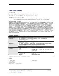
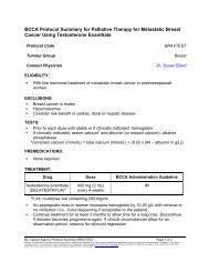
Variations in Cervical Transformation Zone<br />
a)<br />
b)<br />
c)<br />
The location of the squamocolumnar junction<br />
is dependent on the patient’s age, parity,<br />
hormonal status and any previous surgery. If<br />
squamocolumnar junction is visible, sample<br />
with a spatula. If not visible, i.e. in the canal,<br />
sample with elongated end of spatula or<br />
cytobrush.<br />
a) Reproductive age group, nulligravida;<br />
squamocolumnar junction often visible<br />
on ectocervix lateral to os. Os (small,<br />
round or oval). Sample with spatula.<br />
b) Reproductive age group, parous;<br />
squamocolumnar junction often at or<br />
near external os. Sample with spatula.<br />
c) Post menopause. Squamocolumnar<br />
junction often in canal. Cervical os often<br />
smaller. Sample with elongated end of<br />
spatula and cytobrush.
Basic Equipment and Supplies<br />
• examination table<br />
• good illumination<br />
• bi-valve speculum (various sizes)<br />
• pencil for labeling slide<br />
• endocervical brush (call 604-876-4186 in the Lower<br />
Mainland or toll free 1-800-667-5770 to order)<br />
• cytology spray fixative e.g. cytospray (call<br />
Surgipath toll free 1-800-665-7425 to order)<br />
• extended-tip spatula*<br />
• glass microscope slide with frosted end*<br />
• container* for transporting slide to laboratory<br />
• requisition form*<br />
*Supplied free of charge by the <strong>BC</strong> <strong>Cancer</strong> <strong>Agency</strong><br />
Fax request to 604-660-3122<br />
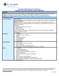
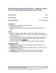
Obtaining the Smear<br />
1. Gently insert a pre-warmed speculum to visualize cervix.<br />
DO NOT USE lubricant jelly on speculum. Lubricant can<br />
obscure cellular detail, interfere with cellular adherence and<br />
cause bacterial over-growth on the slide.<br />
2. Gently cleanse the cervix with cotton pledget if obscured<br />
with discharge or secretions.<br />
3. Identify extent of transformation zone and probable<br />
squamocolumnar junction.<br />
If Squamocolumnar<br />
Junction is Visible<br />
Rotate a spatula through 360°<br />
once to obtain a single specimen.<br />
Fixation is not necessary.<br />
If Not Visible<br />
If squamocolumnar junction is not visible, first use a spatula for the<br />
exocervical specimen; then use a cytobrush or the elongated end of<br />
the spatula for the endocervical sample. Rotate cytobrush 180° only.<br />
Place both specimens on a single slide and fix immediately.<br />
Cautions<br />
• The use of the cytobrush<br />
is not recommended in<br />
pregnant patients.<br />
• If a clinically suspicious<br />
lesion is seen, biopsy<br />
immediately.<br />
• If the patient is menstruating<br />
or infection is present<br />
reschedule exam.<br />
• Irregular bleeding may be a<br />
symptom of gynecological<br />
malignancy. Pelvic<br />
examination with lower<br />
genital tract and appropriate<br />
investigation is indicated.<br />
If cytobrush is used, prompt fixation of the sample is<br />
necessary<br />
Step 1<br />
Patient<br />
Step 2<br />
15cm - 25cm<br />
The use of cotton swabs for<br />
sampling is associated with<br />
cellular trapping and<br />
distortion and is no longer<br />
recommended.
Completing the Requisition Form<br />
To ensure the patient demographics are<br />
up-to-date, the laboratory requires:<br />
• The patient’s current and all previous surnames.<br />
Ensure correct spelling and enter first and<br />
middle names, if applicable. The name on the<br />
Requistion Form and the name on the slide<br />
must match exactly.<br />
• The patient’s PHN number.<br />
• Date of birth (day/month/year).<br />
To ensure accurate report delivery, the<br />
laboratory requires:<br />
• Smear taker’s full address, including postal code<br />
and telephone number.<br />
• Physician or Midwife MSP number<br />
To ensure optimum evaluation of<br />
specimens, the laboratory requires:<br />
• Date of the patient’s last menstrual period (LMP).<br />
• Relevant clinical history e.g., discharge, bleeding,<br />
medications.<br />
• Relevant past history and the reason for the<br />
hysterectomy, previous abnormal Pap smears,<br />
malignancy, or cervical treatment. This<br />
information helps determine appropriate followup<br />
recommendations.<br />
Transporting the Specimen<br />
To ensure that the slides arrive at the<br />
<strong>BC</strong> <strong>Cancer</strong> <strong>Agency</strong> laboratory:<br />
• Labeled slides should be placed in the mailing<br />
containers provided by the <strong>BC</strong> <strong>Cancer</strong> <strong>Agency</strong><br />
laboratory.<br />
For supplies, fax request to 604-660-3122.<br />
• The completed requisition should be folded and<br />
wrapped around each slide-mailing container and<br />
secured with an elastic band. There is no need to<br />
apply a patient identification label to the mailing<br />
container.<br />
• The slide and requisition should be sent by<br />
courier or Canada Post addressed to the <strong>BC</strong>CA:<br />
Slides may be collected and sent in weekly batches.<br />
Cervical <strong>Cancer</strong> Screening Laboratory<br />
c/o Central Processing and Receiving<br />
– Lane Level Laboratory<br />
655 West 12th Avenue<br />
Vancouver, <strong>BC</strong> V5Z 4R4<br />
Phone: 1-877-747-2522 (1-877-PHSA LAB)<br />
Specimen Rejection Policy<br />
Inadequately labeled or unlabeled specimens have<br />
been identified as a significant source of laboratory<br />
error worldwide and for this reason the Cervical<br />
<strong>Cancer</strong> Screening Laboratory (CCSL) must<br />
accurately identify all slides that are processed in the<br />
laboratory. The CCSL will not process unlabeled,<br />
insufficiently labeled or mislabeled slides, a new<br />
specimen will have to be collected.
Optimal and Unsatisfactory Smears<br />
What is an optimal cervical smear<br />
The presence of endocervical cells, metaplastic cells,<br />
and squamous cells suggest a high probability that<br />
the transformation zone has been sampled, which<br />
is necessary for a cervical smear to be considered<br />
optimal.<br />
Cytologists continue to debate the criteria necessary<br />
to ensure that the transformation zone has been<br />
sampled. The presence of squamous metaplastic<br />
cells and endocervical cells and/or atypical cells is<br />
generally regarded as evidence of adequate sampling<br />
of the transformation zone.<br />
What is an unsatisfactory smear<br />
Smears can be considered unsatisfactory for either<br />
technical or interpretative reasons.<br />
Technical Reasons<br />
• Slide was received broken or slide was broken<br />
during handling in the laboratory and is beyond<br />
possible repair<br />
• Slide not labeled with patient’s name<br />
• Mis-match between the name on the slide and the<br />
name on the requisition<br />
• Improperly or illegibly labeled slide<br />
Interpretative Reasons<br />
• 75% or more of the smear is obscured by<br />
inflammatory exudate or blood<br />
• Too few cells are present on the smear (generally<br />
less than one thousand)<br />
• Smear is too thick (cells are piled up on the top<br />
of each other so technologist is unable to examine<br />
individual cells)<br />
• Smear consists mainly of endocervical glandular<br />
cells (smear comes only from endocervical<br />
canal and there is no representation of the<br />
transformation zone)<br />
• Cells are too poorly preserved for adequate<br />
interpretation
Human Papilloma Virus and Cervical <strong>Cancer</strong><br />
The detection of Human Papilloma Virus (HPV)<br />
in the majority of cervical cancer precursor lesions<br />
and invasive cervical carcinomas supports the<br />
assertion that this agent is an essential factor in the<br />
development of cervical cancer. More than 80 types<br />
of HPV have been identified. Approximately 30<br />
types are transmitted principally by skin-to-skin<br />
contact (commonly during sexual activity). About<br />
half of this group have been linked to cervical cancer,<br />
and two (types 16 and 18) account for 70% of this<br />
association in North America. While HPV infection<br />
is very common, only a small percentage of infected<br />
women will develop cervical cancer. Of note is that<br />
the HPV types associated with visible gential warts<br />
do not predispose to invasive cancer. At present the<br />
role of HPV testing for cervical cancer prevention is<br />
being evaluated in British Columbia in a large study<br />
due to start in 2007.<br />
In late 2006, an HPV vaccine protecting against the<br />
most common HPV types associated with cervical<br />
cancer became available in Canada. This for the<br />
first time allows us to move in the direction of<br />
preventing this common infection in the hopes of<br />
further reducing the incidence of cervical cancer.<br />
New Technologies<br />
Recent advances in gynecological cytology have<br />
focused on improving specimen preparation<br />
and processing and on the interpretation of<br />
cytological findings. They will lead to an increase in<br />
screening accuracy and subsequently improve the<br />
detection rate of pre-invasive and invasive cervical<br />
malignancies.<br />
Liquid-Based Cytology<br />
(Thin-Layer Cytology)<br />
The sample is collected with a spatula ± brush in the<br />
same way as for the conventional Pap smear. Instead<br />
of smearing the sample on the slide, the specimen<br />
is washed directly into a vial containing liquid<br />
fixative. Slide preparations are made from the liquid<br />
sample. The cells are fixed more uniformly, mucus<br />
is dissolved, large cell clusters are dispersed and<br />
debris and excessive blood are removed. Random<br />
cell disbursement allows for easier interpretation.<br />
Studies show that liquid-based cytology improves<br />
the detection of atypical cells and reduces the<br />
number of inadequate samples. However, it may<br />
increase the number of false positives. This method<br />
of specimen collection is more costly and transport<br />
is difficult.<br />
It is not currently available in British Columbia.<br />
Machine-Assisted Screening<br />
Computerized screening devices are algorithm-based<br />
decision making instruments. Some automated<br />
screening devices require specially prepared and/or<br />
stained slides, while others can use routinely stained<br />
smears. These machines can be used for primary<br />
screening or as re-screening devices. In the United<br />
States, where 10% of all negative slides must be<br />
re-screened, an automated device was shown to<br />
detect 2 – 3 times more false-negatives than manual<br />
interpretation. In a primary screening mode, up<br />
to 25% of all slides from women with a low<br />
probability of having cervical precancerous lesions<br />
can be scanned by machine only without further<br />
intervention by a cytotechnologist.<br />
10
Appendix I<br />
Current <strong>BC</strong> <strong>Cancer</strong> <strong>Agency</strong> Guidelines for the<br />
Investigation and Management of Women with<br />
Screen Detected Abnormalities<br />
Low–Grade Epithelial Abnormalities<br />
Pap Smear Report Investigation Management<br />
Mild squamous dyskaryosis<br />
Mild endocervical glandular atypia<br />
Repeat smear at 6 month<br />
intervals if abnormal cytology<br />
persists for 2 years, refer to<br />
colposcopy<br />
If mild dysplasia (CIN 1)<br />
confirmed at colposcopy follow<br />
with repeat Pap in 6 months<br />
High-Grade Epithelial Abnormalities<br />
Pap Smear Report Investigation Management<br />
Moderate squamous dyskaryosis<br />
Marked squamous dyskaryosis<br />
Suspicious for squamous cell<br />
carcinoma in situ<br />
Malignant squamous cells<br />
Moderate endocervical glandular<br />
atypia<br />
Marked endocervical glandular<br />
atypia<br />
Cells suspicious for endocervical<br />
carcinoma seen<br />
Refer to colposcopy & directed<br />
biopsy<br />
• If moderate dysplasia/<br />
severe dysplasia/Ca in<br />
Situ, (CIN 2-3) confirmed,<br />
treatment by gynecologist<br />
with appropriate expertise<br />
• If microinvasion present,<br />
refer to gynecologist with<br />
appropriate expertise or<br />
gynecologic oncologist<br />
• If frank invasion present,<br />
refer to gynecologic<br />
oncologist at <strong>BC</strong>CA<br />
Malignant glandular cells seen<br />
11
Appendix II<br />
Management of Women with Low Grade Epithelial<br />
Abnormalities (Mild Dysplasia or CIN 1)<br />
Observation Management<br />
If CIN 1/Mild Dysplasia has been confirmed by colposcopy and biopsy, cervical smears should be taken at<br />
6-month intervals until the abnormality either regresses or progresses. After 2 consecutive negative optimal<br />
smears, smear should be taken at 24-month intervals.<br />
Active Management<br />
Treatment by ablative or excisional methods is not generally recommended for low-grade lesions.<br />
Special Categories<br />
Post Treatment Follow-up (CIN 2/3)<br />
• Ablative therapy: cryotherapy, laser<br />
vaporization,<br />
• Excision therapy: cone biopsy, loop excision<br />
Abnormal smears in pregnancy<br />
(moderate atypia or higher)<br />
Total Hysterectomy for Benign Disease<br />
Colposcopy in 4-6 months<br />
• Complete excision: colpo +/- ECC (depending on<br />
whether satisfactory or not). If all negative, discharge<br />
for follow up Pap smear with GP in 6 months<br />
• Excision incomplete or ablative therapy: repeat<br />
colposcopy with ECC every 6 months times two.<br />
If ECC abnormal, manage per guidelines. If ECC<br />
negative both times, discharge for follow up with GP<br />
in 6 months.<br />
Refer to immediate colposcopy to exclude invasive disease.<br />
Repeat colposcopy in 8-12 weeks during pregnancy to<br />
rule out progression. Reassess lesions with colposcopy<br />
and biopsy 8 weeks post-partum<br />
No further smears required if previous smears showed no<br />
significant squamous abnormality<br />
Further Investigation<br />
When “further investigation” is recommended for abnormal endometrial cells, then the selection of the<br />
methods of investigation rests with the clinician. The method of investigation may consist of endometrial<br />
biopsy, hysteroscopy, ultrasound or dilatation and curettage.<br />
12
Appendix III<br />
Understanding False Negative Rates<br />
Many epidemiologic definitions of false negative,<br />
sensitivity and other similar concepts utilize absolute<br />
(gold standard) knowledge of the true state of the<br />
individual. When dealing with clinical cancer, this<br />
state will most likely be known (e.g., we know<br />
whether someone has symptomatic cancer). The<br />
same definition cannot be used when considering preneoplastic<br />
conditions (dysplasia) since the true state<br />
is only known in individuals who undergo a clinical<br />
investigation. Individuals with normal Pap smears<br />
are not routinely investigated. For these reasons,<br />
determining the precise number of false negatives is<br />
not possible.<br />
In Pap smears and other screening tests, a false<br />
negative rate is often used to refer to what would be<br />
more accurately described as a reclassification rate<br />
for initially negative smears, i.e., what proportion of<br />
smears initially classified as negative are reclassified as<br />
non-negative upon re-screening.<br />
Of the 1998 “negative” smears reviewed, 4.5%<br />
were reclassified as “non-negative.” The cases<br />
reviewed were selected because of their current<br />
cytologic abnormality. Thus, this sample is not<br />
representative of the general “negative” smears.<br />
Furthermore, reviewers had knowledge of the current<br />
smear result at the time of re-screening. These<br />
factors contribute to an over-estimation of the true<br />
misclassification rate.<br />
An alternative statistic that has been calculated<br />
in the literature is the false-negative fraction. In<br />
this case, the number of “negatives” reclassified as<br />
“non-negative” is expressed as a percentage of the<br />
total “non-negatives” (i.e., “non-negatives” that were<br />
initially classified, or subsequently reclassified as<br />
such). CCSP has a false-negative fraction estimate of<br />
2.6% of 1998 smears. This is likely to be an underestimation<br />
of the true false-negative rate, as not all<br />
slides have been reviewed.<br />
13
Appendix IV<br />
Results Terminology<br />
<strong>BC</strong> Cervical <strong>Cancer</strong><br />
Screening Program<br />
CIN/Dysplasia System<br />
The Bethesda System<br />
Unsatisfactory: state reason Unsatisfactory: state reason Unsatisfactory: state reason<br />
Negative, no atypical cells are seen No abnormal cells; metaplasia noted Negative for Intraepithelial Lesion or<br />
Malignancy<br />
Benign changes<br />
Trichomonas vaginalis<br />
Monilia (Candida species)<br />
Cellular changes suggestive of<br />
Herpes simplex viral infection<br />
Benign changes<br />
Radiation effect<br />
Mild squamous dyskaryosis<br />
(+HPV 1 effect)<br />
Mild squamous dyskaryosis<br />
Moderate squamous dyskaryosis<br />
Marked squamous dyskaryosis/<br />
Suspicious squamous cells<br />
Malignant<br />
Malignant squamous cells<br />
Malignant glandular cells<br />
Mild glandular atypia<br />
Moderate glandular atypia<br />
Marked glandular atypia/Suspicious<br />
glandular cells<br />
Other 2<br />
Abnormal cells consistent with reactive atypia<br />
(non-dysplastic)<br />
Trichomonas effect<br />
Yeast effect<br />
Viral effect (Herpes type)<br />
Abnormal cells consistent with reactive atypia<br />
(non-dysplastic)<br />
Inflammatory effect<br />
Irradiation effect<br />
Other<br />
Abnormal cells consistent with reactive atypia<br />
(possibly dysplastic)<br />
Atypical metaplasia<br />
Atypical parakeratosis<br />
Other (add comment)<br />
Abnormal cells consistent with condyloma<br />
(HPV) effect<br />
Mild dysplasia/ 3 CIN 1<br />
Moderate dysplasia/ 3 CIN 2<br />
Severe dysplasia/ 4 CIS/ 3 CIN 3<br />
Abnormal cells consistent with malignancy<br />
Consistent with invasive squamous<br />
carcinoma<br />
Consistent with adenocarcinoma<br />
Type unspecified<br />
Abnormal cells not specifically classified (add<br />
comment)<br />
Benign cellular changes<br />
Trichomonas vaginalis<br />
Fungal organisms morphologically<br />
consistent with Candida spp.<br />
Cellular changes associated with Herpes<br />
Simplex Virus<br />
Benign cellular changes<br />
Reactive cellular changes associated with:<br />
Inflammation<br />
Radiation<br />
Other<br />
ASC-US 5<br />
ASC-H 6<br />
LSIL 7<br />
LSIL 7<br />
HSIL 8<br />
HSIL 8<br />
Carcinoma<br />
Squamous cell carcinoma<br />
Adenocarcinoma<br />
Unspecified<br />
AGC – NOS 9 , AGC – favor neoplastic<br />
AIS 10<br />
Other<br />
1. Human papilloma virus<br />
2. Inconclusive for squamous or glandular differentiation<br />
3. Cervical intraepithelial neoplasia<br />
4. Squamous Carcinoma in situ<br />
5. Atypical squamous cells of undetermined significance<br />
6. Atypical squamous cells – cannot exclude high-grade lesion<br />
7. Low grade squamous intraepithelial lesion<br />
8. High grade squamous intraepithelial lesion<br />
9. Atypical glandular cells (NOS – not otherwise specified)<br />
10. Adenocarcinoma in situ<br />
14
References<br />
Canadian Society of Cytology (1995-1996). Canadian Society of Cytology: Guidelines for Practice and<br />
Quality Assurance in Cytopathology. Canadian Association of Pathologists.<br />
Dorland’s Illustrated Medical Dictionary, 28 th Edition, W.B. Saunders Co., 1994<br />
Health Canada. Quality Management Working Group Cervical <strong>Cancer</strong> Prevention Network (May 1997).<br />
Programmatic guidelines for screening for cancer of the cervix.<br />
Health Canada. Quality Management Working Group Cervical <strong>Cancer</strong> Prevention Network (January 1999).<br />
Programmatic guidelines for screening for cancer of the cervix in Canada. 1-31. [WWWdocument]. URL<br />
http://www.hc-sc.gc.ca/hppb/ahi/cervicalcancer/pubs/screening.pdf<br />
Manitoba Quality Assurance Program, College of Physicians and Surgeons of Manitoba (2000). Cytology<br />
standards of accreditation.<br />
New South Wales Cervical Screening Program (1996). [WWWdocument]. URL http://www.csp.nsw.gov<br />
Nova Scotia Gynaecological <strong>Cancer</strong> Screening Programme (1998). Screening for cancer of the cervix: An<br />
office manual for health professionals. (pp.1-26). Halifax: NSGCSP.<br />
Ontario Cervical Screening Collaborative Group (December 1997). Interim recommendations for follow-up<br />
of pap test results. Ontario Medical Review. 34-36. [WWWdocument]. URL http://www.omaorg/pcomm/<br />
omr/dec97.htm<br />
Cervical <strong>Cancer</strong> Screening Program 2001 Annual Report<br />
Footnotes<br />
1<br />
Cole P, Morrison AS: Basic issues in cancer screening. In Miller AB (Ed): Screening in <strong>Cancer</strong>. Geneva,<br />
International Union Against <strong>Cancer</strong>, 1978, page 7.<br />
2<br />
Miller, AB: Fundamentals of screening: In Screening for <strong>Cancer</strong>, Orlando, Academic Press, 1985, page 3.<br />
15
Educational Materials Order Form<br />
Cervical <strong>Cancer</strong> Screening Program<br />
Material Requests<br />
8th Floor<br />
686 West Broadway<br />
Vancouver, <strong>BC</strong> V5Z 1G1<br />
Fax: 604-660-3645<br />
Please fax this form to the CCSP to receive copies of the following free of charge:<br />
Number Requested:<br />
________________<br />
________________<br />
________________<br />
________________<br />
_______________<br />
_______________<br />
________________<br />
Item Requested:<br />
Technique for Obtaining Cervical Smears – Laminated Instruction Card<br />
Speculum Exam & Pap Smears – Video or DVD<br />
Cervical cancer: protect yourself with regular pap tests – Brochure<br />
HPV & cervical cancer: what you should know, and do – Brochure<br />
Preventing cervical cancer – Booklet<br />
Abnormal pap smear: causes and proper followup – Booklet<br />
Annual Report (most current year)<br />
Name:<br />
____________________________________________________________________________<br />
Address: ____________________________________________________________________________<br />
____________________________________________________________________________<br />
____________________________________________________________________________<br />
MSC#<br />
_____________________<br />
16
Feedback<br />
It is important that we receive your feedback to<br />
ensure that this Manual meets your needs and the<br />
needs of the Cervical <strong>Cancer</strong> Screening Program.<br />
Please use this sheet to forward any comments/<br />
suggestions you may have after using the Seventh<br />
Edition of the Office Manual. Thank you.<br />
Content<br />
________________________________________<br />
________________________________________<br />
________________________________________<br />
________________________________________<br />
________________________________________<br />
________________________________________<br />
________________________________________<br />
________________________________________<br />
Layout<br />
Design:<br />
________________________________________<br />
________________________________________<br />
________________________________________<br />
Order of Topics:<br />
________________________________________<br />
________________________________________<br />
________________________________________<br />
Other:<br />
________________________________________<br />
________________________________________<br />
________________________________________<br />
Illustrations:<br />
________________________________________<br />
________________________________________<br />
________________________________________<br />
Format<br />
Size:<br />
________________________________________<br />
________________________________________<br />
________________________________________<br />
________________________________________<br />
Binding:<br />
________________________________________<br />
________________________________________<br />
________________________________________<br />
________________________________________<br />
Other:<br />
________________________________________<br />
________________________________________<br />
________________________________________<br />
________________________________________<br />
Please Add<br />
________________________________________<br />
________________________________________<br />
________________________________________<br />
________________________________________<br />
Please Delete<br />
________________________________________<br />
________________________________________<br />
________________________________________<br />
________________________________________<br />
Please forward your response to:<br />
Cervical <strong>Cancer</strong> Screening Program<br />
Administration Office<br />
8th Floor, 686 West Broadway<br />
Vancouver, <strong>BC</strong> V5Z 1G1<br />
Fax: 604-660-3645<br />
17