P r o d u c t R a t i o n a l e S u r g i c a l T e c h n i q u e - Biomet
P r o d u c t R a t i o n a l e S u r g i c a l T e c h n i q u e - Biomet
P r o d u c t R a t i o n a l e S u r g i c a l T e c h n i q u e - Biomet

You also want an ePaper? Increase the reach of your titles
YUMPU automatically turns print PDFs into web optimized ePapers that Google loves.
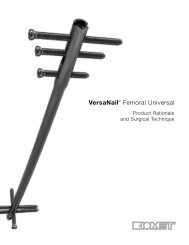
Surgical Technique<br />
Femoral Fractures<br />
Paediatric femoral fractures are typically treated with two nails inserted in a retrograde<br />
fashion from medial and lateral entry portals located above the epiphysis. Very distal<br />
fractures should be treated with an antegrade approach.<br />
Positioning and Fracture Reduction<br />
The patient is positioned on an orthopaedic traction table (Figure 6). An image<br />
intensifier is positioned so that it can be rotated to obtain AP and lateral views. It<br />
should also be possible to visualise the whole femur from the knee to the hip joint.<br />
The entire thigh including the knee is prepared as an operative field. External<br />
manipulation is conducted until adequate reduction is obtained and confirmed by<br />
flouroscopy.<br />
Figure 6<br />
Approach<br />
Make a 2 cm skin incision distally to the required entry hole to provide access for the<br />
instruments, and to prevent any trauma to the skin (Figure 7). You may start either<br />
on the medial or the lateral side. An oblique drill hole is made 2 - 4 cm above the<br />
growth plate by applying a careful angulation movement of the drill bit until the<br />
entry hole is at an angle of at least 60˚ with the axis of the medullary canal (Figure 8).<br />
Make the the entry hole slightly larger than the diameter of the chosen nail.<br />
Figure 7<br />
Figure 8<br />
7