Cyclodextrins Inclusion Complex
Cyclodextrins Inclusion Complex
Cyclodextrins Inclusion Complex

Create successful ePaper yourself
Turn your PDF publications into a flip-book with our unique Google optimized e-Paper software.
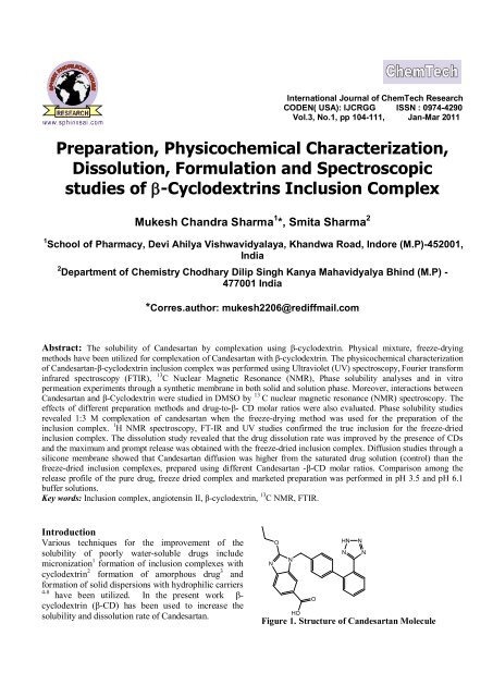
International Journal of ChemTech Research<br />
CODEN( USA): IJCRGG ISSN : 0974-4290<br />
Vol.3, No.1, pp 104-111, Jan-Mar 2011<br />
Preparation, Physicochemical Characterization,<br />
Dissolution, Formulation and Spectroscopic<br />
studies of b-<strong>Cyclodextrins</strong> <strong>Inclusion</strong> <strong>Complex</strong><br />
Mukesh Chandra Sharma 1 *, Smita Sharma 2<br />
1 School of Pharmacy, Devi Ahilya Vishwavidyalaya, Khandwa Road, Indore (M.P)-452001,<br />
India<br />
2 Department of Chemistry Chodhary Dilip Singh Kanya Mahavidyalya Bhind (M.P) -<br />
477001 India<br />
*Corres.author: mukesh2206@rediffmail.com<br />
Abstract: The solubility of Candesartan by complexation using β-cyclodextrin. Physical mixture, freeze-drying<br />
methods have been utilized for complexation of Candesartan with β-cyclodextrin. The physicochemical characterization<br />
of Candesartan-β-cyclodextrin inclusion complex was performed using Ultraviolet (UV) spectroscopy, Fourier transform<br />
infrared spectroscopy (FTIR), 13 C Nuclear Magnetic Resonance (NMR), Phase solubility analyses and in vitro<br />
permeation experiments through a synthetic membrane in both solid and solution phase. Moreover, interactions between<br />
Candesartan and β-Cyclodextrin were studied in DMSO by 13 C nuclear magnetic resonance (NMR) spectroscopy. The<br />
effects of different preparation methods and drug-to-β- CD molar ratios were also evaluated. Phase solubility studies<br />
revealed 1:3 M complexation of candesartan when the freeze-drying method was used for the preparation of the<br />
inclusion complex. 1 H NMR spectroscopy, FT-IR and UV studies confirmed the true inclusion for the freeze-dried<br />
inclusion complex. The dissolution study revealed that the drug dissolution rate was improved by the presence of CDs<br />
and the maximum and prompt release was obtained with the freeze-dried inclusion complex. Diffusion studies through a<br />
silicone membrane showed that Candesartan diffusion was higher from the saturated drug solution (control) than the<br />
freeze-dried inclusion complexes, prepared using different Candesartan -β-CD molar ratios. Comparison among the<br />
release profile of the pure drug, freeze dried complex and marketed preparation was performed in pH 3.5 and pH 6.1<br />
buffer solutions.<br />
Key words: <strong>Inclusion</strong> complex, angiotensin II, β-cyclodextrin, 13 C NMR, FTIR.<br />
Introduction<br />
Various techniques for the improvement of the<br />
solubility of poorly water-soluble drugs include<br />
micronization 1 formation of inclusion complexes with<br />
cyclodextrin 2 formation of amorphous drug 3 and<br />
formation of solid dispersions with hydrophilic carriers<br />
4-8 have been utilized. In the present work βcyclodextrin<br />
(β-CD) has been used to increase the<br />
solubility and dissolution rate of Candesartan.<br />
N<br />
O<br />
N<br />
HO<br />
O<br />
HN N<br />
N<br />
Figure 1. Structure of Candesartan Molecule<br />
N
Mukesh Chandra Sharma et al /Int.J. ChemTech Res.2011,3(1) 105<br />
Candesartan (Figure1) 2-ethoxy-1-({4-[2-(2H-1, 2, 3,<br />
4-tetrazol-5-yl) phenyl] phenyl} methyl)-1H-1, 3benzodiazole-6-carboxylic<br />
acid.it is an orally active<br />
specific angiotensin II, AT1 receptor antagonist, and<br />
clinically effective drug in the treatment of<br />
hypertension. it is slightly soluble in alcohol,<br />
practically insoluble in water. Due to its hydrophobic<br />
nature (octanol/water partition coefficient 9.8 at pH of<br />
7.4). <strong>Cyclodextrins</strong> (CD’s) with their Candesartanershaped<br />
cavities capable to form inclusion complexes<br />
with a wide range of commonly used drugs by taking<br />
the whole molecule or part of it into the cavity and<br />
known to improve the aqueous solubility of drugs.<br />
Many drugs such as hydrocortisone, itraconazole,<br />
omeprazole, mitomycin, nitroglycerin, voriconazole<br />
etc. have been complexed with CD’s and formulated<br />
for enhancing solubility and therapeutic activity. 9-10<br />
Even IRB ß-cyclodextrin (ß-CD) complexes has been<br />
reported to enhance its solubility. 11 In previous years<br />
cyclodextrins (CDs) have been recognized as an<br />
important constituents of pharmaceutical excipients.<br />
They are cyclic oligosaccharides consisting of (α-1, 4)linked<br />
α-D-glucopyranose units, with a relatively<br />
hydrophobic central cavity and a hydrophilic outer<br />
surface. The most abundant natural CDs are αcyclodextrin<br />
(α-CD), β-cyclodextrin (β-CD), and γcyclodextrin<br />
(γ-CD), containing 6, 7, and 8<br />
glucopyranose units, respectively. β-cyclodextrin (β-<br />
CD) has been used in this work. The hydrophilic<br />
exterior surface of the CD molecules makes them<br />
water-soluble, but the hydrophobic cavity provides a<br />
microenvironment for appropriately sized nonpolar<br />
molecules. CDs are capable of forming inclusion<br />
complexes with many drugs by including a whole drug<br />
molecule, or only some non-polar part of it, inside<br />
their cavity. In an aqueous solution, the complexes are<br />
readily dissociated and free drug molecules are in<br />
relatively rapid dynamic equilibrium with drug<br />
molecules bound within the CD cavity 12-16 . These<br />
noncovalent complexes show new physicochemical<br />
characteristics when compared with the guest<br />
molecules. They include better stability, higher<br />
aqueous solubility, increased bioavailability, and less<br />
undesirable side effects. Biological membranes are<br />
hydrophobic and therefore passive diffusion is favored<br />
for relatively hydrophobic molecules. However, the<br />
driving force for the passive diffusion is the high drug<br />
concentration and high drug activity in the aqueous<br />
fluid of the absorption site. Thus, bioavailability of<br />
drugs depends both on their water and lipid solubility.<br />
The formation of drug-CD inclusion complexes can<br />
increase the aqueous solubility of hydrophobic drugs,<br />
hence increasing the driving force for the diffusion<br />
process across the biological membranes. In the<br />
present study, different technique has been used to<br />
prepare inclusion complex of Candesartan with βcyclodextrin.<br />
Prepared inclusion complex was<br />
characterized by Fourier transform infrared (FTIR),<br />
Ultraviolet (UV) and Nuclear Magnetic resonance<br />
(NMR) spectroscopy. The evaluation was performed<br />
by invitro dissolution; diffusion and comparing the<br />
release profile of inclusion complex with marketed<br />
formulation in two buffer solutions Ph 3.5and pH 6.9.<br />
The aim of this study was to improve the<br />
biopharmaceutical properties of Candesartan by the<br />
formation of Candesartan/β-cyclodextrin inclusion<br />
complex.<br />
Materials and Methods<br />
Materials ß-CD (Mole. Wt. 1135) were generous gift<br />
from Gangwal Chemicals Pvt. Ltd. Mumbai.<br />
Candesartan was received as a gift sample from Sun<br />
pharma Baroda Gujarat. Lactose monohydrate,<br />
microcrystalline cellulose, croscarmelose, silicone<br />
dioxide, magnesium stearate and sodium lauryl<br />
sulphate were purchased. All chemicals and solvents<br />
used in this study were of A.R. grade. Freshly prepared<br />
double distilled water was used throughout the work.<br />
Methods<br />
Preparations of Solid Binary Systems-<br />
Candesartan-β-cyclodextrin binary systems were<br />
prepared (1: 3 and 1:5 ratios) as described in detail<br />
below.<br />
Physical Mixture-<br />
Physical mixture (PM) of CD and Candesartan were<br />
prepared by simply mixing powders with a spatula for<br />
15 min and then sieved through 60 #.<br />
Co-evaporation method-<br />
For preparation of complexes by co evaporation<br />
method CD’s and Candesartan were mixed in 1:3<br />
molar ratio, 5 ml of ethanolic solution of Candesartan<br />
was added slowly to 5 ml of the aqueous solution of<br />
CD followed by stirring at 300 rpm using magnetic<br />
stirrer at 37 0 c for 24 hrs. The solvents were then<br />
evaporated at 45-50 0 C. The resultant solids were<br />
pulverized and then sieved through 60 #.<br />
Kneading method-<br />
For preparation of complexes by kneading method, the<br />
CD and Candesartan were taken in 1:3 molar ratio.<br />
The CD was triturated in a mortar with small quantity<br />
of water to obtain a homogeneous paste. Candesartan<br />
was then added slowly while grinding; a small quantity<br />
of methanol was added to facilitate the dissolution of<br />
Candesartan. The mixtures were then grounded for 6
Mukesh Chandra Sharma et al /Int.J. ChemTech Res.2011,3(1) 106<br />
hrs. During this process, an appropriate quantity of<br />
water was added to the mixture to maintain a desired<br />
consistency. The pastes were dried in an oven at 45-50<br />
0 C for 24 hrs. The dried complexes were pulverized<br />
and then sieved through 60 #.<br />
Characterization of Binary Systems<br />
Ultraviolet spectroscopy<br />
UV-visible double beam spectrophotometer,<br />
Shimadzu model 1700 with spectral bandwidth of 1<br />
nm, wavelength accuracy of ± 0.3 nm and The<br />
absorption spectra of the reference and test solutions<br />
were recorded in 1 cm quartz cells over the range of<br />
200– 400 nm. The Candesartan and Candesartan/ β<br />
cyclodextrin samples were prepared in situ in a 3ml<br />
cuvette. A 1μl aliquot of a 0.5 w/v Candesartan 95%<br />
v/v ethanol solution was pipetted into a cuvette<br />
containing a known volume of water (3ml). The<br />
cuvette was then stoppered and shaken. Further<br />
aliquots were then cumulatively added to the cuvette<br />
to give a final Candesartan concentration of 1.0 x 10 -7<br />
μg/ml -1 and the absorbance measured as before. The<br />
procedure was repeated for various concentration of<br />
β-cyclodextrin. The reference solutions were<br />
similarly prepared except than an equivalent aliquot<br />
of 95% ethanol was substituted for the Candesartan<br />
95% v/v ethanol aliquot.<br />
Infrared Spectroscopy<br />
Infrared spectroscopy (IR) spectra of pure Candesartan<br />
and β-CD, as well as their binary products, were<br />
obtained using a IR Magma IR750 by II instrument<br />
using NaCl disc method. Analysis was performed at<br />
room temperature.<br />
13<br />
C NMR studies<br />
NMR spectra were recorded with 500 MHz NMR<br />
machine Solutions of Candesartan, β-cyclodextrin and<br />
Candesartan/ β-cyclodextrin (1:3 and 1:5 ratios) were<br />
prepared by filtering a saturated solution of the<br />
respective material in DMSO through a cotton wool<br />
plug in a 5 mm capillary tube. NMR measurements<br />
were then made using a 500MHz<br />
13 C NMR<br />
spectrometer operating at 500MHz in the pulsed<br />
Fourier transform mode (to an accuracy of ± 0.05ppm).<br />
Each spectrum was referenced to DMSO at 0.005ppm<br />
as an external reference spectrum was recorded for the<br />
preparations. The spectra and chemical shifts values<br />
were recorded.<br />
Evaluation of Binary Systems<br />
Phase solubility study-<br />
Phase-solubility studies were performed by the method<br />
of Higuchi and Connors. 17 Candesartan, in constant<br />
amounts (5 mg) that exceeded its solubility, was<br />
transferred to screw capped vials containing 15 ml of<br />
aqueous solution of ß-CD or at various molar<br />
concentrations(0, 3.0, 6.0, 9.0, 12.0, and 15.0 μm). The<br />
contents were stirred on rotary shaker for 72 hrs. at 37<br />
0 C ± 0.1 0 C and 1200 rpm. The time duration was<br />
fixed based on pilot experiment and found to be<br />
sufficient to achieve equilibrium of mixture. After<br />
reaching equilibrium, samples were filtered through a<br />
0.36 μm membrane filter, suitably diluted and<br />
analyzed spectrophotometrically for drug content at<br />
272 nm UV/Visible spectrophotometer,. Solubility<br />
studies were performed in triplicate.<br />
Formulation studies-<br />
Tablets containing 50 mg of Candesartan were<br />
prepared by direct compression using different<br />
excipients like Lactose monohydrate, colloidal silicon<br />
dioxide, and magnesium stearate. Tablets containing<br />
complexes (equivalent to 50 mg Candesartan) prepared<br />
by kneading and co evaporation method were also<br />
prepared similarly using less quantity of lactose. The<br />
blend was compressed on a six-station single rotary<br />
machine using round-shaped, flat punches to obtain<br />
tablets having thickness 3–4 mm and hardness 3–5<br />
kg/cm2. The tablets were studied in 6 replicates for<br />
release profile of Candesartan using the same method<br />
described in dissolution studies.<br />
Dissolution Studies-<br />
Dissolution studies of Candesartan in powder form,<br />
and <strong>Complex</strong>es with ß-CD were performed to evaluate<br />
drug release profile. Dissolution studies were<br />
performed on USP dissolution apparatus type II with<br />
900 ml dissolution medium 0.1 N HCl (P H 1.2) at 37 0 C<br />
± 0.5 0 C at 75 rpm for 8 hr. At fixed time intervals 5<br />
ml aliquots were withdrawn, filtered, suitably diluted<br />
and assayed for Candesartan content by measuring the<br />
absorbance at 272 nm. (Pilot experimental data<br />
Candesartan icated no change in the l max of<br />
Candesartan due to the presence of CD’s in the<br />
dissolution medium.) Equal volumes of fresh medium<br />
(pre-warmed to 37 0 C) were replaced into the<br />
dissolution medium to maintain constant volume<br />
throughout the test period. Dissolution studies were<br />
performed in 6 replicates, and calculated mean values<br />
of cumulative drug release were used while plotting<br />
the release curves.
Mukesh Chandra Sharma et al /Int.J. ChemTech Res.2011,3(1) 107<br />
Table 1. Solubility of Pure Candesartan Stability constant (Kc), and Correlation Coefficient (R 2 )<br />
as obtained from the Candesartan-β-Cyclodextrin Phase Solubility Diagrams<br />
Medium G Kc (M−1) R 2<br />
pH 4.5<br />
pH 5.0<br />
pH 6.9<br />
0.271<br />
0.281<br />
0.751<br />
218<br />
291<br />
322<br />
0.9319<br />
0.8931<br />
0.9013<br />
Table 2. Chemical shifts for β-cyclodextrin in 1:1 and 1:2 molar ratios with Candesartan<br />
Change in Chemical Shift (∆δ) ppm<br />
Sample Molar a Ratio H(1) H(3) H(5) H(2) H(4) H(6)<br />
Candesartan/βcd 1:1 2.43 3.21 3.71 3.52 3.91 6.76<br />
Candesartan/βcd 1:2 2.67 3.38 4.43 5.42 5.98 6.11<br />
Concentration of<br />
Candesatan (mM/mL)<br />
0.15<br />
0.1<br />
0.05<br />
0<br />
0 0.01 0.02 0.03 0.04 0.05<br />
Concentration of Beta Cyclodextrine (mM/mL)<br />
Figure2. Phase solubility diagram of Candesartan-β cyclodextrin at different pH<br />
Results and Discussion<br />
Phase Solubility Studies-<br />
The phase solubility diagram β-cyclodextrin –<br />
Candesartan system in water can be characterized as<br />
AL type phase solubility curve, which suggests that the<br />
molar ratio of the complex is 1:3 the stability constant<br />
was found to be 78.65.Results of the phase solubility<br />
studies are shown in Figure 2 that presents the<br />
solubility profiles of Candesartan as a function of<br />
increasing concentrations of β-CD in aqueous solution<br />
at different pH values (4.5, 5.0, and 6.9). As can be<br />
seen, the solubility of the drug increased linearly with<br />
the increase of β-CD concentration (≤3 μM), giving<br />
rise to A-type solubility diagrams. This linear<br />
candesartan-β-CD correlation, with a slope of less than<br />
1, suggests the formation of a 1:1 (mol/mol)<br />
candesartan-β-CD inclusion complex, at the different<br />
studied pH values. The calculated stability constant<br />
values were 139, 314, and 219 M−1, respectively, at<br />
pH (a) 4.5, (b) 5.0, and (c) 6.9 (Table 1). Figure 2<br />
Candesartan icating that Candesartan -β- CD<br />
pH 4.5<br />
pH 5.0<br />
pH 6.9<br />
complexes (1:1 molar ratio) are sufficiently stable. In<br />
fact, values of obtained stability constants are always<br />
within the range of 100 to 1000 M −1 , which is believed<br />
to Candesartan an ideal value. The solubility<br />
differences of candesartan as a function of the method<br />
used for the preparation of the solid binary products,<br />
that is, physical mixture and freeze-dried products was<br />
also studied. It has been found that when the freezedried<br />
product candesartan-β-CD (1:2 ratio) was used,<br />
the drug solubility increased by a factor of 3 compared<br />
with that of the physical mixture. Moreover, it was 6fold<br />
higher than that of the pure candesartan.<br />
Ultra Violet Spectroscopy studies<br />
The UV spectrum for Candesartan consists of two<br />
peaks, one at 272nm and other at 311 nm<br />
(not very prominent) and a point of inflexion at 258<br />
nm. The molar absorptivities at these wavelengths<br />
were determined as 0.651, 0.872 respectively. The<br />
point of inflexion at 242nm (ε = 0.652) has been<br />
assigned to butane group by reference to the UV
Mukesh Chandra Sharma et al /Int.J. ChemTech Res.2011,3(1) 108<br />
spectrum for butane l group bonded to a nonconjugated<br />
molecule shows Candesartan ring system<br />
structure obtained with UV. In the spectrum for<br />
Candesartan, the absence of a peak at 280 nm and the<br />
FT-IR studies<br />
The IR spectra of β-cyclodextrin, Candesartan and βcyclodextrin<br />
– Candesartan complex (1:1 molar ratio)<br />
is shown Figure 3. Two biphenyl peaks at 1571 cm -1<br />
and 1621 cm -1 characterize the spectrum of<br />
Candesartan. A broad band at 1680cm -1 characterizes<br />
the spectrum for β cyclodextrin, which is due to the<br />
glycoside linkages. The spectrum for the physical mix<br />
and kneaded preparation are more or less the<br />
summation of those for the hydroxyl group peaks of<br />
Candesartan at 1823 cm -1 & 1781cm -1 .In contrast in<br />
the spectrum for the Candesartan / β-cyclodextrin<br />
freeze dried preparation the Candesartan hydroxyl<br />
peaks are Candesartan invidually observed and instead<br />
appear as a single broad band around 1431cm -1 . The<br />
cyclodextrin glycoside peak is unchanged in the<br />
presence of Candesartan both as freeze preparation and<br />
as a physical mixture.<br />
13 C- NMR Studies<br />
In each of the spectra, whether in the presence or<br />
absence of Candesartan, the signals due to H(2) are<br />
clearly visible around 5.0 ppm, whereas in the range<br />
0.00<br />
presence of a slightly less prominent one at 311nm<br />
may be due to conjugation between the tetrazole group<br />
and the Candesartanole ring.<br />
4.0-3.5 ppm, the spectral patterns from the<br />
cyclodextrin signals in the presence of Candesartan are<br />
quite different from those observed in its absence.<br />
Inspection of the spectra shows that the differences are<br />
not due to new signal multiplicities, but instead, arise<br />
from changes to the chemical shifts of the signals, in<br />
particular, changes to the signals from H(3) and H(5).<br />
The signals have move downfield and these<br />
movements are consistent irrespective of the<br />
Candesartan/β-cyclodextrin molar ratio. However, the<br />
δ values in the 1:3 and 1:5 mixtures show that a<br />
change in molar ratio does not similarly affect each<br />
signal. In the absence of Candesartan the H (3) signals<br />
from β-cyclodextrin give rise to a singlet at 3.63 ppm.<br />
In the presence of Candesartan the β-cyclodextrin H<br />
(3) signals are shifted up field, such that one signal of<br />
the triplet is obscured by the signal due to H (6) and<br />
other two signals appears at 3.9 ppm, on the up field<br />
side of the H. In the absence of Candesartan the H<br />
signals are almost completely obscured by H signal. In<br />
the presence of Candesartan β-cyclodextrin H signals<br />
are shifted up field so that they are completely visible<br />
around 2.54 ppm. (Figure 4)<br />
Figure 3. FT-IR spectrum of β-cyclodextrin, Candesartan and Candesartan-β-cyclodextrin<br />
inclusion complex
Mukesh Chandra Sharma et al /Int.J. ChemTech Res.2011,3(1) 109<br />
Figure 4. 1 H NMR of (i) β-cyclodextrin (ii) Candesartan -β-cyclodextrin<br />
In Vitro Dissolution Studies<br />
The dissolution studies was carried out with<br />
Candesartan and it’s complexes and physical mixture<br />
using dissolution medium 0.1 N HCl. 30 min<br />
(dissolved within 60 min), time to dissolve 50% drug<br />
(t50%) and mean dissolution time are reported in Table<br />
3. The data revealed the onset of dissolution of pure<br />
Candesartan was very low in (29.22 % within 30 min).<br />
It is evident that the dissolution rate of pure<br />
Candesartan is very low (49.46 % in 4 hr.) <strong>Inclusion</strong><br />
complexes CHH, CNB, IOH and COOH significantly<br />
enhanced dissolution rate of Candesartan (85–91%) 8<br />
hrs) 18. Moreover, candesartan diffusion rate was higher<br />
from the control-saturated solution. These results<br />
confirm that β-CDs do not interact significantly with<br />
the silicone membrane. Moreover, although drug<br />
diffusion was very similar for all the freeze-dried<br />
products, candesartan diffusion rate slightly decreased
Mukesh Chandra Sharma et al /Int.J. ChemTech Res.2011,3(1) 110<br />
as the CD amount increased. This behaviour is the<br />
consequence of different drug thermodynamic activity<br />
in the studied systems. In fact, for saturated solutions,<br />
the thermodynamic activity is constant despite the<br />
amount of dissolved drug. In addition, only free<br />
molecules can diffuse through the rate-limiting<br />
membrane 19. Therefore, the drug activity is only related<br />
to its free molecules in solution. In the presence of β-<br />
CD, candesartan solubility increases because of the<br />
formation of the inclusion complexes, but the actual<br />
thermodynamic activity is lower than that of the drug<br />
alone. Similar results were recently reported by<br />
different authors. 20-23 . The diffusion profile obtained<br />
from the saturated solution is very irregular (R 2 =<br />
0.9583) with high standard deviations. This is a<br />
consequence of the higher instability of this system<br />
when compared with the candesartan-β-CD binary<br />
products, which gave a very regular candesartan<br />
diffusion profile (always R 2 > 0.873) with standard<br />
deviation less than 2 %. Therefore, data obtained from<br />
the diffusion study suggest that β-CD is able to both<br />
stabilize the system and regularize the diffusion<br />
profile.<br />
Conclusion-<br />
Results obtained during this study showed that β-CD is<br />
able to improve candesartan dissolution properties.<br />
The best results were obtained from freeze-dried<br />
product, in which a true inclusion of candesartan with<br />
β-CD was confirmed by studies both in the solid and<br />
liquid phase. Despite their different solubility, drug<br />
diffusion through a model silicone membrane was<br />
higher for the saturated drug solution than the freezedried<br />
inclusion complexes that were able to stabilize<br />
the system leading to a more regular diffusion profile.<br />
Table 3. Dissolution Efficiency and Dissolution Percentage Values at 25and 90 Minutes and<br />
Time to dissolve 50% Drug for Candesartan and Candesartan-β-Cyclodextrin Systems*<br />
DE25 DP35 DE50 DP90 T50%<br />
Candesartan 2.0 3.53 18.79 48.91 >70<br />
PM 11.56 18.82 38.71 59.84 55<br />
FD 35.73 69.64 74.13 86.60 < 35<br />
*DE Candesartanicates dissolution efficiency; DP, dissolution percentage; t 50%, time to dissolve 50% of<br />
drug; CANDESARTAN, Candesartan; PM, physical mixture; and FD, freeze-dried. DE was calculated<br />
from the area under the dissolution curve at 15 and 60 minutes and is expressed as a percentage of the<br />
area of the rectangle described by 100% dissolution at the same time.<br />
% Drug released<br />
120<br />
100<br />
80<br />
60<br />
40<br />
20<br />
0<br />
0 20 40 60 80 100 120 140<br />
Time(min)<br />
Figure 5. Dissolution Profiles of Candesartan and<br />
its complexes with β-Cyclodextrin in simulated<br />
gastric fluid<br />
% Drug released<br />
120<br />
100<br />
80<br />
60<br />
40<br />
20<br />
0<br />
0 20 40 60 80 100 120 140<br />
Time(min)<br />
Figure 6. Dissolution Profiles of Candesartan and<br />
its complexes with β-<strong>Cyclodextrins</strong>.
Mukesh Chandra Sharma et al /Int.J. ChemTech Res.2011,3(1) 111<br />
References<br />
1. M.K.Gupta, A .Vanwert, R.H. Bogner, J Pharm<br />
Sci., 92,551. (2003)<br />
2. C. Cavallari, B. Abertini, R. Gonzalez,<br />
L.Rodriguez, Eur J Phar Biopharm., 65, 73(2002).<br />
3. O.I. Corrigan, Thermochim Acta., 248,245-258.<br />
(1995)<br />
4. W.L. Chiou, S.Riegalman, J Pharm Sci., 60, 1281-<br />
1302. (1971)<br />
5. A.A Ambike, K.R.Mahadik, A .Paradkar, Int J<br />
Pharm., 282,151-162. (2004)<br />
6. A. Paradkar, A.A. Ambike, B.K.Jadhav, K.R.<br />
Mahadik, Int J Pharm, 271,281-286(2004)<br />
7. G .Van den Mooter, M .Wuyts, N. Blaton,Eur J<br />
Pharm Sci.,12,261-269(2001)<br />
8. J. Jung, S. Yoo, S. Lee, K. Kim, D. Yoon, K.<br />
Lee,Int J Pharm., 187,209-218(1999).<br />
9. J .Szejtli, Med. Res. Rev., 14, 353–386, (1994)<br />
10. J.Szejtli, IUPAC, Pure Appl. Chem., 76, 1825-<br />
1845 (2004)<br />
11. R.Hirlekar, Kadam V, AAPS Pharma Sci Tech.,<br />
10, 276-281(2009)<br />
12. D .Duchene, Paris Editions de Santè., 351-<br />
407(1992).<br />
*****<br />
13. K.H. Frömming, J.Szejtli , Kluwer Academic<br />
Publishers (1994).<br />
14. D.O. Thompson, Crit Rev Ther Drug Carrier Syst.,<br />
104,14-1, (1997)<br />
15. M. Masson, T. Loftsson, G. Masson, E.S.<br />
Stefànsson, J Control Release., 59,107-118(1999)<br />
16. J. Szejtli, L.Szente, Die Pharmazie.,36 (10), 694-<br />
698 (1981)<br />
17. T.Higuchi, K.Connors, Adv. Anal. Chem. Instrum.,<br />
4, 17–123, (1965)<br />
18. L. Lehmann, E. Kleinpeter, 1 H NMR J Incl<br />
Phenom., 27, 232(1991)<br />
19. Characterization and Dissolution Properties of<br />
Nimesulide-Cyclodextrin Binary Systems.<br />
AAPS Pharm Sci Tech., 4 (1) (2003)<br />
20. P.R. Vavia , N.A. Adhage, Drug Dev Candesartan<br />
Pharm., 25,543,(1999)<br />
21. S.Baboota, S.P.Agarwal, Die pharmazie., 58,<br />
73,(2003)<br />
22. J.Millic Askarabic, D.C. Rajic, L.J. Tasic, Djuric<br />
S, Kosa P , Pintye-Hodi K. Drug<br />
Develop.Candesartan.Pharm., 23, 1123 (1997)<br />
23. K.Jarho, A. Urtti, V.J. Stella, Jarvinen T. Int J<br />
Pharm Pharmacol.,48,264- 270,(1996)