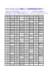
Pneumoperitoneum Caused by Ruptured Gas-Containing Pyogenic ...
Pneumoperitoneum Caused by Ruptured Gas-Containing Pyogenic ...
Pneumoperitoneum Caused by Ruptured Gas-Containing Pyogenic ...

Create successful ePaper yourself
Turn your PDF publications into a flip-book with our unique Google optimized e-Paper software.
<strong>Pneumoperitoneum</strong>, pyogenic liver abscess<br />
11<br />
<strong>Pneumoperitoneum</strong> <strong>Caused</strong> <strong>by</strong> <strong>Ruptured</strong> <strong>Gas</strong>-<strong>Containing</strong><br />
<strong>Pyogenic</strong> Liver Abscess: A Case Report and Literature Review<br />
Chih-Chung Chao 1 , Sung-Yuan Hu 2,3,4,5 , Ying-Hock Teng 1,3<br />
<strong>Pneumoperitoneum</strong> reflects intra-abdominal visceral perforation in 85 to 95 % of all occurrences.<br />
In 5 to 15 % of cases, however, pneumoperitoneum does not reflect perforation and results from another<br />
source. Herein, we report a rare case of surgery indicated pneumoperitoneum caused <strong>by</strong> the rupture<br />
of gas-containing pyogenic liver abscess (GPLA) in a newly-diagnostic diabetic 57-year-old man. He<br />
presented the symptoms mimicking intra-abdominal visceral perforation. He recovered uneventfully after<br />
surgical intervention, drainage, full-course of antibiotics and strict control of blood glucose. Cultures of<br />
blood and ascites grew Klebsiella pneumoniae.<br />
Key words: gas-containing pyogenic liver abscess (GPLA), pneumoperitoneum, rupture<br />
Introduction<br />
Acute abdomen is a common and emergent<br />
condition in the emergency department (ED).<br />
Indeed, it is necessary to be investigated<br />
immediately and treated it without delay.<br />
<strong>Pneumoperitoneum</strong> is the term used to describe<br />
the presence of free air within peritoneal cavity<br />
but outside the viscera. In the major cases of<br />
pneumoperitoneum, it is the result of intraabdominal<br />
visceral perforation, especially the<br />
hollow organs (1) . <strong>Gas</strong>tric and duodenal ulcers<br />
account for the majority of these cases (2) . Generally,<br />
prompt surgical intervention is required in these<br />
patients. <strong>Ruptured</strong> GPLA is a rare cause of surgery<br />
indicated pneumoperitoneum (3-6) .<br />
Case Report<br />
A 57 year-old man presented to the ED with<br />
fever, productive cough and general weakness for<br />
1 week. He denied any systemic disease in the<br />
past except herbal drugs for polydipsia, polyuria,<br />
polyphagia and body weight lost from 78 to 50 kg<br />
in recent 3 years. On arrival of ED, vital signs<br />
were body temperature of 36.1℃, blood pressure<br />
164/106 of mmHg, heart rate of 96 beats/min<br />
and respiratory rate of 18 breaths/min. Physical<br />
examination revealed coarse crackles over the<br />
right lung field, soft abdomen on palpation, normal<br />
peristalsis on auscultation and there were no<br />
localized tenderness, rebounding pain or percussion<br />
pain over the right flank and right lower chest<br />
regions. First look of the upright chest X-ray<br />
(CXR) (Fig. 1) was increased infiltration over right<br />
lung field. Laboratory investigations were white<br />
blood cell counts 33960/μL with 90% neutrophils<br />
and 2.5% bands, C-reaction protein 31.6 mg/dL,<br />
glucose 838 mg/dL, HbA1c 14.6%, aspartate<br />
aminotransferase 117 U/L, alanine aminotransferase<br />
Received: August 22, 2011 Accepted for publication: December 17, 2011<br />
From the 1 Department of Emergency Medicine, Chung Shan Medical University Hospital; 2 Division of Toxicology, Department<br />
of Emergency Medicine, Taichung Veterans General Hospital; 3 School of Medicine, Chung Shan Medical University; 4 Institute<br />
of Medicine, Chung Shan Medical University; 5 National Taichung Nursing College, Taiwan (R.O.C.)<br />
Address reprint requests and correspondence: Dr. Sung-Yuan Hu<br />
Division of Toxicology, Department of Emergency Medicine, Taichung Veterans General Hospital<br />
160 Section 3, Chung-Kang Road, Taichung, Taiwan (R.O.C.)<br />
Tel: (04)23592525 ext 3670 Fax: (04)23594065<br />
E-mail: song9168@pie.com.tw
12<br />
J Emerg Crit Care Med. Vol. 23, No. 1, 2012<br />
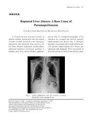
Fig. 1<br />
The upright chest X-ray showed small air bubbles (black<br />
arrow) superimposing at upper liver area, which were<br />
suggestive of a gas-containing pyogenic liver abscess (GPLA)<br />
90 U/L, total bilirubin 0.8 mg/dL, alkaline<br />
phosphatase 123 U/L. Then he was admitted<br />
under the impression of newly-diagnostic diabetes<br />
mellitus (DM), pneumonia and impairment of liver<br />
function test, nature to be determinated.<br />
Abdominal sonography was scheduled on next<br />
day. However, sudden onset of abdominal pain<br />
over right upper quadrant (RUQ) with vomiting<br />
occurred on the second day. Physical examination<br />
revealed diffuse tenderness, especially over RUQ,<br />
with muscle rigidity and rebounding pain, but<br />
the absence of Murphy’s sign. First look of the<br />
second upright CXR (Fig. 2) showed bilateral<br />
subphrenic free air and right side pleural effusion.<br />
Second look of the first (Fig. 1) and the second<br />
(Fig. 2) upright CXR revealed small air bubbles<br />
superimposed at upper liver area was suggestive<br />
of GPLA. Abdominal computed tomography (CT)<br />
scan (Fig. 3) disclosed a big GPLA in right lobe of<br />
liver, 11.6 x 10 x 6.8 cm in size, intra-abdominal<br />
free air, ascites and right side pleural effusion. An<br />
emergency surgical intervention was conducted<br />
under the tentative diagnosis of secondary<br />
peritonitis due to ruptured GPLA.<br />
After surgical intervention, peritoneal lavage,<br />
drainage, antibiotic treatment and strict control of<br />
blood glucose, the infection was well controlled and<br />
he was discharged smoothly on the postoperative<br />
14th day. Cultures of blood and ascites yielded<br />
Klebsiella pneumoniae.<br />
Discussion<br />
<strong>Pneumoperitoneum</strong> usually can be detected<br />
<strong>by</strong> upright chest and left lateral decubitus of<br />
abdomen radiographs, but CT scan is more sensitive<br />
to detect than upright CXR (7) . In 5 to 15% of<br />
cases, pneumoperitoneum does not reflect intra-
<strong>Pneumoperitoneum</strong>, pyogenic liver abscess<br />
13<br />
Fig. 2<br />
The upright chest X-ray showed right side pleural effusion,<br />
bilateral subphrenic free air (white arrows), and small air bubbles<br />
(black arrow) superimposing at upper liver area, which were<br />
suggestive of a GPLA with free air in the peritoneum<br />
Fig. 3<br />
A contrast-enhanced abdominal computed tomographic scan<br />
showed a GPLA in right lobe of liver, 11.6 x 10 x 6.8 cm in size<br />
(black arrow), intra-abdominal free air (white arrow) and ascites
14<br />
J Emerg Crit Care Med. Vol. 23, No. 1, 2012<br />
abdominal visceral perforation and results from<br />
another source and such a situation is termed<br />
spontaneous pneumoperitoneum (SP) or nonsurgical<br />
pneumoperitoneum (NSP) (1,8) . SP and NSP can be<br />
classified according to the source as 5 categories<br />
as below (Table 1): (1) Thoracic; (2) Abdominal;<br />
(3) Gynecologic; (4) Miscellaneous and idiopathic;<br />
(5) Pseudopneumoperitoneum (1-2,4-9) . Most cases of<br />
NSP occurred as a procedural complication or as<br />
a complication of medical intervention. The most<br />
common sources of NSP are from thoracic route (3,9) .<br />
When abdominal pain and distension are minimal<br />
and peritoneal signs, fever, and leukocytosis are<br />
absent, NSP should be considered.<br />
P y o g e n i c l i v e r a b s c e s s ( P L A ) i s n o t<br />
uncommon and sometimes life-threatening. PLA<br />
is associated with high mortality as the diagnosis<br />
is often delayed. In recent decades, its recognition<br />
Table 1 Causes of spontaneous and nonsurgical pneumoperitoneum<br />
Category<br />
Causes<br />
Thoracic Mechanical ventilation Cardiopulmonary resuscitation<br />
Mask ventilation<br />
Bronchoscopy<br />
Pneumothorax<br />
Asthma<br />
Atelectasis<br />
Chronic obstructive pulmonary disease<br />
Bullous emphysema<br />
Bronchopulmonary fistula<br />
Pulmonary sepsis, tuberculosis Blunt trauma<br />
Blast injury<br />
Tracheal rupture<br />
Increased intrathoracic pressure such as cough, retching<br />
Barotrauma<br />
Valsava maneuver<br />
Quick decompression while scuba diving<br />
Procedure related such as median sternotomy, heart transplant<br />
Abdominal Open laparotomy Laparoscopic procedure<br />
Peritoneal dialysis<br />
Endoscopic procedures<br />
Blunt, penetrating trauma Colon contrast examination<br />
Splenic embolization<br />
Pneumatosis cystoides intestinalis<br />
Diverticulosis<br />
Spontaneous bacterial peritonitis<br />
Pneumocholecystitis<br />
<strong>Gas</strong>-containing pyogenic liver abscess<br />
Collagen vascular disease Subclinical perforated viscus<br />
<strong>Gas</strong>tric emphysema<br />
<strong>Gas</strong>-producing organism infection<br />
Gynecologic Coitus Postpartum knee-chest exercise<br />
Orogenital sex<br />
Vaginal insufflation and douching<br />
Ovarian cancer<br />
Gynecologic examination procedures<br />
<strong>Gas</strong>-producing organism infection<br />
Miscellaneous and Idiopathic Cocaine use<br />
Aerophagia<br />
Amyloidosis<br />
Dental extraction<br />
Adenotonsillectomy<br />
Tracheostomy<br />
Scleroderma without pneumatosis cystoides intestinalis
<strong>Pneumoperitoneum</strong>, pyogenic liver abscess<br />
15<br />
has clearly improved through the advance of<br />
more sensitive and specific imaging techniques,<br />
such as ultrasonography and CT scan, with their<br />
relevant therapeutic implications (10) . Improvement<br />
of diagnosis and treatment methods over the<br />
past more than 60 years, so the mortality rate of<br />
PLA has decreased from 79.6% to 1.47-15% and<br />
mortality rate of PLA was 10.9% during 1996-2004<br />
in Taiwan (11,12) . However, only 10% of PLA<br />
patients presented with the classic triad of fever,<br />
jaundice, and RUQ abdominal tenderness (13) . Based<br />
upon the lack of specificity of clinical symptoms<br />
and signs, as well as laboratory parameters, so<br />
promptly diagnosing PLA remains a task for the<br />
for emergency physicians because the time for<br />
evaluation of PLA patients is often limited (14) .<br />
Clues for diagnosis of PLA in the ED can be<br />
classified as 5 categories as below (Table 2): 1.<br />
Presenting symptoms; 2. Physical examination<br />
findings; 3. Predisposing factors; 4. Laboratory<br />
abnormalities; 5. Abnormal findings in chest and<br />
abdomen radiographs (13-15) . Among these clues,<br />
fever and RUQ abdominal pain are the most<br />
typical symptoms; RUQ abdominal tenderness or<br />
jaundice is the most common physical examination<br />
findings (13-15) . The first evaluation of an occult liver<br />
abscess may be improved <strong>by</strong> a history directed<br />
to identification predisposing conditions, mainly<br />
immunosuppression and hepatobiliary disorders. 14<br />
PLA should be considered when physicians met<br />
patients with typical or several clues of PLA.<br />
The most appropriate treatment for PLA is<br />
antibiotic therapy with percutaneous drainage<br />
under ultrasonic guidance if indicated (11,16) . DM is<br />
the most common underlying disease in patients<br />
with PLA (11,16) . 85.5% of patients with GPLA and<br />
60.9% of patients with ruptured PLA had DM (3,4,17) .<br />
In Taiwan, PLA caused <strong>by</strong> Klebsiella pneumoniae<br />
has long been recognized, and its incidence was<br />
reported to range from 50% to 88% of the over<br />
all PLA (3,12) . Klebsiella pneumoniae is the most<br />
common pathogen of GPLA, PLA and ruptured<br />
PLA (3,4,12,17-18) . The incidence of rupture in PLA is<br />
about 5.5% (3,4) . The occurrence of gas formation in<br />
PLA is 7%-24% (18) . Although gas-forming bacterial<br />
infections are generally caused <strong>by</strong> anaerobic<br />
organisms, but majority of all GPLA have been<br />
reported to be caused <strong>by</strong> aerobic organism such as<br />
Table 2 Clues for diagnosis of pyogenic liver abscess in the emergency department<br />
Category<br />
Presenting symptoms<br />
Physical examination<br />
findings<br />
Predisposing factors<br />
Abnormal findings in chest and<br />
abdomen radiographs<br />
Laboratory abnormalities<br />
Clues<br />
Fever, Abdominal pain, Chills, Nausea, Vomiting, Anorexia, Weight loss,<br />
Malaise, Diarrhea, Chest pain, Cough<br />
Jaundice, RUQ tenderness, Hepatomegaly, Splenomegaly, Ascites, Sepsis<br />
syndrome, Muscle rigidity, Rebounding pain<br />
DM, Hepatobiliary disorders (including liver transplantation), HIV<br />
infection, Concomitant neoplasm, Alcoholism, Recent abdominal<br />
surgery (in previous recent weeks), Chronic brucellosis, Steroidal and<br />
Immunosuppressive therapy<br />
Elevated right hemidiaphragm, Right basilar infiltration, Right pleural<br />
effusion, Air bubbles over liver area, <strong>Pneumoperitoneum</strong><br />
Hyperbilirubinemia, Elevation of blood liver enzymes or alkaline<br />
phosphatase, Leukocytosis, Hypoalbuminemia, Anemia
16<br />
J Emerg Crit Care Med. Vol. 23, No. 1, 2012<br />
Table 3 Poor prognostic factors of pyogenic liver abscess<br />
APACHE II score at admission ≥15 Compromised immune status<br />
Malignancy<br />
Diabetes mellitus<br />
Uremia<br />
Senility<br />
Hyperbilirubinemia 1 High level of blood creatinine 2<br />
High level of blood urea nitrogen 3<br />
Hyperglycemia<br />
MDR isolates<br />
Bacteremia<br />
Polymicrobial infection 4 Anaerobic infection 4<br />
Non-K. pneumoniae infection 4<br />
Antibiotics alone<br />
Diagnostic delay<br />
Multiple abscesses<br />
Bi-lobe involvement<br />
<strong>Gas</strong>-forming liver abscess<br />
Alveolar gas pattern and pneumoperitoneum as viewed on radiographs<br />
Globular configuration, shaggy margin, alveolar internal structure, and total gas content on CT scans.<br />
APACHE = Acute Physiology And Chronic Health Evaluation, MDR = multi-drug resistant, K. pneumoniae<br />
= Klebsiella pneumoniae, 1 = blood total bilirubin > 20.52 μmol/L, 2 = blood creatinine > 115 μmol/L, 3 =<br />
blood urea nitrogen>7.86 mmol/L, 4 = isolated pathogen growing in blood or abscess cultures<br />
Klebsiella pneumonia and Escherichia coli (18) . The<br />
impairment of local perfusion may also inhibit the<br />
removal of gas from the infected tissue and cause<br />
the GPLA (5) . However, if rupture of PLA occurs<br />
and signs of acute peritonitis present, surgery<br />
is the only treatment for this condition (5) . Poor<br />
prognostic factors of PLA are listed at Table 3 (19,20) .<br />
Multivariate analysis revealed that gas-forming<br />
abscess, multi-drug resistant isolates, anaerobic<br />
infection, blood urea nitrogen level >7.86 mmol/l,<br />
and APACHE II score ≥15 were associated with<br />
high mortality (20) .<br />
I n s u m m a r y, i n s o m e o f t h e c a s e s o f<br />
pneumoperitoneum, the physicians didn’t make<br />
a proper diagnosis until surgical intervention.<br />
<strong>Pneumoperitoneum</strong> with acute abdominal pain is<br />
mostly secondary to hollow organ perforation, but<br />
there are still some other causes which present the<br />
similar clinical condition. Rupture of GPLA is a<br />
rare one of them and can mimic intra-abdominal<br />
visceral perforation. When physicians meet patients<br />
who have pneumoperitoneum with acute abdominal<br />
pain and the clinical clues of PLA exist. It must<br />
be borne in mind that rupture of GPLA is one<br />
of the differential diagnoses. Further abdominal<br />
sonography, even abdominal CT scan is needed to<br />
establish the proper diagnosis for immediate and<br />
definite managements.<br />
References<br />
1. Mularski RA, Sippel JM, Osbrone ML.<br />
<strong>Pneumoperitoneum</strong>: a review of nonsurgical<br />
causes. Crit Care Med 2000;28:2638-44.<br />
2. Mularski RA, Ciccolo ML, Rappaport WD.<br />
Nonsurgical causes of pneumoperitoneum.<br />
West J Med 1999;170:41-6.<br />
3. Lee CH, Leu HS, Wu TS, Su LH, Liu JW. Risk<br />
factors of spontaneous rupture of liver abscess<br />
caused <strong>by</strong> Klebsiella pneumoniae. Diagn<br />
Microbiol Infect Dis 2005;52:79-84.<br />
4. Chou FF, Sheen-Chen SM, Lee TY. Rupture<br />
of pyogenic liver abscess. Am J <strong>Gas</strong>troenterol<br />
1995;90:767-70.<br />
5. Ukikusa M, Inomoto T, Kitai T, et al.<br />
<strong>Pneumoperitoneum</strong> following the spontaneous
<strong>Pneumoperitoneum</strong>, pyogenic liver abscess<br />
17<br />
rupture of a gas-containing pyogenic liver<br />
abscess: report of a case. Surg Today<br />
2001;31:76-9.<br />
6. S h u m H C, G a u J S, L i a o T H, e t a l.<br />
S p o ntaneously ruptured gas-containing<br />
pyogenic liver abscess: an unusual case of<br />
pneumoperitoneum. Chin J Radiol 2004;29: 203-6.<br />
7. Stapkis JC, Thickman D. Diagnosis of<br />
pneumoperitoneum: abdominal CT vs.<br />
upright chest film. J Comput Assist Tomogr<br />
1992;16:713-6.<br />
8. Williams NM, Watkin DF. Spontaneous<br />
pneumoperitoneum and other nonsurgical<br />
causes of intraperitoneal free gas. Postgrad<br />
Med J 1997;73:531-7.<br />
9. Chen WT, Lee BC, Tsai MJ. <strong>Pneumoperitoneum</strong><br />
without hollow organ perforation: a case report.<br />
J Taiwan Emerg Med 2011;13:48-52.<br />
10. Jiménez E, Tiberio G, Sánchez J, Jiménez FJ,<br />
Jiménez G. <strong>Pyogenic</strong> hepatic abscesses: 16<br />
years experience in its diagnosis and treatment.<br />
Enferm Infecc Microbiol Clin 1998;16: 307-11.<br />
11. Stain SC, Yellin AE, Donovan AJ, Brien HW.<br />
<strong>Pyogenic</strong> liver abscess: modern treatment. Arch<br />
Surg 1991;126:991-6.<br />
12. Tsai FC, Huang YT, Chang LY, Wang JT.<br />
<strong>Pyogenic</strong> liver abscess as endemic disease,<br />
Taiwan. Emerg Infect Dis 2008;14;1592-600.<br />
13. Johannsen EC, Sifri CD, Madoff LC. <strong>Pyogenic</strong><br />
liver abscesses. Infect Dis Clin North Am<br />
2000;14:547-63.<br />
14. Hernández JL, Ramos C. <strong>Pyogenic</strong> hepatic<br />
abscess: clues for diagnosis in the emergency<br />
room. Clin Microbiol Infect 2001;7:567-70.<br />
15. Rahimian J, Wilson T, Oram V, Holzman<br />
RS. <strong>Pyogenic</strong> liver abscess: recent trends<br />
in etiology and mortality. Clin Infect Dis<br />
2004;39:1654-9.<br />
16. Yu SC, Ho SS, Lau WY, et al. Treatment of<br />
pyogenic liver abscess: prospective randomized<br />
comparison of catheter drainage and needle<br />
aspiration. Hepatology 2004;39:932-8.<br />
17. Chou FF, Sheen-Chen SM, Chen YS, Lee TY.<br />
The comparison of clinical course and results<br />
of treatment between gas-forming and nongas-forming<br />
pyogenic liver abscess. Arch Surg<br />
1995;130:401-5.<br />
18. Lee HL, Lee HC, Guo HR, Ko WC, Chen<br />
KW. Clinical significance and mechanism of<br />
gas formation of pyogenic liver abscess due<br />
to Klebsiella pneumoniae. J Clin Microbiol<br />
2004;42:2783-5.<br />
19. Lee TY, Wan YL, Tsai CC. <strong>Gas</strong>-containing<br />
liver abscess: radiological findings and clinical<br />
Significance. Abdom imaging 1994;19:47-52.<br />
20. Chen SC, Tsai SJ, Chen CH, et al. Predictors<br />
of mortality in patients with pyogenic liver<br />
abscess. Neth J Med 2008;66:196-203.
18<br />
J Emerg Crit Care Med. Vol. 23, No. 1, 2012<br />
<br />
<br />
1 2,3-5 1,3<br />
85-95%5-15%<br />
57<br />
<br />
<br />
1008221001217<br />
1<br />
2 3 4 <br />
5<br />
<br />
40705160<br />
<br />
(04)235925253670(04)23594065<br />
E-mail: song9168@pie.com.tw