- Page 2 and 3:
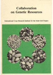
Cover: Ex-Bornu, an important landr
- Page 4 and 5:
Correct citation: I C R I S A T (In
- Page 6 and 7:
Pearl Millet Entomology Insect Pest
- Page 8 and 9:
Potential and Prospects of Pearl Mi
- Page 10 and 11:
tropical countries i t w i l l be v
- Page 12:
Opening Session
- Page 15 and 16: globusum has globose caryopses, and
- Page 17 and 18: Area, Production, and Productivity:
- Page 19 and 20: area increased substantially in Raj
- Page 21 and 22: apply complex fertilizer at 20-60 k
- Page 23 and 24: Diseases. Diseases are endemic to p
- Page 25 and 26: levels of adoption. The relative co
- Page 27 and 28: Socioeconomic Factors Management. T
- Page 30 and 31: Pearl Millet in African Agriculture
- Page 32 and 33: The poor performance in food produc
- Page 34 and 35: 900 800 700 600 Mean 500 400 300 20
- Page 36 and 37: population pressures in recent year
- Page 38 and 39: Economics of Millet Production In 1
- Page 40 and 41: ather than to yield increases, show
- Page 42: Simpara, M. B. 1985. La recherche s
- Page 45 and 46: making it possible to grow a number
- Page 47 and 48: in about 30 accessions. Unlike mono
- Page 49 and 50: genome male-sterile lines, and A A
- Page 51 and 52: Pearl m i l l e t (Pennisetum ameri
- Page 53 and 54: Vasil, V., and Vasil, I . K . 1981a
- Page 55 and 56: Introduction Pearl millet (Penniset
- Page 57 and 58: Clean sorghum o r m i l l e t G r i
- Page 60 and 61: peas, peanuts, or baobab leaves. Wh
- Page 62 and 63: Beer Two major kinds of beer are pr
- Page 64 and 65: properties for pearl millets. It is
- Page 68 and 69: the germ (McDonough 1986). The high
- Page 70 and 71: Table 6. Nitrogen distribution in s
- Page 72 and 73: Banigo, E.O.I., de Man, J.B., and D
- Page 75 and 76: Discussion The discussion of the pa
- Page 77: Improvement through Plant Breeding
- Page 81 and 82: Diversity and Utilization of Pearl
- Page 83 and 84: with protruding grains, but in the
- Page 85 and 86: Table 2. Pearl millet accessions as
- Page 87 and 88: lese millets constitute two distinc
- Page 89 and 90: turn, P. purpureum, (Dujardin and H
- Page 91 and 92: more to useful genetic diversificat
- Page 93 and 94: Hanna, W . W . , Wells, H . D . , a
- Page 107 and 108: Varietal Improvement of Pearl Mille
- Page 109 and 110: the weedy form can significantly in
- Page 111 and 112: Table 3. Evaluation of heterosis in
- Page 113 and 114: Table 4. Grain yield of local 3/4 p
- Page 115 and 116: ased on selection of drought-resist
- Page 117:
Marchais, L., and Pernes, J. 1985.
- Page 120 and 121:
Andrews 1984), there are operationa
- Page 122 and 123:
S h o r t - t e r m g o a l s I n t
- Page 124 and 125:
Table 4a. Prediction equations, adv
- Page 126 and 127:
Table 4c. Prediction equations, adv
- Page 128 and 129:
Nebraska B s y n t h e t i c o r i
- Page 130 and 131:
6 P o p u l a t i o n c r o s s e s
- Page 133 and 134:
Pearl Millet Hybrids H . R . Dave 1
- Page 135 and 136:
Table 2. Early released hybrids in
- Page 137 and 138:
Table 6. Performance of third phase
- Page 139 and 140:
Breeding Pearl Millet Male-Sterile
- Page 141 and 142:
Table 1. Male-sterile lines of pear
- Page 143 and 144:
Senegal. This source is reported to
- Page 145 and 146:
possible solutions to produce high
- Page 147 and 148:
selected from IP 2696 has recently
- Page 149:
Burton, G.W., and Athwal, D.S. 1967
- Page 153:
Biological Factors Affecting Pearl
- Page 157:
Moderator's Overview Biological Fac
- Page 160 and 161:
Introduction Downy mildew of pearl
- Page 162 and 163:
2 3 1 2 4 6 I 5 3 1 A s e x u a l s
- Page 164 and 165:
however, that wind plays an importa
- Page 166 and 167:
Figure 4. Figure assembly to demons
- Page 168 and 169:
in sterilized distilled water. Leav
- Page 170 and 171:
Frederiksen, R.A., and Rosenow, D .
- Page 172 and 173:
Tasugi, H. 1933. Studies on Japanes
- Page 174 and 175:
(Decarvalho 1949), Nigeria (King an
- Page 176 and 177:
Table 1. Differential and stable do
- Page 178 and 179:
ness against certain Phycomycetes (
- Page 180 and 181:
Large s c a l e f i e l d s c r e e
- Page 182 and 183:
References A I C M I P ( A U India
- Page 184 and 185:
Sun, M . H . , Chang, S.S., and Tsa
- Page 186 and 187:
Introduction Ergot (Claviceps fusif
- Page 188 and 189:
antidotale (Thakur and Kanwar 1978)
- Page 190 and 191:
Table 1. Performance of some select
- Page 192 and 193:
Table 4. Performance of some select
- Page 194 and 195:
Siddiqui, M . R . , and Khan, I . D
- Page 196 and 197:
184
- Page 198 and 199:
186
- Page 200 and 201:
188
- Page 202 and 203:
190
- Page 204 and 205:
192
- Page 206 and 207:
194
- Page 208 and 209:
Sahelian Zone Tunisia 0 km 1000 Mor
- Page 210 and 211:
eflexa, and other scarab beetle spe
- Page 212 and 213:
200 Partially Burning, Bagging, and
- Page 214 and 215:
Table 1. Predators, parasites, and
- Page 216 and 217:
References Appert, J. 1957. Les Par
- Page 219 and 220:
Role and Utilization of Microbial A
- Page 221 and 222:
located through the root phloem. In
- Page 223 and 224:
They are not strong competitors in
- Page 225:
Fred, E.B., Baldwin, I . L . , and
- Page 229:
Climatic and Edaphic Limitations to
- Page 232 and 233:
much larger, about 2-4 kPa. Differe
- Page 234 and 235:
over the range that occurs in the f
- Page 236 and 237:
224 top 2 m holds from 100-250 mm o
- Page 238 and 239:
The conservative nature of qD is ex
- Page 240 and 241:
228 Table 4. Root and shoot charact
- Page 242 and 243:
Research on all these responses sho
- Page 245 and 246:
Making Millet Improvement Objective
- Page 247 and 248:
Table 1. Percentage of cultivated a
- Page 249 and 250:
• Well adapted, early-maturity, l
- Page 251 and 252:
in tests conducted by ICRISAT on fa
- Page 253 and 254:
ing a relatively good year in the S
- Page 255 and 256:
stress, as well as to genetic diffe
- Page 257:
Norman, D . W . , Newman, M . D . ,
- Page 260 and 261:
919.0 1212.0, Total area: 11.19 m i
- Page 262 and 263:
Table 4. Economics of pearl millet
- Page 264 and 265:
Azospirillum as a Seed Inoculant Az
- Page 267 and 268:
Management Practices to Increase Yi
- Page 269 and 270:
1500 1300 1100 900 700 500 300 100
- Page 271 and 272:
a physical form similar to SSP and
- Page 273 and 274:
Tillage The beneficial effects of t
- Page 275 and 276:
tivars of different maturity groups
- Page 277 and 278:
as fertility. The nitrogen contribu
- Page 279 and 280:
Greenland, D.J. 1958. Nitrate fluct
- Page 281 and 282:
Breeding for Adaptation to Environm
- Page 283 and 284:
15 S i k a r D i s t r i c t 15 Nia
- Page 285 and 286:
Table 2. Percentage of variation in
- Page 287 and 288:
Table 3. Correlation of the drought
- Page 289 and 290:
Selection for Traits Correlated to
- Page 291:
Discussion Squire's paper drew on d
- Page 295 and 296:
Androgenesis and Enzymatic Diversit
- Page 297 and 298:
Research on Pearl Millet Hybrids in
- Page 299 and 300:
La couleur des grains est variable.
- Page 301 and 302:
processed in different ways dependi
- Page 303 and 304:
Genetic Divergence in Landraces of
- Page 305 and 306:
Pearl Millet Regional Trials by CIL
- Page 307 and 308:
cultural practices such as early pl
- Page 309 and 310:
Host-Plant Resistance to Insect Pes
- Page 311 and 312:
of synthetics and composite varieti
- Page 313 and 314:
Pearl Millet Microbiology Potential
- Page 315 and 316:
available phosphate level in the so
- Page 317 and 318:
Pearl Millet Production in Drier Ar
- Page 319 and 320:
with cowpea system was the most eff
- Page 321 and 322:
Pearl Millet Pathology Disease Inci
- Page 323 and 324:
diseases hitherto undescribed are c
- Page 325 and 326:
Stunt and Counter-Stunt Symptoms in
- Page 327 and 328:
promising sources of resistance: Se
- Page 329 and 330:
69-188 mm for total seedling length
- Page 331:
un certain nombre de contraintes d'
- Page 335 and 336:
Priorities for Crop Protection Rese
- Page 337 and 338:
• those falling into other discip
- Page 339 and 340:
Striga Breeding for resistance to S
- Page 341:
ting improved cultivars to existing
- Page 345 and 346:
Survey of Pearl Millet Production a
- Page 347 and 348:
Table 4. Major constraints to produ
- Page 349 and 350:
Table 6. Extent of cultivation of r
- Page 351 and 352:
Table 10. Area planted to released
- Page 353:
Table 12. Production area and produ
- Page 357 and 358:
Participants Botswana Louis Mazhani
- Page 359 and 360:
Madhya Pradesh G.S. Chauhan Millet
- Page 361 and 362:
M . N . Prasad Professor & Head Cot
- Page 363 and 364:
S.K. Manzo Plant Pathologist Depart
- Page 365 and 366:
ICRISAT Center S. Appa Rao Botanist
- Page 367 and 368:
RA-00110