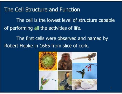
The Cell Structure and Function
The Cell Structure and Function
The Cell Structure and Function

Create successful ePaper yourself
Turn your PDF publications into a flip-book with our unique Google optimized e-Paper software.
<strong>The</strong> <strong>Cell</strong> <strong>Structure</strong> <strong>and</strong> <strong>Function</strong><br />
<strong>The</strong> cell is the lowest level of structure capable<br />
of performing all the activities of life.<br />
<strong>The</strong> first cells were observed <strong>and</strong> named by<br />
Robert Hooke in 1665 from slice of cork.<br />
1
<strong>The</strong> <strong>Cell</strong> Thoery<br />
Proposed by Matthais Schleiden <strong>and</strong> <strong>The</strong>odor<br />
Schwann in 1839:<br />
-All living things are made up of cells.<br />
-<strong>Cell</strong>s are the smallest working unit of all<br />
living things.<br />
-All cells come from preexisting cells through<br />
cells division.<br />
2
Some organisms consist of a single cells =<br />
unicellular organism, others are multicellular<br />
aggregates of specialized cells.<br />
3
Whether multicellular or unicellular, all<br />
organisms must accomplish the same functions:<br />
uptake <strong>and</strong> processing of nutrients<br />
excretion of wastes<br />
response to environmental stimuli<br />
<strong>and</strong> reproduction among others.<br />
4
How We Study <strong>Cell</strong>s<br />
-microscope<br />
-cell fractionation<br />
Most cells are between<br />
1-100 µm in diameter which<br />
can be visualized by light<br />
microscope.<br />
5
Light microscope (LM)<br />
In the LM, visible light is passed through the<br />
specimen <strong>and</strong> then through glass lenses.<br />
<strong>The</strong> lenses refract light such that the image is<br />
magnified into the eyes or the video screen.<br />
magnification <strong>and</strong> resolving power<br />
6
Magnification = the ratio of an object’s image to its<br />
real size.<br />
Magnification of LM ~ X1,000<br />
Resolving power = the measure of the image clarity.<br />
It is the minimum distance two points can be<br />
separated <strong>and</strong> still viewed as two separate points.<br />
Resolution is limited by the shortest wavelength<br />
of the light ~ one half of the wavelength used.<br />
<strong>The</strong> minimum resolution of a light microscope<br />
is about 2 microns, the size of a small bacterium.<br />
7
Electron microscope (EM)<br />
-focuses a beam of electrons through the specimen<br />
or onto its surface<br />
-theoretically, the resolution of a modern EM could<br />
reach 0.1 nanometer (nm), but the practical limit is<br />
closer to about 2 nm<br />
-Transmission <strong>and</strong> Scanning electron microscope<br />
9
Transmission electron microscope (TEM)<br />
-study internal ultrastructure of cells<br />
-electron beam was aimed through the thin section<br />
of specimen<br />
-the image was focused <strong>and</strong> magnified by<br />
electromagnet (instead of glass lenses)<br />
-to enhance the image,<br />
the thin section of<br />
preserved cells are<br />
stained with atoms of<br />
heavy metals<br />
-the micrograph is 2<br />
dimensional<br />
10
Scanning electron microscope (SEM)<br />
-study the surface structure of the cells<br />
-the sample surface is covered with the thin film of<br />
gold<br />
-the electron beam excites the electrons on the<br />
sample surface<br />
-the secondary electrons<br />
are collected <strong>and</strong> focused<br />
on a screen<br />
-the image appeared 3-<br />
dimensional<br />
11
<strong>Cell</strong> Fractionation<br />
-to separate the organelles of cells for functional<br />
study<br />
-the disrupted cells are centrifuged at different<br />
speed <strong>and</strong> duration to fractionate components of<br />
different sizes<br />
12
<strong>Cell</strong> fractionation prepares quantities of<br />
specific cell components/organelles for functional<br />
analysis = biochemical studies.<br />
Microscopes are a major tool in cytology = the<br />
study of cell structures.<br />
Cytology coupled with biochemistry, the study<br />
of molecules <strong>and</strong> chemical processes in metabolism,<br />
developed modern cell biology.<br />
13
A Panoramic View of the <strong>Cell</strong><br />
Basic features of cells:<br />
-All cells are bounded by a plasma membrane.<br />
-<strong>The</strong> semifluid substance within the membrane is<br />
the cytosol, containing the organelles.<br />
-All cells contain chromosomes which carry genes in<br />
the form of DNA.<br />
-All cells also have ribosomes, tiny organelles that<br />
make proteins using the instructions contained in<br />
genes.<br />
14
-A major difference between prokaryotic <strong>and</strong><br />
eukaryotic cells is the location of chromosomes.<br />
-eukaryotic cell, chromosomes are contained in<br />
a membrane-enclosed organelle, the nucleus.<br />
-prokaryotic cell, the DNA is concentrated in<br />
the nucleoid without a membrane separating it from<br />
the rest of the cell.<br />
15
Bacteria: a prokaryotic cell<br />
16
Problems with <strong>Cell</strong> Size<br />
In general, eukaryotic cells are much bigger than prokaryotic<br />
cells:<br />
Most bacteria are 1-10 microns in diameter.<br />
Eukaryotic cells are typically 10-100 microns in<br />
diameter.<br />
-A large cell requires "much more" in terms of the<br />
cellular components.<br />
-Uptake from the environment is also a problem for<br />
large cells: there is less surface area compared to the<br />
volume.<br />
-Distribution of nutrients from one portion of a large<br />
cell to another is also a problem, simply because of the<br />
distance required for the nutrients to travel.<br />
17
Thus most cells are small: sufficient surface area to<br />
accommodate the volume.<br />
Larger organisms do not generally have larger cells than smaller<br />
organisms, - just simply more cells.<br />
18
<strong>The</strong> Plasma Membrane<br />
-double layer of phospholipids<br />
-various proteins are attached to it<br />
-carbohydrate side chains are found only on the outer surface<br />
of plasma membrane<br />
19
<strong>The</strong> Plasma Membrane<br />
- function of plasma membrane = selective barrier that<br />
allows passage of oxygen, nutrients, <strong>and</strong> wastes for the<br />
whole volume of the cell.<br />
20
Overview of Animal <strong>Cell</strong><br />
21
Overview of Plant <strong>Cell</strong><br />
22
<strong>The</strong> Nucleus <strong>and</strong> Its<br />
Envelope<br />
-<strong>The</strong> nucleus<br />
contains most of the<br />
genes in a eukaryotic<br />
cell.<br />
Some genes<br />
are located in<br />
mitochondria <strong>and</strong><br />
chloroplast<br />
-<strong>The</strong> nucleus<br />
averages about 5<br />
microns in diameter.<br />
23
-<strong>The</strong> nucleus is<br />
enclosed by a<br />
nuclear envelope<br />
which is a double<br />
membrane of 20 -40<br />
nm apart.<br />
-Where the double<br />
membranes are<br />
fused, a nuclear<br />
pore complex allows<br />
large<br />
macromolecules <strong>and</strong><br />
particles to pass<br />
through.<br />
24
-<strong>The</strong> nuclear side of<br />
the envelope is lined<br />
by the nuclear<br />
lamina, a network of<br />
intermediate<br />
filaments that<br />
maintain the shape<br />
of the nucleus.<br />
25
Level of Chromatin Packing<br />
In the nucleus,<br />
the DNA <strong>and</strong> the<br />
associated proteins are<br />
organised into firous<br />
material called<br />
chromatin.<br />
In a normal cell<br />
they appear as diffuse<br />
mass.<br />
26
However, when the<br />
cell prepares to divide, the<br />
chromatin fibers coil up to<br />
be seen as separate<br />
structures, chromosomes.<br />
27
Nucleolus = a region (in<br />
the nucleus) of densely<br />
stained fibers <strong>and</strong><br />
granules adjoining<br />
chromatin.<br />
In the nucleolus,<br />
rRNA is synthesized <strong>and</strong><br />
assembled with proteins<br />
from the cytoplasm to<br />
form ribosomal subunits.<br />
<strong>The</strong> subunits pass<br />
from the nuclear pores to<br />
the cytoplasm where<br />
they combine to form<br />
ribosomes.<br />
28
Ribosomes<br />
-particles consisted of proteins <strong>and</strong> ribosomal RNA<br />
(rRNA)<br />
-function = protein synthesis<br />
-prokaryotes = 70S eukaryotes = 80S<br />
29
Eukaryotes (80s ribosome):<br />
large subunit = 60S<br />
45 proteins<br />
rRNA = 28S, 8S <strong>and</strong> 5S<br />
small subunit = 40S<br />
30 proteins<br />
rRNA = 18S<br />
30
-free ribosomes in cytosol synthesize proteins that<br />
function within cytosol<br />
-bound ribosomes are attached to the outside of the<br />
endoplasmic reticulum or nuclear envelope:<br />
membrane protein, organelle proteins or secretory<br />
protein<br />
31
Bound <strong>and</strong> free ribosomes are structurally<br />
identical <strong>and</strong> can alternate between the two roles.<br />
32
<strong>The</strong> Endomembrane System<br />
<strong>The</strong> collection of membranes inside <strong>and</strong> around<br />
a eukaryotic cell.<br />
<strong>The</strong>se membranes are related either through<br />
direct physical contact or by transfer of vesicles (sac<br />
of membrane).<br />
In spite of these links, these membranes have<br />
diverse functions <strong>and</strong> structures:<br />
nuclear envelope, endoplasmic reticulum,<br />
Golgi apparatus, lysosomes,<br />
vacuoles, <strong>and</strong> the plasma membrane.<br />
33
Endoplasmic reticulum (ER)<br />
-ER consists of a network of<br />
membranous tubules <strong>and</strong> sacs<br />
called cisternae. (cisterna = a<br />
reservoir for a liquid)<br />
-the network are interconnected<br />
- <strong>The</strong> ER membrane is<br />
continuous with the nuclear<br />
envelope <strong>and</strong> the cisternal space<br />
of the ER is continuous with the<br />
space between the two<br />
membranes of the nuclear<br />
envelope.<br />
34
-smooth <strong>and</strong> rough ER (with &<br />
without ribosomes)<br />
-Smooth ER:<br />
synthesis of lipid (oils,<br />
phospholipids, <strong>and</strong> steroids)<br />
glycogen metabolism in<br />
the liver cells<br />
detoxification of drugs<br />
<strong>and</strong> poisons<br />
store calcium for muscle<br />
contraction<br />
35
Rough ER: ribosomes are<br />
attached to the outside<br />
-is abundant in cells that<br />
secrete protein<br />
-synthesis secretory<br />
proteins, cell membrane protein<br />
<strong>and</strong> organelle protein<br />
(proteins are targeted to determined<br />
location according to the sorting signals.<br />
Sorting signals are encoded in a.a<br />
sequences or in the attached<br />
oligosaccharides)<br />
-synthesis of<br />
phospholipids <strong>and</strong> ER<br />
associated protein<br />
36
-proteins are synthesized from<br />
the bound ribosomes<br />
-threaded into the cisternal<br />
space through a pore formed<br />
by a protein in the ER<br />
membrane<br />
-protein folds into its native<br />
conformation<br />
-an oligosaccharide is attached<br />
to the protein = glycosylation<br />
-those proteins are wrapped in<br />
the transport vesicles that bud<br />
from the ER<br />
37
<strong>The</strong> Golgi apparatus:<br />
-major sites for carbohydrate symthesis<br />
-sorting <strong>and</strong> dispatching station for the products of the ER<br />
-consists of flatten membranous sacs, cisternae<br />
-unlike ER cisternae, the Golgi cisternae are not physically<br />
connected.<br />
38
-a Golgi stacks has polarity: cis face (the receiving<br />
side) <strong>and</strong> trans face (the shipping side)<br />
39
-transport vesicle from ER fuses to the cis face to transfer the<br />
material to the Golgi<br />
-proteins <strong>and</strong> lipids are altered; glycosylation <strong>and</strong><br />
phosphorylation (tagging the sorting signal)<br />
-oligosaccharides portion of the glycoproteins are modified:<br />
Golgi removes some sugar monomers <strong>and</strong> substitutes others<br />
-some polysaccharides are synthesized in the Golgi e.g pectin<br />
<strong>and</strong> cellulose of the plant cell wall <strong>and</strong> most of the<br />
glycosaminoglycans of animal extracellular matrix<br />
-Golgi products (that will be secreted) depart from the trans<br />
face of the Golgi by transport vesicle for the correct docking.<br />
40
Relationships among Organelles of the<br />
Endomembrane System<br />
-secretory proteins,<br />
lysosomes, vacuoles<br />
<strong>and</strong> membrane are<br />
synthesized by the<br />
rough ER, then<br />
transported through<br />
the Golgi as a vesicle.<br />
-during this process, their molecular compositions<br />
<strong>and</strong> metabolic functions are modified.<br />
41
Lysosomes: principal sites of intracellular digestion<br />
-contain hydrolytic enzymes (required acidic pH) to digest<br />
proteins, polysaccharides, fats <strong>and</strong> nucleic acids<br />
(if those hydrolases leak out of the lysosmes, they are not likely to do<br />
damage unless the cells become acidic)<br />
42
-lysosomal enzymes <strong>and</strong> membranes are made by rough<br />
ER <strong>and</strong> transferred to the Golgi for processing<br />
-lysosomal membranes are highly glycosylated to protect<br />
them from lysosomal proteases<br />
43
-Food particles engulfed as a food vacuole (phagocytosis)<br />
or an endosome (product of receptor-mediated<br />
endocytosis) is fused with the lysosome.<br />
-<strong>The</strong> digestion products are passed to cytosol <strong>and</strong><br />
become nutrient for the cell.<br />
44
-autophagy = process which cells recycle its own<br />
organic material<br />
-the organelles are fused with a lysosome<br />
-after digestion, the organic monomers are<br />
returned to the cytosol for reuse<br />
45
Vacuoles:<br />
-membrane-bound sacs<br />
-diverse functions in cell maintenance<br />
food vacuoles formed by phagocytosis <strong>and</strong><br />
digested by lysosomes<br />
contractile vacuoles (in protists) pump excess<br />
water out of the cells<br />
central vacuole (a versatile compartment in<br />
plants) stores protein <strong>and</strong> metabolic by-products,<br />
reservoir of inorganic ions, pigments<br />
46
Mitochondria <strong>and</strong> Chloroplast<br />
-both are energy transformers of cells<br />
mitochondria = cellular respiration<br />
chloroplast = photosynthesis<br />
-both are not part of the endomembrane system<br />
-most of their proteins are synthesized by the free<br />
ribosomes in the cytosol<br />
-a few of the proteins are synthesized from their own<br />
ribosomes<br />
48
-both organelles contain small quantity of DNA that<br />
direct the synthesis of polypeptides produced by the<br />
internal ribosomes<br />
-both organelles grow <strong>and</strong> reproduce as<br />
semiautonomous organelles<br />
49
Mitochondria<br />
-1-10 µm long<br />
-some cells contain a single large mitochondrion but<br />
most cells contain several mitochondria<br />
-enclosed by two membrane: outer <strong>and</strong> inner<br />
membrane with different permeability<br />
-cristae = fold innermembrane to increase the<br />
surface area<br />
-matrix <strong>and</strong> intermembrane space<br />
51
-matrix contains:<br />
-ds circular DNA<br />
-prokaryote like ribosome (70S)<br />
-enzymes in TCA cycle<br />
-enzymes for β-oxidation of fatty acid<br />
Glucose <strong>and</strong> fatty acids are catabolized in the<br />
matrix of mitochondria.<br />
-innermembrane of mitochondria contains:<br />
-electron transport chain<br />
-ATP synthase<br />
<strong>The</strong> energy from catabolism in the matrix is<br />
converted into ATP.<br />
52
Chloroplast<br />
-is one of the generalized plant structure called<br />
plastids<br />
-found in mesophyll cells of the leaves <strong>and</strong> in algae<br />
-2-4 µm wide <strong>and</strong> 5-10 µm long<br />
53
-2 membranes: inner <strong>and</strong> outer membrane<br />
-stroma ~ matrix of mitochondria<br />
*ds circular DNA<br />
*70S ribosomes<br />
*enzyme for carbohydrate biosynthesis<br />
54
-thylakoids contain photosynthetic machinery of the<br />
chloroplast:<br />
*light absorbing pigment<br />
*electron carriers<br />
*ATP synthesizing apparatus<br />
55
Microbodies<br />
-electron-dense cytoplasmic particles bound by a single<br />
membrane<br />
-contain oxidative enzymes: D-amino acid oxidase,<br />
ureate oxidase <strong>and</strong> catalase<br />
-they are not formed in the<br />
Golgi complex<br />
-they are self replicating, like<br />
the mitochondria<br />
-e.g. peroxisomes, glyoxisomes<br />
56
Peroxisomes<br />
-single membrane<br />
-contain enzymes that transfer<br />
hydrogen from various<br />
substrate to oxygen <strong>and</strong><br />
produce H 2 O 2 as intermediate<br />
product<br />
-but peroxisomes contain<br />
another enzyme that convert<br />
H 2 O 2 to H 2 O<br />
-some peroxisomes break down fatty acids to smaller molecules<br />
that are transported to mitochondria for fuel<br />
Glyoxysomes = specialized peroxisomes (found in fat storing<br />
tissues of plant seeds) convert fatty acid to sugar which can be<br />
used as energy for seedling.<br />
57
<strong>The</strong> Cytoskeleton<br />
-a network of fibers extending throughout the cytoplasm<br />
-function:<br />
provide mechanical strength to the cell<br />
establish cell shape<br />
locomotion (several types of cell motility)<br />
intracellular transport of organelles<br />
-3 main types of fiber:<br />
1.microtubules: determine the positions of membraneenclosed<br />
organelles <strong>and</strong> intracellular transport<br />
2.microfilament: determine the shape of the cell <strong>and</strong><br />
necessary for the whole cell locomotion<br />
3.intermediate filament: provide mechanical strength<br />
<strong>and</strong> resistance to shear stress<br />
58
Cytoskeleton <strong>and</strong> <strong>Cell</strong> Motility<br />
<strong>Cell</strong> motility requires interaction of<br />
cytoskeleton with proteins called motor molecules<br />
in ATP dependent manner.<br />
Sliding of<br />
neighboring<br />
microtubules moves<br />
cilia <strong>and</strong> flagella.<br />
In muscle contraction, motor molecules slide<br />
microfimaments.<br />
60
Walking of organelles along microtubules<br />
Motor molecule attached to receptor on<br />
organelles can “walk” the organelles along<br />
microtubules or microfilaments.<br />
e.g. migration of<br />
neurotransmitters<br />
containing vesicles to<br />
the tips of nerve cell<br />
axon.<br />
61
Microtubules<br />
-are straight, hollow<br />
cylinders<br />
-have a diameter of<br />
about 25 nm<br />
-are variable in length<br />
but can grow 1000 times as<br />
long as they are thick<br />
-are built by the<br />
assembly of dimers of alpha<br />
tubulin <strong>and</strong> beta tubulin.<br />
-are found in both<br />
animal <strong>and</strong> plant cells<br />
62
-grow at each end by the<br />
polymerization of tubulin<br />
dimers (powered by the<br />
hydrolysis of GTP)<br />
-shrink at each end by<br />
the release of tubulin dimers<br />
(depolymerization)<br />
-participate in a wide<br />
variety of cell activities. Most<br />
involve motion. <strong>The</strong> motion is<br />
provided by protein "motors"<br />
that use the energy of ATP to<br />
move along the microtubule.<br />
63
Microtubule motors<br />
<strong>The</strong>re are two major groups of microtubule motors:<br />
kinesins (most of these move toward the plus end<br />
of the microtubules) <strong>and</strong><br />
dyneins (which move toward the minus end).<br />
Examples:<br />
<strong>The</strong> rapid transport of organelles, like vesicles <strong>and</strong><br />
mitochondria, along the axons of neurons takes place<br />
along microtubules with their plus ends pointed toward<br />
the end of the axon. <strong>The</strong> motors are kinesins.<br />
<strong>The</strong> migration of chromosomes in mitosis <strong>and</strong><br />
meiosis takes place on microtubules that make up the<br />
spindle fibers. Both kinesins <strong>and</strong> dyneins are used as<br />
motors<br />
64
-function of microtubules:<br />
*shape <strong>and</strong> support the cell<br />
*resist compression<br />
*serve as track along which organelle can<br />
move via motor molecule<br />
*responsible for separation of chromosome<br />
during cell division<br />
*component of centrosome, centrioles, cilia<br />
<strong>and</strong> flagella<br />
65
Centrosomes <strong>and</strong> Centrioles<br />
Centrosomes (microtubule-organizing center) = a<br />
region near the nucleus from which microtubules<br />
sprouts.<br />
66
Centrioles<br />
-each centrosomes contain<br />
a pair of centrioles<br />
-found in animal cells<br />
-composed of 9 sets of<br />
triplet microtubules<br />
arranged in a ring<br />
-centrioles replicate before<br />
cell division<br />
-may help organize<br />
microtubule assembly<br />
but centrioles are not essential for this function:<br />
centrosomes of most plants lack centrioles.<br />
67
Cilia <strong>and</strong> Flagella<br />
-both cilia <strong>and</strong> flagella are constructed from<br />
microtubules<br />
-both provide either locomotion for the cell or move<br />
fluid pass the cell<br />
-found in prokaryotes <strong>and</strong> eukaryotes<br />
-cilia <strong>and</strong> flagella differ in their beating pattern<br />
cilia sweep<br />
mucus carrying<br />
trapped debris<br />
from the lungs.<br />
68
A flagellum has an undulating motion that<br />
generates force in the same direction as the<br />
flagellum’s axis.<br />
69
Cilia works like oars. <strong>The</strong> alternating power <strong>and</strong><br />
recovery strokes generating force in a direction<br />
perpendicular to the cilium’s axis.<br />
70
-celia <strong>and</strong> flagella have the same ultrastructure =<br />
microtubules core sheathed in an extension of<br />
plasma membrane<br />
-“9+2” arrangement of microtubules (9 doublets +<br />
2single)<br />
-both are anchored in the cell by a basal body,<br />
whose structure is identical to a centriole<br />
72
-each microtubule<br />
connects to the next by a<br />
motor molecule dynein<br />
-dynein is responsible for<br />
the bending of cilia <strong>and</strong><br />
flagella<br />
-addition of phosphate group from ATP to dynein<br />
causes conformational change in this protein which<br />
result in grabing, moving <strong>and</strong> releasing of the outer<br />
microtubule<br />
73
Microfilament or Actin Filament<br />
-a twisted double chain of<br />
actin subunits of 7 nm<br />
diameter<br />
-bear tension (pulling force)<br />
-found in all eukaryotic cells<br />
-combine with other protein to form a threedimensional<br />
network beneath the plasma<br />
membrane<br />
74
-Bundles of microfilament<br />
make up core of microvilli (to<br />
increase cell surface area) of<br />
the animal cells specialized in<br />
transport materials across<br />
plasma membrane.<br />
75
-this network make the semisolid consistency of the<br />
cortex = gel state in the outer cytoplasmic layer<br />
-the interior cytoplasm is the more fluid state<br />
(sol)<br />
-cytoplasm can be converted between gel-sol due<br />
to the reversible assembly of microfilaments into<br />
the networks<br />
Amoeboid movement<br />
:Amoeba, white blood<br />
cells<br />
76
Cytoplasmic streaming: plant cells<br />
-a circular flow of cytoplasm within cells<br />
-this movement is a result of actin-myosin<br />
interactions <strong>and</strong> sol-gel transformations<br />
-speed the distribution of materials within the cells<br />
77
Muscle fiber<br />
-made of actin filaments arranged parallel to one<br />
another along the length of a muscle cell<br />
-a motor molecule myosin is interdigitated with actin<br />
filament<br />
-muscle contraction results from the sliding of actin<br />
<strong>and</strong> myosin<br />
78
Intermediate filament<br />
-cytoplasmic ropelike fibers with average 10 nm in diameter<br />
(<strong>and</strong> thus are "intermediate" in size between actin filaments<br />
(8 nm) <strong>and</strong> microtubules (25 nm)<br />
-<strong>The</strong>re are several types of intermediate filament, each<br />
constructed from one or more proteins characteristic of it.<br />
keratins are found in epithelial cells <strong>and</strong> also form hair<br />
<strong>and</strong> nails (over 20 different kinds of keratins have been found)<br />
nuclear lamins form a meshwork that stabilizes the<br />
inner membrane of the nuclear envelope<br />
neurofilaments strengthen the long axons of neurons;<br />
vimentins provide mechanical strength to muscle (<strong>and</strong><br />
other) cells.<br />
79
Despite their chemical diversity,<br />
intermediate filament play similar<br />
roles in the cells: providing a<br />
supporting framework within the<br />
cells<br />
80
-other intermediate filaments make up the nuclear<br />
lamina that lines the interior of the nuclear envelope<br />
81
<strong>The</strong> vimentin intermediate filament network (red) extends into<br />
the lamella where tetramers colocalize with the actin-bundling<br />
protein, fimbrin (green). <strong>The</strong>se fluorescent foci are specialized<br />
focal adhesions found in osteoclasts <strong>and</strong> macrophages called<br />
podosomes<br />
82
-intermediate filaments strengthen the long extension<br />
(axon) of nerve cells that transmit impulse<br />
83