Conjugation
Conjugation
Conjugation

You also want an ePaper? Increase the reach of your titles
YUMPU automatically turns print PDFs into web optimized ePapers that Google loves.
Modern Microbial Genetics, Second Edition. Edited by Uldis N. Streips, Ronald E. Yasbin<br />
Copyright # 2002 Wiley-Liss, Inc.<br />
ISBNs: 0-471-38665-0 Hardback); 0-471-22197-X Electronic)<br />
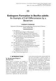
19<br />
<strong>Conjugation</strong><br />
RONALD D. PORTER<br />
Department of Biochemistry and Molecular Biology, The Pennsylvania State University, University Park,<br />
Pennsylvania 16802<br />
I. Introduction ................................... 464<br />
A. C. Regulation of F factor fertility. .............. 464<br />
II. <strong>Conjugation</strong> by the E. coli FFactor............... 465<br />
A. Overview ................................... 465<br />
B. Structure of the F Factor ..................... 465<br />
C. Regulation of F Factor Fertility ............... 467<br />
D. Establishment of Cell Contact . . ............... 468<br />
E. DNA Mobilization and Transfer............... 469<br />
F. Separation of the Mating Pair . . ............... 471<br />
III. Hfr <strong>Conjugation</strong> and Chromosomal Transfer ....... 471<br />
A. How Hfr Strains Arise . ...................... 471<br />
B. Properties of Hfr's ........................... 474<br />
C. Recombination after Hfr conjugation. . . ........ 475<br />
IV. F-prime <strong>Conjugation</strong> and Merodiploids ............ 478<br />
A. The Generation of F-primes................... 478<br />
B. <strong>Conjugation</strong> Properties of F-primes ............ 481<br />
V. <strong>Conjugation</strong> of Fertility-Inhibited F-like Plasmids . . . 482<br />
VI. Nonconjugative, Mobilizable Plasmids ............. 483<br />
VII. Broad Host Range Self-transmissible Plasmids ...... 484<br />
VIII. Chromosome Mobilization by Non-F Plasmids ..... 486<br />
IX. Plasmid-Based <strong>Conjugation</strong> in Other Bacteria....... 488<br />
A. Salmonella.................................. 488<br />
B. Pseudomonas................................ 489<br />
1. Chromosome Mobilization in<br />
P. aeruginosa ............................. 489<br />
2. Chromosome Mobilization in P. putida ....... 490<br />
3. R-primes in Pseudomonads . . ............... 490<br />
C. Streptomyces................................ 490<br />
D. Gram-Positive Cocci Streptococcus, etc.) ....... 493<br />
E. Other Plasmid-Based <strong>Conjugation</strong> Systems ...... 494<br />
X. Conjugative Transposons . . ...................... 495<br />
XI. Conclusion .................................... 496<br />
XII. Appendix: <strong>Conjugation</strong>al Mapping . ............... 496
464 PORTER<br />
I. INTRODUCTION<br />
<strong>Conjugation</strong> is the mode of gene transfer<br />
that involves the transfer of DNA between<br />
two live bacterial cells that are in direct contact.<br />
Although conjugation in nature most<br />
often simply involves the transfer of plasmid<br />
DNA from donor to recipient cell, chromosomal<br />
DNA can be transferred under certain<br />
circumstances. Much of the discussion in<br />
this chapter will focus on the F factormediated<br />
conjugation system of Escherichia<br />
coli as this system serves as a prototype for<br />
conjugation in Gram-negative bacteria. The<br />
less well-characterized Gram-positive conjugation<br />
systems will be described later in the<br />
chapter.<br />
There are many aspects of the discovery of<br />
conjugation in E. coli by Lederberg and Tatum<br />
1946) that were strongly influenced by<br />
elements of serendipity. The choice of the K-<br />
12 strain for use in the initial experiments is<br />
one of the most striking examples of such<br />
happy chance. The protocol involved mixing<br />
two isolates that each had at least two nutritional<br />
deficiencies so that cells where one<br />
marker had reverted would not be scored as<br />
recombinants. Although the K-12 strain was<br />
chosen primarily because of the availability<br />
of isolates with more than one counterselectable<br />
nutritional marker, it also happened to<br />
contain a self-transmissible plasmid. The<br />
particular self-transmissible plasmid in K-<br />
12, the F factor, was also unusual in that it<br />
both constitutively expresses conjugal transfer<br />
functions see below) and contains several<br />
transposable elements that allow it to interact<br />
with the bacterial chromosome.<br />
A. C. Regulation of F factor fertility<br />
Lederberg later estimated that only about<br />
five percent of randomly selected E. coli isolates<br />
would have given recombinants with the<br />
selection protocol that was initially employed.<br />
It was also happy chance that the<br />
nutritional genetic markers in the isolates<br />
selected were closely clustered on the<br />
chromosome in each strain so that optimal<br />
yields of recombinants were readily obtained<br />
without the requirement for multiple crossovers.<br />
Although the work of the Avery<br />
group with transformation in Streptococcus<br />
pneumoniae Avery et al., 1944) had set<br />
the stage, there were still many scientists<br />
who were reluctant to believe that the lowly<br />
bacteria could engage in any form of sexual<br />
activity. The work done by Lederberg and<br />
others who soon followed convincingly<br />
demonstrated that bacteria were organisms<br />
where genetics could be productively practiced.<br />
There are two distinct requirements that<br />
must be met in order for conjugation to<br />
occur. The first of these requirments is that<br />
the cells be able to engage in a specific contact<br />
cycle. The second is that some DNA in<br />
the donor cell be capable of undergoing<br />
mobilization. Plasmids that encode all of the<br />
necessary gene products to enable the potential<br />
donor cell to carry out a specific contact<br />
cycle with a suitable recipient cell are said<br />
to be conjugative. Plasmids whose DNA can<br />
be prepared for transfer to a recipient cell<br />
are called mobilizable. Both of these capabilities<br />
do not always reside on the same plasmid,<br />
however, and neither ability alone<br />
is sufficient for conjugal DNA transfer. Inability<br />
to carry out either or both of these<br />
functions classifies a plasmid as being nonconjugative<br />
and/or nonmobilizable. A plasmid<br />
may simply lack one or both of these<br />
abilities as it was originally isolated, or it may<br />
have lost one or both of these abilities through<br />
mutation. Plasmids that are mobilizable, but<br />
nonconjugative, are often efficiently transferred<br />
to recipient cells when other plasmids<br />
present in the donor cell provide the necessary<br />
cell contact functions. Plasmids that are both<br />
conjugative and mobilizable are termed selftransmissible<br />
Clark and Warren, 1979).<br />
It should be noted, however, that the word<br />
mobilization is also often used to describe<br />
the situation where a plasmid, generally a<br />
self-transmissible one, is able to affect the<br />
conjugational transfer of donor cell chromosomal<br />
DNA to a recipient cell. In fact the<br />
word mobilization is most often used in this<br />
sense. While plasmid mobilization refers to a
CONJUGATION 465<br />
plasmid's possession of the ability to transfer<br />
a copy of its own DNA to a recipient cell<br />
whenever a mating pair has been formed,<br />
chromosome mobilization in conjugation<br />
occurs as the result of some kind of physical<br />
association between the donor cell chromosome<br />
and the plasmid undergoing conjugational<br />
transfer. These plasmid/chromosome<br />
associations can be very stable in the case<br />
of Hfr strains or very transient in the case<br />
of cointegrates formed as intermediates in<br />
transposition; these examples will be discussed<br />
later in the chapter. Regardless of the<br />
degree of stability involved, the chromosome<br />
is passively carried along to the recipient cell<br />
as the result of its covalent association with<br />
the plasmid during chromosomal mobilization.<br />
The word mobilization will be used<br />
both ways in this chapter, and the student<br />
should make every effort not to interchange<br />
the two meanings of the word.<br />
II. CONJUGATION BY THE<br />
E. COLI FFACTOR<br />
A. Overview<br />
E. coli cells totally lacking the presence of the<br />
F factor in any form are called F cells. The<br />
F factor can, however, exist in a cell in three<br />
different forms. First, cells containing an autonomously<br />
replicating F plasmid are called<br />
F ‡ cells. Such cells efficiently transfer the F<br />
plasmid to a suitable recipient but rarely<br />
transfer donor cell chromosomal DNA.<br />
Second, the F factor is able to integrate<br />
into the donor cell chromosome to give rise<br />
to an Hfr high frequency of recombination)<br />
cell that can efficiently transfer donor cell<br />
chromosomal DNA to a recipient cell by<br />
conjugation. Third, F-prime plasmids arise<br />
when the integrated F factor in an Hfr<br />
carries some chromosomal DNA with it as<br />
it is recombined out of the chromosome and<br />
returns to the autonomously replicating<br />
state. F-primes are transferred to a suitable<br />
recipient in much the same manner as a wildtype<br />
F factor. Although the establishment of<br />
the mating pair and the initiation of DNA<br />
transfer is identical in all three cases, the<br />
ability of these three donor types to transfer<br />
chromosomal DNA to a recipient cell differs<br />
considerably. These three different types of F<br />
factor±containing cells will be discussed in<br />
more detail later in the chapter.<br />
In the case of F factor±mediated conjugation,<br />
contact initially occurs between the tip<br />
of the donor cell's F factor±encoded sex pilus<br />
and the exterior envelope of the recipient<br />
cell. Direct contact, presumably achieved by<br />
basal disassembly of the pilus, produces an<br />
unstable mating pair. Multiple cell interactions<br />
frequently give rise to mating cell<br />
aggregates that may contain up to 20 cells<br />
Achtman et al., 1978a). A picture and diagrammatic<br />
representation of E. coli mating<br />
aggregates are shown in Figure 1. Although<br />
some DNA transfer between the cells may<br />
occur at these early stages, most DNA transfer<br />
occurs between pairs of cells specifically<br />
stabilized within the mating aggregate. Cells<br />
are called exconjugants after mating pair<br />
dissociation, and recipient cells that have<br />
received DNA from donor cells are called<br />
merozygotes. These merozygotes become<br />
transconjugants after the donor DNA has<br />
become stabilized in the recipient cell. This<br />
stabilization of donor DNA can occur either<br />
by recombination with recipient DNA or,<br />
in the case of transferred plasmid DNA, by<br />
establishment of the transferred plasmid<br />
DNA as an independent replicon in the<br />
recipient cell. The various steps in this overall<br />
process will be discussed in more detail<br />
below. A number of excellent recent review<br />
articles dealing with F factor±mediated conjugation<br />
are available for additional study<br />
Willetts and Skurray, 1980, 1987; Willetts<br />
and Wilkens, 1984; Ippen-Ihler and Minkley,<br />
1986, Frost et al., 1994, Firth et al., 1996).<br />
B. Structure of the F Factor<br />
The F factor is a 100 kb plasmid that can<br />
be divided into four fairly distinct regions<br />
see Fig. 2). The region that is labeled inc,<br />
rep is the portion of the F factor that is<br />
involved in the vegetative replication of the<br />
plasmid. Mini-F plasmids containing only<br />
this region can be constructed, and these
466 PORTER<br />
Fig. 1. Hfr times F mating aggregates. An interpretative diagram is shown within each micrograph of mating<br />
E. coli cells. The Hfr cells in these diagrams are drawn with thin walls while the F cells are drawn with thick<br />
walls. The cells for which the Hfr versus F assignment was uncertain are shown in white, and F pili that are<br />
thought to connect cells are indicated. Reproduced from Achtman et al., 1978a, with permission of the<br />
publisher.)<br />
mini-F derivatives demonstrate all of the<br />
replication properties of the parent plasmid.<br />
It is this region of the plasmid that determines<br />
the F factor's incompatibility properties<br />
regarding other plasmids in the same cell<br />
see Perlin, this volume).<br />
The F factor also contains a region where<br />
four transposable elements are clustered.<br />
In addition to the two copies of IS3 and the<br />
copy of IS2, there is a copy of Tn1000,<br />
also known as gd, whose transposition properties<br />
are very similar to those of Tn3. As
CONJUGATION 467<br />
Fig. 2. Map of the Escherichia coli F factor. The four<br />
major regions of the F factor indicated on the figure<br />
are 1) the inc,rep region which determines vegetative<br />
replication and plasmid incompatibility properties,<br />
2) the tra region which stretches from oriT to<br />
IS3 and provides conjugative and DNA mobilization<br />
functions, 3) the region containing the four transposable<br />
elements that facilitate interactions between<br />
the F factor and other DNA molecules, and 4) the<br />
``silent region'' between IS2 and the inc,rep region.<br />
we will see later in this chapter, it is these<br />
transposable elements that are primarily responsible<br />
for the ability of the F factor to<br />
interact with other DNA molecules, including<br />
the chromosome, in the cell. The third<br />
region of the F factor is sometimes referred<br />
to as the silent region as few distinct<br />
genetic functions have been shown to reside<br />
there.<br />
The approximately 35 kb region of the F<br />
factor labeled tra is the fourth region, and it<br />
is involved in making the F factor a selftransmissible<br />
plasmid. This tra region is<br />
very similar in organization to the tra regions<br />
of many F-like R factors. It contains the oriT<br />
site, at which DNA transfer is initiated, and<br />
DNA sequence analysis Frost et al., 1994)<br />
indicates the presence of 36 open reading<br />
frames with most of the likely genes designated<br />
tra and some trb. Three of the translated<br />
genes traM, traJ, and artA) produce<br />
separate transcripts, but all of the other<br />
genes form a single operon starting with<br />
traY. Although this huge operon has secondary<br />
promoters, the traY promoter appears to<br />
be dominant under conjugative conditions.<br />
The overall structure of the tra regulon is<br />
shown in Figure 3.<br />
C. Regulation of F Factor Fertility<br />
The main tra operon starting with traY) is<br />
positively regulated by the product of the<br />
separately transcribed traJ gene Willetts,<br />
1977; Gaffney et al., 1983). A number of<br />
mechanisms have been proposed to explain<br />
the need for TraJ protein in the efficient expression<br />
of the traY promoter, but it is currently<br />
thought that TraJ protein binding<br />
works by providing sufficient superhelicity<br />
for transcription initiation Gaudin and Silverman,<br />
1993). In most F-like plasmids, the<br />
traJ gene is normally negatively regulated<br />
by the finO and finP gene products fin ˆ<br />
fertility inhibition). A virtue of the F factor<br />
in genetic studies, however, is its lack of fertility<br />
inhibition due to its lack of a functional<br />
finO gene. The tra genes and conjugal ability<br />
are therefore constitutively expressed unless<br />
finO is provided in-trans by a fin ‡ F-like plasmid.<br />
This constitutive expression of fertility<br />
functions was an important element in the<br />
Fig. 3. Map of the F factor transfer region. The top line gives a size scale in kilobase pairs, while the second<br />
line show restriction enzyme sites that are not relevant to our discussion here. The third line represents the<br />
genes with tra genes shown in uppercase letter and trb genes shown in lowercase letters; the oriT site, the IS3-<br />
containing finO gene, and the finP transcript are also shown. The remaining lines show the various genes<br />
grouped by function. Reproduced from Frost et al., 1994, with permission of the publisher.)
468 PORTER<br />
discovery of conjugation by Lederberg and<br />
Tatum 1994). The initially mysterious lack<br />
of fertility regulation in F is due to an IS3<br />
insertion, which traditionally marks one end<br />
of the tra regulon, in the finO gene Yoshioka<br />
et al., 1987). finP is transcribed from the antisense<br />
strand in the mRNA leader region of<br />
the traJ gene, but the finP transcript apparently<br />
does not code for a protein Johnson<br />
et al., 1981). The overlap of the finP RNA<br />
and the leader region of the traJ mRNA is<br />
diagrammatically illustrated in Figure 4. The<br />
binding of these two RNA molecules places<br />
the translational start signals for traJ within<br />
an RNA duplex region which would presumably<br />
preclude translation initiation Finlay<br />
et al., 1986). The presence of FinO protein<br />
has been shown to greatly extend the halflife<br />
of finP RNA even in the absence of traJ<br />
mRNA Lee et al., 1992), and the FinO protein<br />
is capable of binding to secondary structural<br />
features of both RNA species to<br />
promote the formation of an RNA duplex<br />
van Biesen and Frost, 1994; Ghetu et al.,<br />
1999). It is only when the FinO protein stabilizes<br />
this RNA duplex that the translation of<br />
the traJ mRNA is precluded.<br />
Fig. 4. Location of the F factor finP gene. The position<br />
of the finP transcript is shown relative to the<br />
traM and traJ genes. The coordinate positions indicated<br />
are relative to the start of the traJ mRNA.<br />
The finP transcript starts leftward at position 111<br />
and extends to roughly position 34. It should be<br />
noted that the finP transcript is complementary to<br />
much of the leader region of the traJ mRNA, and<br />
overlaps slightly with the coding region for the TraJ<br />
protein.<br />
The other separately transcribed tra gene,<br />
traM, is located very close to oriT and appears<br />
to be negatively autoregulated by means of<br />
binding sites for its own gene product in the<br />
dual promoter region Penfold et al., 1996).<br />
The demonstration that traM is also positively<br />
regulated by TraY protein binding<br />
Penfold et al., 1996) has indicated that the<br />
long suspected possibility of traJ regulation<br />
of traM is the result of an indirect effect.<br />
D. Establishment of Cell Contact<br />
A typical F factor-containing E. coli cell will<br />
possess one to three F-encoded sex) pili on<br />
its surface. The F pilus is a hollow cylinder<br />
with an exterior diameter of 8 nm and an<br />
interior diameter of 2 nm Folkhard et al.,<br />
1979). These sex pili may be up to 20 mm<br />
in length, and are often visualized microscopically<br />
by the adsorption of male-specific<br />
phages. The F sex pili consist of many molecules<br />
of a single protein, pilin, which is encoded<br />
by the traA gene Frost et al., 1984).<br />
The traQ gene product apparently converts<br />
the initial 121 amino acid traA polypeptide<br />
into the functional 70 amino acid polypeptide,<br />
perhaps while acting as a chaperone<br />
during its insertion into the inner membrane<br />
Maneewannakul et al., 1993). The N-terminal<br />
amino acid of the mature pilin is acetylated<br />
by the product of the traX gene, but the<br />
conjugation properties of traX mutants lacking<br />
this acetylation seem largely unaffected<br />
Moore et al., 1993). Claims for the phosphorylation<br />
and glycosylation of pilin have<br />
not been substantiated. At least 13 additional<br />
genes are required for the assembly of a functional<br />
pilus, but their specific roles are not<br />
known.<br />
The initiation of mating pair formation<br />
requires that the tip of the F pilus make contact<br />
with a specific receptor site on the surface<br />
of the recipient cell. Although the exact<br />
nature of that receptor is not known, mutations<br />
that render a cell incapable of functioning<br />
as a recipient in conjugation Con )<br />
generally map in either ompA or in genes<br />
involved in lipopolysaccharide LPS) synthesis.<br />
It appears that LPS participates in the
CONJUGATION 469<br />
initiation of mating pair formation, and it has<br />
also been shown that Zn ‡‡ is required at the<br />
earliest stages of this process. The expression<br />
of the ompA gene in the recipient cell, on the<br />
other hand, is necessary for the stabilization<br />
of the mating pair. It remains possible, however,<br />
that the OmpA protein is also part of<br />
the receptor site, as added LPS-OmpA protein<br />
complexes block mating pair formation<br />
more effectively than LPS alone Achtman<br />
et al., 1978b). The ability of LPS or LPS-<br />
OmpA protein complexes to prevent mating<br />
pair formation presumably results from their<br />
ability to interact with F pili and thereby<br />
prevent the pili from making contact with<br />
recipient cells.<br />
Although DNA transfer can occur through<br />
an extended F pilus Ou and Anderson, 1970;<br />
Harrington and Rogerson, 1990), little DNA<br />
transfer normally occurs before mating pair<br />
stabilization. The F pilus has been shown to<br />
retract upon the attachment of male-specific<br />
phages Jacobson, 1972), and it is generally<br />
assumed that the initiation of direct envelopeto-envelope<br />
contact between donor and recipient<br />
cell involves the retraction of the pilus<br />
by disassembly at its base within the donor<br />
cell. The direct contact between cells yields an<br />
unstable mating pair where little DNA transfer<br />
occurs, and it is likely that the pilus is<br />
no longer required after unstable mating<br />
pair formation has occurred Achtman et al.,<br />
1978a). The conversion of this unstable<br />
mating pair to a stable mating pair where<br />
DNA transfer can occur efficiently involves<br />
the participation of the traG and traN gene<br />
products, as mutations in either of these genes<br />
result in inefficient mating pair formation<br />
without reducing the extent of conjugal<br />
DNA replication Manning et al., 1981).<br />
The exact nature of the final surface-to-surface<br />
interaction between two mating cells is<br />
not well understood, but the traD gene product<br />
may be involved in the formation of a<br />
pore between the two inner membranes Panicker<br />
and Minkley, 1985).<br />
Effective mating pair formation between<br />
two F factor-containing donor cells is prevented<br />
by a phenomenon called surface exclusion.<br />
Surface exclusion requires the traS<br />
and traT gene products which are located in<br />
the inner and outer membranes, respectively<br />
Achtman et al., 1977, 1979; Minkley and<br />
Willetts, 1984; Cheah et al., 1986). Although<br />
pilus to envelope contacts between donor<br />
cells do occur, surface exclusion prevents<br />
mating pair stabilization between two donor<br />
cells as well as the initiation of donor conjugal<br />
DNA synthesis see below). Mating between<br />
two donor cells can be achieved,<br />
however, by a procedure called F phenocopy<br />
mating. This involves growing the cell<br />
to be used as the recipient into stationary<br />
phase so that tra expression, and therefore<br />
surface exclusion, is minimized. It is also<br />
interesting to note that the traT gene product<br />
becomes a major component of the outer<br />
membrane and plays a role in serum resistance<br />
and in reducing the susceptibility of<br />
cells to phagocytosis Moll et al., 1980;<br />
Aguero et al., 1984). Although serum resistance<br />
is not directly relevant to conjugation<br />
mechanism, this tra-dependent phenotype<br />
provides a selective advantage to F factorcontaining<br />
cells in some environments.<br />
E. DNA Mobilization and Transfer<br />
For DNA to actually be transferred from<br />
donor to recipient cell, the plasmid DNA in<br />
the donor cell must go through a series of<br />
processing steps that we refer to as mobilization.<br />
As replacement synthesis of the transferred<br />
strand of donor cell plasmid DNA is<br />
generally concurrent with DNA transfer, the<br />
entire process preparation for transfer, or<br />
mobilization, and transfer itself) is sometimes<br />
also referred to as donor conjugal DNA synthesis<br />
DCDS). Four or five tra gene products<br />
are involved in DCDS, and several events<br />
must occur before the actual DNA synthesis<br />
and transfer begins. First, one strand of the<br />
DNA is nicked at the F factor oriT origin of<br />
transfer) site. This nicking was shown by<br />
infecting cells with a bacteriophage l derivative<br />
carrying the oriT site, and examining the<br />
DNA of the progeny phage Everett and Willetts,<br />
1980). l oriT was used as a convenient<br />
means of packaging the DNA of interest as it
470 PORTER<br />
is much easier to isolate the appropriate<br />
DNA, with and without nicks, from virions<br />
than to purify a minority species of nicked<br />
plasmid DNA from cell lysates. It was found<br />
that 5±10% of the l oriT phage contained a<br />
nick within oriT when Flac was present in the<br />
infected cell; there was no nicking observed<br />
when Flac was not present.<br />
These and other in vivo studies indicated<br />
that this strand-specific nicking reaction at<br />
oriT required the traY gene product plus the<br />
product of a gene called traZ that later<br />
turned out to be a secondary translation<br />
product of the traI gene Everett and Willetts,<br />
1980; Traxler and Minkley, 1987). Studies<br />
to determine the precise location of the<br />
nick at oriT and to carry out the nicking<br />
reaction in vitro Thompson et al., 1989;<br />
Matson and Morton, 1991; Reygers et al.,<br />
1991) revealed that the complete traI gene<br />
product possesses both the oriT nicking activity<br />
and the strand separation activity<br />
known as E. coli DNA helicase I Abdel-<br />
Monem et al., 1983) that is responsible for<br />
separating the two DNA strands during<br />
transfer. The traI nickase-helicase apparently<br />
becomes covalently linked to the 5 0<br />
end of the nicked DNA strand during the<br />
nicking reaction and may play a role in plasmid<br />
recircularization after transfer is complete<br />
Reygers et al, 1991; Matson et al.,<br />
1993). The traY gene product and the integration<br />
host factor IHF) of E. coli both<br />
have binding sites in the oriT region, and<br />
both are needed for efficient oriT nicking<br />
by the TraI protein in vitro under more<br />
physiologically relevant conditions Nelson<br />
et al., 1995). The nicking reaction at oriT is<br />
constitutive in that it occurs in the absence of<br />
either mating pair formation or DCDS<br />
Everett and Willetts, 1980), so a nick at<br />
oriT does not automatically lead to the initiation<br />
of DCDS. It has been suggested that<br />
the binding of TraY protein and IHF at<br />
oriT permit the binding of TraI protein to<br />
form a complex referred to as a relaxosome<br />
Howard et al, 1995). The presence of such<br />
relaxosomes is a common attribute of many<br />
plasmids whose DNA can be self-mobilized.<br />
The role of the traM gene product has not<br />
been well defined. It is not required for piliation<br />
or nicking at oriT, but it is required for<br />
DNA transfer and replacement strand synthesis<br />
in the donor cell. It thus seems to<br />
trigger the start of DCDS at a nicked oriT<br />
site in response to a signal arising after the<br />
tip of the F pilus contacts a suitable recipient<br />
cell Everett and Willetts, 1980). There are<br />
three TraM protein binding sites near oriT,<br />
but these are on the 3 0 side of the nick site<br />
and therefore do not involve the leading end<br />
of the transferred strand. These TraM binding<br />
sites clearly play a role in traM autoregulation,<br />
and the ability of TraM protein to<br />
also bind TraD protein Disque-Kochem<br />
and Dreiseikelmann, 1997) may serve to indicate<br />
a role for TraM protein in positioning<br />
the nicked DNA at the transfer portal of<br />
which TraD protein is thought to be a part<br />
Panicker and Minkley, 1985).<br />
A single strand of F factor DNA is transferred<br />
to the recipient cell starting with the 5 0<br />
end from the nicked oriT site Rupp and<br />
Ihler, 1968; Ohki and Tomizawa, 1968).<br />
The transfer of the displaced strand to the<br />
recipient cell is normally accompanied by<br />
DNA polymerase III-mediated replacement<br />
synthesis in the donor cell, but transfer<br />
can occur in the absence of this synthesis<br />
Sarathy and Siddiqi, 1973; Kingsman and<br />
Willetts, 1978). This replacement DNA synthesis<br />
requires priming by RNA polymerase<br />
Kingsman and Willetts, 1978), and this requirement<br />
may reflect blockage of the 3 0 end<br />
by one of the proteins involved in DCDS.<br />
A new complementary strand for the<br />
entering donor DNA is synthesized by the<br />
recipient cell's normal DNA synthesis machinery.<br />
It now appears that the necessary<br />
priming for this complementary strand synthesis<br />
is achieved by a special promoter<br />
called Frpo that allows host cell RNA polymerase<br />
to initiate a transcript on singlestranded<br />
DNA that can be continued by<br />
DNA polymerase III holoenzyme Masai<br />
and Arai, 1997). This special promoter and<br />
possibly others like it also appear to allow<br />
for the rapid expression of a number of genes
CONJUGATION 471<br />
from the leading region of the transferred F<br />
factor DNA in the recipient cell before complementary<br />
DNA strand synthesis has been<br />
accomplished. While the role of many of<br />
these leading region genes has not been determined,<br />
this group includes ssf, the F factor's<br />
SSB or single-stranded DNA-binding<br />
protein gene, and the psiB gene whose product<br />
acts to prevent SOS induction by the<br />
entering single-stranded DNA in the recipient<br />
cell during DNA transfer Bailone et al.,<br />
1988; Bagdasarian et al., 1992).<br />
The stabilization of the F factor in the<br />
recipient cell is a recA-independent process<br />
Clark, 1967) that typically requires the<br />
transfer of both ends of oriT Everett and<br />
Willetts, 1982). Despite earlier evidence for<br />
the transfer of single-stranded concatemers<br />
of F factor DNA Ohki and Tomizawa,<br />
1968; Matsubara, 1968), the observed requirements<br />
for recircularization favor the<br />
transfer of unit length DNA strands Willetts<br />
and Skurray, 1987). The TraI protein bound<br />
to the 5 0 end of the transferred strand could<br />
simply re-ligate that 5 0 end to the 3 0 end when<br />
it arrives in a direct reversal of the original<br />
oriT nicking reaction if the 3 0 end remains<br />
unobstructed during the course of replacement<br />
DNA synthesis in the donor cell. The<br />
fact that transfer can occur in the absence of<br />
replacement DNA synthesis in the donor cell<br />
indicates that this reaction can most likely<br />
occur. While the distinct priming event for<br />
initiation of replacement strand synthesis in<br />
the donor Kingsman and Willetts, 1978)<br />
indicates that the 3 0 end is not initially involved<br />
in rolling circle replication, that result<br />
does not rule out a later extension event<br />
involving that 3 0 end. If the free 3 0 end in<br />
the donor cell is not preserved throughout<br />
the transfer event, then circularization to<br />
complete transfer would presumably involve<br />
another nicking reaction at the reconstituted<br />
oriT site. While there is no experimental evidence<br />
that directly supports or contradicts<br />
this second possibility, the fact that some<br />
oriT mutations yield reduced nicking efficiency<br />
without reducing termination or circularization<br />
efficiency Gao et al., 1994)<br />
makes it seem unlikely that this is the primary<br />
mechanism. The student is referred to<br />
Wilkens and Lanka 1993) for a more extensive<br />
discussion of this subject. Figure 5<br />
shows a model for the transfer of F factor<br />
DNA during conjugation.<br />
F. Separation of the Mating Pair<br />
The destabilization of the mating pair and its<br />
separation are poorly understood. Mechanical<br />
disruption of the mating pairs leaves<br />
little apparent lasting damage Low and<br />
Wood, 1965), and it is therefore possible<br />
that mating pair disruption is sometimes a<br />
spontaneous and random process. In Hfr by<br />
F matings, where the transfer of the tra<br />
regulon to the recipient occurs only after<br />
100‡ minutes of DNA transfer see below),<br />
the mating pairs aggregates) do not show<br />
detectable levels of separation for at least<br />
60 minutes Achtman et al., 1978a). When<br />
F ‡ or F-prime cells are used as donors, however,<br />
an intact tra regulon is quickly transferred<br />
to the recipient and mating aggregates<br />
rapidly decay Achtman et al., 1978a). Although<br />
there is no firm evidence that initial<br />
mating pair stability is the same in these two<br />
situations, it seems reasonable to speculate<br />
that the expression of transferred tra genes in<br />
the recipient cell may play an active role in<br />
mating pair disaggregation.<br />
III. HFR CONJUGATION AND<br />
CHROMOSOMAL TRANSFER<br />
A. How Hfr Strains Arise<br />
The integration of the F factor into the E.<br />
coli chromosome gives rise to Hfr strains<br />
that efficiently transfer, or mobilize, donor<br />
cell chromosomal markers to recipient cells.<br />
The first Hfr strains were isolated by Cavalli-<br />
Sforza HfrC) and Hayes HfrH), and many<br />
other Hfr strains have subsequently been<br />
isolated. Although Hfr's representing a minimum<br />
of 20 clearly distinct sites of F factor<br />
integration have been described Low, 1972),<br />
it appears that a limited number of integration<br />
sites are highly favored. There are<br />
four transposable elements on the F factor
472 PORTER<br />
Fig. 5. Model for conjugative transfer of F. A specific strand of the plasmid thick line) is nicked at oriT<br />
by the TraYI nickase-helicase and transferred in the 5 0 -to-3 0 direction through a pore formed between<br />
juxtaposed donor and recipient cells. The plasmid strand retained in the donor is shown as a thin line. The<br />
termini of the transferred strand are attached to the cell membrane. DNA helicase I from the traI gene) is<br />
bound to the membrane near the pore, and its migration along the transferred strand provides the motive<br />
force for displacing the strand into the recipient cell. New F factor DNA broken lines) is synthesized in both<br />
donor and recipient cells by DNA polymerase III. The model assumes that a primer is required for the DNA<br />
synthesis and that single-stranded DNA-binding protein small circles) coats the single-stranded DNA.<br />
Reproduced from Willetts and Skurray, 1987, with permission of the publisher.)<br />
Davidson et al., 1975)Ðtwo copies of IS3,<br />
one copy of IS2, and one copy of Tn1000<br />
also known as gdÐand some Hfr formation<br />
definitely involves recA-dependent recombination<br />
between an F factor-borne transposable<br />
element and a homologue in the<br />
cell's chromosome. It has in fact been shown<br />
that the sites of Hfr formation largely correlate<br />
with known positions of IS elements in<br />
the E. coli chromosome Umeda and Ohtsubo,<br />
1989). Hfr's arise in recA cells at<br />
considerably lower frequencies Deonier<br />
and Mirels, 1977; Cullum and Broda, 1979),<br />
but the mechanistic basis for this recAindependent<br />
Hfr formation is not known.<br />
Hfr's vary greatly in their stability; excision<br />
of the integrated F factor from some<br />
chromosomal locations is essentially never<br />
observed, but F ‡ cells arise at high frequency<br />
with some Hfr strains Low, 1973). The most<br />
unstable Hfr's are generally those whose integration<br />
involved gd, but the basis for the<br />
variations in stability of other Hfr's is not<br />
known. The relative position and orientation<br />
of the integrated F factor for many of the<br />
commonly used Hfr's is shown in Figure 6.<br />
The limited ability of an autonomous<br />
nonintegrated) F factor F ‡ ) to transfer<br />
the donor chromosome cannot be explained<br />
solely by the frequency with which Hfr's<br />
arise in an F ‡ culture. A second component<br />
of F ‡ -mediated chromosome transfer involves<br />
a process called conduction. Conduction<br />
is a type of passive mobilization that<br />
can involve any replicon, including the<br />
chromosome, present in an F ‡ cell. When<br />
the Tn1000 present on the F factor initiates<br />
replicative transposition see Whittle and
CONJUGATION 473<br />
Fig. 6. Map positions of integrated sex factors for some E. coli Hfr strains. Each arrowhead indicates the<br />
position and orientation of integration of the sex factor on the 100 minute map of the E. coli chromosome.<br />
The location of some chromosomal genetic markers is also shown. The sequence of markers transferred<br />
from a given strain begins behind the arrowhead. Thus HfrC located at about 13 minutes) transfers counter<br />
clockwise from the point of origin purE then lac then argF ) while HfrH located at about 98 minutes)<br />
transfers clockwise. Reproduced from Low, 1987, with permission of the publisher.)<br />
Salyers, this volume) to another replicon in<br />
the donor cell, an intermediate step in the<br />
transposition process is a cointegrate structure<br />
where the two copies of Tn1000 serve<br />
as the boundaries between the F factor and<br />
the other replicon. Normally such a cointegrate<br />
structure is rapidly resolved into<br />
separate replicons in the F ‡ cell by the<br />
Tn1000-encoded resolvase. The F factor is<br />
unchanged by this process, but the other<br />
replicon has a newly added copy of<br />
Tn1000. If, however, DNA transfer is initiated<br />
during the cointegrate or replicon<br />
fusion stage of this transposition, the replicon<br />
that is covalently linked to the F factor<br />
by Tn1000 will also be involved in DNA<br />
transfer.<br />
When all or part of another replicon is<br />
transferred to a conjugal recipient by such a<br />
series of events, we say that it has been ``conducted''<br />
by the F factor. This process was<br />
first described when it was shown that the<br />
low frequency transfer of pBR322 from<br />
donor to recipient was always accompanied<br />
by the addition of a copy of Tn1000 to the<br />
transferred pBR322 plasmid Guyer, 1978).<br />
The transposition of Tn1000 from the F<br />
factor to the chromosome can similarly<br />
result in the transfer of donor cell chromosomal<br />
DNA to a recipient cell where it is<br />
available for recombination with the recipient<br />
cell chromosome. Although the rapid<br />
resolution of any such transposition intermediates<br />
in the donor cell precludes their<br />
identification as Hfr's, such replicon fusions<br />
between the F factor and the donor cell<br />
chromosome temporarily function as Hfr's<br />
before they are resolved. The Tn1000-based<br />
conduction of a plasmid by the F factor is<br />
shown diagrammatically in Figure 7. Other<br />
transient associations between the F factor<br />
and the chromosome may also promote
474 PORTER<br />
Fig. 7. Plasmid conduction. The cell in the upper left-hand corner has a copy of the F factor and a<br />
nonmobilizable plasmid called pX. The F factor copy of g-d is indicated by the thicker portion of the line<br />
and the cellular nucleoid is shown as a cross-hatched circle. In step I, g-d transposition to pX is initiated with<br />
the formation of a cointegrate. Step II shows that this donor cell has formed a mating pair with a suitable<br />
recipient cell before resolution of the cointegrate, and step III shows transfer of the plasmid cointegrate to<br />
the recipient cell. In step IV, resolution of the cointegrate occurs independently in both donor and recipient<br />
cell; pX now has a copy of g-d in both cases. By using a double selection scheme that allows the growth of<br />
only those recipient cells that express a pX-borne gene typically a drug resistance determinant), a collection<br />
of g-d insertion mutants can be obtained for any DNA fragment carried by pX.<br />
chromosome transfer by F ‡<br />
Goto et al., 1984).<br />
donor cells<br />
B. Properties of Hfr's<br />
In any typical Hfr strain, the integrated F<br />
factor resides at a particular location within<br />
the chromosome, and oriT is pointed in one<br />
of the two possible directions. This fact leads<br />
to one of the two most important descriptive<br />
properties of Hfr conjugation: orientation of<br />
transfer. The orientation of transfer depends<br />
on whether oriT is pointed clockwise or<br />
counterclockwise on the E. coli genetic<br />
map, and determines the order in which<br />
chromosomal markers will be transferred<br />
by the donor. For example, one Hfr strain<br />
might transfer a set of hypothetical markers<br />
in the order A then B then C then D, while<br />
another Hfr strain would transfer D then C<br />
then B then A.
CONJUGATION 475<br />
The second important descriptive property<br />
of Hfr conjugation is referred to as the<br />
gradient of transfer. The gradient of transfer<br />
was originally thought to occur because mating<br />
pairs undergo spontaneous random disruption<br />
Jacob and Wollman, 1961), but<br />
subsequent work has indicated that the time<br />
dependence of the marker transfer gradient<br />
is not correlated with the time dependence of<br />
mating pair disaggregation Wood, 1968;<br />
Achtman et al., 1978a). Whatever the actual<br />
mechanism, the net result is that markers<br />
transferred early are transferred at a higher<br />
frequency than markers that are transferred<br />
later. The gradient of transfer dictates that<br />
marker transfer efficiency will depend on the<br />
marker's position relative to that of the integrated<br />
F factor.<br />
One of the initial questions that arose<br />
during the characterization of Hfr conjugation<br />
dealt with the nature of the transfer<br />
event. The initial data could be explained by<br />
assuming that a uniformly sized piece of<br />
donor DNA was always transferred and that<br />
recombination with the recipient chromosome<br />
began at the proximal end of the transferred<br />
donor DNA segment. In that situation<br />
the gradient of transfer would result from a<br />
cessation of recombination with time and<br />
length as processing continued along the fragment.<br />
It could also be explained by assuming<br />
that different sized segments of donor DNA<br />
were transferred and that recombination was<br />
limited by the size of the piece that had been<br />
transferred. The latter hypothesis was shown<br />
to be correct on the basis of experiments involving<br />
a phenomenon called zygotic induction<br />
Wollman et al., 1956).<br />
When an Hfr that is lysogenic for bacteriophage<br />
l is mated with an F recipient,<br />
l DNA is transferred to the recipient cell<br />
without the l cI-encoded repressor. If the<br />
recipient cell is nonlysogenic, there is no l<br />
cI-encoded repressor present, and the entering<br />
l DNA therefore undergoes induction to<br />
yield a burst of phage in the recipient cell or<br />
zygote without the need for any recombination.<br />
This was initially observed in experiments<br />
where an HfrHl ‡ ) strain was mated<br />
with an F l ) strain when it was found<br />
that Gal ‡ transconjugants were essentially<br />
undetectable. This was the result of the tight<br />
linkage between the E. coli gal operon and<br />
the l prophage location on the chromosome;<br />
rarely were gal genes transferred to the recipient<br />
without the simultaneous transfer<br />
of l as the gal operon and the primary bacteriophage<br />
l integration site are very closely<br />
linked 0.3 minutes on a 100 minute scale) on<br />
the E. coli chromosome. At the same time,<br />
however, recombinants involving markers<br />
closer to the HfrH point of origin, such<br />
as Thr ‡ 17minutes earlier) or Lac ‡ 9 minutes<br />
earlier), were readily detectable in those<br />
crosses. It was concluded that those transconjugants<br />
resulted from the transfer of<br />
shorter pieces of donor DNA that did not<br />
involve the transfer of the l prophage.<br />
The nearly uniform rate of transfer of<br />
DNA from each Hfr makes conjugation a<br />
powerful tool for genetic mapping over very<br />
long distances. Transfer is initiated at the<br />
oriT site of the integrated F factor and proceeds<br />
in the direction dictated by the orientation<br />
of its integration. Transfer initiates<br />
rapidly within about 3 minutes of mixing<br />
cells) and proceeds at a reasonably uniform<br />
rate Wood, 1968) of about 45,000 base pairs<br />
per minute, making time of entry a good<br />
criterion for determining the distance of a<br />
marker from the Hfr origin Low, 1987).<br />
Since transfer is initiated in the middle of<br />
the integrated F factor, the recipient cells<br />
remain F Hayes, 1953) unless the entire<br />
donor chromosome is transferred and the<br />
F factor is subsequently integrated. Complete<br />
transfer is rare, but can be detected by<br />
selecting recombinants for a marker transferred<br />
late. Chromosomal DNA transferred<br />
by an Hfr can also recombine with homologous<br />
plasmid-borne DNA in the recipient<br />
Porter, 1982). Chromosomal mapping by<br />
conjugation will be discussed in more detail<br />
in the Appendix at the end of this chapter.<br />
C. Recombination after Hfr <strong>Conjugation</strong><br />
Once the transfer of variable portions of the<br />
Hfr chromosome had been established by
476PORTER<br />
the zygotic induction experiments described<br />
above, it became possible to estimate the<br />
efficiency of recombination events after<br />
donor DNA transfer. Among recombinants<br />
selected for a distal marker, more than 50%<br />
inherit any given nonselected proximal<br />
marker from the donor. This serves to indicate<br />
that there is a greater than 50% probability<br />
of a donor marker being recombined<br />
into the recipient cell chromosome when<br />
the presence of a more distal donor marker<br />
serves to clearly show that the proximal<br />
marker has been transferred. Inheritance of<br />
more distal markers in these selected recombinants<br />
is less frequent, however, probably<br />
due at least in part to subsequent interruption<br />
of DNA transfer.<br />
Very long linkage groups are typically<br />
observed by genetic criteria in Hfr conjugation.<br />
One study of marker linkage in Hfr<br />
conjugation estimated a 20% probability of<br />
a crossover per ``minute'' one ``minute'' is<br />
1% of the E. coli chromosomeÐabout 45<br />
kilobase pairs) of transferred DNA Low,<br />
1965), while another study estimated an<br />
even lower frequency of crossovers Pittard<br />
and Walker, 1967). The net result of these<br />
long linkage groups is a low frequency of<br />
recombination between two closely linked<br />
proximal markers. As an example, you<br />
might determine the frequency of Thr ‡ ,<br />
Leu ‡ , and Pro ‡ transconjugants among<br />
those selected for Gal ‡ from a cross between<br />
HfrH and a multiply marked F recipient.<br />
Although Gal ‡ transconjugants that are<br />
plus for all three of these non selected<br />
proximal markers would be common, recombinants<br />
that are plus for one or two of<br />
these markers and minus for the others<br />
would be considerably less frequent. Although<br />
you would find that more than<br />
50% of the Gal ‡ transconjugants were<br />
``plus'' for any of the three proximal markers<br />
scored individually, such classes of recombinant<br />
would show considerable overlap<br />
for these three markers because the more<br />
closely linked markers appear to be frequently<br />
recombined into the recipient chromosome<br />
as a group.<br />
Markers very near the Hfr origin, however,<br />
are not frequently inherited. The rare<br />
inheritance of very early markers less than<br />
one to two minutes from the origin) from the<br />
donor was proposed to be due to a length<br />
exclusion effect whereby the earliest sequences<br />
transferred were somehow not available<br />
for recombination Low, 1965). However,<br />
crossovers do occur frequently in this very<br />
early region Pittard and Walker, 1967), suggesting<br />
that increased crossover frequency<br />
leads to the reduced recovery of these markers.<br />
An anti-pairing effect of the leading F<br />
factor DNA has also been suggested Pittard<br />
and Walker, 1967). In contrast, the probability<br />
of crossover per minute of transferred<br />
DNA is somewhat less for very late markers;<br />
this effect leads to physically larger linkage<br />
groups Verhoff and DeHaan, 1966).<br />
The long linkage groups observed genetically<br />
are at variance with the results of physical<br />
studies of recombination following<br />
conjugation. Differentially labeled donor<br />
and recipient DNA become covalently associated,<br />
but only short pieces mostly about<br />
0.4 kb) of single-stranded donor DNA<br />
appeared to be integrated Siddiqi and Fox,<br />
1973). Incorporation of double-stranded<br />
donor DNA was not detected Siddiqi and<br />
Fox, 1973), even though the transferred<br />
single-stranded DNA is rapidly converted<br />
to the double-stranded state in the recipient<br />
cell. The method used for detection of<br />
inserted double-stranded DNA, however, required<br />
that the light density donor DNA<br />
initially be found in association with heavy<br />
density recipient DNA so as to distinguish it<br />
from unrecombined donor DNA. That criterion<br />
would be valid if the double-stranded<br />
insertions were short relative to the broken<br />
fragments of recipient DNA after cell lysis,<br />
but it would not be valid for double-stranded<br />
insertions whose length might be comparable<br />
to or greater than the recipient DNA fragments<br />
produced by cell lysis and subsequent<br />
sample manipulations. It is now accepted<br />
that large segments of double-stranded<br />
donor DNA are incorporated into the recipient<br />
chromosome see below), and the single-
CONJUGATION 477<br />
stranded insertions seen by Siddiqi and Fox<br />
1973) may simply represent heteroduplex<br />
regions generated by branch migration and<br />
resolution of Holliday junctions at the crossover<br />
sites.<br />
Smith 1991) reevaluated a great deal of<br />
published linkage data in light of an improved<br />
understanding of recombination<br />
mechanism. He suggested that most recombination<br />
events in E. coli cells with a functional<br />
RecBCD pathway occur by means<br />
of RecBCD enzyme entry at both the<br />
leading end of the transferred DNA and<br />
the broken end generated by termination<br />
of transfer. The RecBCD enzyme then processes<br />
through the DNA in DNA helicase<br />
mode until it encounters a Chi recombinational<br />
hotspot an asymmetric 8 base sequence<br />
that occurs about every 5 kbp in E.<br />
coli DNA). Nicking at those Chi sites results<br />
in the displacement of a single-stranded<br />
DNA tail by continued RecBCD enzyme<br />
helicase action, and the resulting singlestranded<br />
tail allows the binding of RecA<br />
protein for recombination initiation. This<br />
``long chunk'' mechanism produces a crossover<br />
at each end of the donor DNA fragment<br />
so that essentially the entire donor<br />
sequence is incorporated into the recipient<br />
cell genome. Smith then proposes that there<br />
is also a ``short chunk'' mechanism that accounts<br />
for situations where most of the<br />
donor DNA sequence proximal to the<br />
selected marker is not integrated into the<br />
recipient cell chromosome. The action of<br />
RecBCD enzyme at the broken or distal<br />
end of the donor DNA fragment is envisioned<br />
to be the same in this ``short chunk''<br />
case, but the second crossover event does not<br />
involve RecBCD-dependent recombination<br />
at the leading end. The speculation is that<br />
recombination within the transferred donor<br />
DNA fragment is promoted by the RecF<br />
recombination pathway by which recombination<br />
may be initiated by means of the inherent<br />
partial single-strandedness of recently<br />
transferred donor DNA. This speculation<br />
is consistent with published observations<br />
that strains with an active RecF pathway<br />
recBCD sbcBC ) show considerably reduced<br />
linkage following Hfr conjugation as<br />
compared to recBCD ‡ strains where the<br />
RecBCD pathway is active and thought to<br />
predominate Mahajan and Datta, 1979;<br />
Lloyd and Thomas, 1983).<br />
Lloyd and Buckman 1995) subsequently<br />
carried out a study of recombinants formed<br />
after Hfr conjugation that involved analyzing<br />
the effect of both distance from the origin of<br />
transfer and numerous recombination genes.<br />
Their results were consistent with a mechanism<br />
such as Smith's RecBCD-dependent<br />
``long chunk'' model for most recombinants<br />
where the amount of Hfr donor DNA transferred<br />
was in the range of 500 kbp or less.<br />
While Smith's model would require termination<br />
of DNA transfer to allow entry of the<br />
RecBCD enzyme at the leading or oriT end,<br />
they suggest that at least some of these ``long<br />
chunk'' events may involve non-RecBCDdependent<br />
initiation events utilizing transient<br />
single-strandedness near the leading end<br />
while DNA transfer is still occurring as originally<br />
proposed by Lloyd and Thomas<br />
1984). They also observed, however, that<br />
many of the so-called short chunk recombinants<br />
where donor markers proximal to the<br />
selected marker were not incorporated arose<br />
within sectored colonies that also contained<br />
recombinants showing the much longer ``long<br />
chunk'' linkage groups. They propose that<br />
these sectored colonies may have arisen<br />
from secondary recombination events involving<br />
the displaced recipient DNA sequence<br />
after the recombined donor sequence has<br />
undergone one round of chromosomal replication.<br />
As nonsectored short chunk recombinants<br />
might have arisen from secondary<br />
recombination events occurring prior to recipient<br />
cell chromosome replication, they<br />
regard such secondary recombination events<br />
are being the probable source for many of the<br />
short chunk recombinants.<br />
A somewhat different story emerged when<br />
the selected marker was such that donor<br />
DNA segments of 1000 kbp or more had<br />
to be transferred Lloyd and Buckman,<br />
1995). While very long linkage groups still
478 PORTER<br />
predominated under those conditions, it<br />
appeared that fewer of the proximal or<br />
leading end crossover events involved donor<br />
DNA sequence transferred at the earliest<br />
times. There is no simple, straightforward<br />
explanation for this phenomenon, but it was<br />
suggested that recombinants involving longer<br />
transfer times may more often involve proximal<br />
end initiation at single-stranded gaps<br />
that may occur further from the proximal/<br />
leading end than RecBCD-dependent events<br />
occurring after DNA transfer has been terminated.<br />
The student is referred to the original<br />
work Lloyd and Buckman, 1995) for a<br />
discussion of how a number of rec gene<br />
mutants affect linkage parameters. All of the<br />
preceding discussion assumes a need for an<br />
even number of crossover events to produce a<br />
viable recombinant, and most of that discussion<br />
has been focused on scenarios involving<br />
the minimum number of two such events. It<br />
should be noted, however, that none of the<br />
data rules out at least the occasional appearance<br />
of recombinants involving a larger, but<br />
still even, number of such crossover events.<br />
When recombination between closely<br />
linked markers in the lacZ gene was measured<br />
in transconjugants, it was found that<br />
there was very little correlation between recombination<br />
frequency and the map order of<br />
the alleles as determined by deletion mapping<br />
Norkin, 1970). This phenomenon lack<br />
of correlation between recombination frequency<br />
and physical distance for closely<br />
linked markers) is referred to as marker<br />
effects. Such marker effects are thought to<br />
result from gene conversion events involving<br />
the nonrandom correction of nucleotide base<br />
mismatches in heteroduplex DNA produced<br />
by recombination. The dramatic marker<br />
effects in Hfr conjugation Norkin, 1970)<br />
provide a strong argument for the generation<br />
of heteroduplex DNA during conjugational<br />
recombination, and this is strengthened by<br />
the fact that such marker effects subsequent<br />
to Hfr conjugation have been shown to be<br />
dependent on the mismatch correction genes<br />
in E. coli Feinstein and Low, 1986). These<br />
heteroduplex regions contain one strand<br />
from each of two different parental DNA<br />
molecules, and would only occur where a<br />
single strand of donor DNA became integrated<br />
into the recipient DNA homoduplex<br />
perhaps as part of a recombination initiation<br />
event) or where heteroduplex DNA<br />
had been generated by branch migration of<br />
a Holliday junction. As the equivalent of a<br />
crossover event must involve one of two<br />
closely linked markers for recombinants to<br />
be observed, it is not surprising that such<br />
marker effects are observed.<br />
Although the recombination that occurs<br />
after Hfr conjugation is classified as homologous<br />
or general, there are other considerations<br />
that may have a bearing on the<br />
nature and distribution of recombination<br />
events that occur. fre frequent recombination<br />
exchange) regions where genetic exchanges<br />
by the RecF pathway are clustered<br />
on the E. coli genome have also been suggested<br />
Bressler et al., 1978, 1981). As already<br />
discussed above, the location of Chi<br />
sites may affect the distribution of genetic<br />
exchanges when the RecBCD pathway is<br />
involved in conjugational recombination.<br />
Therefore genetic exchanges between donor<br />
and recipient DNA are probably not entirely<br />
random following Hfr conjugation.<br />
IV. F-PRIME CONJUGATION<br />
AND MERODIPLOIDS<br />
A. The Generation of F-primes<br />
F-prime factors see Low, 1972; Holloway<br />
and Low, 1987and 1996 for reviews) can<br />
arise from Hfr's by a number of different<br />
mechanisms. Those mechanisms include illegitimate<br />
recombination events those with<br />
no known mechanistic basis), recombination<br />
between IS elements that flank the integrated<br />
F factor, recombination between an IS<br />
element within an integrated F factor and<br />
another copy of the same IS element in<br />
flanking chromosomal sequence, recombination<br />
between homologous chromosomal sequences<br />
flanking the integrated F factor, or<br />
abortive intramolecular transposition where<br />
the resolution step of Tn1000 transposition
CONJUGATION 479<br />
Fig. 8. Formation of F42lac by abortive transposition. The strain in which F42lac arose contained an F<br />
factor that had integrated at an IS3 between proA,B and lac in the E. coli chromosome. A: The integrated F<br />
factor, shown as a heavy line, has looped around to bring its copy of g-d into close proximity with the<br />
chromosome at a point between lac and proC. B: g-d has begun transposition by breakage of the DNA at the<br />
chromosomal target site and ligation of single strands of g-d at each end of the target site break. C: A<br />
hypothetical intermediate where the molecules have been realigned and replication of g-d is indicated by the<br />
arrows. D: Completed transposition event where the replicated copies of g-d have undergone site-specific<br />
recombination at their internal resolution sites. F42lac formation, however, occurred when the final resolution<br />
step panel C to panel D) failed to happen.<br />
does not occur. A schematic representation<br />
of the abortive intramolecular transposition<br />
event that gave rise to a particular F-prime<br />
called F42lac is shown in Figure 8. Early<br />
studies with F13 a particular F-prime that<br />
arose in an Hfr13 strain) demonstrated that<br />
the chromosomal genes present on the F-<br />
prime were missing from the chromosome<br />
Scaife and Pekhov, 1964). This observation<br />
indicates that F-prime formation is mechanistically<br />
equivalent to a chromosomal deletion<br />
involving the production of two smaller<br />
circles from one larger circle in a manner<br />
that is roughly analogous to prophage excision<br />
by the Campbell model Low, 1972).<br />
Such deletion events would normally result<br />
in the loss of the smaller circle, but it is<br />
preserved as a newly generated F-prime<br />
when the deleted sequence includes F factor<br />
replication functions. Type I F-primes incorporate<br />
host DNA from only one side of<br />
the integrated F factor and leave behind part<br />
of the F factor. Type II F-primes incorporate<br />
host DNA from both sides of the integrated<br />
F factor and retain the complete F factor<br />
see Fig. 9).<br />
Although the strain in which the F-prime<br />
arises, the primary F-prime strain, may initially<br />
contain both an intact and a deleted<br />
version of the chromosome, the cells remain<br />
functionally haploid and may require the F-<br />
prime for viability when only the deleted
480 PORTER<br />
Fig. 9. Generation of type I and type II F-primes. The insertion of the F factor heavy line with arrow to<br />
indicate oriT ) within the chromosome thin line) as found in an Hfr is shown at the top of the figure. On the<br />
left side of the figure, a recombination event between the flanking chromosomal DNA and a site within<br />
the integrated F factor gives rise to a type I F-prime and leaves some F factor DNA in the chromosome.<br />
On the right side of the figure, recombination between flanking chromosomal DNA on either side of the<br />
integrated F factor gives rise to a type II F-prime with the concurrent production of a chromosomal deletion.<br />
version of the chromosome is present Scaife<br />
and Pekhov, 1964). Such primary F-prime<br />
strains do not promote efficient chromosome<br />
mobilization because the chromosome lacks<br />
the sequences needed for high-frequency<br />
homologous recombination with the F-<br />
prime Pittard and Ramakrishnan, 1964;<br />
Scaife and Pekhov, 1964; Berg and Curtiss,<br />
1967). Transfer of an F-prime to a recipient<br />
with an intact chromosome produces a secondary<br />
F-prime strain which is merodiploid,<br />
or partially diploid, for the host chromosomal<br />
DNA carried by the F-prime. Such<br />
merodiploids are frequently used for genetic<br />
complementation experiments, but recA<br />
mutants should be used to preclude recombination,<br />
which can lead to confusing results.<br />
Recombination between the chromosomal<br />
and F-prime copies of host DNA, and<br />
subsequent segregation which may not<br />
be necessary if the recombination event is<br />
nonreciprocal) can convert an initial heterogenote<br />
into a homogenote. Such ``homogenotization''<br />
is commonly used to move<br />
alleles between strains Jacob and Wollman,<br />
1961; Low, 1972). With the use of an appropriate<br />
selection or screening procedure, an F-<br />
prime can be used to pick up a mutant allele<br />
from the chromosome of one strain by<br />
homogenotization. The F-prime carrying<br />
that particular mutant allele can then be<br />
mated into another strain where it is transferred<br />
to the chromosome by a reversal of<br />
the homogenotization protocol. After spontaneous<br />
or acridine orange promoted curing<br />
of the F-prime Miller, 1972), the second<br />
strain contains the desired mutant chromosomal<br />
allele from the first strain with very<br />
little perturbation of the surrounding chromosomal<br />
region. The tra-dependent enhanced<br />
recombination properties of F-primes see<br />
Section IVB below) cause them to demonstrate<br />
homogenotization more frequently<br />
than most other plasmids Yancey and Porter,<br />
1985).<br />
Although the isolation of primary F-prime<br />
strains is not straightforward Berg and Curtiss,<br />
1967), a number of methods have been<br />
described for directly obtaining secondary F-<br />
prime strains Holloway and Low, 1996).<br />
These methods all involve Hfr times F-<br />
matings, and the first requirement is an Hfr
CONJUGATION 481<br />
strain where the F factor is integrated near<br />
the chromosomal segment that is to be<br />
carried by the F-prime being sought. The<br />
underlying assumption is that F-primes will<br />
have arisen at a low frequency in any given<br />
Hfr strain, and that one merely needs a<br />
means of selecting for the transfer of such<br />
spontaneously arising F-primes into a suitable<br />
recipient cell.<br />
When the marker to be selected for incorporation<br />
into an F-prime is one that is transferred<br />
late late-situated) by the Hfr strain<br />
being used, early interruption of a mating<br />
often yields F-primes in the selected recipients.<br />
As the interruption of the mating<br />
precludes the transfer of a complete donor<br />
chromosome, the late-situated marker can<br />
only be transferred to the recipient as part<br />
of an F-prime. A more general method involves<br />
the use of recA recipient strains in<br />
such matings Low, 1968). No recombination<br />
can occur between donor and recipient<br />
chromosomes in the recA recipient unless<br />
recA ‡ is introduced from the donor, and this<br />
is usually prevented by interruption of the<br />
mating. Therefore the transferred marker<br />
can only be selected in the recipient if it is<br />
part of a functional replicon such as an F-<br />
prime. Double male strains with two integrated<br />
copies of the F factor have also been<br />
used in F-prime isolation Clark et al., 1969).<br />
When such a strain is mated with a suitable<br />
recipient, F-primes containing the chromosomal<br />
region between the two F factor insertion<br />
sites can be isolated at a relatively high<br />
frequency.<br />
Although F-primes involving essentially<br />
every region of the E. coli chromosome<br />
have been described Low, 1972), the process<br />
is far from random and certain F-primes<br />
repeatedly appear Hadley and Deonier,<br />
1979, 1980). This preferential formation of<br />
certain specific F-primes appears to be due<br />
to the presence and positioning of transposable<br />
elements Timmons et al., 1983; Umeda<br />
and Ohtsubo, 1989), and recombination involving<br />
the multiple copies of ribosomal<br />
RNA operons rrn) present in the E. coli<br />
chromosome have also been shown to sometimes<br />
participate in F-prime formation Blazey<br />
and Burns, 1983). When transposable<br />
elements are involved, the necessary circularization<br />
can occur by general recombination<br />
between two copies of the transposable element<br />
or by incomplete replicative transposition<br />
events initiated by a single copy Hadley and<br />
Deonier, 1979, 1980; see the chapter by<br />
Streeps, this volume and Figs. 8 and 9). These<br />
same types of recombination events may also<br />
have a major impact on the stability of F-<br />
primes, particularly large ones. Although F-<br />
primes containing up to 30% of the E. coli<br />
chromosome have been reported Low,<br />
1972), deletion derivatives frequently appear<br />
in such cultures because the presence of such<br />
large F-primes slows the growth rate of the<br />
host cell Simons et al., 1980). It is probable<br />
that such deletions also frequently occur via<br />
recombination events involving transposable<br />
elements.<br />
B. <strong>Conjugation</strong> Properties of F-primes<br />
Due to general recombination in Rec ‡<br />
secondary F-prime strains, there is an equilibrium<br />
between the integrated and autonomous<br />
state of the F-prime. Because of their<br />
ability to reintegrate by homologous recombination,<br />
F-primes were initially described as<br />
F factors that could ``remember'' where they<br />
had been integrated Richter, 1957). F-<br />
primes recombine both into and out of the<br />
chromosome more frequently than an unaltered<br />
F factor because the recombination<br />
events involve extensive stretches of homology;<br />
dozens to hundreds of kilobases as<br />
compared to the one to two kilobases of IS<br />
insertion sequence) homology typically involved<br />
in F factor integration and excision.<br />
In its autonomous state the F-prime promotes<br />
conjugation and self-transfer like the<br />
F ‡ factor. Such conjugation establishes the<br />
F-prime in the recipient cell as an independent<br />
replicon repliconation), but the host<br />
DNA carried by the F-prime can recombine<br />
with recipient DNA even when complete repliconation<br />
does not occur e.g., if part of<br />
the F DNA is not transferred, as frequently<br />
happens with large F-primes). The
482 PORTER<br />
integrated form of the F-prime behaves like<br />
an Hfr, and thereby serves as the basis of F-<br />
prime mobilization of the chromosome. This<br />
is the usage of the word mobilization where a<br />
plasmid transfers part or all of the donor cell<br />
chromosome to the recipient cell by means of<br />
a physical association between plasmid and<br />
chromosome. F-primes generally mobilize<br />
the chromosome of a Rec ‡ secondary donor<br />
cell quite effectively, and transfer efficiencies<br />
that are only down 5-to 100-fold from Hfrmediated<br />
chromosome transfer are often<br />
achieved. F-prime mobilization of the<br />
chromosome can be used to move specific<br />
markers by conjugation when a suitable Hfr<br />
is not readily available.<br />
The usefulness of F-primes for both<br />
chromosome mobilization and homogenotization<br />
is accentuated by their enhanced recombination<br />
properties. This phenomenon<br />
was first shown with lplac5 specialized<br />
transduction where the transducing phage<br />
recombines with F42lac see Fig. 8) about<br />
30 times more efficiently than with a<br />
chromosomal lac gene Porter et al., 1978).<br />
This recombination enhancement depends<br />
on both constitutive expression of the F<br />
factor tra regulon Porter, 1981) and the<br />
presence of a functional RecBCD enzyme<br />
exonuclease V) in the recipient cell Porter<br />
et al., 1978, 1982). Only the oriT site must be<br />
in-cis on the same DNA molecule as) to the<br />
recombining lac genes Seifert and Porter,<br />
1984), and it appears that the action of<br />
trans-acting tra gene products specifically<br />
traY and traI) atoriT is crucial in allowing<br />
enhanced recombination involving F42lac or<br />
any oriT-containing plasmid Carter and<br />
Porter, 1991; Carter et al., 1992). The exact<br />
role of the RecBCD enzyme in promoting<br />
enhanced recombination is not known, but<br />
it has been shown that this same recombination<br />
enhancement phenomenon participates<br />
in recombination between F42lac and chromosomal<br />
lac genes in lac merodiploids Yancey<br />
and Porter, 1985). This tra- and RecBCD<br />
enzyme-dependent enhancement therefore<br />
facilitates both chromosome mobilization<br />
and homogenotization by promoting increased<br />
recombinational interaction between<br />
fertile F-primes and the host cell chromosome.<br />
V. CONJUGATION OF<br />
FERTILITY-INHIBITED F-LIKE<br />
PLASMIDS<br />
Although the conjugation properties of the F<br />
factor are representative of those seen with a<br />
variety of self-transmissible plasmids that<br />
have been examined, it is atypical in that its<br />
fertility inhibition system is inoperative. As<br />
mentioned previously, this is because the<br />
finO gene of the F factor has been inactivated<br />
by an IS3 insertion. This was fortunate<br />
for the development of bacterial genetics, but<br />
it does not appear to be common in nature.<br />
There are a large number of plasmids,<br />
however, whose tra regulon structure is similar<br />
to that of the F factor while their fertility<br />
inhibition systems remain functional. R<br />
factor plasmids such as R1 and R100 are<br />
among the well-characterized members of<br />
this F-like group. This situation poses two<br />
questions regarding the general subject of<br />
fertility inhibition. The first is why fertility<br />
inhibition is so commonly found in F-like<br />
plasmids. The second is how significant<br />
levels of conjugal transfer are achieved under<br />
fertility inhibition conditions.<br />
There is no definitive answer to the first<br />
question, but there is a reasonably good<br />
speculation that can be advanced. The production<br />
of pilin and all of the other tra<br />
proteins that are synthesized when tra expression<br />
is derepressed must exert a significant<br />
energy drain on the plasmid-containing<br />
cell. Although this is of little consequence in<br />
a laboratory setting, it could represent a significant<br />
selective disadvantage to a cell that<br />
is attempting to compete in a natural setting.<br />
The conferral of serum resistance by the expression<br />
of the surface exclusion proteins<br />
could represent a significant selective advantage<br />
for tra expression in some situations,<br />
but the surface exclusion genes of the F<br />
factor are expressed from a secondary promoter<br />
even when the main tra operon
CONJUGATION 483<br />
promoter for traY to traI; see fig. 3) is not<br />
functioning Cheah et al., 1986).<br />
The answer to the second question stems<br />
from the nature of biological regulation.<br />
Such regulation is essentially never absolute<br />
as demonstrated by the fact that a low level<br />
of lacZ expression occurs even when lacI<br />
repression is functioning. This is also the<br />
case with the fin system, and a minority of<br />
the cells in a plasmid-bearing population, are<br />
always expressing the tra regulon even when<br />
fertility inhibition is fully functional. This<br />
minority of conjugally proficient cells is<br />
capable of initiating conjugal plasmid transfer<br />
whenever a population of suitable recipient<br />
cells in encountered.<br />
The functional donor cells quickly establish<br />
mating pairs and achieve plasmid transfer<br />
to a limited number of recipient cells. In<br />
those transconjugant recipient cells, an expression<br />
race between fin and tra ensues.<br />
Initially tra expression wins this race, and<br />
the primary recipient cells the cells that<br />
just recieved a copy of the plasmid) readily<br />
become functional donor cells. These primary<br />
recipient cells then function as secondary<br />
donor cells. As this process continues, a<br />
mating cascade develops that results in<br />
transfer of the plasmid to a majority of the<br />
suitable recipient cells that are available.<br />
Eventually fin expression ``catches up,'' and<br />
the newly expanded population of plasmidbearing<br />
cells returns to the state where only a<br />
small minority of the cells are conjugally<br />
proficient at any given time. This scenario<br />
provides a means of increasing genetic variation<br />
in the overall population without excessive<br />
energy expenditure by the plasmidbearing<br />
cells.<br />
This whole process is analogous to the<br />
situation observed in the classic PAJAMA<br />
experiment Pardee et al., 1959). In the<br />
PAJAMA experiment the name comes from<br />
PArdee, JAcob, and Monod), there is an expression<br />
race between the lacI gene and the<br />
structural genes of the lac operon lacZ, Y,<br />
and A) when a lacI ‡ lacZ ‡ version of the E.<br />
coli lac region is transferred to a lacI lacZ<br />
recipient cell by Hfr conjugation. The burst of<br />
b-galactosidase synthesis seen in that situation<br />
indicates that the lacZ gene was expressed<br />
more rapidly than the lacI gene in<br />
the recipient cell. Eventually lacI expression<br />
catches up, and repression of the lac structural<br />
genes is established.<br />
The net result in the fin-tra case is a situation<br />
where a moderate reduction in mating<br />
proficiency is probably more than compensated<br />
for by the energy savings experienced<br />
by the general population. The particular<br />
subset of cells that will be expressing tra at<br />
any given time under fertility inhibition conditions<br />
is random, so no particular cell experiences<br />
the selective growth disadvantage<br />
conferred by tra expression for an extended<br />
period of time.<br />
VI. NONCONJUGATIVE,<br />
MOBILIZABLE PLASMIDS<br />
Many smaller plasmids possess a system that<br />
frequently provides them with the ability to<br />
achieve efficient conjugal transfer without<br />
carrying the considerable number of genes<br />
that are required for the specific cell contact<br />
cycle. Whenever these plasmids are present<br />
in the same cell as an appropriate conjugative<br />
plasmid, the ability to mobilize their<br />
DNA allows efficient DNA transfer after<br />
the formation of a stable mating pair has<br />
been achieved by the other plasmid. Note<br />
that here we are using the word mobilization<br />
in the sense of preparing plasmid DNA for<br />
transfer, not in the sense of transferring the<br />
donor cell chromosome.<br />
While numerous nonconjugative but mobilizable<br />
plasmids are found both in nature<br />
and in the scientific literature, the colicin<br />
E1 ColE1) plasmid see Perlin, this volume)<br />
will serve as a good example of such a plasmid<br />
for our purposes here. This 6.8 kb plasmid<br />
achieves high copy number, and its<br />
dnaA-independent origin of vegetative replication<br />
has been used in the construction<br />
of many of the common E. coli cloning<br />
vectors. The plasmid contains a nic or bom<br />
basis of mobility) site that functions much<br />
like the oriT site of the F factor. Three specific<br />
proteins were found to be associated
484 PORTER<br />
with purified ColE1 DNA, and treatment of<br />
this DNA with ionic detergents or proteases<br />
caused a relaxation of the superhelical DNA<br />
to an open circular form Clewell and Helinski,<br />
1969, 1970). The nick responsible for<br />
the relaxation occurs at a unique site Lovett<br />
et al., 1974), and one of the proteins becomes<br />
covalently associated with the 5 0 end of the<br />
broken strand during relaxation Guiney<br />
and Helinski, 1975). It was later inferred<br />
that the nick introduced during relaxation<br />
occurs at the nic site, as plasmid derivatives<br />
with cis-acting mutations incapable of relaxation<br />
are also incapable of mobilization<br />
Warren et al., 1978).<br />
ColE1 also contains a mob mobilization)<br />
region that contains four genes designated<br />
mbeA through mbeD where ``mbe'' specifies<br />
mobilization for ColE1 Boyd et al., 1989).<br />
Three of these mbe gene products are the<br />
proteins involved in the relaxation complex,<br />
and it is the MbeA protein that becomes covalently<br />
bound to the nicked DNA as is seen<br />
with TraI protein at the F factor oriT site. The<br />
role of the fourth Mbe protein is unknown.<br />
The proteins encoded by these mob the<br />
generic term) genes are specifically required<br />
for mobilization, however, as their functions)<br />
cannot be provided by the conjugative<br />
plasmid that is responsible for mating pair<br />
formation. The variation in mob-type gene<br />
specificities among a number of characterized<br />
mobilizable and self-transmissible plasmids is<br />
thought to be the result of differences in the<br />
oriT-like sites carried by these plasmids Willetts<br />
and Wilkens, 1984).<br />
The ability of traI derivatives of the F<br />
factor to mobilize ColE1 Willetts and<br />
Maule, 1979) serves to make two additional<br />
important points. The student will recall see<br />
above) that the traI gene of the F factor<br />
encodes the DNA helicase involved in strand<br />
separation during the transfer of F factor<br />
DNA Abdel-Monem et al., 1983), and<br />
traI F factors cannot transfer their DNA<br />
despite the fact that they can produce stable<br />
mating pairs. The first point is that the conjugative<br />
plasmid does not have to be capable<br />
of transferring its own DNA in order to<br />
permit transfer of the mobilizable plasmid.<br />
The second point is that the mobilizable<br />
plasmid must use a non-F factor host cell<br />
function or provide its own function for<br />
strand separation during DNA transfer.<br />
The complexity of the interactions between<br />
conjugative and mobilizable plasmids<br />
is illustrated by the variations in mobilization<br />
efficiency achieved in the presence of<br />
different conjugative plasmids. ColE1, for<br />
example, is efficiently mobilized in the presence<br />
of IncF, IncI, or IncP conjugative plasmids,<br />
while mobilization is inefficient in the<br />
presence of IncW plasmids Reeves and Willetts,<br />
1974; Warren et al., 1979). RSF1010 is<br />
another mobilizable plasmid that is efficiently<br />
mobilized by IncP conjugative plasmids<br />
while it is not mobilized at all by IncF<br />
plasmids Willetts and Crowther, 1981). It<br />
therefore appears that interaction between<br />
mob-type proteins and an oriT-like site is a<br />
necessary, but not a sufficient, condition for<br />
achieving mobilization. A systematic study<br />
involving all combinations of six distinct<br />
conjugative plasmids and four mobilizable<br />
plasmids revealed that interactions between<br />
the relaxosome of the mobilizable plasmid,<br />
the traD-like portal or coupling proteins)<br />
available, and the particular pilus type provided<br />
by the conjugative plasmid all play an<br />
important role in mobilization efficiency<br />
Cabezon et al., 1997).<br />
VII. BROAD HOST RANGE<br />
SELF-TRANSMISSIBLE<br />
PLASMIDS<br />
While the F factor is capable of conjugal<br />
transfer to and maintenance in E. coli and<br />
its close relatives see Section VIIA below), a<br />
variety of self-transmissible plasmids have a<br />
much broader host range for both conjugation<br />
and replication. The best-characterized<br />
plasmid of this type is known as RK2, RP1,<br />
and RP4 as the three independently isolated<br />
plasmids are thought to be essentially identical.<br />
This plasmid can move between and<br />
be stably maintained in a large number<br />
of different Gram-negative bacteria Guiney,
CONJUGATION 485<br />
1984). In addition to differences in replication<br />
competence in different organisms, however,<br />
plasmids differ in their ability to promote<br />
mating pair formation and DNA transfer<br />
across boundaries involving species, genus<br />
and kingdom. RP4 can promote DNA transfer<br />
by conjugation to a variety of different<br />
Gram-positive bacteria Guiney et al., 1985;<br />
Trieu-Cuot et al., 1987), but selection of<br />
transconjugants in those experiments requires<br />
the use of a shuttle plasmid containing<br />
an origin or replication that is functional in<br />
the Gram-positive recipients Trieu-Cuot<br />
et al., 1987). ColE1-derived plasmids can<br />
also be mobilized from E. coli to filamentous<br />
cyanobacteria of the genus Anabaena when<br />
RK2-promoted mating pair formation<br />
occurs Wolk et al., 1984). Several plasmids<br />
including the F factor can promote conjugal<br />
DNA transfer from E. coli to the yeast Saccharomyces<br />
cerevisiae when the plasmid being<br />
transferred contains a yeast origin of replication<br />
Heinemann and Sprague, 1989; Bates<br />
et al., 1998), but RP4 does this much more<br />
efficiently than the F factor Bates et al.,<br />
1998). Thus there are also plasmid-dependent<br />
differences in mating pair formation and<br />
DNA transfer efficiency that influence such<br />
DNA transfers across classification boundaries<br />
as well as in host range for DNA replication.<br />
The conjugation properties of these broad<br />
host range plasmids show both similarities<br />
and differences with the F factor conjugation<br />
system that are worthy of note. First, RP4<br />
contains a nic or oriT-like site where plasmid-specific<br />
proteins act to form a relaxosome<br />
as part of the DNA mobilization<br />
process. It also carries the genes involved in<br />
producing specialized conjugative pili and<br />
promoting mating pair formation and stabilization.<br />
However, RK2 encodes a pilus that<br />
is thinner and more rigid than the F pilus<br />
Bradley, 1980). One obvious result of this<br />
difference in pilus structure is that RK2 is<br />
only capable of mating pair formation on a<br />
solid surface while the F factor mates equally<br />
well under either solid or liquid media conditions<br />
Bradley et al., 1980). Also, the genes<br />
of the tra regulon of RK2 are not contiguous<br />
as they are with the F factor in that they are<br />
divided between two regions called Tra1 and<br />
Tra2 that constitute almost half of the<br />
60 kbp plasmid Pansegrau et al., 1994).<br />
Not all of the genes in these two Tra regions<br />
are essential for plasmid transfer between<br />
two E. coli cells those required for such<br />
transfer are called core genes), and it seems<br />
possible that at least some of the other genes<br />
in these regions are involved in transfers involving<br />
more distantly related organisms.<br />
Self-transmissible broad host range plasmids<br />
such as RK2 must overcome several<br />
barriers in order to cross genus and species<br />
boundaries. The initial barrier to be overcome<br />
is stable mating pair formation and<br />
DNA mobilization. The presence of a restriction<br />
enzyme system in the recipient cell<br />
that degrades incoming plasmid DNA potentially<br />
constitutes a second barrier to successful<br />
plasmid transfer and establishment.<br />
As already mentioned, the third possible barrier<br />
is a replication defect that would preclude<br />
the autonomous replication of the<br />
transferred plasmid in the new recipient<br />
host cell. All of these steps have been shown<br />
to play a role in determining effective plasmid<br />
host range.<br />
One obvious way in which a surface barrier<br />
could operate would be to prevent the<br />
donor pilus tip from making suitable contact<br />
with the surface of a potential recipient cell.<br />
Purified F sex pili bind to E. coli cells but not<br />
to Pseudomonas aeruginosa cells Helmuth<br />
and Achtman, 1978), and it has been shown<br />
that the F factor cannot promote plasmid<br />
transfer to P. aeruginosa Guiney, 1982). Experiments<br />
with a well-characterized series of<br />
LPS mutants of Salmonella minnesota have<br />
been used to demonstrate how this surface<br />
barrier phenomenon can operate. Although<br />
the progressive reduction in the length of the<br />
LPS sugar side chain had no effect on the<br />
ability of the F factor or RK2 to transfer to<br />
S. minnesota recipients, the ability of R64 an<br />
IncI plasmid) to transfer dropped off in parallel<br />
to the reduction in sugar side chain<br />
length Guiney, 1984). It is also possible
486PORTER<br />
that future studies of surface phenomena will<br />
uncover situations where mating pair formation<br />
and stabilization are inhibited despite<br />
efficient contact by the tip of the pilus.<br />
Restriction endonuclease degradation in<br />
the recipient cell can also potentially limit<br />
the host range of a self-transmissible plasmid.<br />
The resident DNA chromosomal and<br />
plasmid) already in a restriction endonuclease-containing<br />
cell is resistant to restriction<br />
because of specific methylation by a<br />
corresponding modification enzyme, and<br />
newly replicated cellular DNA is temporarily<br />
hemimethylated methylated on one strand<br />
Ðthe parental one) and still protected. Although<br />
plasmid DNA entering as a singlestrand<br />
would not be subject to degradation<br />
by most restriction endonucleases, the<br />
double-stranded DNA resulting from complementary<br />
strand synthesis would be unmodified<br />
and susceptible to degradation.<br />
One solution for the plasmid would be to<br />
eliminate possible restriction enzyme recognition<br />
sites. If RK2 did actually go through a<br />
process of ``shedding'' restriction enzyme<br />
recognition sites, the mechanism whereby it<br />
was accomplished remains a mystery. Digestion<br />
of RK2 with restriction enzymes recognizing<br />
a hexanucleotide sequence revealed<br />
that only one of 18 enzymes tested cut it at<br />
the expected frequency, while many of these<br />
enzymes cut this 60 kb plasmid only once or<br />
twice Lanka et al., 1983). The single EcoRI<br />
site present in RK2 resulted in a 10-fold<br />
reduction in its ability to transfer into a<br />
strain expressing EcoRI, and the introduction<br />
of several additional EcoRI sites into<br />
the plasmid caused transfer into an EcoRI<br />
expressing strain to drop to nearly undetectable<br />
levels Guiney, 1984).<br />
An alternative mechanism for dealing with<br />
restriction activity in the recipient cell is<br />
exemplified by the ardA gene originally described<br />
for the ColIb-P9 plasmid and subsequently<br />
found to be present on a variety of<br />
self-transmissible plasmids from enteric bacteria<br />
Chilley and Wilkens, 1995). When expressed<br />
in the recipient cell, the product of<br />
this gene interferes with the action of the<br />
type I restriction endonucleases commonly<br />
found in enteric bacteria and thus provides<br />
protection for the entering plasmid.<br />
Inability of a conjugally transferred plasmid<br />
to replicate in its new host comprises<br />
the third major host range barrier. The<br />
fact, however, that self-transmissible plasmids<br />
seemingly often promote mating pair<br />
formation at low efficiency with organisms<br />
where plasmid replication is not possible,<br />
can be exploited in experiments utilizing<br />
nonreplicating ``suicide'' plasmids to introduce<br />
genetic information into an organism<br />
by conjugation. The introduction of a transposon<br />
by this method allows efficient transposon<br />
mutagenesis of organisms where no<br />
other method for introducing DNA is available.<br />
This method can also be used to make<br />
very specific mutants in such organisms by<br />
reintroducing cloned fragments of chromosomal<br />
DNA that have been modified in<br />
vitro. Recombination with the conjugally<br />
introduced DNA fragment can result in the<br />
replacement of the corresponding sequences<br />
originally present in the chromosome of the<br />
recipient cell.<br />
VIII. CHROMOSOME<br />
MOBILIZATION BY NON-F<br />
PLASMIDS<br />
Gram-negative bacteria commonly contain<br />
plasmids, many of which have conjugative<br />
or mobilizable properties, or both. Some of<br />
these plasmids possess a transfer system that<br />
is highly analogous to that of the F factor<br />
e.g., overlapping fin specificity or specific<br />
tra gene complementation), but transfer<br />
systems quite distinct from F are also found.<br />
The interrupted tra regulon structure of<br />
RK2 has already been mentioned Pansegrau<br />
et al., 1994), and the narrow host range<br />
IncP plasmid, R91, has also been shown to<br />
have two separate tra regions Moore and<br />
Krishnapillai, 1982). tra regulons significantly<br />
smaller than the 30 ‡ kb regions seen<br />
with F and F-like plasmids have also been<br />
described. The entire tra regulon of R46, the<br />
IncN plasmid on which the SOS-enhancing
CONJUGATION 487<br />
muc genes were originally found see DNA<br />
repair chapter by Yasbin, this volume), has<br />
been cloned on a 22.5 kb fragment Brown<br />
and Willetts, 1981), and a 19.4 kb fragment<br />
from the closely related pCU1 plasmid was<br />
sufficient to confer self-transmissibility on<br />
the cloning vector Thatte and Iyer, 1983).<br />
Tn5 mutagenesis studies with pCU1 have<br />
suggested that the tra regulon may not even<br />
occupy the entire 19.4 kb fragment Thatte<br />
and Iyer, 1983).<br />
Although most such plasmids rarely affect<br />
the transfer of donor chromosomal DNA<br />
to the recipient, there are three possible<br />
mechanisms by which chromosomal DNA<br />
mobilization or transfer can theoretically be<br />
achieved. Here we are using the word mobilization<br />
to refer to plasmid-mediated transfer<br />
of donor cell chromosomal DNA; we are not<br />
using it to describe the preparation of plasmid<br />
DNA for transfer. First, donor DNA<br />
may be transferred to a recipient by an Hfrlike<br />
mechanism after a self-transmissible<br />
plasmid has stably integrated into the donor<br />
cell chromosome. Second, donor DNA can<br />
be transferred by an F-prime-like mechanism.<br />
If the self-transmissible plasmid carries<br />
substantial amounts of DNA sequence homologous<br />
to the donor cell chromosome as<br />
exemplified by E. coli F-primes), the plasmid<br />
can recombine in and out of the donor cell<br />
chromosome. A recA-dependent recombinational<br />
equilibrium is established between the<br />
autonomous and integrated states of the<br />
plasmid, and transfer of donor chromosomal<br />
DNA is achieved when conjugation initiates<br />
while the plasmid is in the integrated state.<br />
Third, the chromosome can be ``conducted''<br />
Clark and Warren, 1979) to the recipient<br />
cell when the plasmid has become involved<br />
in the formation of a transposition cointegrate<br />
with the donor cell chromosome. This<br />
third situation generally represents a transient<br />
intermediate in replicative transposition<br />
to or from the plasmid, and is therefore typically<br />
a low-frequency event.<br />
Although low-level chromosome mobilization<br />
often occurs without firm evidence<br />
for plasmid integration, bonafide Hfr formation<br />
has only rarely been documented Holloway,<br />
1979). Hfr-like strains can be derived<br />
with a number of different plasmids, however,<br />
by a phenomenon called integrative suppression.<br />
When strains containing a dnaA ts<br />
mutation are grown at high temperature,<br />
various plasmids integrate at low frequency<br />
into the chromosome at apparently random<br />
locations to form Hfr-like strains in which<br />
the integrated plasmid provides the basis<br />
for chromosomal replication. The DNA<br />
replication defect of the dnaA ts mutation is<br />
suppressed, hence the term integrative suppression.<br />
The F factor, a number of F-like<br />
plasmids that normally do not integrate into<br />
the chromosome, and some I-like plasmids<br />
Datta and Barth, 1976) have all been shown<br />
to be capable of forming Hfr-like strains by<br />
integrative suppression.<br />
Chromosome mobilization by non-F<br />
plasmids often appears more akin to mobilization<br />
by F-primes a recombinational<br />
equilibrium between the autonomous and<br />
integrated states of a homology bearing plasmid)<br />
than stable Hfr-like mobilization. The<br />
same plasmid often gives considerably different<br />
results in different bacterial species. This<br />
may often be due to differing degrees and<br />
amounts of homology; for example, several<br />
derivatives of plasmid RK2 carrying differing<br />
amounts of sequence homologous to<br />
the E. coli chromosome were tested for their<br />
ability to mobilize the donor cell chromosome.<br />
The amount of homology between<br />
the plasmid and the chromosome was directly<br />
correlated with the ability of the plasmid<br />
to mobilize the chromosome by a recA ‡<br />
-dependent mechanism Grinter, 1981).<br />
The third mechanism for chromosome<br />
mobilization is conduction by the formation<br />
of transposition cointegrates between a selftransmissible<br />
plasmid and the donor cell<br />
chromosome. The actual mechanistic basis<br />
of conduction has been most clearly demonstrated<br />
for the conduction of pBR322<br />
a mob , and therefore nonmobilizable, derivative<br />
of ColE1) to recipient cells by the<br />
F factor. In this situation, Tn1000 gd)<br />
forms cointegrates between the F factor and
488 PORTER<br />
pBR322, and pBR322 transferred by this<br />
mechanism contains a copy of the transposable<br />
element left by resolution of the cointegrate<br />
in the recipient Guyer, 1978; see Fig.<br />
7). Mutations in genes on nonmobilizable<br />
plasmids can thus be readily obtained Sancar<br />
et al., 1981). It is widely assumed that the<br />
donor cell chromosome can be conducted by<br />
the same mechanism.<br />
This mechanism has been shown to be implicated<br />
in the mobilization actually conduction)<br />
of the chromosome of a variety of gramnegative<br />
bacteria by a plasmid called R68.45<br />
Willetts et al., 1981). The parent R68 plasmid<br />
another name for RK2) mobilizes the<br />
chromosome very inefficiently, but the<br />
R68.45 derivative was isolated on the basis<br />
of efficient mobilization of the Pseudomonas<br />
aeruginosa chromosome. R68 contains a<br />
single copy of IS21 that transposes very infrequently,<br />
but the two tandem copies of IS21 in<br />
R68.45 allow very efficient transposition to<br />
occur. As there was no detectable sequence<br />
homology between R68.45 and the P. aeruginosa<br />
chromosome, it was concluded that efficient<br />
chromosome mobilization was the<br />
result of high-frequency cointegrate formation<br />
between the plasmid and the chromosome.<br />
Other self-transmissible plasmids<br />
containing a suitable transposable element<br />
may be able to act similarly. The student is<br />
referred to a review article by Holloway<br />
1979) for a detailed discussion of chromosome<br />
mobilization by plasmids.<br />
IX. PLASMID-BASED<br />
CONJUGATION IN OTHER<br />
BACTERIA<br />
While much of the effort expended on our<br />
understanding of conjugation systems has<br />
involved work with E. coli, conjugational<br />
phenomena have been described in a wide<br />
variety of other organisms. Some of these<br />
additional systems involve conjugative and/<br />
or mobilizable plasmids, but conjugative<br />
transposons have emerged in recent years as<br />
an increasingly important aspect of conjugation.<br />
This section will deal with plasmidbased<br />
conjugation in other bacteria while<br />
the conjugative transposons will be treated<br />
separately in the next section.<br />
A. Salmonella<br />
Although a variety of natural isolates of<br />
Salmonella sp. contain self-transmissible<br />
plasmids, these plasmids have not been extensively<br />
used in the development of Salmonella<br />
genetics. Instead, the F factor from E.<br />
coli K-12 has been imported and utilized to<br />
develop Hfr strains in S. typhimurium and S.<br />
abony. Many of the initial F-containing<br />
strains of Salmonella were largely infertile<br />
because the strains also contained Fin ‡ plasmids,<br />
but the curing of those plasmids or the<br />
use of F factor fertility mutants resulted in<br />
F-containing Salmonella strains that were<br />
fully as fertile as their E. coli counterparts<br />
Sanderson et al., 1983).<br />
In some strains of S. typhimurium now<br />
more correctly referred to as S. enterica serovar<br />
Typhimurium), the F factor integrated<br />
at only one position on the chromosome,<br />
and it was postulated that this was due to<br />
the presence of a sex factor affinity site where<br />
the F factor could readily recombine at that<br />
location in the chromosome Sanderson<br />
et al., 1972). Although the basis for this sex<br />
affinity site in some strains of S. typhimurium<br />
is not known, a sex affinity site in the E. coli<br />
chromosome was shown to be due to the<br />
presence of gd Guyer et al., 1980). The Hfr<br />
collection for S. typhimurium and S. abony is<br />
not as extensive as that for E. coli, but it has<br />
been used extensively in chromosomal mapping<br />
in these organisms Sanderson, 1996).<br />
The power of chromosomal mapping by<br />
conjugation in Salmonella has been greatly<br />
extended by the use of a technique employing<br />
Tn10 insertions positioned around<br />
the chromosome Chumley et al., 1979). An<br />
Flac with a temperature-sensitive replication<br />
defect and containing a copy of Tn10 is<br />
introduced into a strain carrying Tn10<br />
at the desired location on the Salmonella<br />
chromosome. When the temperature is then<br />
raised, Lac ‡ survivors will result from the<br />
integration of the Flac by recombination
CONJUGATION 489<br />
between the two copies of Tn10. Although<br />
these pseudo-Hfr's are fairly unstable, a<br />
fresh isolate functions quite effectively in<br />
mobilizing donor chromosomal DNA to a<br />
recipient cell. Variations on this technique<br />
have also been used in Pseudomonas see<br />
below), and may well be developed in a<br />
number of additional organisms.<br />
B. Pseudomonas<br />
The pseudomonads are a diverse group of<br />
microorganisms, and that diversity is reflected<br />
in the variety of conjugation systems<br />
that have been developed for use with various<br />
members of this group. A large number<br />
of self-transmissible and mobilizable Pseudomonas<br />
plasmids have been described, and<br />
some of these have been extensively studied<br />
because they carry groups of genes involved<br />
in the metabolism of complex organic compounds<br />
that bacteria normally cannot metabolize.<br />
Although conjugation has been<br />
reported in a number of Pseudomonas species,<br />
the discussion here will be restricted to<br />
chromosome mobilization in P. aeruginosa<br />
and P. putida where conjugation has been<br />
utilized most extensively for genetic mapping.<br />
Even with these two species, extensive variations<br />
in conjugative behavior have been described,<br />
and plasmids that may mobilize the<br />
chromosome of one strain are often unable to<br />
function effectively in other strains of the<br />
same species. The student is referred to a<br />
review article Holloway, 1986) for a more<br />
detailed discussion of conjugation in pseudomonads.<br />
The student should note that the<br />
following discussion of chromosome mobilization<br />
in Pseudomonas deals with plasmidmediated<br />
transfer of the donor cell chromosome<br />
rather than the preparation of plasmid<br />
DNA for transfer.<br />
1. Chromosome mobilization in P. aeruginosa<br />
Although a wide variety of chromosome mobilizing<br />
plasmids have been found in various<br />
pseudomonads, the beginnings of a defined<br />
conjugation system in a pseudomonad occurred<br />
when it was found that a plasmid<br />
from P. aeruginosa strain PAT could mobilize<br />
the chromosome of P. aeruginosa strain<br />
PAO Holloway and Jennings, 1958). The<br />
plasmid, called FP2, can mobilize the host<br />
cell chromosome unidirectionally from a<br />
single chromosomal site. Subsequent usage<br />
of this plasmid has allowed time-of-entry<br />
style conjugational mapping see Appendix)<br />
to be done quite effectively for about 25% of<br />
the P. aeruginosa chromosome. Other plasmids<br />
of the FP series with different origin<br />
sites for chromosome mobilization have<br />
been described, but their use in mapping<br />
studies has not been extensive. The mechanism<br />
by which the FP plasmids mobilize the<br />
P. aeruginosa chromosome remains unclear.<br />
Transposon insertions in the chromosome<br />
have also been used to provide the basis for<br />
chromosome mobilization in P. aeruginosa.<br />
This is mechanistically similar to the system<br />
described for Salmonella above, and involves<br />
a self-transmissible plasmid containing a<br />
transposon recombining with another copy<br />
of the same transposon in the donor cell<br />
chromosome. R18 and R91±5 are self-transmissible<br />
plasmids that carry Tn1 in opposite<br />
orientations relative to the plasmid's origin<br />
of transfer, and it has been shown that these<br />
plasmids can mobilize the P. aeruginosa<br />
chromosome each of the plasmids in the<br />
opposite direction) starting from a variety<br />
of chromosomal Tn1 insertions Krishnapillai<br />
et al., 1981). This methodology contributed<br />
to the development of a genetic map for<br />
P. aeruginosa.<br />
Genetic mapping in P. aeruginosa has also<br />
been greatly facilitated by the use of a group<br />
of ECM enhanced chromosome-mobilizing)<br />
plasmids that is typified by an IncP1 plasmid<br />
called R68.45 which contains a tandem duplication<br />
of an insertion sequence called<br />
IS21. The transposition of IS21 is greatly<br />
increased when in this tandem duplication<br />
arrangement, and this allows for reasonably<br />
frequent conduction of the donor cell<br />
chromosome when DNA transfer initiates<br />
while a transposition cointegrate exists.<br />
R68.45 can mobilize all regions of the<br />
P. aeruginosa strain PAO chromosome, and<br />
time-of-entry experiments have been done
490 PORTER<br />
for the entire chromosome by this methodology<br />
Haas and Holloway, 1978; Royle<br />
et al., 1981).<br />
2. Chromosome mobilization in P. putida<br />
Although plasmids analogous to the FP<br />
plasmids of P. aeruginosa have not been<br />
found in P. putida, a number of genetic<br />
mapping systems based on chromosomal<br />
mobilization have been described. The earliest<br />
system involved chromosome mobilization<br />
by a hybrid of the Pseudomonas OCT<br />
and XYL plasmids, and the combining of<br />
data from this system with transduction<br />
data allowed the generation of a primitive<br />
chromosomal map for P. putida PpG1 Mylroie<br />
et al., 1977). Other conjugational mapping<br />
work has involved the use of ECM<br />
plasmids such as R68.45 with P. putida strain<br />
PPN, but a plasmid called pMO22 has been<br />
used to develop a much more powerful mapping<br />
system.<br />
R91±5 is a Fin derivative of R91 that has<br />
been used to mobilize the chromosome of<br />
Tn1containing strains of P. aeruginosa see<br />
above). This plasmid cannot replicate in P.<br />
putida, and transconjugants with this organism<br />
cannot be achieved in matings with P.<br />
aeruginosa PAO donors. It was found, however,<br />
that plasmid markers could be recovered<br />
at a low frequency in P. putida<br />
recipients when pMO22, an R91±5 derivative<br />
containing Tn501, was mated from a P.<br />
aeruginosa PAO donor. Such transconjugants<br />
turn out to have the entire pMO22<br />
plasmid integrated into the P. putida<br />
chromosome, and subsequently function effectively<br />
as Hfr-like donors in matings with<br />
other P. putida strains Dean and Morgan,<br />
1983). A number of distinct chromosomal<br />
insertion sites for pMO22 were found, and<br />
the resulting collection of Hfr-like strains has<br />
been instrumental in the development of a<br />
genetic map for P. putida strain PPN. Although<br />
the insertion of either Tn501 or Tn7<br />
into R91±5 allows such Hfr-like strains to be<br />
derived, it appears that the Tn1 already present<br />
in R91±5 provides the means for the<br />
actual chromosomal insertion event. The<br />
basis for the coordinate involvement of<br />
Tn501 or Tn7 with Tn1 in the integration<br />
process remains a mystery.<br />
3. R-primes in pseudomonads<br />
R-primes are derivatives of R factor plasmids<br />
that have acquired additional DNA<br />
from the chromosome of a host bacterium.<br />
These R-primes are largely analogous to the<br />
F-primes of E. coli discussed above, and the<br />
extended host range capabilities of many R<br />
factors has allowed R-primes to be generated<br />
and utilized in a variety of bacterial genera<br />
Holloway and Low, 1987, 1996). They have<br />
been particularly useful in pseudomonads<br />
where the extensive use of self-transmissible<br />
R factors has provided a framework for the<br />
isolation of a variety of R-primes.<br />
R68.45 and other similar ECM plasmids<br />
have been extensively used for generating<br />
R-primes containing P. aeruginosa chromosomal<br />
DNA. The resulting R-primes contain<br />
the chromosomal DNA between the two<br />
copies of IS21 present in the original R68.45<br />
plasmid. These can be selected for by genetic<br />
complementation after transfer into suitably<br />
marked Rec strains of P. aeruginosa, orby<br />
transfer into P. putida. Genetic complementation<br />
still occurs in P. putida, but a lack<br />
of sufficient nucleotide base homology prevents<br />
recombination between the R-primeborne<br />
P. aeruginosa DNA and the P. putida<br />
chromosome. This is analogous to the situation<br />
that exists when E. coli/S. typhimurium<br />
merodiploids are made. R-primes can also be<br />
obtained from strains of P. putida containing<br />
integrated derivatives of R91±5 by mechanisms<br />
that appear to be highly analogous<br />
to F-prime production in Hfr strains of E.<br />
coli.<br />
C. Streptomyces<br />
The importance of members of the genus<br />
Streptomyces in commercial antibiotic production<br />
has led to extensive efforts to develop<br />
genetic systems for these organisms.<br />
As a result the first conjugative plasmid<br />
from a Gram-positive organism was found<br />
in S. coelicolor A32) Vivian, 1971). The
CONJUGATION 491<br />
plasmid, SCP1, was characterized on the<br />
basis of its ability to influence the exchange<br />
of chromosomal genes and to produce the<br />
antibiotic methylenomycin. Although it<br />
does not appear to carry much in the way<br />
of important genetic information for the cell,<br />
it is unusual in that it is a linear doublestranded<br />
DNA plasmid of approximately<br />
350 kbp Kinashi and Shimaji-Murayama,<br />
1991).<br />
The various conjugative behavior patterns<br />
of the SCP1 plasmid of S. coelicolor are<br />
somewhat analogous to those of the F factor<br />
of E. coli. Strains lacking SCP1 SCP1 ;<br />
originally called UF for ultrafertility) correspond<br />
to F strains E. coli. Attempted matings<br />
between two such SCP1 strains that<br />
also lack all other known fertility plasmids<br />
yield verifiable recombinants for chromosomal<br />
markers at extremely low levels Bibb<br />
and Hopwood, 1981). Such recombinants<br />
were not detectable in attempted crosses between<br />
two strains of S. lividans that were<br />
totally lacking in fertility plasmids Hopwood<br />
et al., 1983). SCP1 ‡ strains, also called<br />
IF for initial fertility, appear to correspond<br />
to F ‡ strains of E. coli in that transfer of the<br />
presumably autonomous plasmid is several<br />
orders of magnitude roughly 10 3 to 10 5<br />
times) more frequent than the detectable<br />
transfer of chromosomal markers. The fact<br />
that recombinants from SCP1 ‡ times SCP1<br />
crosses tend to preferentially inherit chromosomal<br />
markers from the donor strain has led<br />
to the conclusion that most of the transfer<br />
occurs as the result of Hfr-like cells, such as<br />
NF see below), present in the SCP1 ‡ culture<br />
Hopwood et al., 1973).<br />
A variety of Hfr-like derivatives of SCP1 ‡<br />
strains of S. coelicolor have been described.<br />
The original such strain was called NF, for<br />
normal fertility, and it has been determined<br />
that the SCP1 plasmid integrates by means<br />
of recombination between a copy of an insertion<br />
sequence now called IS446) on the<br />
plasmid and one of two closely positioned<br />
copies of IS446 on the S. coelicolor chromosome<br />
Kendall and Cullum, 1986). The integration<br />
mechanism also involves the deletion<br />
of both chromosomal and plasmid sequence,<br />
but exactly how these deletions arise during<br />
integration remains unclear. Such strains can<br />
effectively transfer chromosomal genes to a<br />
suitable recipient, but the donor properties<br />
of these strains differ from those of E. coli<br />
Hfr's in two principal ways Hopwood et al.,<br />
1969). First, the transfer appears to be bidirectional<br />
in that markers on either side of the<br />
integrated SCP1 plasmid are transferred<br />
with equal efficiency. Second, all of the recombinants<br />
from such NF times UF crosses<br />
become NF, whereas the vast majority of<br />
recombinants from an Hfr times F cross<br />
in E. coli remain F . Although unidirectional<br />
donors that appear to involve the<br />
integration of SCP1-prime plasmids see<br />
below) at alternate chromosomal sites have<br />
been described, these also involve transfer of<br />
the donor character to the recipient in all<br />
cases Vivian and Hopwood, 1973).<br />
It has been proposed that these two variations<br />
from Hfr-style chromosomal mobilization<br />
could be explained by either of<br />
two models Hopwood et al., 1985). The<br />
first would have a unidirectional transfer of<br />
DNA, perhaps single-stranded as in E. coli,<br />
emanating from the integrated SCP1 with<br />
the proviso that transfer normally involves<br />
a DNA length greater than that of the<br />
chromosome. With this model the unidirectional<br />
transfer observed in some situations<br />
might be explained on the basis of a recombination<br />
hotspot of the SCP1 integration site<br />
in that particular donor strain. The second<br />
model is that passive conduction to the recipient<br />
of the entire chromosome occurs as<br />
an extension of the fact that the SCP1 plasmid<br />
itself is transferred to the recipient as an<br />
intact double-stranded DNA molecule. Although<br />
experimental evidence to discriminate<br />
between these two models is not<br />
available, the second model is made attractive<br />
by the types of marker transfer gradients<br />
that are observed in NF times UF crosses.<br />
Various SCP1-prime plasmids have also<br />
been described for S. coelicolor Hopwood<br />
and Wright, 1976). Although the mechanistic<br />
details of their formation have not been
492 PORTER<br />
determined, their conjugative properties generally<br />
correspond to those of F-primes in E.<br />
coli and R-primes in a variety of Gram-negative<br />
bacteria. These SCP1-prime plasmids<br />
can provide for the mobilization of donor<br />
cell chromosomal markers that are not actually<br />
present on the SCP1-prime, and both<br />
stable and unstable Hfr-like strains appear<br />
to be formed by the integration of these<br />
plasmids into the chromosome Vivian and<br />
Hopwood, 1973).<br />
In addition to SCP1, a variety of other<br />
fertility plasmids from S. coelicolor have<br />
been characterized. The first such plasmid<br />
described was a 31 kb self-transmissible plasmid<br />
called SCP2 Bibb et al., 1977). This<br />
plasmid promotes little, if any, transfer of<br />
chromosomal markers, but variants called<br />
SCP2* spontaneously arise that are capable<br />
of low frequency chromosomal transfer.<br />
Unlike the case with SCP1, neither Hfr-like<br />
nor F-prime-like donors involving SCP2<br />
have been found in vivo. The smaller size of<br />
SCP2, however, has allowed the in vitro construction<br />
of SCP2-primes that can mediate<br />
the transfer of the host cell chromosome<br />
Hopwood et al., 1985).<br />
One of the more interesting phenotypes<br />
associated with Streptomyces conjugation<br />
is called pocking. These matings typically<br />
involve the mixing of two strains, often<br />
starting with spores, on an agar surface,<br />
followed by mycelial growth, mating, and<br />
the subsequent transfer of resulting spores<br />
to selective media to obtain recombinants.<br />
The regions of the mating plate where plasmid<br />
transfer has occurred are visible as<br />
regions of reduced growth or pocksÐit may<br />
be useful to think of pocks on a Streptomyces<br />
mating plate as being similar in appearance<br />
to turbid bacteriophage plaques.<br />
Plasmid mutants that give rise to small pocks<br />
also show less extensive plasmid transfer,<br />
and it has been hypothesized that such small<br />
pocks arise when the plasmid is incapable of<br />
intramycelial transfer in the recipient strain,<br />
referred to as spread, while retaining the ability<br />
to achieve intermycelial transfer between<br />
the donor and recipient strains. Although a<br />
definitive relationship between between plasmid<br />
transfer and pocking has not been demonstrated,<br />
the weight of evidence suggests<br />
that the two phenomena are intimately associated<br />
Hopwood et al., 1985).<br />
A clue as to the basis of pocking appeared<br />
when it was found that insertions into a<br />
certain nonreplicative region of pIJ101, a<br />
small self-transmissible plasmid also used as<br />
a Streptomyces cloning vector, gave derivatives<br />
that were incapable of transforming S.<br />
lividans Kieser et al., 1982). It was originally<br />
thought that pIJ101 must contain one or<br />
more kil genes whose expression results in<br />
cell death unless they are repressed by plasmid-borne<br />
kor, or kill override, genes Kendall<br />
and Cohen, 1987). It was thought that<br />
the original insertion mutant of pIJ101 had<br />
inactivated a kor gene, and it seemed reasonable<br />
to conclude that this had made it impossible<br />
for S. lividans cells containing that<br />
particular plasmid derivative to survive.<br />
One might then explain pocking as cell<br />
growth inhibition by a temporary expression<br />
of kil genes in recipient cells before establishment<br />
of kor repression allows cell growth to<br />
resume. It has not been possible to document<br />
the presence of bonafide kil genes, however,<br />
and it is now thought that the kor effect<br />
stems from its role in transcriptional regulation<br />
of plasmid genes involved in transfer<br />
Stein, et al., 1989). The burst of plasmid<br />
gene expression when this kor repression is<br />
temporarily alleviated immediately following<br />
transfer might contribute to both increased<br />
conjugal proficiency and reduced growth<br />
rate on the part of new recipient cells.<br />
Space considerations do not allow a discussion<br />
of the numerous small self-transmissible<br />
Streptomyces plasmids that have been<br />
characterized, but the pIJ101 plasmid serves<br />
to illustrate a couple of additional important<br />
features of conjugation in Streptomyces.<br />
First, only a single tra gene found on pIJ101<br />
is required for conjugal function on the part<br />
of the plasmid Kendall and Cohen, 1987),<br />
so it seems likely that trans-acting host cell<br />
functions must be involved in the conjugal<br />
transfer of at least some of the smaller Strep-
CONJUGATION 493<br />
tomyces plasmids. Second, a mutational<br />
analysis of the pIJ101 tra gene has shown<br />
that plasmid transfer and chromosome mobilizing<br />
ability are differentially dependent<br />
on the gene product as some mutations<br />
affect chromosome mobilizing ability to a<br />
much greater extent than plasmid transfer<br />
Pettis and Cohen, 2000). In summary, a<br />
great deal remains to be learned about<br />
conjugation mechanism in these interesting<br />
organisms. For further details, the student<br />
is referred to Hopwood and Kieser<br />
1993).<br />
D. Gram-Positive Cocci Streptococcus,<br />
etc.)<br />
Although the streptococci as a group are<br />
more noted for the transformation systems<br />
that have been developed in S. pneumoniae<br />
and S. sanguis, three distinctive types of conjugation<br />
system have also been described for<br />
this group of organisms. Two of these systems<br />
appear to involve plasmid conjugation and<br />
associated phenomena while the third involves<br />
a unique type of conjugal transfer of<br />
chromosomal elements called conjugative<br />
transposons Section X). One type of plasmid<br />
conjugation system normally utilizes sex<br />
pheromones and operates at high efficiency,<br />
but the other type of plasmid conjugation<br />
system operates at low efficiency phenomena<br />
and does not involve the participation of sex<br />
pheromones. The pheromone-enhanced conjugation<br />
systems seem largely limited to the<br />
enterococci see Perlin, this volume).<br />
Sex pili have not been observed in conjugative<br />
streptococci, and conjugal transfer<br />
frequently depends on the co-precipitation<br />
of donor and recipient cells onto a solid<br />
surface. High-efficiency mating in liquid culture<br />
can be obtained, however, with a few<br />
plasmid-containing Enteroccoccus faecalis<br />
strains, whose mating is controlled by small<br />
peptide sex pheromones see Clewell, 1993<br />
and 1999 for overviews). In this type of plasmid<br />
conjugation system, the recipient cells<br />
produce a pheromone that elicits a mating<br />
response from donor cells. The induction of<br />
the donor mating response by pheromones<br />
increases the level of plasmid transfer by<br />
about 10,000-fold as compared to the level<br />
observed in the uninduced state Dunny<br />
et al., 1979; Ike and Clewell, 1984). There<br />
are a number of these pheromones, and<br />
each appears to be keyed to a specific plasmid<br />
Dunny et al., 1979). Genomic analysis<br />
has recently revealed that a number of the<br />
common pheromones are processed from the<br />
signal sequence segments of surface lipoproteins<br />
Clewell et al., 2000), and a chromosomal<br />
gene of Enterococcus faecalis called<br />
eep for enhanced expression of pheromone)<br />
has been identified that may be involved in<br />
the specific processing of these signal sequence<br />
peptides An et al., 1999).<br />
After receiving the plasmid, the recipient<br />
cell ceases production of the particular pheromone<br />
associated with that plasmid, but continues<br />
to produce other pheromones Dunny<br />
et al., 1979; Ike et al., 1983). The transconjugant,<br />
as well as the initial donor cell, also<br />
produces a new plasmid-encoded peptide<br />
that functions as a competitive inhibitor of<br />
the corresponding pheromone. It has been<br />
speculated that the inhibitor prevents the<br />
plasmid-containing cells from responding to<br />
a concentration of pheromone that is too low<br />
to result in effective mating aggregate formation.<br />
The nomenclature used for the components<br />
of these systems is standardized. As an<br />
example, cells containing plasmid pAD1 respond<br />
to a pheromone called cAD1 and produce<br />
an inhibitor that is called iAD1.<br />
A pheromone-induced donor cell produces<br />
a surface adhesion protein, referred<br />
to as the aggregation substance Ehrenfeld<br />
et al., 1986). This donor aggregation substance<br />
binds to a surface receptor called the<br />
enterococcal binding substance to promote<br />
clumping, and the ability of low concentrations<br />
of lipoteichoic acid to prevent this<br />
clumping suggests that it may be a component<br />
of the binding substance Ehrenfeld et al.,<br />
1986). This enterococcal binding substance is<br />
present on potential donors and potential<br />
recipients alike, and pure cultures of donor<br />
cells exhibit clumping when treated with exogenous<br />
pheromone.
494 PORTER<br />
Although successful pheromone-mediated<br />
plasmid transfer has thus far only been observed<br />
between E. faecalis cells, one of the E.<br />
faecalis sex pheromones, called cAM373,<br />
is also produced by a variety of Staphylococcus<br />
aureus isolates and by a few isolates of<br />
other species of streptococci Clewell et al.,<br />
1985). These cAM373-producing isolates<br />
can induce clumping with E. faecalis strains<br />
bearing the corresponding plasmid, pAM373,<br />
but potential recipients containing a functionally<br />
replicating plasmid were not obtained.<br />
DNA transfer apparently occurred,<br />
however, as conjugative transposons see<br />
Section X) carried by pAM373 were found<br />
to have successfully integrated into the<br />
Staphylococcus aureus chromosome after<br />
such matings. Thus the sex pheromone-mediated<br />
clumping phenomenon initially described<br />
for E. faecalis may have a much<br />
broader role in genetic exchange between<br />
Gram-positive bacteria.<br />
Initial genetic analysis with one of these E.<br />
faecalis plasmids, pAD1, involved the use of<br />
Tn917 insertions to delineate a 31.2 kb<br />
region of the plasmid that is necessary for<br />
high-efficiency plasmid conjugation involving<br />
pheromones Ehrenfeld and Clewell,<br />
1987). Insertions in part of this region reduced<br />
or eliminated plasmid transfer without<br />
affecting the clumping response, while insertions<br />
in other regions prevented cell aggregation<br />
as well as plasmid transfer) in liquid<br />
cultures. While a number of regulatory genes<br />
and the gene producing the aggregation substance<br />
have been studied in some detail for<br />
several of these plasmids see Clewell, 1999),<br />
very little is still known about the genes and<br />
gene products that are directly involved in<br />
plasmid transfer. Interestingly, the oriT analogue<br />
for pAD1 is located within a group of<br />
genes thought to function in the vegetative<br />
replication of the plasmid An and Clewell,<br />
1997).<br />
Considerably less is known about the plasmid<br />
conjugation system that does not involve<br />
sex pheromones see Macrina and<br />
Archer, 1993, for a review). The lack of inducible<br />
aggregation results in much lower<br />
plasmid transfer efficiencies, but many of<br />
these plasmids demonstrate a much broader<br />
host range. One example is the E. faecalis<br />
plasmid pAMb1 which transfers to Lactobacillus<br />
casei, Staphylococcus aureus, and Bacillus<br />
subtilis, as well as to a variety of other<br />
species of streptococci. The size of pAMb1,<br />
approximately 26 kb, is much less than that<br />
of the conjugative plasmids that are pheromome<br />
responsive, but the nature of the genes<br />
whose products mediate the actual transfer<br />
event has not been determined. Some of<br />
these conjugative streptococcal plasmids of<br />
both types pheromone induced and nonpheromone<br />
induced) can mobilize nonconjugative<br />
plasmids Dunny and Clewell,<br />
1975; Smith et al., 1980), and others chromosomal<br />
DNA Franke et al., 1978). The actual<br />
basis of mobilization is not yet characterized,<br />
however, for any of these plasmids.<br />
E. Other Plasmid-Based <strong>Conjugation</strong><br />
Systems<br />
It is not our purpose here to catalog all bacterial<br />
conjugation systems, and many that<br />
have been described in less well-known organisms<br />
have not been characterized in any<br />
detail. The intriguing Ti plasmid conjugation<br />
system from Agrobacterium is covered separately<br />
by Ream this volume), and a discussion<br />
of plasmid conjugation systems in anaerobes,<br />
particularly the genera Bacteroides and Clostridium,<br />
may be found in the article by Macrina<br />
1993) as well as by Whittle and Salyers,<br />
this volume. We do want to note, however,<br />
the existence of conjugation in thermophiles<br />
and hence in the Archaea as well. Since the<br />
first report of a conjugative plasmid in an<br />
archaeobacterium of the genus Sulfolobus<br />
Schleper, et al., 1995), it has been shown<br />
that conjugative plasmids are quite common<br />
in that organism Prangishvili et al, 1998;<br />
Stedman et al. 2000) and that transfer of<br />
chromosomal markers can be promoted<br />
Schmidt, et al., 1999). Plasmid conjugation<br />
has also been demonstrated in a eubacterial<br />
thermophile of the genus Thermus with the<br />
additional possibility of Hfr-like transfer suggested<br />
Ramirez-Arcos, et al., 1998).
CONJUGATION 495<br />
X. CONJUGATIVE<br />
TRANSPOSONS<br />
These interesting elements can both transpose<br />
to new locations in the cell in which<br />
they reside and insert into a replicon in another<br />
cell after mediating their own transfer<br />
by conjugation without any stabilized existence<br />
as a plasmid. They will not be dealt with<br />
extensively here because they are covered in<br />
considerable detail by Whittle and Salyers,<br />
this volume. The student is also referred<br />
to review articles by Salyers et al. 1995),<br />
Scott and Churchward 1995), and Rice<br />
1998).<br />
The first two of these elements described<br />
were Tn916 from Enterococcus faecalis<br />
Franke and Clewell, 1981) and Tn5253, originally<br />
known as the Vcat-tet) element,<br />
from Streptococcus pneumoniae Shoemaker<br />
et al., 1980). The much larger Tn5253<br />
element turns out to contain a copy of the<br />
18 kbp Tn916 element, and such composite<br />
Fig. 10. Proposed model for Tn916 transposition<br />
and conjugation. a: Staggered nicks in the sequences<br />
flanking the initial position of the element result in<br />
the production of nonreplicating circular intermediate<br />
I with six potentially mismatched bases. While<br />
this double-stranded circular form may be transferred<br />
directly to the recipient and then possibly<br />
undergo correction of the mismatched bases, it is<br />
perhaps more likely that nicking at the presumptive<br />
oriT-like site within the element allows transfer of a<br />
single strand to the recipient cell where complementary<br />
strand synthesis then occurs; circular intermediate<br />
II results in either case. b: Cleavage of circular<br />
intermediate II and the target is followed by ligation<br />
to produce an integrated element that is flanked by<br />
mismatched coupling sequences until one round of<br />
replication has occurred. Reproduced from Scott<br />
and Churchward, 1995 with permission from the<br />
``Annual Review of Microbiology'' Volume 49 Q<br />
1995 by Annual Reviews.)<br />
structures seem to be relatively common<br />
among conjugative transposons. The intensively<br />
studied Tn916 element seems to be<br />
the most common such element among the<br />
Gram-positive cocci, and many of the other<br />
elements that have been described are related<br />
to it. Many of these conjugative transposons<br />
carry multiple drug-resistance determinants,<br />
but tetracyline resistance is almost always<br />
observed to be present in such elements.<br />
Intercellular transfers by these elements<br />
only occur at low frequency during filter<br />
solid surface) matings in the laboratory,<br />
but interspecies transfer is quite common<br />
Courvalin, 1994). Tn916 or Tn916-like<br />
elements presumably of Gram-positive<br />
origin have been found in a number of<br />
Gram-negative species, but distinctively different<br />
conjugative transposons are quite<br />
commonly found in the Gram-negative<br />
genus Bacteriodes.<br />
A model for the excision, conjugation, and<br />
insertion of Tn916 is shown in Figure 10<br />
from Scott and Churchward, 1995). An<br />
element-encoded integrase protein is needed<br />
for the excision and integration steps while<br />
the element-encoded excision protein only<br />
plays a role in the excision step. These proteins<br />
show some interesting functional similarities<br />
to the well-known bacteriophage l<br />
intregase and excision proteins. The fact<br />
that some strains of Lactococcus lactis can<br />
serve as a recipient but not a donor for the<br />
conjugative transposition of Tn916 has led<br />
to the speculation that an IHF-like host<br />
factor is also required for excision Bringel<br />
et al., 1991). The six base pair staggered cuts<br />
made to initiate excision are called coupling<br />
sequences, and the excised element is circularized<br />
even though base-pairing within these<br />
coupling sequences is unlikely. The integration<br />
reaction does not duplicate the target<br />
site as generally seen for more conventional<br />
transposons, but it does generate flanking<br />
mismatched heteroduplexes that can be eliminated<br />
either by mismatch correction or by a<br />
round of recipient replicon replication. A<br />
role for the double-stranded circular intermediate<br />
in the integration reaction is
496PORTER<br />
supported by the observation that such circular<br />
intermediates purified from E. coli<br />
strains can integrate subsequent to protoplast<br />
transformation in Bacillus subtilis<br />
Scott et al., 1988). Tn916 does not seem to<br />
be very site specific with regard to the integration<br />
reaction, but this is not the case with<br />
all conjugative transposons.<br />
The conjugation part of the process is<br />
much less well understood than the excision<br />
and integration reactions. One study exploring<br />
concomitant transfer of chromosomal<br />
markers suggested the occurrence of something<br />
approximating a complete cell fusion<br />
event Torres et al., 1991), but a direct correlation<br />
of chromosomal marker transfer<br />
with conjugative transposition was not established.<br />
At the same time the indirect<br />
mobilization of donor cell plasmids by conjugative<br />
transposons in several systems see<br />
Salyers et al., 1995) suggests that at least<br />
pore formation between donor and recipient<br />
cell is mediated by conjugative transposons.<br />
There is no evidence for either the involvement<br />
of sex pili as seen with plasmid conjugation<br />
in the Gram-negative bacteria or the<br />
involvement of adhesion proteins such as are<br />
seen with pheromone-mediated plasmid conjugation<br />
in E. faecalis. It has been suggested<br />
that a single-strand of the excised doublestranded<br />
circular element is transferred and<br />
then duplicated in the recipient cell before<br />
integration Scott et al., 1994), and oriT-like<br />
function has been demonstrated for a 466 bp<br />
segment of Tn916 Jaworski and Clewell,<br />
1995). Tn5 insertion analysis indicated that<br />
about 11 kbp located largely towards one<br />
end of Tn916 consists of genes required for<br />
intercellular transfer but not intracellular<br />
transfer of the element Senghas et al.,<br />
1988), and there are 11 putative open reading<br />
frames in that region. One of those open<br />
reading frames possesses significant similarity<br />
to the mbeA gene of ColE1 whose product<br />
acts at the ColE1 nic site Flannagan<br />
et al., 1994), but nothing else is known about<br />
these putative conjugation genes. The traA<br />
gene of Tn916 is located at the other end of<br />
the element with the genes for the integase<br />
and excisase, and traA seems to be a positive<br />
regulator of other genes involved in the conjugative<br />
process Jaworski et al., 1996). It<br />
has been suggested that concerted regulation<br />
of traA and the int-Tn and xis-Tn genes may<br />
serve to coordinate the transposition and<br />
conjugation functions of the element Jaworski<br />
et al., 1996).<br />
XI. CONCLUSION<br />
Bacterial conjugation systems are both diverse<br />
and complex. In the Gram-negative<br />
bacteria, conjugation is generally a plasmid<br />
phenomenon that only occasionally results<br />
in the transfer of some portion of the donor<br />
cell chromosome. F factor-mediated transfer<br />
of the chromosome can be systematically<br />
achieved with E. coli and some of its close<br />
relatives, but it is achieved much less frequently<br />
in a variety of Gram-negative<br />
bacteria when plasmids other than the F<br />
factor are utilized. Although conjugation is<br />
also known to often involve plasmids in<br />
Gram-positive organisms, the conjugative<br />
transposons found in both Gram-positive<br />
and Gram-negative bacteria provide what<br />
seems to be an exception to the requirement<br />
for plasmid involvement in all conjugation<br />
systems. Conjugative donor cell chromosome<br />
transfer is certainly achieved in the<br />
streptomycetes, but there are certainly mechanistic<br />
differences as compared to the E. coli<br />
F factor system. Nevertheless, this most ascetically<br />
appealing of the bacterial gene<br />
transfer processes undoubtedly plays a very<br />
important role in the production and maintenance<br />
of genetic variability in bacteria.<br />
XII. APPENDIX:<br />
CONJUGATIONAL MAPPING<br />
A variety of techniques have been developed<br />
for genetic mapping in bacteria. Both transformation<br />
and generalized transduction are<br />
very useful for fine-structure mapping in organisms<br />
where these gene transfer modes operate<br />
at reasonably high efficiency. However,<br />
conjugation using Hfr-type donors remains<br />
the single most powerful technique for mapping<br />
large segments of the bacterial chromo-
CONJUGATION 497<br />
some. Despite the power of this method,<br />
conjugational mapping has only been done<br />
in a handful of organisms where such Hfrtype<br />
donors have been available. The prototype<br />
system for such chromosomal mapping<br />
involves the use of Hfr's based on the E. coli<br />
F factor. This mapping system has been used<br />
in some Salmonella species as well as in E.<br />
coli itself. The principles are the same for the<br />
Hfr-like systems that have been derived for<br />
Pseudomonas aeruginosa and P. putida. The<br />
conjugation system in Streptomyces coelicolor<br />
has also been used for mapping purposes,<br />
but the mapping strategy in that organism<br />
differs considerably from that employed in<br />
the Gram-negative organisms. The discussion<br />
here will be limited to the principles<br />
used in the E. coli-like systems.<br />
The mapping strategy often used in Hfrstyle<br />
conjugation is based on a principle<br />
called time of entry. The rate at which<br />
DNA is transferred from donor to recipient<br />
cell is fairly constant for any given E. coli Hfr<br />
strain, even though some variation between<br />
Hfr's can be observed. The time after the<br />
initiation of mating pair formation at which<br />
any given chromosomal marker enters the<br />
recipient depends on its position relative to<br />
the integrated F factor in terms of both distance<br />
and direction. The E. coli chromosomal<br />
map is calibrated in minutes to reflect<br />
this time-of-entry phenomenon. The map<br />
was originally 90 minutes in length, but it<br />
was re-calibrated to 100 minutes in the mid-<br />
1970s in order to more accurately reflect<br />
numerous mapping data on various parts of<br />
the chromosome. Map positions given in the<br />
older literature have to be evaluated in light<br />
of this re-calibration.<br />
The actual time-of-entry data obtained reflect<br />
both the gradient of transfer and orientation<br />
of transfer that were discussed in the<br />
chapter. An actual experiment involves the<br />
following steps. First, the Hfr and F strain<br />
are grown to a specified cell density generally<br />
1±2 10 8 cells per ml) at 378C. Temperature<br />
has a strong effect on fertility F<br />
factor tra expression is minimal at 308C<br />
and below) and transfer velocity, so it is<br />
important to ensure that both strains are<br />
kept at 378C both prior to and during<br />
mating. This means that you must adjust<br />
relative cell density at an early stage of<br />
growth so that both strains reach the desired<br />
level at the same time. Second, the two<br />
strains are mixed, generally at a ratio of<br />
one Hfr cell per 10 F cells, and incubation<br />
is continued at 378C. Third, samples of the<br />
mating culture are taken periodically for<br />
mating interruption. This is called the blendor<br />
technique, but a real blendor is seldom<br />
used. Although vigorous vortex mixing is<br />
sometimes used to disrupt mating pairs, the<br />
method of choice involves a special tubeholding<br />
attachment for a sabre saw. The<br />
underlying principle is that only so much<br />
donor DNA transfer can occur in any such<br />
defined time period. Plating agar is frequently<br />
present during the interruption<br />
step, and further initiation of mating is prevented<br />
by both dilution and cell immobilization<br />
in the agar after plating.<br />
The interrupted samples of mating culture<br />
are plated on the appropriate media for<br />
scoring the desired recombinants. This involves<br />
both a selection for the recombinant<br />
F cells e.g., omitting arginine from minimal<br />
media to select Arg ‡ recombinants)<br />
and a counterselection for the Hfr donor.<br />
Most matings done for mapping purposes<br />
involve Str s Hfr strains sensitive to the antibiotic<br />
streptomycin) and Str r recipients so<br />
that streptomycin can be used as the counterselective<br />
agent. The data obtained are<br />
then plotted as recombinants per Hfr or recombinants<br />
per ml versus minutes of mating<br />
before blending. Some hypothetical time-ofentry<br />
data are shown in Figure 11.<br />
For each genetic marker scored, the timeof-entry<br />
curve can be broken down into<br />
three phases as shown for marker A in<br />
Figure 11. The initial phase is the baseline<br />
portion of the curve where the scored marker<br />
has not yet reached any recipients and no<br />
recombinants can scored. The second phase<br />
involves the part of the curve where the recombinants<br />
are beginning to appear and<br />
the slope of the curve is rapidly changing.
498 PORTER<br />
Fig. 11. Time-of-entry curve for a hypothetical mating interruption experiment. An Hfr times F mating is<br />
initiated, and samples of the mating culture are periodically interrupted and plated on plates that select for F<br />
recipient cells that have received genetic marker A, B, or C from an Hfr donor cell. The time of entry for any<br />
given marker correlates to its chromosomal location, and the distance of any selected marker from the Hfr<br />
origin of transfer oriT ) can be determined as described in the text. The three phases of the time-of-entry<br />
curve for marker A, as described in the text, are shown by numbers.<br />
The third phase of the curve occurs when the<br />
slope has stabilized and the number of scorable<br />
recombinants is rapidly rising. This is<br />
the part of the curve that is used to determine<br />
an actual time of entry for a given marker.<br />
Eventually such curves would show a shoulder<br />
and plateau as the recombinant level<br />
maximizes, but this portion of the curve is<br />
not terribly informative and is generally<br />
ignored.<br />
The time of entry for a given marker is<br />
derived by an extrapolation to the abscissa<br />
from the earliest part of phase three of the<br />
curve where recombinants are just beginning<br />
to appear in significant numbers. The value<br />
obtained, in minutes of mating before interruption,<br />
provides a good initial indication of<br />
the distance between the marker and the site<br />
of F factor integration. This value is often<br />
compared to the time of entry achieved in<br />
similar matings for known markers in that<br />
region of the chromosome. The use of more<br />
than one Hfr is frequently used to firm up<br />
the approximate position before using P1<br />
transduction to map the marker with precision.<br />
Although the initiation of DNA transfer is<br />
not actually synchronous, the rapid rise in<br />
recombinants at the time-of-entry point often<br />
closely approximates a step function for<br />
genetic markers that are transferred early<br />
by the Hfr. The degree of apparent synchrony<br />
with which transfer occurs is lessened<br />
with markers further from the F factor site<br />
of integration. This is reflected in an increase<br />
in the phase two region of the curve and<br />
a reduction in the slope observed in phase<br />
three of the time-of-entry curve. These effects<br />
can be seen for markers B and C in Figure<br />
11. Although useful data can be obtained for
CONJUGATION 499<br />
distances up to and beyond 50 minutes, the<br />
use of an Hfr that transfers the marker of<br />
interest prior to 30 minutes is preferable for<br />
mapping purposes.<br />
All of the discussion to this point has assumed<br />
that a direct selection is available for<br />
the marker being transferred to the F recipient.<br />
When such a direct selection is not<br />
available, a select-then-screen strategy must<br />
be employed. Recombinants for an available<br />
selectable marker are obtained at various<br />
time points, and these are tested for the nonselectable<br />
marker by replica plating or some<br />
other suitable screening procedure. If the<br />
marker of interest is between the site of F<br />
factor integration and the selected marker,<br />
transfer of the marker of interest will be<br />
observed in roughly half of the recombinants.<br />
High levels of co-transfer > 75%) indicate<br />
tight linkage between the selected<br />
marker and the screened marker. When the<br />
nonselected marker of interest is transferred<br />
after the selected marker, co-transfer of the<br />
two markers will seldom be observed. This<br />
type of procedure allows the nonselectable<br />
marker to be assigned to the particular region<br />
of the chromosome between two such selectable<br />
markers. The power and ease of drugresistance<br />
selection has made use of Hfr<br />
strains with mapped transposon insertions a<br />
very popular method of carrying out this type<br />
of select-then-screen protocol.<br />
When the marker of interest is a conditional<br />
lethal e.g., auxotrophy or temperature<br />
sensitivity), a print mating procedure<br />
can be employed to make the initial assignment<br />
of marker position to a defined region<br />
of the chromosome. A plate is prepared with<br />
about 20 Hfr strains patched on the plate at<br />
positions corresponding to the appropriate F<br />
factor integration site on the chromosome.<br />
The clockwise-transferring Hfr's and counterclockwise-transferring<br />
Hfr's are generally<br />
arranged in two concentric circles to simplify<br />
interpretation of the actual print mating<br />
plate. A Str r F strain carrying the conditional<br />
lethal marker to be mapped is grown<br />
up and spread on a streptomycin-containing<br />
plate. This plate must either select<br />
against the conditional lethal marker or be<br />
incubated under conditions, such as high<br />
temperature, that provide the necessary selection.<br />
Then a fresh replica of the Hfr plate<br />
is printed on the lawn of F cells by replica<br />
plating. After incubation under suitable conditions,<br />
this print mating plate is read. For<br />
each ring of Hfr's, the gradient of transfer<br />
should be apparent. There will be little or no<br />
growth at positions where the corresponding<br />
Hfr is transferring the marker late while<br />
strong patches will result from Hfr's that<br />
transfer the marker early. For each set of<br />
Hfr's clockwise and counterclockwise) a<br />
sharp breakpoint between early and late<br />
transfer will be evident. This allows the<br />
marker to be assigned to the region of the<br />
chromosome between the F factor integration<br />
site for the earliest clockwise-transferring<br />
and the earliest counterclockwisetransferring<br />
Hfr. Data of this type greatly<br />
facilitates the choice of Hfr's for subsequent<br />
interrupted mating experiments.<br />
When possible, the use of a print mating<br />
and a subsequent interrupted mating experiment<br />
provides the most effective means<br />
of localizing a marker on the E. coli chromosome.<br />
The availability of numerous Hfr<br />
strains containing well-mapped transposon<br />
insertions makes it possible to map essentially<br />
any genetic marker on the basis of<br />
time of entry. The student is referred to<br />
an article by Low 1991) for a more extensive<br />
discussion of conjugational mapping<br />
methods.<br />
LITERATURE CITED<br />
Abdel-Monem M, Taucher-Scholz G, Klinkert M-Q<br />
1983): Identification of Escherichia coli DNA helicase<br />
I as the traI gene product of the F sex factor.<br />
Proc Natl Acad Sci USA 80:4659±4663.<br />
Achtman M, Kennedy N, Skurray R 1977): Cell-cell<br />
interactions in conjugating Escherichia coli: Role of<br />
traT protein in surface exclusion. Proc Natl Acad Sci<br />
USA 74:5104 ±5108.<br />
Achtman M, Morelli G, Schwuchow S 1978a): Cell-cell<br />
interactions in conjugating Escherichia coli: Role of F<br />
pili and fate of mating aggregates. J Bacteriol<br />
135:1053±1061.<br />
Achtman M, Schwuchow S, Helmuth R, Morelli R,<br />
Manning G 1978b): Cell-cell interactions in
500 PORTER<br />
conjugating Escherichia coli: Con mutants and stabilization<br />
of mating aggregates. Mol Gen Genet<br />
164:171±183.<br />
Achtman M, Manning PA, Edelbluth C, Herrlich P<br />
1979): Export without proteolytic processing of inner<br />
and outer membrane proteins encoded by F sex factor<br />
tra cistrons in Escherichia coli minicells. Proc Natl<br />
Acad Sci USA 76:4837± 4841.<br />
Aguero CW, Aron L, DeLuca AG, Timmis KN, Cabello<br />
FC 1984): A plasmid-encoded outer membrane protein,<br />
TraT, enhances resistance of Escherichia coli to<br />
phagocytosis. Infect Immun 46:740±746.<br />
An FY, Clewell DB 1997): The origin of transfer oriT)<br />
of the enterococcal, pheromone-responding, cytolysin<br />
plasmid pAD1 is located within the repA determinant.<br />
Plasmid 37:87±94.<br />
An FY, Sulavik MC, Clewell DB 1999): Identification<br />
and characterization of a determinant eep) on the<br />
Enterococcus faecalis chromosome that is involved in<br />
the production of the peptide sex pheromone cAD1. J<br />
Bacteriol 181:5915±5921.<br />
Avery OT, MacLeod CM, McCarty M 1944): Studies<br />
on the chemical nature of the substance inducing<br />
transformation of pneumococcal types: Induction of<br />
transformation by a desoxyribonucleic acid fraction<br />
isolated from pneumococcus type III. J Experimental<br />
Med 79:137±158.<br />
Bagdasarian M, Bailone A, Angulo JF, Scholz P, Bagdasarian<br />
MM, Devoret R 1992): PsiB, and anti-SOS<br />
protein, is transiently expressed by the F sex factor<br />
during its transmission to an Escherichia coli K-12<br />
recipient. Mol Microbiol 6:885±893.<br />
Bailone A, Backman A, Sommer S, Celerier J, Bagdasarian<br />
MM, Bagdasarian M, Devoret R 1988): PsiB<br />
polypeptide prevents activation of RecA protein in<br />
Escherichia coli. Mol Gen Genet 214:389±395.<br />
Bates S, Cashmore AM, Wilkins BM 1998): IncP plasmids<br />
are unusually effective in mediating conjugation<br />
of Escherichia coli and Saccharomyces cerevisiae: Involvement<br />
of the Tra2 mating system. J Bacteriol<br />
180:6538±6543.<br />
Berg CM, Curtiss III R 1967): Transposition derivatives<br />
of an Hfr strain of Escherichia coli K-12. Genetics<br />
56:503±525.<br />
Bibb MI, Freeman RF, Hopwood DA 1977): Physical<br />
and genetical characterization of a second sex factor,<br />
SCP2, for Streptomyces coelicolor A32). Mol Gen<br />
Genet 154:155±166.<br />
Bibb MI, Hopwood DA 1981): Genetic studies of the<br />
fertility plasmid SCP2 and its SCP2* variants in<br />
Streptomyces coelicolor A32). J Gen Microbiol<br />
126:427± 442.<br />
Blazey DL, Burns RO 1983): recA-dependent recombination<br />
between rRNA operons generates type II F 0<br />
plasmids. J Bacteriol 156:1344 ±1348.<br />
Boyd AC, Archer JAK, Sherratt DJ 1989): Characterization<br />
of the ColE1 mobilization region and its protein<br />
products. Mol Gen Genet 217:488± 498.<br />
Bradley DE 1980): Morphological and serological relationships<br />
of conjugative pili. Plasmid 4:155±169.<br />
Bradley DE, Taylor DE, Cohen DR 1980): Specification<br />
of surface mating systems among conjugative<br />
drug resistance plasmids in Escherichia coli K-12. J<br />
Bacteriol 143:1466±1470.<br />
Bresler SE, Krivonogov SV, Lanzov VA 1978): Scale of<br />
the genetic map and genetic control of recombination<br />
after conjugation in Escherichia coli K-12. Mol Gen<br />
Genet 166:337±346.<br />
Bresler SE, Goryshin I-Yu, Lanzov VA 1981): The<br />
process of general recombination in Escherichia coli<br />
K-12: structure of intermediate products. Mol Gen<br />
Genet 183:139±143.<br />
Bringel F, Van Alstine GL, Scott JR 1991): A host<br />
factor absent from Lactococcus lactis subspecies lactis<br />
MG1363 is required for conjugative transposition.<br />
Mol Microbiol 5:2893±2993.<br />
Brown AMC, Willetts NS 1981): A physical and genetic<br />
map of the IncN plasmid R46. Plasmid 5:188±201.<br />
Cabezon E, Sastre JI, de la Cruz F 1997): Genetic<br />
evidence of a coupling role for the TraG protein<br />
family in bacterial conjugation. Mol Gen Genet<br />
254:400± 406.<br />
Carter JR, Porter RD 1991): traY and traI are required<br />
for oriT-dependent enhanced recombination between<br />
lac-containing plasmids and lplac5. J Bacteriol<br />
173:1027±1034.<br />
Carter JR, Patel DR, Porter RD 1992): The role of oriT<br />
in tra-dependent enhanced recombination between<br />
mini-F-lac-oriT and lplac5. Genet Res Camb<br />
59:157±165.<br />
Cheah K-C, Ray A, Skurray R 1986): Expression of F<br />
plasmid traT: Independence of traY ! Z promoter<br />
and traJ control. Plasmid 16:101±107.<br />
Chilley PM, Wilkens BM 1995): Distribution of the<br />
ardA family of antirestriction genes on conjugative<br />
plasmids. Microbiol Reading 141:2157±2164<br />
Chumley FG, Menzel R, Roth JR 1979): Hfr formation<br />
directed by Tn10. Genetics 91:639±655.<br />
Clark AJ 1967): The beginning of a genetic analysis of<br />
recombination-proficiency. J Cell Physiol 70 Suppl<br />
1):165±180.<br />
Clark AJ, Maas WK, Low B 1969): Production of a<br />
merodiploid strain from a double male strain of E.<br />
coli K12. Mol Gen Genet 105:1±15.<br />
Clark AJ, Warren GJ 1979): Conjugal transmission of<br />
plasmids. Ann Rev Genet 13:99±125.<br />
Clewell DB 1993): Sex pheromones and the plasmidencoded<br />
mating response in Enterococcus faecalis. In<br />
Clewell DB ed): ``Bacterial <strong>Conjugation</strong>'' New York:<br />
Plenum Press, pp 349±367.<br />
Clewell DB 1999): Sex pheromone systems in enterococci.<br />
In Dunny GM, Winans SC eds): ``Cell-Cell<br />
Signaling in Bacteria.'' Washington, DC: ASM Press,<br />
pp 42±65.
CONJUGATION 501<br />
Clewell DB, Helinski DR 1969): Supercoiled circular<br />
DNA-protein complex in Escherichia coli: Purification<br />
and induced conversion to an open circular<br />
DNA form. Proc Natl Acad Sci USA 62:1159±1166.<br />
Clewell DB, Helinski DR 1970): Properties of a supercoiled<br />
deoxyribonucleic acid-protein relaxation complex<br />
and strand specificity of the relaxation event.<br />
Biochem 9:4428± 4440.<br />
Clewell DB, An FY, White BA, Gawron-Burke C<br />
1985): Streptococcus faecalis sex pheromome<br />
cAM373) also produced by Staphylococcus aureus<br />
and identification of a conjugative transposon<br />
Tn918). J Bacteriol 162:1212±1220.<br />
Clewell DB, An FY, Flannagan SE, Antiporta M,<br />
Dunny GM 2000): Enterococcal sex pheromone precursors<br />
are part of signal sequences for surface lipoproteins.<br />
Mol Microbiol 35:246±247.<br />
Courvalin P 1994): Transfer of antibiotic resistance<br />
genes between Gram-positive and Gram-negative bacteria.<br />
Antimicrob Agents Chemother 38:1447±1451.<br />
Cullum J, Broda P 1979): Chromosome transfer and<br />
Hfr formation by F in rec ‡ and recA strains of Escherichia<br />
coli K-12. Plasmid 2:358±365.<br />
Datta N, Barth PT 1976): Hfr formation by I pilusdetermining<br />
plasmids in Escherichia coli K-12. J Bacteriol<br />
125:811±817.<br />
Davidson N, Deonier RC, Hu S, Ohtsubo E 1975):<br />
Electron microscope heteroduplex studies of sequence<br />
relations among plasmids of Escherichia coli: X. Deoxyribonucleic<br />
acid sequence organization of F and<br />
F-primes, and the sequences involved in Hfr formation.<br />
In Schlessinger D ed): ``Microbiology 1974''.<br />
Washington, DC: ASM Press, pp 56±65.<br />
Dean HF, Morgan AF 1983): Integration of R91-5 ::<br />
Tn501 into the Pseudomonas putida PPN chromosome<br />
and genetic circularity of the chromosomal map. J<br />
Bacteriol 153:485± 497.<br />
Deonier RC, Mirels L 1977): Excision of F plasmid<br />
sequences by recombination at directly repeated insertion<br />
sequence 2 elements: involvement of recA. Proc<br />
Natl Acad Sci USA 74:3965±3969.<br />
Disque-Kochem C, Dreiseikelmann B 1997): The cytoplasmic<br />
DNA-binding protein TraM binds to the<br />
inner membrane protein TraD in vitro. J Bacteriol<br />
179:6133±6137.<br />
Dunny GM, Clewell DB 1975): Transmissible toxin<br />
hemolysin) plasmid in Streptococcus faecalis and its<br />
mobilization of a noninfectious drug resistance plasmid.<br />
J Bacteriol 124:784 ±790.<br />
Dunny GM, Craig RA, Carron RL, Clewell DB 1979):<br />
Plasmid transfer in Streptococcus faecalis. Production<br />
of multiple sex pheromones by recipients. Plasmid<br />
2:454± 465.<br />
Ehrenfeld EE, Kessler RE, Clewell DB 1986): Identification<br />
of pheromone-induced surface proteins in<br />
Streptococcus faecalis and evidence for a role for lipoteichoic<br />
acid in formation of mating aggregates. J<br />
Bacteriol 168:6±12.<br />
Ehrenfeld EE, Clewell DB 1987): Transfer functions of<br />
the Streptococcus faecalis plasmid pAD1: Organization<br />
of plasmid DNA encoding response to sex pheromone.<br />
J Bacteriol 169:3473±3481.<br />
Everett R, Willetts N 1980): Characterization of an in<br />
vivo system for nicking at the origin of conjugal DNA<br />
transfer of the sex factor F. J Mol Biol 136:129±150.<br />
Everett R, Willetts N 1982): Cloning, mutation, and<br />
location of the F origin of conjugal transfer. EMBO<br />
J 1:747±753.<br />
Feinstein SI, Low KB 1986): Hyper-recombining recipient<br />
strains in bacterial conjugation. Genetics<br />
113:13±33.<br />
Finlay BB, Frost LS, Paranchych W, Willetts NS 1986):<br />
Nucleotide sequences of five IncF plasmid finP alleles.<br />
J Bacteriol 167:754±757.<br />
Firth N, Ippen-Ihler K, Skurray RA 1996): Structure<br />
and function of the F factor and mechanism of conjugation,<br />
p.2377±2401. In Neidhardt FC, Curtiss III R,<br />
Ingraham JL, Lin ECC, Low KB, Magasanik B, Reznikoff<br />
WS, Riley M, Schaechter M, Umbargar HE<br />
eds): ``Escherichia coli and Salmonella: Cellular and<br />
Molecular Biology''. Washington, DC: ASM Press.<br />
Flannagan SE, Zitzow LA, Su YA, Clewell DB 1994):<br />
Nucleotide sequence of the 18 kp conjugative transposon<br />
Tn916 from Enterococcus faecalis. Plasmid<br />
32:350±354.<br />
Folkhard W, Leonard KR, Malsey S, Marvin DA, Dubochet<br />
J, Engel A, Achtman M, Helmuth R 1979): X-<br />
ray diffraction and electron microscope studies of the<br />
structure of bacterial F pili. J Mol Biol 130:145±160.<br />
Franke AE, Dunny GM, Brown BL, An F, Oliver DR,<br />
Damle SP, Clewell DB 1978): Gene transfer in<br />
Streptococcus faecalis: Evidence for the mobilization<br />
of chromosomal determinants by transmissible plasmids,<br />
p. 45±47. In Schlessinger D ed): ``Microbiology<br />
1978''. Washington, DC: ASM Press, pp 45±47.<br />
Franke AE, Clewell DB 1981): Evidence for a chromosome-borne<br />
resistance transposon Tn916) inStreptococcus<br />
faecalis that is capable of ``conjugal'' transfer<br />
in the absence of a conjugative plasmid. J Bacteriol<br />
145:494±502.<br />
Frost LS, Paranchych W, Willetts NS 1984): DNA<br />
sequence of the F traALE region that includes the<br />
gene for F pilin. J Bacteriol 160:395± 401.<br />
Frost LS, Ippen-Ihler K, Skurray RA 1994): Analysis<br />
of the sequence and gene products or the transfer<br />
region of the F sex factor. Microbiol Rev 58:162±210.<br />
Gaffney D, Skurray R, Willetts N 1983): Regulation of<br />
the F conjugation genes studied by hybridization and<br />
tra-lacZ fusion. J Mol Biol 168:103±122.<br />
Gao Q, Luo Y, Deonier RC 1994): Initiation and termination<br />
of DNA transfer at F plasmid oriT. Mol<br />
Microbiol 11:449± 458.<br />
Gaudin HM, Silverman PM 1993): Contribution of<br />
promoter context and structure to regulated expression<br />
of the F plasmid traY promoter in Escherichia<br />
coli K-12. Mol Microbiol 8:335±342.
502 PORTER<br />
Ghetu AF, Gubbins MJ, Oikkawa K, Kay CM, Frost<br />
LS, Glover JNM 1999): The FinO repressor of bacterial<br />
conjugation contains two RNA binding regions.<br />
Biochem 38:14036±14044.<br />
Goto N, Shoji A, Horiuchi S, Nakaya R 1984): Conduction<br />
of nonconjugative plasmids by F 0 lac is not<br />
necessarily associated with transposition of the g-d<br />
sequence. J Bacteriol 159:590±596.<br />
Grinter NJ 1981): Analysis of chromosome mobilization<br />
using hybrids between plasmid RP4 and a fragment<br />
of bacteriophage l carrying IS1. Plasmid<br />
5:267±276.<br />
Guiney DG 1982): Host range of conjugation and replication<br />
functions of the Escherichia coli sex plasmid<br />
Flac: Comparison with the broad host range plasmid<br />
RK2. J Mol Biol 162:699±703.<br />
Guiney DG 1984): Promiscuous transfer of drug resistance<br />
in Gram-negative bacteria. J Infect Dis 149:320±<br />
329.<br />
Guiney DG, Helinski DR 1975): Association of protein<br />
with the 5 0 terminus of the broken DNA strand in the<br />
relaxed complex of plasmid ColE1. J Biol Chem<br />
250:8796±8803.<br />
Guiney DG, Chikami G, Deiss C, Yakobson E 1985):<br />
The origin of plasmid DNA transfer during bacterial<br />
conjugation. In Helinski DR, Cohen SN, Clewell DB,<br />
Jackson DA, Hollaender A eds): ``Plasmids in Bacteria''.<br />
New York: Plenum Press, pp 521±534.<br />
Guyer MS 1978): The g-d sequence of F is an insertion<br />
sequence. J Mol Biol 125:233±247.<br />
Guyer MS, Reed RR, Steitz JA, Low KB 1980): Identification<br />
of a sex-factor-affinity site in E. coli as g-d.<br />
Cold Spring Harbor Symp Quant Biol 45:135±140.<br />
Haas D, Holloway BW 1978): Chromosome mobilization<br />
by the R plasmid R68.45: A tool in Pseudomonas<br />
genetics. Mol Gen Genet 158:229±237.<br />
Hadley RG, Deonier RC 1979): Specificity in formation<br />
of type II F 0 plasmids. J Bacteriol 139:961±976.<br />
Hadley RG, Deonier RC 1980): Specificity in the formation<br />
of Dtra F-prime plasmids. J Bacteriol<br />
143:680±692.<br />
Harrington LC, Rogerson AC 1990): The F pilus of<br />
Escherichia coli appears to support stable DNA transfer<br />
in the absence of wall-to-wall contact between<br />
cells. J Bacteriol 172:7263±7264.<br />
Hayes W 1953): The mechanism of genetic recombination<br />
in Escherichia coli. Cold Spring Harbor Symp<br />
Quant Biol 18:75±93.<br />
Heinemann JA, Sprague Jr GF 1989): Bacterial conjugative<br />
plasmids mobilize DNA transfer between bacteria<br />
and yeast. Nature 340:205±209.<br />
Helmuth R, Achtman M 1978): Cell-cell interactions in<br />
conjugating Escherichia coli: Purification of F pili<br />
with biological activity. Proc Natl Acad Sci USA<br />
75:1237±1241.<br />
Holloway BW 1979): Plasmids that mobilize the bacterial<br />
chromosome. Plasmid 2:1±19.<br />
Holloway BW 1986): Chromosome mobilization and<br />
genomic organization in Pseudomonas. In Gunsalus<br />
IC, Sokatch JR, Ornston LN eds): ``The Bacteria: A<br />
Treatise on Structure and Function'', Vol 10. ``The<br />
Biology of Pseudomonas''. Orlando, FL: Academic<br />
Press, pp 217±249.<br />
Holloway BW, Jennings PA 1958): An infectious fertility<br />
factor for Pseudomonas aeruginosa. Nature<br />
181:855±856.<br />
Holloway B, Low KB 1987): F-prime and R-prime<br />
factors. In Neidhardt FC, Ingraham JL, Low KB,<br />
Magasanik B, Schaechter M, Umbargar HE eds):<br />
``Escherichia coli and Salmonella typhimurium: Cellular<br />
and Molecular Biology''. Washington, DC: ASM<br />
Press, pp 1145±1153.<br />
Holloway B, Low KB 1996): F-prime and R-prime<br />
factors. In Neidhardt FC, Curtiss III R, Ingraham<br />
JL, Lin ECC, Low KB, Magasanik B, Reznikoff<br />
WS, Riley M, Schaechter M, Umbargar HE eds):<br />
``Escherichia coli and Salmonella: Cellular and Molecular<br />
Biology''. Washington, DC: ASM Press, pp<br />
2413±2420.<br />
Hopwood DA, Harold RJ, Vivian A, Ferguson HM<br />
1969): A new kind of fertility variant in Streptomyces<br />
coelicolor. Genetics 62:461± 477.<br />
Hopwood DA, Chater KF, Dowding JE, Vivian A<br />
1973): Advances in Streptomyces coelicolor genetics.<br />
Bacteriol Rev 37:371± 405.<br />
Hopwood DA, Wright HM 1976): Genetic studies on<br />
SCP1-prime strains of Streptomyces coelicolor A32).<br />
J Gen Microbiol 95:107±120.<br />
Hopwood DA, Kieser T, Wright HM, Bibb MJ 1983):<br />
Plasmids, recombination and chromosome mapping<br />
in Streptomyces lividans 66. J Gen Microbiol<br />
129:2257±2269.<br />
Hopwood DA, Lydiate DJ, Malpartida F, Wright HM<br />
1985): Conjugative sex plasmids of Streptomyces. In<br />
Helinski DR, Cohen SN, Clewell DB, Jackson DA,<br />
Hollaender A eds): ``Plasmids in Bacteria''. New<br />
York: Plenum Press, pp 615±634.<br />
Hopwood DA, Kieser T 1993): Conjugative plasmids of<br />
Streptomyces. In Clewell DB eds): ``Bacterial <strong>Conjugation</strong>''.<br />
New York: Plenum Press, pp 293±311.<br />
Howard MT, Nelson WC, Matson SW 1995): Stepwise<br />
assembly of a relaxosome at the F plasmid origin of<br />
transfer. J Biol Chem 270:28381±28386.<br />
Ike Y, Craig RC, White BA, Yagi Y, Clewell DB 1983):<br />
Modification of Streptococcus faecalis sex pheromones<br />
after acquisition of plasmid DNA. Proc Natl<br />
Acad Sci USA 80:5369±5373.<br />
Ike Y, Clewell DB 1984): Genetic analysis of the pAD1<br />
pheromone response in Streptococcus faecalis, using<br />
transposon Tn917 as an insertional mutagen. J Bacteriol<br />
158:777±783.<br />
Ippen-Ihler K, Minkley Jr EG 1986): The conjugation<br />
system of F, the fertility factor of Escherichia coli.<br />
Ann Rev Genet 20:593±624.
CONJUGATION 503<br />
Jacob F, Wollman EL 1961): Sexuality and the genetics<br />
of bacteria. New York: Academic Press.<br />
Jacobson A 1972): Role of F pili in the penetration of<br />
bacteriophage f1. J Virol 10:835±843.<br />
Jaworski DD, Clewell DB 1995): A functional origin of<br />
transfer oriT) on the conjugative transposon Tn916.J<br />
Bacteriol 177:6644 ±6651.<br />
Jaworski DD, Flannagan SE, Clewell DB 1996): Analysis<br />
of traA, int-Tn, and xis-Tn mutations in the<br />
conjugative transposon Tn916. Plasmid 36:201±208<br />
Johnson D, Everett R, Willetts N 1981): Cloning of F<br />
DNA fragments carrying the origin of transfer oriT<br />
and the fertility inhibition gene finP. J Mol Biol<br />
153:187±202.<br />
Kendall K, Cullum J 1986): Identification of a DNA<br />
sequence associated with plasmid integration in Streptomyces<br />
coelicolor A32). Mol Gen Genet<br />
202:240±245.<br />
Kendall KJ, Cohen SN 1987): Plasmid transfer in<br />
Streptomyces lividans: Identification of a kil-kor<br />
system associated with the transfer region of pIJ101.<br />
J Bacteriol 169:4177±4183.<br />
Kieser T, Hopwood DA, Wright HM, Thompson CJ<br />
1982): pIJ101, a multi-copy broad host-range Streptomyces<br />
plasmid: Functional analysis and development<br />
of DNA cloning vectors. Mol Gen Genet<br />
185:223±238.<br />
Kinashi H, Shimaji-Murayama M 1991): Physical characteristics<br />
of SCP1, a giant linear plasmid from Streptomyces<br />
coelicolor. J Bacteriol 173:5123±5129.<br />
Kingsman A, Willetts N 1978): The requirements for<br />
conjugal DNA synthesis in the donor strain during<br />
Flac transfer. J Mol Biol 122:287±300.<br />
Krishnapillai VN, Royle P, Lehrer J 1981): Insertions<br />
of the transposon Tn1 into the Pseudomonas aeruginosa<br />
chromosome. Genetics 97:495±511.<br />
Lanka E, Lurz R, Furste JP 1983): Molecular cloning<br />
and mapping of SphI restriction fragments of plasmid<br />
RP4. Plasmid 10:303±307.<br />
Lederberg J, Tatum EL 1946): Gene recombination in<br />
Escherichia coli. Nature 158:558.<br />
Lee SH, Frost LS, Paranchych W 1992): FinOP repression<br />
of the plasmid involves extension of the half-life<br />
of FinP antisense RNA by FinO. Mol Gen Genet<br />
235:131±139.<br />
Lloyd RG, Thomas A 1983): On the nature of the<br />
RecBC and RecF pathways of conjugal recombination<br />
in Escherichia coli. Mol Gen Genet 190:156±161.<br />
Lloyd RG, Thomas A 1984): A molecular model for<br />
conjugational recombination in Escherichia coli K12.<br />
Mol Gen Genet 197:328±336.<br />
Lloyd RG, Buckman C 1995): <strong>Conjugation</strong>al recombination<br />
in Escherichia coli: Genetic analysis of recombinant<br />
formation in Hfr x F-crosses. Genetics<br />
139:1123±1148.<br />
Lovett MA, Guiney DG, Helinski DR 1974): Relaxation<br />
complexes of plasmids ColE1 and ColE2:<br />
Unique site of the nick in the open circular DNA of<br />
the relaxed complexes. Proc Natl Acad Sci USA<br />
71:3854±3857.<br />
Low B 1965): Low recombination frequency for<br />
markers very near the origin in conjugation in E.<br />
coli. Genet Res 6:469± 473.<br />
Low B 1968): Formation of merodiploids in matings<br />
with a class of Rec recipient strains of Escherichia<br />
coli K12. Proc Natl Acad Sci USA 60:160±167.<br />
Low KB 1972): Escherichia coli K-12 F-prime factors,<br />
old and new. Bacteriol Rev 36:587±607.<br />
Low B 1973): Rapid mapping of conditional and auxotrophic<br />
mutations in Escherichia coli K-12. J Bacteriol<br />
113:798±812.<br />
Low KB 1987): Mapping techniques and determination<br />
of chromosome size. In Neidhardt FC, Ingraham JL,<br />
Low KB, Magasanik B, Schaechter M, Umbargar HE<br />
eds): ``Escherichia coli and Salmonella typhimurium:<br />
Cellular and Molecular Biology''. Washington, DC:<br />
ASM Press, pp 1184±1189.<br />
Low KB 1991): <strong>Conjugation</strong>al methods for mapping<br />
with Hfr and F-prime strains. Methods Enzymol<br />
204:43±62.<br />
Low B, Wood TH 1965): A quick and efficient method<br />
for interruption of bacterial conjugation. Genet Res<br />
6:300±303.<br />
Macrina FL 1993): Conjugal transfer in anaerobic bacteria.<br />
In Clewell DB ed): ``Bacterial <strong>Conjugation</strong>''.<br />
New York: Plenum Press, pp 331±348.<br />
Macrina FL, Archer GL 1993): <strong>Conjugation</strong> and broad<br />
host range plasmids in streptococci and staphylococci.<br />
In Clewell DB ed): ``Bacterial <strong>Conjugation</strong>''. New<br />
York: Plenum Press, pp 313±329.<br />
Mahajan SK, Datta AR 1979): Mechanisms of recombination<br />
by the RecBC and the RecF pathways<br />
following conjugation in Escherichia coli. Mol Gen<br />
Genet 169:67±78.<br />
Maneewannakul K, Maneewannakul S, Ippen-Ihler K<br />
1993): Synthesis of F pilin. J Bacteriol 175:1384 ±<br />
1391.<br />
Manning PA, Morelli G, Achtman M 1981): traG protein<br />
of the F sex factor of Escherichia coli and its role<br />
in conjugation. Proc Natl Acad Sci USA<br />
78:7487±7491.<br />
Masai H, Arai K 1997): Frpo: A novel single-stranded<br />
DNA promoter for transcription and for primer RNA<br />
synthesis of DNA replication. Cell 89:897±907.<br />
Matson SW, Morton BS 1991): Escherichia coli DNA<br />
helicase I catalyzes a site-and strand-specific nicking<br />
reaction at the F plasmid oriT. J Biol Chem<br />
266:16232±16237.<br />
Matson SW, Nelson WC, Morton BS 1993): Characterization<br />
of the reaction product of the oriT nicking<br />
reaction catalyzed by Escherichia coli DNA helicase I.<br />
J Bacteriol 175:2599±2606.
504 PORTER<br />
Matsubara K 1968): Properties of sex factor and related<br />
episomes isolated from purified Escherichia coli<br />
zygote cells. J Mol Biol 38:89±108.<br />
Miller JH 1972): ``Experiments in Molecular Genetics.''<br />
Cold Spring Harbor, NY: Cold Spring Harbor Laboratory<br />
Press.<br />
Minkley EG Jr, Willetts NS 1984): Overproduction,<br />
purification, and characterization of the F traT protein.<br />
Mol Gen Genet 196:225±235.<br />
Moll A, Manning PA, Timmis K 1980): Plasmid-determined<br />
resistance to serum bactericidal activity: A<br />
major outer membrane protein, the traT gene product<br />
is responsible for plasmid-specified serum resistance<br />
in Escherichia coli. Infect Immun 28:359±367.<br />
Moore D, Hamilton CM, Maneewannakul K, Mintz Y,<br />
Frost LS, Ippen-Ihler K 1993): The Escherichia coli<br />
K-12 F plasmid gene traX is required for acetylation<br />
of F pilin. J Bacteriol 175:1375±1383.<br />
Moore RJ, Krishnapillai V 1982): Tn7 and Tn501 insertions<br />
into Pseudomonas aeruginosa plasmid R91-5:<br />
mapping of two transfer regions. J Bacteriol<br />
149:276±283.<br />
Mylroie JR, Friello DA, Siemens TV, Chakrabarty AM<br />
1977): Mapping of Pseudomonas putida chromosomal<br />
genes with a recombinant sex factor plasmid.<br />
Mol Gen Genet 157:231±237.<br />
Nelson WC, Howard MT, Sherman JA, Matson SW<br />
1995): The traY gene product and Integration Host<br />
Factor stimulate Escherichia coli DNA helicase I-<br />
catalyzed nicking at the F plasmid oriT. J Biol Chem<br />
270:28374 ±28380.<br />
Norkin LC 1970): Marker-specific effects in genetic<br />
recombination. J Mol Biol 51:633±655.<br />
Ohki M, Tomizawa J 1968): Asymetric transfer of<br />
DNA strands in bacterial conjugation. Cold Spring<br />
Harbor Symp Quant Biol 33:651±657.<br />
Ou JT, Anderson TF 1970): Role of pili in bacterial<br />
conjugation. J Bacteriol 102:648±654.<br />
Panicker MM, Minkley EG Jr 1985): DNA transfer<br />
occurs during a cell surface contact stage of F sex<br />
factor-mediated bacterial conjugation. J Bacteriol<br />
162:584±590.<br />
Pansegrau W, Lanka E, Barth PT, Figurski DH, Guiney<br />
DG, Haas D, Helinski DR, Schwab H, Stanisich VA,<br />
Thomas CM 1994): Complete nucleotide sequence of<br />
Birmingham IncPa plasmids: Compilation and comparative<br />
analysis. J Mol Biol 239:623±663.<br />
Pardee AB, Jacob F, Monod J 1959): The genetic control<br />
and cytoplasmic expresssion of inducibility in the<br />
synthesis of b-galactosidase by E. coli. J Mol Biol<br />
1:165±178.<br />
Penfold SS, Simon J, Frost LS 1996): Regulation of the<br />
expression of the traM gene of the F sex factor of<br />
Escherichia coli. Mol Microbiol 20:549±558.<br />
Pettis GS, Cohen SN 2000): Mutational analysis of the<br />
tra locus of the broad-host-range Streptomyces plasmid<br />
pIJ101. J Bacteriol 182:4500± 4504.<br />
Pittard J, Ramakrishnan T 1964): Gene transfer by F 0<br />
strains of Escherichia coli: IV. Effect of a chromosomal<br />
deletion on chromosome transfer. J Bacteriol<br />
88:367±373.<br />
Pittard J, Walker EM 1967): <strong>Conjugation</strong> in Escherichia<br />
coli: recombination events in terminal regions of<br />
transferred deoxyribonucleic acid. J Bacteriol<br />
94:1656±1663.<br />
Porter RD 1981): Enhanced recombination between<br />
F42lac and lplac5: Dependence on F42lac fertility<br />
functions. Mol Gen Genet 184:355±358.<br />
Porter RD 1982): Recombination properties of P1dlac.<br />
J Bacteriol 152:345±350.<br />
Porter RD, McLaughlin T, Low KB 1978): Transduction<br />
versus ``conjuduction'': Evidence for multiple<br />
roles for exonuclease V in genetic recombination in<br />
Escherichia coli. Cold Spring Harbor Symp Quant<br />
Biol 43:1043±1047.<br />
Porter RD, Welliver RA, Witkowski TA 1982): Specialized<br />
transduction with lplac5: Dependence on recB. J<br />
Bacteriol 150:1485±1488.<br />
Prangishvili D, Albers S-V, Holz I, Arnold HP, Stedman<br />
K, Klein T, Singh H, Hiort J, Schweier A, Kristjansson<br />
JK, Zillig W 1998): <strong>Conjugation</strong> in Archaea:<br />
Frequent occurrence of conjugative plasmids in Sulfolobus.<br />
Plasmid 40:190±202.<br />
Ramirez-Arcos S, Fernandez-Herrero LA, Marin I, Berenguer<br />
J 1998): Anaerobic growth, a property horizontally<br />
transferred by an Hfr-like mechanism among<br />
extreme thermophiles. J Bacteriol 180:3137±3143.<br />
Reeves P, Willetts N 1974): Plasmid specificity of the<br />
origin of transfer of sex factor F. J Bacteriol 120:125±<br />
130.<br />
Reygers U, Wessel R, Muller H, Hoffman-Berling H<br />
1991): Endonuclease activity of Escherichia coli<br />
DNA helicase I directed against the transfer origin<br />
of the F factor. EMBO J 10:2689±2694.<br />
Rice LB 1998): Tn916 family conjugative transposons<br />
and dissemination of antimicrobial resistance determinants.<br />
Antimicrob Agents Chemother 42:1871±<br />
1877.<br />
Richter A 1957): Complementary determinants on an<br />
Hfr phenotype in E. coli K12. Genetics 42:391.<br />
Royle PL, Matsumoto H, Holloway BW 1981): Genetic<br />
circularity of the Pseudomonas aeruginosa PAO<br />
chromosome. J Bacteriol 145:145±155.<br />
Rupp WD, Ihler G 1968): Strand selection during bacterial<br />
mating. Cold Spring Harbor Symp Quant Biol<br />
33:647±650.<br />
Salyers AA, Shoemaker NB, Stevens AM, Li L-Y<br />
1995): Conjugative transposons: An unusual and diverse<br />
set of integrated gene transfer elements. Microbiol<br />
Rev 59:579±590.<br />
Sancar A, Wharton RP, Seltzer S, Kacinski BM, Clarke<br />
ND, Rupp WD 1981): Identification of the uvrA gene<br />
product. J Mol Biol 148:45±62.
CONJUGATION 505<br />
Sanderson KE, Ross H, Zeigler L, Makela PH 1972):<br />
F ‡ , Hfr, and F 0 strains of Salmonella typhimurium and<br />
Salmonella abony. Bacteriol Rev 36:608±637.<br />
Sanderson KE, Kadam SK, MacLachlan PR 1983):<br />
Derepression of F factor function in Salmonella typhimurium.<br />
Can J Microbiol 29:1205±1212.<br />
Sanderson KE 1996): F-mediated conjugation, F ‡<br />
strains, and Hfr strains of Salmonella typhimurium<br />
and Salmonella abony. In Neidhardt FC, Curtiss III<br />
R, Ingraham JL, Lin ECC, Low KB, Magasanik B,<br />
Reznikoff WS, Riley M, Schaechter M, Umbargar<br />
HE eds): ``Escherichia coli and Salmonella: Cellular<br />
and Molecular Biology''. Washington, DC: ASM<br />
Press, pp 2406±2412.<br />
Sarathy PV, Siddiqi O 1973): DNA synthesis during<br />
bacterial conjugation: II. Is DNA replication in the<br />
Hfr obligatory for chromosome transfer J Mol Biol<br />
78:443± 451.<br />
Scaife J, Pekhov AP 1964): Deletion of chromosomal<br />
markers in association with F-prime factor formation<br />
in Escherichia coli K12. Genet Res 5:495± 498.<br />
Schleper C, Holz I, Janekovic D, Murphy J, Zillig W<br />
1995): A multicopy plasmid of the extremely thermophilic<br />
archaeon Sulfolobus effects its transfer to recipients<br />
by mating. J Bacteriol 177:4417± 4426.<br />
Schmidt KJ, Beck KE, Grogan DW 1999): UV stimulation<br />
of chromosomal marker exchange in Sulfolobus<br />
acidocaldarius: Implications for DNA repair, conjugation<br />
and homologous recombination at extremely<br />
high temperatures. Genetics 152:1407±1415.<br />
Scott JR, Kirchman PA, Caparon MG 1988): An intermediate<br />
in transposition of the conjugative transposon<br />
Tn916. Proc Natl Acad Sci USA 85:4809±4813.<br />
Scott JR, Bringel F, Marra D, Van Alstine G, Rudy CK<br />
1994): Conjugative transposition of Tn916: Preferred<br />
targets and evidence for conjugative transfer of a<br />
single strand and for a double-stranded circular intermediate.<br />
Mol Microbiol 11:1099±1108.<br />
Scott JR, Churchward GG 1995): Conjugative transposition.<br />
Annu Rev Microbiol 49:367±397.<br />
Shoemaker NB, Smith MD, Guild WR 1980): DNA seresistant<br />
transfer of chromosomal cat and tet insertions<br />
by filter mating in pneumococcus. Plasmid<br />
3:80±87.<br />
Seifert HS, Porter RD 1984): Enhanced recombination<br />
between lplac5 and F42lac: Identification of cis- and<br />
trans-acting factors. Proc Natl Acad Sci USA<br />
81:7500±7504.<br />
Senghas E, Jones JM, Yamamoto M, Gawron-Burke C,<br />
Clewell DB 1988): Genetic organization of the bacterial<br />
conjugative transposon Tn916. J Bacteriol<br />
170:245±249.<br />
Siddiqi O, Fox MS 1973): Integration of donor DNA in<br />
bacterial conjugation. J Mol Biol 77:101±123.<br />
Simons RW, Hughes KT, Nunn WD 1980): Regulation<br />
of fatty acid degradation in Escherichia coli: Dominance<br />
studies with strains merodiploid in gene fadR. J<br />
Bacteriol 143:726±730.<br />
Smith MD, Shoemaker NB, Burdett V, Guild WR<br />
1980): Transfer of plasmids by conjugation in<br />
Streptococcus pneumoniae. Plasmid 3:70±79.<br />
Smith GR 1991): <strong>Conjugation</strong>al recombination in E.<br />
coli: Myths and mechanisms. Cell 64:19±27.<br />
Stedman KM, She Q, Phan H, Holz I, Singh H, Prangishvili<br />
D, Garrett R, Zillig W 2000): pING family of<br />
conjugative plasmids from the extremely thermophilic<br />
archaeon Sulfolobus islandicus: Insights in recombination<br />
and conjugation in Crenarchaeaota. J Bacteriol<br />
182:7014 ±7020.<br />
Stein DS, Kendall KJ, Cohen SN 1989): Identification<br />
and analysis of transcriptional regulatory signals for<br />
the kil and kor loci of Streptomyces plasmid pIJ101.<br />
J Bacteriol 171:5768±5775.<br />
Thatte V, Iyer VN 1983): Cloning of a plasmid region<br />
specifying the N transfer system of conjugation in E.<br />
coli. Gene 21:227±236.<br />
Thompson TL, Centola MD, Deonier RC 1989): Location<br />
of the nick at oriT of the F plasmid. J Mol Biol<br />
207:505±512.<br />
Timmons MS, Bogardus AM, Deonier RC 1983): Mapping<br />
of chromosomal IS5 elements that mediate type<br />
II F-prime plasmid excision in Escherichia coli K-12. J<br />
Bacteriol 153:395± 407.<br />
Torres OR, Korman RZ, Zahler SA, Dunny GM 1991):<br />
The conjugative transposon Tn925: Enhancement of<br />
conjugal transfer by tetracycline in Enterococcus faecalis<br />
and mobilization of chromosomal genes in Bacillus<br />
subtilis and E. faecalis. Mol Gen Genet<br />
225:395± 400.<br />
Traxler BA, Minkley EG Jr 1987): Revised genetic map<br />
of the distal end of the F transfer operon: Implications<br />
for DNA helicase I, nicking at oriT, and conjugal<br />
DNA transport. J Bacteriol 169:3251±3259.<br />
Trieu-Cuot P, Carlier C, Martin P, Courvalin P 1987):<br />
Plasmid transfer by conjugation from Escherichia coli<br />
to gram-positive bacteria. FEMS Microbiol Lett<br />
48:289±294.<br />
Umeda M, Ohtsubo E 1989): Mapping of insertion<br />
elements IS1, IS2 and IS3 on the Escherichia coli K-<br />
12 chromosomes: Role of the insertion elements in<br />
formation of Hfrs and F 0 factors and in rearrangement<br />
of bacterial chromosomes. J Mol Biol 208:601±<br />
614.<br />
van Biesen T, Frost LS 1994): The FinO protein of IncF<br />
plasmids binds FinP antisense RNA and its target,<br />
traJ mRNA, and promotes duplex formation. Mol<br />
Microbiol 14:427±436.<br />
Verhoff C, DeHaan PG 1966): Genetic recombination<br />
in Escherichia coli: I. Relation between linkage of<br />
unselected markers and map distance. Mutation Res<br />
3:101±110.<br />
Vivian A 1971): Genetic control of fertility in Streptomyces<br />
coelicolor A32): Plasmid involvement in the<br />
interconversion of UF and IF strains. J Gen Microbiol<br />
69:353±364.
506PORTER<br />
Vivian A, Hopwood DA 1973): Genetic control of<br />
fertility in Streptomyces coelicolor A32): New kinds<br />
of donor strains. J Gen Microbiol 76:147±162.<br />
Warren GJ, Twigg AJ, Sherratt DJ 1978): ColE1 plasmid<br />
mobility and relaxation complex. Nature<br />
274:259±261.<br />
Warren GJ, Saul MW, Sherratt DJ 1979): ColE1 plasmid<br />
mobility: Essential and conditional functions.<br />
Mol Gen Genet 170:103±107.<br />
Wilkens B, Lanka E 1993): DNA processing and replication<br />
during plasmid transfer between Gram-negative<br />
bacteria. In Clewell DB ed), ``Bacterial <strong>Conjugation</strong>''.<br />
New York: Plenum Press, pp 105±136.<br />
Willetts N 1977): The transcriptional control of fertility<br />
in F-like plasmids. J Mol Biol 112:141±148.<br />
Willetts N, Maule J 1979): Investigations of the F conjugation<br />
gene traI: traI mutants and ltraI transducing<br />
phages. Mol Gen Genet 169:325±336.<br />
Willetts NS, Skurray R 1980): The conjugative system<br />
of F-like plasmids. Ann Rev Genet 14:41±76.<br />
Willetts N, Crowther C 1981): Mobilization of the nonconjugative<br />
IncQ plasmid RSF1010. Genet Res<br />
37:311±316.<br />
Willetts NS, Crowther C, Holloway BW 1981): The<br />
insertion sequence IS21 of R68.45 and the molecular<br />
basis for mobilization of the bacterial chromosome.<br />
Plasmid 6:30±52.<br />
Willetts N, Skurray R 1987): Structure and function of<br />
the F factor and mechanism of conjugation. In Neidhardt<br />
FC, Ingraham JL, Low KB, Magasanik B,<br />
Schaechter M, Umbargar HE eds), ``Escherichia coli<br />
and Salmonella typhimurium: Cellular and Molecular<br />
Biology''. Washington, DC: ASM press, pp<br />
1110±1133.<br />
Willetts NS, Wilkins B 1984): Processing of plasmid<br />
DNA during bacterial conjugation. Microbiol Rev<br />
48:24± 41.<br />
Wolk CP, Vonshak A, Kehoe P, Elhai J 1984): Construction<br />
of shuttle vectors capable of conjugative<br />
transfer from Escherichia coli to nitrogen fixing filamentous<br />
cyanobacteria. Proc Natl Acad Sci USA<br />
81:1561±1565.<br />
Wollman E-L, Jacob F, Hayes W 1956): <strong>Conjugation</strong><br />
and genetic recombination in Escherichia coli K-12.<br />
Cold Spring Harbor Symp Quant Biol 21:141±162.<br />
Wood TH 1968): Effects of temperature, agitation, and<br />
donor strain on chromosome transfer in Escherichia<br />
coli K-12. J Bacteriol 96:2077±2084.<br />
Yancey SD, Porter RD 1985): General recombination<br />
in Escherichia coli K-12: In vivo role of RecBC<br />
enzyme. J Bacteriol 162:29±34.<br />
Yoshioka Y, Ohtsubo H, Ohtsubo E 1987): Repressor<br />
gene finO in plasmids R100 and F: Constitutive transfer<br />
of plasmid F is caused by insertion of IS3 into F<br />
finO. J Bacteriol 169:619±623.