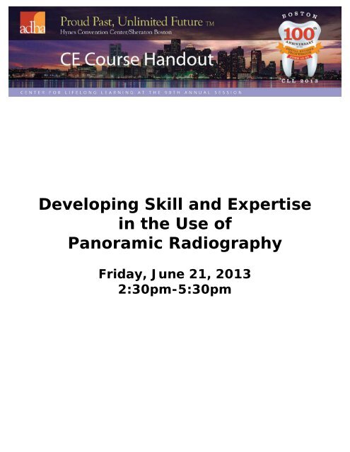
Developing Skill and Expertise in the Use of Panoramic Radiography
Developing Skill and Expertise in the Use of Panoramic Radiography
Developing Skill and Expertise in the Use of Panoramic Radiography

Create successful ePaper yourself
Turn your PDF publications into a flip-book with our unique Google optimized e-Paper software.
<strong>Develop<strong>in</strong>g</strong> <strong>Skill</strong> <strong>and</strong> <strong>Expertise</strong><br />
<strong>in</strong> <strong>the</strong> <strong>Use</strong> <strong>of</strong><br />
<strong>Panoramic</strong> <strong>Radiography</strong><br />
Friday, June 21, 2013<br />
2:30pm-5:30pm
5/13/2013<br />
Welcome!<br />
<strong>Develop<strong>in</strong>g</strong> <strong>Skill</strong> <strong>and</strong> <strong>Expertise</strong><br />
<strong>in</strong> <strong>the</strong> <strong>Use</strong> <strong>of</strong> <strong>Panoramic</strong><br />
<strong>Radiography</strong><br />
Today’s Presentation:<br />
•Part I: The Art & Science <strong>of</strong> <strong>Panoramic</strong><br />
<strong>Radiography</strong><br />
(50 m<strong>in</strong>)<br />
<br />
•Part II: Anatomy & Pathology (50 m<strong>in</strong>)<br />
• <br />
•Part III: Interpretation <strong>and</strong> Identification Activity<br />
(50 m<strong>in</strong>)<br />
Learn<strong>in</strong>g Objectives<br />
• Describe <strong>the</strong> fundamentals <strong>of</strong> panoramic imag<strong>in</strong>g<br />
• Describe <strong>the</strong> purpose <strong>and</strong> use <strong>of</strong> panoramic radiography<br />
• List <strong>the</strong> four basic components common to most panoramic x-<br />
ray mach<strong>in</strong>es<br />
• Expla<strong>in</strong> <strong>the</strong> concept <strong>of</strong> <strong>the</strong> focal trough<br />
• Identify <strong>the</strong> planes used to position <strong>the</strong> arches correctly with<strong>in</strong><br />
<strong>the</strong> focal trough<br />
• Describe correct patient position<strong>in</strong>g errors seen on panoramic<br />
images<br />
• Identify common position<strong>in</strong>g errors <strong>and</strong> <strong>the</strong> necessary measures<br />
needed to correct such errors<br />
• Identify hard tissue anatomic l<strong>and</strong>marks <strong>of</strong> <strong>the</strong> maxilla <strong>and</strong><br />
m<strong>and</strong>ible<br />
• Identify s<strong>of</strong>t tissue <strong>and</strong> air space images on a radiograph<br />
• Identify artifacts on a radiograph<br />
• Identify pathology<br />
• Discuss <strong>the</strong> advantages <strong>and</strong> disadvantages <strong>of</strong> panoramic<br />
imag<strong>in</strong>g<br />
1
5/13/2013<br />
References<br />
• Frommer, HH, & Stabulas-Savage, JJ (2011). Radiology for <strong>the</strong><br />
Dental Pr<strong>of</strong>essional, 9 th Ed., Mosby Elsevier, St. Louis, MO.<br />
• Har<strong>in</strong>g, JI & L<strong>in</strong>d, LJ, (1993). Radiographic Interpretation for <strong>the</strong><br />
Dental Hygienist, Saunders, Philadelphia, PA.<br />
• Iannucci & Howerton (2011). Dental <strong>Radiography</strong> Pr<strong>in</strong>ciples <strong>and</strong><br />
Techniques, 4 th Ed, Mosby Elsevier, St. Louis, MO.<br />
• Langl<strong>and</strong>, OE, Langlais, RP & Preece, JW (2002). Pr<strong>in</strong>ciples <strong>of</strong><br />
Dental Imag<strong>in</strong>g, 2 nd Ed, Lipp<strong>in</strong>cott, Williams & Wilk<strong>in</strong>s,<br />
Baltimore, MD.<br />
• Matteson SR, Tyndall DA, Burkes EJ et al: The radiology <strong>of</strong><br />
benign <strong>and</strong> malignant lesion, Dent Radiogr Photogr 57:35-52,<br />
78-84, 1985.<br />
• Preece, John W., (2009). University <strong>of</strong> Texas Health Sciences,<br />
San Antonio, TX.<br />
• Thomson, EM & Johnson, ON, (2007). Essentials <strong>of</strong> Dental<br />
<strong>Radiography</strong>, 9 th Ed., Pearson, Upper Saddle River, NJ.<br />
• White, SC & Pharoah, MJ (2009). Oral Radiology Pr<strong>in</strong>ciples <strong>and</strong><br />
Interpretation, 6 th Ed., Mosby Elsevier, St. Louis, MO.<br />
History<br />
• First <strong>in</strong>troduced 1959 by S.S.<br />
White Corp. as <strong>the</strong>ir<br />
"Panorex" unit<br />
• Designed based on work<br />
done <strong>in</strong> 1949 by Dr. Y.V.<br />
Paastero, a F<strong>in</strong>nish dentist<br />
• Essential part <strong>of</strong> dental<br />
diagnosis<br />
• Complement to <strong>in</strong>traoral<br />
films<br />
• Panorama = "unobstructed<br />
view <strong>of</strong> a region <strong>in</strong> any<br />
direction"<br />
<strong>Use</strong>s <strong>in</strong> Dentistry<br />
Benefits<br />
• Broad coverage <strong>of</strong> <strong>the</strong> facial<br />
bones & teeth<br />
• Low patient radiation dose<br />
• Convenience <strong>of</strong> <strong>the</strong><br />
exam<strong>in</strong>ation for <strong>the</strong> patient<br />
• <strong>Use</strong> <strong>in</strong> patients unable to<br />
open <strong>the</strong>ir mouths<br />
• Short time required to make<br />
a panoramic image vs. FMX<br />
• Patients don’t m<strong>in</strong>d!<br />
<strong>Use</strong>s<br />
• Impacted teeth<br />
• Eruption patterns<br />
• Growth & Development<br />
• Detect diseases, lesions<br />
<strong>and</strong> conditions <strong>of</strong> <strong>the</strong> jaws<br />
• Exam<strong>in</strong>e <strong>the</strong> extent <strong>of</strong> large<br />
lesions<br />
• Evaluate trauma<br />
Not Recommended for<br />
Dx:<br />
• Caries<br />
• Periodontal disease<br />
• Periapical lesions<br />
2
5/13/2013<br />
<strong>Panoramic</strong> Imag<strong>in</strong>g<br />
• A s<strong>in</strong>gle tomographic image <strong>of</strong> <strong>the</strong> facial structures <strong>in</strong>clud<strong>in</strong>g:<br />
• Maxilla<br />
• M<strong>and</strong>ible<br />
• All <strong>the</strong> support<strong>in</strong>g structures<br />
• A tomographic series is comprised <strong>of</strong> multiple cuts<br />
(exposures) 0.5cm apart<br />
• Requires rotat<strong>in</strong>g, slit-beam exposure with rotat<strong>in</strong>g (reciprocat<strong>in</strong>g)<br />
receptor<br />
• Can be film or digital sensors<br />
Courtesy <strong>of</strong> Mosby, Inc., Elsevier Pub, 2009..<br />
Image Layer -or-<br />
Focal Trough -or-<br />
Focal Plane<br />
• A narrow, specific, curved, fixed space between <strong>the</strong> x-ray<br />
source (x-ray tube) <strong>and</strong> <strong>the</strong> film<br />
• Can vary <strong>in</strong> shape from one unit to ano<strong>the</strong>r, but is specifically<br />
shaped to record <strong>the</strong> human jaw<br />
• Is where <strong>the</strong> images projected onto <strong>the</strong> radiograph are recorded<br />
clearly<br />
• When <strong>the</strong> patient is improperly positioned with<strong>in</strong> this space, it is<br />
<strong>the</strong> source <strong>of</strong> many errors that can result <strong>in</strong> an undiagnostic<br />
image<br />
3
5/13/2013<br />
<strong>Panoramic</strong> Units<br />
•Many different k<strong>in</strong>ds <strong>of</strong> units available today<br />
•Variables <strong>in</strong>clude:<br />
• # <strong>and</strong> locations <strong>of</strong> <strong>the</strong> centers <strong>of</strong> rotation<br />
• Fixed or adjustable focal trough<br />
• Type <strong>and</strong> shape <strong>of</strong> film transport mechanism<br />
• Manual or automatic sett<strong>in</strong>g controls<br />
• Head-position<strong>in</strong>g devices/bite blocks<br />
• Wall-mounted or free-st<strong>and</strong><strong>in</strong>g<br />
• kVP <strong>and</strong> mA ranges<br />
• Sit or st<strong>and</strong> patient position<br />
<strong>Panoramic</strong> Units<br />
•Analog film units<br />
• Film size 5.5" or 6.5”<br />
• Fast film<br />
• Intensify<strong>in</strong>g screens<br />
• 1 pano exposure = 4 BWX exposure (F-speed film)<br />
•Digital units<br />
•Charge-coupled device (CCD)<br />
•Photostimulatable phosphor plate (PSP)<br />
•DICOM (2004)<br />
•All have common position<strong>in</strong>g requirements<br />
• Mid-Saggital Plane<br />
• Frankfort Plane -OR- Ala-Tragus L<strong>in</strong>e<br />
• Each mach<strong>in</strong>e has its own unique technique for operat<strong>in</strong>g, which can be easily<br />
learned<br />
Midsagittal<br />
Plane<br />
• Should be perpendicular to<br />
<strong>the</strong> floor<br />
• Be positioned evenly<br />
between <strong>the</strong> eyes<br />
• Go down center <strong>of</strong> nose,<br />
philtrum <strong>and</strong> down middle<br />
<strong>of</strong> bite block<br />
Controls “head tilt<strong>in</strong>g” <strong>and</strong><br />
“twist<strong>in</strong>g” <strong>of</strong> head<br />
4
5/13/2013<br />
Head Twisted: One side <strong>of</strong> <strong>the</strong> mouth is magnified <strong>and</strong> ramus<br />
is distorted, more <strong>of</strong> <strong>the</strong> sp<strong>in</strong>e appears on one side <strong>of</strong> film<br />
Frankfort<br />
Plane:<br />
• An imag<strong>in</strong>ary l<strong>in</strong>e from<br />
<strong>the</strong> floor <strong>of</strong> <strong>the</strong> orbit<br />
(eye) to <strong>the</strong> external<br />
audiology meatus.<br />
• Should be parallel to<br />
<strong>the</strong> floor<br />
Controls ch<strong>in</strong> position<strong>in</strong>g<br />
(high/low)<br />
Ala-Tragus L<strong>in</strong>e<br />
• *An imag<strong>in</strong>ary l<strong>in</strong>e from<br />
<strong>the</strong> ala <strong>of</strong> <strong>the</strong> nose to<br />
<strong>the</strong> external auditory<br />
meatus<br />
Controls ch<strong>in</strong> position<strong>in</strong>g<br />
(high/low)<br />
5
5/13/2013<br />
Common Position<strong>in</strong>g Requirements:<br />
Bite Block<br />
Anterior teeth should be positioned <strong>in</strong> <strong>the</strong> proper<br />
groove(s).<br />
Common Position<strong>in</strong>g Requirements:<br />
Bite Block<br />
• Bit<strong>in</strong>g too far forward:<br />
• Anterior teeth appear<br />
narrow <strong>and</strong> smaller<br />
• The sp<strong>in</strong>e appears <strong>in</strong><br />
radiograph<br />
• Bit<strong>in</strong>g too far back:<br />
• Anterior teeth appear<br />
widened <strong>and</strong> blurred<br />
• Condyles <strong>of</strong> m<strong>and</strong>ible<br />
are not visible<br />
Common Position<strong>in</strong>g Requirements: Bite<br />
Block<br />
• Bit<strong>in</strong>g too far forward:<br />
• Anterior teeth appear<br />
narrow <strong>and</strong> smaller<br />
• The sp<strong>in</strong>e appears <strong>in</strong><br />
radiograph<br />
• Bit<strong>in</strong>g too far back:<br />
• Anterior teeth appear<br />
widened <strong>and</strong> blurred<br />
• Condyles <strong>of</strong> m<strong>and</strong>ible<br />
are not visible<br />
Courtesy <strong>of</strong> Mosby, Inc., Elsevier Pub, 2009..<br />
6
5/13/2013<br />
The patient’s anterior teeth appear to be narrow <strong>and</strong> smaller on <strong>the</strong><br />
panoramic image when patient’s teeth are positioned too far forward<br />
on <strong>the</strong> bite-block. (More <strong>of</strong> <strong>the</strong> sp<strong>in</strong>e is on image also.)<br />
Anterior teeth appear widened <strong>and</strong> blurred on <strong>the</strong> panoramic image when <strong>the</strong><br />
patient’s teeth are positioned too far back on <strong>the</strong> bite-block.<br />
Common Position<strong>in</strong>g Requirements:<br />
Focal Trough Position<br />
• Some units will have adjustments to<br />
assure position<strong>in</strong>g with<strong>in</strong> <strong>the</strong> Focal<br />
Trough (or Image Layer)<br />
7
5/13/2013<br />
Common Position<strong>in</strong>g Requirements:<br />
Ch<strong>in</strong> Position<br />
Should not be angled up or down<br />
Controlled by <strong>the</strong> correct alignment <strong>of</strong> <strong>the</strong><br />
Frankfort Plane or Ala-Tragus L<strong>in</strong>e<br />
Ch<strong>in</strong> too high: An “exaggerated frown l<strong>in</strong>e” is seen on a<br />
panoramic image when <strong>the</strong> patient’s ch<strong>in</strong> is tipped up. (Ch<strong>in</strong><br />
rest is too high, Frankfort plane is not parallel to <strong>the</strong> floor)<br />
Ch<strong>in</strong> too low: exaggerated smile<br />
8
5/13/2013<br />
Common Position<strong>in</strong>g Requirements:<br />
Head Tilt<br />
• Head position<strong>in</strong>g devices<br />
should be firm enough to<br />
prevent tipp<strong>in</strong>g or<br />
rotat<strong>in</strong>g<br />
• Correct midsagittal<br />
orientation should be<br />
ma<strong>in</strong>ta<strong>in</strong>ed throughout<br />
<strong>the</strong> whole procedure<br />
• Can also prevent patient<br />
movement dur<strong>in</strong>g<br />
exposure<br />
Head Tilted: One side <strong>of</strong> <strong>the</strong> mouth is enlarged, overall image<br />
is tilted <strong>and</strong> slightly distorted.<br />
Common Position<strong>in</strong>g Requirements:<br />
Lead Apron<br />
Should not have a thyroid<br />
collar (<strong>in</strong>terferes with<br />
primary beam)<br />
Place on patient with <strong>the</strong><br />
long side on <strong>the</strong> patient’s<br />
back<br />
9
5/13/2013<br />
A lead apron artifact appears as a large cone-shaped<br />
radiopacity obscur<strong>in</strong>g <strong>the</strong> m<strong>and</strong>ible.<br />
Common Position<strong>in</strong>g Requirements:<br />
Patient Posture<br />
• Sp<strong>in</strong>e should be kept erect<br />
• “St<strong>and</strong> tall”<br />
• Shoulders relaxed<br />
• H<strong>and</strong>s on grips/h<strong>and</strong>les<br />
• Cross h<strong>and</strong>s if need more<br />
room<br />
• Prevents sp<strong>in</strong>e from be<strong>in</strong>g<br />
superimposed on anterior<br />
teeth<br />
(No slump<strong>in</strong>g!)<br />
Patient slumped: If <strong>the</strong> patient is not st<strong>and</strong><strong>in</strong>g erect (slumped), a<br />
superimposition <strong>of</strong> <strong>the</strong> cervical sp<strong>in</strong>e (arrows) may be seen at <strong>the</strong> center<br />
<strong>of</strong> <strong>the</strong> panoramic image.sp<strong>in</strong>e shadow ghost appears on anterior teeth<br />
10
5/13/2013<br />
Common Position<strong>in</strong>g Requirements:<br />
Tongue Position<br />
If <strong>the</strong> patient does not place his or her tongue on <strong>the</strong> ro<strong>of</strong> <strong>of</strong> <strong>the</strong><br />
mouth <strong>and</strong> hold it <strong>the</strong>re throughout <strong>the</strong> imag<strong>in</strong>g procedure, a<br />
radiolucent shadow will be superimposed over <strong>the</strong> image <strong>of</strong> <strong>the</strong> apices<br />
<strong>of</strong> maxillary teeth.<br />
O<strong>the</strong>r Considerations<br />
These items should be removed to allow visualization <strong>of</strong><br />
all structures <strong>and</strong> prevent creat<strong>in</strong>g ghost images:<br />
• Dentures/pros<strong>the</strong>tic appliances<br />
• Jewelry (<strong>in</strong>clud<strong>in</strong>g necklaces)<br />
• Oral/facial/ear pierc<strong>in</strong>gs<br />
• Hear<strong>in</strong>g aids/glasses<br />
•Hairp<strong>in</strong>s<br />
• Bib cha<strong>in</strong>s<br />
Large hoop earr<strong>in</strong>gs (1) <strong>and</strong> ghost images (2). The ghost<br />
image <strong>of</strong> <strong>the</strong> earr<strong>in</strong>g appears on <strong>the</strong> opposite side <strong>of</strong> <strong>the</strong> image<br />
<strong>and</strong> is enlarged <strong>and</strong> laterally distorted.<br />
*<br />
11
5/13/2013<br />
<strong>Panoramic</strong> Anatomy<br />
Why is Anatomy<br />
Important<br />
• You must underst<strong>and</strong> what is normal to be<br />
able to dist<strong>in</strong>guish between:<br />
• Normal<br />
• Variation <strong>of</strong> Normal<br />
• Disease<br />
• Why is it difficult with panoramic<br />
• Complex structures <strong>of</strong> <strong>the</strong> mid-face<br />
• Super-imposition <strong>of</strong> <strong>the</strong> structures<br />
• Large number <strong>of</strong> potential artifacts<br />
• Out <strong>of</strong> practice<br />
Anatomy: basic concepts<br />
• Underst<strong>and</strong><strong>in</strong>g density changes<br />
• Air obscures hard tissue<br />
• S<strong>of</strong>t tissue obscures air<br />
• Hard tissue obscures s<strong>of</strong>t tissue<br />
• Ghost images obscure everyth<strong>in</strong>g<br />
1
5/13/2013<br />
<strong>Panoramic</strong> Radiographic Anatomy:<br />
Air spaces<br />
1. Palatoglossal air space 2. nasopharyngeal air space 3. oropharyngeal air<br />
space<br />
<strong>Panoramic</strong> Radiographic Anatomy:<br />
Air spaces<br />
<strong>Panoramic</strong> Radiographic Anatomy:<br />
Air Spaces<br />
1. palatoglossal air space 2. nasopharyngeal air space 3. oropharyngeal air<br />
space (AIR OBSCURES HARD TISSUE)<br />
2
5/13/2013<br />
<strong>Panoramic</strong> Radiographic Anatomy:<br />
S<strong>of</strong>t tissues<br />
1. tongue 2. s<strong>of</strong>t palate <strong>and</strong> uvula 3. lip l<strong>in</strong>e 4. ear<br />
<strong>Panoramic</strong> Radiographic Anatomy:<br />
S<strong>of</strong>t Tissues<br />
<strong>Panoramic</strong> Radiographic Anatomy:<br />
S<strong>of</strong>t Tissues<br />
1.dorsum <strong>of</strong> tongue 2. s<strong>of</strong>t palate <strong>and</strong> uvula 3. ear. (SOFT TISSUE OBSCURES<br />
AIR) (HARD TISSUE OBSCURES SOFT TISSUE)<br />
3
5/13/2013<br />
<strong>Panoramic</strong> Radiographic Anatomy:<br />
Ghost Images<br />
b<br />
a<br />
a<br />
a, Ghost image <strong>of</strong> contralateral side <strong>of</strong> <strong>the</strong> m<strong>and</strong>ible. b, Ghost image <strong>of</strong> <strong>the</strong><br />
cervical sp<strong>in</strong>e.<br />
<strong>Panoramic</strong> Radiographic Anatomy:<br />
Ghost Images<br />
Ghost images (GHOST IMAGES OBSCURE EVERYTHING)<br />
Break for activity<br />
4
5/13/2013<br />
Radiographic Anatomy: <strong>Panoramic</strong><br />
Maxilla <strong>and</strong> surround<strong>in</strong>g structures<br />
1. mastoid process 2. styloid process 3. external auditory meatus 4. glenoid fossa 5.<br />
articular em<strong>in</strong>ence 6. lateral pterygoid plate 7. pterygomaxillary fissure 8. maxillary<br />
tuberosity 9. <strong>in</strong>fraorbital foramen 10. orbit 11. <strong>in</strong>cisive canal 12. <strong>in</strong>cisive foramen 13.<br />
anterior nasal sp<strong>in</strong>e 14. nasal cavity <strong>and</strong> conchae 15. nasal septum 16. hard palate 17.<br />
maxillary s<strong>in</strong>us 18. floor <strong>of</strong> maxillary s<strong>in</strong>us 19. zygomatic process <strong>of</strong> maxilla 20. zygomatic<br />
arch 21. hamular process<br />
Radiographic Anatomy: <strong>Panoramic</strong><br />
Maxilla <strong>and</strong> surround<strong>in</strong>g structures<br />
10<br />
1. external auditory meatus 2. zygomatic process <strong>of</strong> maxilla 3. <strong>in</strong>fraorbital foramen 4. orbit<br />
5. anterior nasal sp<strong>in</strong>e 6. nasal septum 7. nasal conchae 8. hard palate 9. zygomatic<br />
process <strong>of</strong> maxilla 10. pterigomaxillary fissure<br />
Radiographic Anatomy: <strong>Panoramic</strong><br />
Maxilla <strong>and</strong> surround<strong>in</strong>g structures<br />
1. glenoid fossa 2. articular em<strong>in</strong>ence 3. maxillary tuberosity 4. maxillary s<strong>in</strong>us<br />
5. zygoma<br />
5
5/13/2013<br />
Radiographic Anatomy: <strong>Panoramic</strong><br />
M<strong>and</strong>ible <strong>and</strong> surround<strong>in</strong>g structures<br />
1. condyle 2. m<strong>and</strong>ibular notch 3. coronoid process 4. m<strong>and</strong>ibular foramen 5. l<strong>in</strong>gula 6<br />
m<strong>and</strong>ibular canal 7. mental foramen 8. hyoid bone 9. mental ridge 10. mental fossa 11.<br />
l<strong>in</strong>gual foramen 12. genial tubercles 13. <strong>in</strong>ferior border <strong>of</strong> m<strong>and</strong>ible 14. mylohyoid ridge<br />
15. <strong>in</strong>ternal oblique ridge 16. external oblique ridge<br />
Radiographic Anatomy: <strong>Panoramic</strong><br />
M<strong>and</strong>ible <strong>and</strong> surround<strong>in</strong>g structures<br />
1. condyle 2. m<strong>and</strong>ibular notch 3. coronoid process 4. m<strong>and</strong>ibular foramen 5.<br />
mental foramen 6. genial tubercles 7. styloid process<br />
<strong>Panoramic</strong> Radiographic<br />
Anatomy: M<strong>and</strong>ible <strong>and</strong> surround<strong>in</strong>g structures<br />
1. m<strong>and</strong>ibular canal 2. hyoid 3. external oblique ridge 4. angle <strong>of</strong> m<strong>and</strong>ible<br />
6
5/13/2013<br />
Radiographic Anatomy: <strong>Panoramic</strong><br />
M<strong>and</strong>ible <strong>and</strong> surround<strong>in</strong>g structures<br />
1. <strong>in</strong>ferior border <strong>of</strong> m<strong>and</strong>ible 2. subm<strong>and</strong>ibular fossa 3. external oblique ridge 4.<br />
s<strong>of</strong>t tissue <strong>of</strong> ear<br />
Radiographic Pathology<br />
Describ<strong>in</strong>g Radiographic Lesions<br />
LESION<br />
Radiolucent<br />
Radiopaque<br />
Mixed<br />
Well-Def<strong>in</strong>ed<br />
Poorly Def<strong>in</strong>ed<br />
S<strong>in</strong>gle<br />
Multiple<br />
Asymptomatic<br />
Symptomatic<br />
Tooth Associated<br />
Not tooth associated<br />
Hard Tissue<br />
S<strong>of</strong>t Tissue<br />
7
5/13/2013<br />
Benign Tumors<br />
• Benign lesions grow<strong>in</strong>g <strong>in</strong> bone<br />
tend to be round or oval<br />
• They tend to grow slowly <strong>and</strong><br />
displace adjacent tissues <strong>and</strong>/or<br />
teeth.<br />
Malignant Tumors<br />
• Symptoms:<br />
• Ill-def<strong>in</strong>ed Radolucent <strong>and</strong>/or Radiopaque, moth<br />
eaten appearance<br />
• Fast grow<strong>in</strong>g<br />
• Perforation <strong>of</strong> bone<br />
• Pa<strong>in</strong> <strong>and</strong> swell<strong>in</strong>g<br />
• Loose teeth<br />
• Altered sensations<br />
Radiolucent Periapical<br />
• Periapical Abscess<br />
• Periapical Granuloma<br />
• Periapical Cyst<br />
8
5/13/2013<br />
Radiolucent/Radiopaque Periapical:<br />
Periapical Cemento-Osseous Dysplasia<br />
• Radiolucent<br />
early/Radiopaque late<br />
with radiolucent rim<br />
• Associated with apex <strong>of</strong><br />
m<strong>and</strong>ibular anterior teeth<br />
• Teeth are vital<br />
• Asymptomatic<br />
• Common <strong>in</strong> middle aged<br />
African American women<br />
• Tx: None<br />
Unilocular Radiolucent Pericoronal:<br />
Dentigerous Cyst<br />
• Tx: surgical removal<br />
• Recurrence: unlikely<br />
Unilocular Radiolucent Pericoronal:<br />
Adenomatoid Odontogenic Tumor<br />
(AOT)<br />
• Well Circumscribed<br />
Radiolucent Unilocular or<br />
Radiolucent with<br />
Radiopaque flecks<br />
• Tx: Surgical Removal<br />
• Recurrence: none<br />
9
5/13/2013<br />
Radiolucent Unilocular:<br />
Nasopalat<strong>in</strong>e Duct Cyst<br />
• Corticated border<br />
• Tx: Surgical Removal<br />
• Recurrence: Rare<br />
Residual Cyst<br />
• Unilocular Radiolucent<br />
– Corticated or noncorticated<br />
border<br />
• Tx: surgical removal<br />
• Recurrence: unlikely<br />
Multilocular Radiolucent<br />
Lesions<br />
• “COMA”:<br />
– Central Giant Cell Granuloma<br />
– Odontogenic Keratocyst (OKC)<br />
– Myxoma<br />
– Ameloblastoma<br />
10
5/13/2013<br />
Central Giant Cell Granuloma<br />
• Radiolucent Well-Def<strong>in</strong>ed<br />
Multilocular or Unilocular<br />
– Corticated or noncorticated<br />
border<br />
• Dx: Biopsy<br />
• Tx: Curettage<br />
Odontogenic Keratocyst (OKC)<br />
• Radiolucent, well-def<strong>in</strong>ed<br />
smooth borders<br />
• Sclerotic or corticated<br />
border<br />
• Tx: Surgical Excision <strong>and</strong><br />
osseous curettage<br />
• Recurrence: 30%<br />
Odontogenic Myxoma<br />
• Radiolucent Multilocular<br />
or Unilocular<br />
• Irregular or scalloped<br />
marg<strong>in</strong>s<br />
• Th<strong>in</strong> boney trabelculae<br />
arranged at right angles<br />
• Tx: Curettage<br />
• Recurrence: 25%<br />
11
5/13/2013<br />
Ameloblastoma<br />
• Multilocular radiolucent<br />
“soap bubble” or<br />
“honeycomb”<br />
• Tx: Depends on size <strong>of</strong><br />
lesion<br />
• Small: aggressive curettage<br />
or bloc resection<br />
• Large: bloc or segmental<br />
resection<br />
• Recurrence: 55-90%<br />
Malignant Tumors<br />
• Symptoms:<br />
• Ill-def<strong>in</strong>ed Radolucent <strong>and</strong>/or Radiopaque, moth<br />
eaten appearance<br />
• Fast grow<strong>in</strong>g<br />
• Perforation <strong>of</strong> bone<br />
• Pa<strong>in</strong> <strong>and</strong> swell<strong>in</strong>g<br />
• Loose teeth<br />
• Altered sensations<br />
• Poorly def<strong>in</strong>ed mixed<br />
Radiolucent/Radiopaque<br />
lesion<br />
– Sunburst or sunray<br />
appearance <strong>in</strong> 20% <strong>of</strong><br />
cases<br />
• Tx: Radical surgical<br />
excision, Radiation <strong>and</strong><br />
chemo<strong>the</strong>rapy<br />
• Prognosis Poor (30-50%)<br />
survival rate<br />
Osteosarcoma<br />
12
5/13/2013<br />
Chondrosarcoma<br />
• Poorly Def<strong>in</strong>ed<br />
Radiolucent lesion or<br />
mixed Radiolucent/<br />
Radiopaque lesion<br />
• Expansile, with<br />
widen<strong>in</strong>g <strong>of</strong> <strong>the</strong> PDL<br />
• Tx: Wide excision,<br />
Chemo<strong>the</strong>rapy <strong>and</strong><br />
radiation<br />
• Prognosis Poor: less<br />
than 20% after 5<br />
years, metastasis<br />
usually to lungs or<br />
o<strong>the</strong>r bones<br />
Metastatic Tumors <strong>of</strong> <strong>the</strong><br />
Jaw<br />
• Poorly Def<strong>in</strong>ed<br />
Radiolucent Lesion<br />
• Tx: Surgical Excision,<br />
chemo<strong>the</strong>rapy <strong>and</strong><br />
radiation<br />
• Poor Prognosis: 10%<br />
after five years<br />
www.orad.org<br />
13
5/13/2013<br />
Radiopacities<br />
Radiopacities:<br />
Idiopathic Osteosclerosis<br />
• Idiopathic= unknown<br />
cause<br />
• Sclerosis= scar-like<br />
• Area <strong>of</strong> <strong>in</strong>creased<br />
density <strong>of</strong> bone<br />
• Tx: None necessary<br />
Radiopacities:<br />
Condens<strong>in</strong>g Osteitis<br />
• Condens<strong>in</strong>g Osteitis<br />
• Tooth with deep<br />
restoration, welldef<strong>in</strong>ed<br />
radiolucent<br />
lesion, radiopaque<br />
“halo” around lesion<br />
• Bone may or may not<br />
go back to normal<br />
after extraction<br />
14
5/13/2013<br />
Radiopacities:<br />
Odontoma, Compound<br />
• Radiopaque lesion <strong>of</strong>ten<br />
with radiolucent zone<br />
surround<strong>in</strong>g<br />
• Often related to impacted<br />
teeth<br />
• Tx: Surgical Removal<br />
• Recurrence: none<br />
Radiopacities:<br />
Odonotoma, Complex<br />
• Radiopaque lesion <strong>of</strong>ten<br />
with radiolucent zone<br />
surround<strong>in</strong>g<br />
• Often related to<br />
impacted teeth<br />
• Tx: Surgical Removal<br />
• Recurrence: none<br />
Radiopacities:<br />
Cementoblastoma<br />
• Slow grow<strong>in</strong>g radiopaque<br />
lesion associated with<br />
tooth root<br />
• Often has f<strong>in</strong>e radiolucent<br />
border<br />
• Tx: surgical removal with<br />
extraction or root<br />
amputation<br />
15
5/6/2013<br />
Interpretation &<br />
Identification<br />
Special Thanks to<br />
John W. Preece, DMD, MS<br />
University <strong>of</strong> Texas Health Sciences<br />
San Antonio, Tx<br />
Co-author <strong>of</strong> Pr<strong>in</strong>ciples <strong>of</strong> Dental Imag<strong>in</strong>g, 2 nd Ed,<br />
Lipp<strong>in</strong>cott, Williams & Wilk<strong>in</strong>s, Baltimore, MD.<br />
1
5/6/2013<br />
Left<br />
2
5/6/2013<br />
Left<br />
Left<br />
3
5/6/2013<br />
Left<br />
Left<br />
4
5/6/2013<br />
Case<br />
History<br />
Chief compla<strong>in</strong>t<br />
A 45-year-old high school teacher seeks your<br />
advice regard<strong>in</strong>g his “swollen jaw.”<br />
History <strong>of</strong> Present Illness<br />
He has been aware <strong>of</strong> persistent <strong>and</strong><br />
gradually <strong>in</strong>creas<strong>in</strong>g jaw swell<strong>in</strong>g for<br />
many years. He has had no symptoms.<br />
He wishes to know if <strong>the</strong> lesion is<br />
malignant. If benign, he will decl<strong>in</strong>e<br />
treatment for religious reasons.<br />
5
5/6/2013<br />
Past Medical History<br />
He has no systemic diseases <strong>and</strong> no<br />
allergies. He is tak<strong>in</strong>g no medications.<br />
He does not smoke or dr<strong>in</strong>k.<br />
How would you describe <strong>the</strong> <strong>in</strong>traoral changes<br />
Copyright © 2008 by Saunders, an impr<strong>in</strong>t <strong>of</strong> Elsevier Inc.<br />
6
5/6/2013<br />
How would you describe <strong>the</strong> <strong>in</strong>traoral changes<br />
Next step<br />
Copyright © 2008 by Saunders, an impr<strong>in</strong>t <strong>of</strong> Elsevier Inc.<br />
Describe <strong>the</strong> radiographic appearance<br />
Copyright © 2008 by Saunders, an impr<strong>in</strong>t <strong>of</strong> Elsevier Inc.<br />
7
5/6/2013<br />
The biopsy specimen showed revealed Ameloblastoma<br />
Copyright © 2008 by Saunders, an impr<strong>in</strong>t <strong>of</strong> Elsevier Inc.<br />
Follow-up<br />
Initially <strong>the</strong> patient refused treatment but returned 7 years later with<br />
advanced disease. Because <strong>of</strong> <strong>the</strong> size <strong>and</strong> persistent growth <strong>of</strong> <strong>the</strong><br />
tumor mass, <strong>the</strong> patient consented to a hemim<strong>and</strong>ibulectory procedure<br />
that he tolerated well <strong>and</strong> recovered without complications.<br />
Copyright © 2008 by Saunders, an impr<strong>in</strong>t <strong>of</strong> Elsevier Inc.<br />
8
5/6/2013<br />
9
5/6/2013<br />
Left<br />
Ignore<br />
this area<br />
10
5/6/2013<br />
11
5/6/2013<br />
12
5/6/2013<br />
13
5/6/2013<br />
14
5/6/2013<br />
15
5/6/2013<br />
16
5/6/2013<br />
Left<br />
Ano<strong>the</strong>r Case<br />
•45 year old female<br />
•“Unusual sensation” <strong>in</strong><br />
upper left<br />
•Vitality read<strong>in</strong>g <strong>of</strong> teeth<br />
“ambiguous”<br />
•Now what<br />
17
5/6/2013<br />
O<strong>the</strong>r ill-def<strong>in</strong>ed lesions<br />
•4 months later,<br />
symptoms not<br />
improved<br />
•Now what<br />
18