Eur. Arch. Psychiatry Clin. Neurosci. - MDC
Eur. Arch. Psychiatry Clin. Neurosci. - MDC
Eur. Arch. Psychiatry Clin. Neurosci. - MDC

Create successful ePaper yourself
Turn your PDF publications into a flip-book with our unique Google optimized e-Paper software.
<strong>Eur</strong> <strong>Arch</strong> <strong>Psychiatry</strong> <strong>Clin</strong> <strong>Neurosci</strong> (2012) 262:87–91<br />
DOI 10.1007/s00406-011-0217-3<br />
SHORT COMMUNICATION<br />
Repetitive magnetic stimulation of human-derived neuron-like<br />
cells activates cAMP-CREB pathway<br />
Julian Hellmann • Rene Jüttner • Clarisse Roth •<br />
Malek Bajbouj • Imke Kirste • Isabella Heuser •<br />
Karen Gertz • Matthias Endres • Golo Kronenberg<br />
Received: 2 December 2010 / Accepted: 3 May 2011 / Published online: 12 May 2011<br />
Ó Springer-Verlag 2011<br />
Abstract Repetitive transcranial magnetic stimulation<br />
(rTMS) is a non-invasive neurostimulatory technique<br />
widely used in research, diagnostics, and neuro-psychiatric<br />
therapy. Despite its growing popularity, basic molecular<br />
mechanisms underlying the clinical effects of rTMS have<br />
remained largely under-researched. Here, we present a<br />
human-derived neuronal cell culture system responsive to<br />
rTMS effects. SH-SY5Y neuroblastoma cells were differentiated<br />
by retinoic acid treatment for 10 days, resulting in<br />
a neuronal phenotype characterized by upregulation of<br />
neuronal marker proteins and generation of an action<br />
potential in response to depolarizing current step injection.<br />
Repetitive magnetic stimulation of these cells resulted in<br />
increased intracellular cAMP levels and increased phosphorylation<br />
of transcription factor CREB. Pretreatment<br />
with ketamine (1 lM) potentiated, while pretreatment with<br />
lithium (2 mM) attenuated this cellular response to repetitive<br />
magnetic stimulation. In conclusion, we introduce<br />
J. Hellmann C. Roth M. Bajbouj I. Kirste I. Heuser <br />
G. Kronenberg (&)<br />
Klinik und Hochschulambulanz für Psychiatrie und<br />
Psychotherapie, Charité Campus Benjamin Franklin,<br />
Eschenallee 3, 14050 Berlin, Germany<br />
e-mail: golo.kronenberg@charite.de<br />
J. Hellmann K. Gertz M. Endres G. Kronenberg<br />
Klinik und Poliklinik für Neurologie, Charité<br />
Universitätsmedizin Berlin, Charitéplatz 1,<br />
10117 Berlin, Germany<br />
J. Hellmann K. Gertz M. Endres G. Kronenberg<br />
Center for Stroke Research Berlin, Charité Universitätsmedizin<br />
Berlin, Charitéplatz 1, 10117 Berlin, Germany<br />
R. Jüttner<br />
Developmental Neurobiology, Max-Delbrück-Center,<br />
Robert-Rössle-Str. 10, 13092 Berlin, Germany<br />
here a novel in vitro system responding to rTMS at the<br />
level of second messenger signaling. The use of humanderived<br />
cells with neuron-like properties will prove useful<br />
for further studies on the cellular effects of rTMS.<br />
Keywords rTMS cAMP CREB SH-SY5Y<br />
neuroblastoma cells<br />
Introduction<br />
Repetitive transcranial magnetic stimulation (rTMS) represents<br />
a relatively novel and versatile tool with a number of<br />
diagnostic, research, and therapeutic implications [15, 17].<br />
rTMS is a non-invasive technique for brain stimulation,<br />
which makes use of the principle of electromagnetic induction<br />
as first described by Michael Faraday. The direct effects<br />
of rTMS are therefore largely confined to cortical neurons in<br />
the proximity of an externally placed magnetic coil [3].<br />
Despite the widespread use of rTMS in the clinical<br />
setting, very little is known about the molecular mechanisms<br />
behind it. Importantly, there is currently no model<br />
system available in basic neuro-psychiatric research to<br />
study neurobiological mechanisms of rTMS in vitro. In<br />
antidepressant pharmacotherapy, it is widely accepted that<br />
key biological mechanisms underlying therapeutic effects<br />
involve plasticity-associated signal transduction pathways<br />
converging on CREB, leading to modulation of the transcription<br />
of cAMP responsive genes [4, 12, 14, 21].<br />
Especially in hippocampus, CREB-regulated gene transcription<br />
has been shown to increase growth factor activity<br />
and to promote regenerative processes such as dendritic<br />
sprouting and neurogenesis [2].<br />
So far, only few studies have investigated the biological<br />
effects of repetitive magnetic stimulation (rTMS) on<br />
123
88 <strong>Eur</strong> <strong>Arch</strong> <strong>Psychiatry</strong> <strong>Clin</strong> <strong>Neurosci</strong> (2012) 262:87–91<br />
cultured cells. Notably, these studies generally failed to<br />
demonstrate effects on second messenger production or<br />
cell signaling, which were expected from studies in animals<br />
[9]. Moreover, previous investigations in vitro used either<br />
non-neuronal, undifferentiated neuronal or non-human cell<br />
systems [18–20]. The paucity of observed effects in these<br />
earlier reports may be due in large part to the use of<br />
undifferentiated or non-neuronal cells. Here, we hypothesized<br />
that to become fully useful for the study of rTMS,<br />
cells have to develop neuronal characteristics first. Therefore,<br />
in this study, we employed SH-SY5Y neuroblastoma<br />
cells after retinoic acid (RA)-induced differentiation as a<br />
neuronal, human-derived culture system. After providing<br />
data on neuronal differentiation, we show that rTMS<br />
induces the formation of cAMP and subsequent phosphorylation<br />
of CREB. Furthermore, we used the anesthetic<br />
ketamine and mood stabilizer lithium as two examples to<br />
demonstrate that our system is also suitable to study<br />
interactions between drug treatment and rTMS.<br />
Materials and methods<br />
Cell culture<br />
Human SH-SY5Y neuroblastoma cells (Geweberesourcenzentrum<br />
Braunschweig, Germany) were seeded at an initial<br />
density of 4 9 10 5 cells/cm 2 for cAMP and pCREB experiments<br />
and 1-5 9 10 5 cells/cm 2 for electrophysiological and<br />
immunofluorescence studies. Cells were cultured as described<br />
in detail previously [7]. Briefly, cells were grown in<br />
minimum essential medium containing Earle’s salts, 100<br />
units/ml penicillin, 100 lg/ml streptomycin and 10% fetal<br />
bovine serum (Biochrom, Berlin, Germany). Retinoic acid<br />
(RA; Sigma–Aldrich, Taufkirchen, Germany) was applied at<br />
a final concentration of 10 lM. Lithium aspartate (Sigma)<br />
was added to a final concentration of 2 mM, and ketamine<br />
(CuraMED, Karlsruhe, Germany) was added to a final concentration<br />
of 1 lM.<br />
Repetitive magnetic stimulation (rTMS)<br />
Cells were placed on the magnetic coil of a conventional<br />
Magstim Pro TM device. Stimulation parameters were set to<br />
either 60 or 180 series at maximum intensity, resulting in a<br />
total of 600 or 1,800 stimuli, respectively, applied in series of<br />
10 pulses at 5 Hz with a 10-s interval between series. In the<br />
sham-stimulation condition, cells were removed from the<br />
incubator and exposed to the same environment as the stimulated<br />
cells for the same period of time. Serum was removed<br />
from culture medium 24 h before stimulation. Drugs were<br />
applied in serum-free medium 1 h prior to stimulation.<br />
Analysis of intracellular cAMP levels<br />
Cells from four wells of a 96-well plate were pooled as one<br />
sample (*5 9 10 5 cells). Intracellular cAMP levels were<br />
determined by a non-acetylation method using a cAMPspecific<br />
enzyme-linked immunosorbent assay (RPN 225,<br />
GE Healthcare, Buckinghamshire, UK).<br />
Western blot analysis<br />
SDS–PAGE and Western blotting were performed as<br />
described in detail elsewhere [7, 8]. In brief, cells were<br />
lysed with ice-cold mammalian protein extraction reagent<br />
(Pierce Biotechnology, Rockford, Ill., U.S.A.) and cellular<br />
debris was precipitated by centrifugation at 25,000g and<br />
4°C for 25 min. Protein concentration was determined<br />
using BCA assay (Pierce Biotechnology). Equal amounts<br />
of protein were loaded on sodium dodecyl sulfate polyacrylamide<br />
gels (10–20%) and blotted onto PVDF membranes<br />
(Millipore, Schwalbach, Germany). Blots were<br />
probed with the following antibodies: anti-GFAP (1:500;<br />
Sigma), polyclonal anti-neurofilament (1:1,000; Sigma),<br />
anti-MAP2 (1:500; Sigma), anti-b-actin (HRP-labeled,<br />
1:10,000; Sigma), anti-CREB, and anti-phospho-CREB<br />
(both 1:1,000; Cell Signaling Technologies, Danvers, MA,<br />
USA). Densitometric quantification was performed using<br />
the LAS 3000 imaging system and Aida image analysis<br />
software, version 4.1 (Raytest, Straubenhardt, Germany).<br />
Immunofluorescence<br />
Cells were cultured on poly-L-lysine-coated glass coverslips.<br />
Fixation was with 4% paraformaldehyde (PFA) in<br />
TBS buffered to pH 7.4. Cells were stained with anti-<br />
MAP2 antibody (1:500; Sigma) and RhodX-conjugated<br />
secondary antibody (anti-mouse; 1:250; JacksonImmuno-<br />
Research, West Grove, PA). Nuclear dye Sytox Green<br />
(Invitrogen, Karlsruhe, Germany) was used at a concentration<br />
of 1:5,000. All confocal microscopy was performed<br />
using a spectral confocal microscope (TCS SP2; Leica,<br />
Nussloch, Germany).<br />
Electrophysiology<br />
RA-treated cells were analyzed by whole-cell patch-clamp<br />
recordings for passive membrane properties and voltagegated<br />
currents in voltage-clamp mode as well as for generation<br />
of action potentials in current-clamp mode using a<br />
patch-clamp amplifier (EPC-9, HEKA Elektronik,<br />
Lambrecht, Germany) as described in detail previously<br />
[22]. The recording pipette solution contained (in mM): 120<br />
KCl, 4 NaCl, 5 ethylene glycol-bis(b-aminoethyl ether)<br />
123
<strong>Eur</strong> <strong>Arch</strong> <strong>Psychiatry</strong> <strong>Clin</strong> <strong>Neurosci</strong> (2012) 262:87–91 89<br />
N,N,N 0 ,N 0 -tretraacetic acid (EGTA), 5 N-2-hydroxyethylpiperzine-N<br />
0 -2-ethanesulfonic acid (HEPES), 5 glucose,<br />
0.5 CaCl 2 and 4 MgCl 2 buffered to pH 7.3. During the<br />
experiment, cells were bathed in a solution containing (in<br />
mM): 145 NaCl, 3 KCl, 20 N-2-hydroxyethylpiperzine-N 0 -<br />
2-ethanesulfonic acid (HEPES), 20 glucose, 2 CaCl 2 , 1<br />
MgCl 2 adjusted to pH 7.3. Signals were sampled at a rate of<br />
10 kHz using WinTida software (HEKA Elektronik,<br />
Lambrecht, Germany) and Bessel filtered at 3 kHz.<br />
Statistical analyses<br />
Values are presented as mean ± SEM. All numerical<br />
analyses were performed using the Graph Pad Prism Ò<br />
program. Differences between means were analyzed by<br />
analysis of variance (ANOVA). P \ 0.05 was considered<br />
statistically significant.<br />
Results<br />
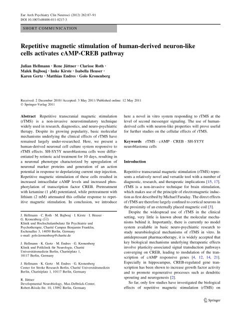
SH-SY5Y cells adopt neuronal characteristics<br />
upon differentiation with retinoic acid<br />
The SH-SY5Y neuroblastoma cell line is widely used as an<br />
in vitro model to study biochemical and functional properties<br />
of neurons. Importantly, it is a human-derived cell<br />
line [1]. SH-SY5Y cells were differentiated for 10 days in<br />
the presence of RA. Subsequent histological analysis consistently<br />
yielded strong MAP2-immunoreactivity in all cells<br />
throughout the cultures, indicating neuronal differentiation<br />
(Fig. 1a). Similarly, immunoblot analysis of undifferentiated<br />
(0 DIV) and differentiated (10 DIV) SH-SY5Y cells<br />
revealed increasing levels of neuronal marker proteins<br />
neurofilament and MAP2 with differentiation whereas<br />
expression of glial marker protein GFAP receded (Fig. 1b).<br />
Differentiated SH-SY5Y cells are able to produce<br />
action potentials<br />
The physiological properties of SH-SY5Y cells treated for<br />
10 days with RA were characterized using whole-cell<br />
patch-clamp recordings. Analysis of passive membrane<br />
properties revealed a whole-cell membrane capacitance of<br />
13.0 ± 1.2 pF and an input resistance of 1270.1 ±<br />
113.6 MX. Furthermore, activation of voltage-gated<br />
channels by depolarizing pulses induced voltage-gated<br />
Na -currents of 499.1 ± 85.2 pA and voltage-gated<br />
K -currents of 725.1 ± 85.9 pA. Importantly, cells generated<br />
an action potential (AP) upon current injection of at<br />
least 30 pA (Fig. 1c).<br />
Repetitive magnetic stimulation (rTMS) increases<br />
intracellular cAMP levels and induces CREB<br />
phosphorylation in differentiated SH-SY5Y cells<br />
SH-SY5Y cells were differentiated in the presence of RA<br />
for 10 days. Cells were subjected to 180 consecutive series<br />
of rTMS or sham stimulation (sham rTMS) as described<br />
above. rTMS resulted in a significant increase in intracellular<br />
cAMP levels both in vehicle-treated cells and in cells<br />
that had been pretreated with ketamine. By contrast, lithium<br />
treatment attenuated this effect of rTMS on intracellular<br />
cAMP levels (Fig. 1d).<br />
The transcription factor CREB (cAMP response element-binding<br />
protein) is activated by phosphorylation.<br />
Chronic antidepressant treatment has previously been<br />
shown to increase CREB phosphorylation in mice [21].<br />
Here, we studied the effects of either 60 or 180 consecutive<br />
series of rTMS on the levels of phosphorylated<br />
CREB (pCREB); 180 consecutive series of rTMS significantly<br />
increased pCREB levels. Ketamine pretreatment<br />
potentiated, while lithium treatment largely inhibited the<br />
effects of rTMS on the amount of pCREB (Fig. 1e, f).<br />
Total levels of CREB protein were not affected by either<br />
1-h pretreatment with drugs or the stimulation procedure<br />
(not shown).<br />
Discussion<br />
To study the molecular effects of rTMS, it would be beneficial<br />
to have a cell culture system that responds to magnetic<br />
stimulation in a parallel fashion to in vivo responses.<br />
While many animal models exist, an in vitro model,<br />
although more removed from the in vivo system, carries<br />
some additional advantages. In vitro models allow for a<br />
higher throughput at a greatly reduced cost. Also, human<br />
cells such as SH-SY5Y could then be studied rather than<br />
rodent neurons, and the different responses between the<br />
two evaluated. Lastly, stimulating neurons in vitro strips<br />
away the additional complications of penetration through<br />
skin and bone and allows the pure study of rTMS directly<br />
on the tissue of interest.<br />
In a hallmark study in rats, Ji and co-workers demonstrated<br />
effects of rTMS on immediate early gene expression<br />
and CREB phosphorylation in several brain areas [9].<br />
Since activation of CREB may promote neuroplastic<br />
responses such as synaptogenesis and neurogenesis, these<br />
in vivo findings link rTMS to a host of neurobiological<br />
mechanisms, which may be therapeutically harnessed [11,<br />
23]. Importantly, long-term rTMS has also been shown to<br />
increase the expression of CREB target gene BDNF in rat<br />
hippocampus [13].<br />
123
90 <strong>Eur</strong> <strong>Arch</strong> <strong>Psychiatry</strong> <strong>Clin</strong> <strong>Neurosci</strong> (2012) 262:87–91<br />
A B Neuronal Markers<br />
C<br />
0daysRA<br />
10daysRA<br />
Current Clamp Recordings<br />
10 days RA<br />
MAP2<br />
GFAP<br />
Neurofilament<br />
β-actin<br />
D E CREB Activation<br />
F<br />
vehicle lithium ketamine<br />
Intracellular cAMP levels<br />
*<br />
*<br />
*<br />
* *<br />
*<br />
sham rMS<br />
0.8<br />
* *<br />
1,5<br />
rel. to<br />
vehicle<br />
cAMP levels<br />
unstimulated<br />
1,0<br />
vehicle lithium ketamine<br />
vels rel. to<br />
xpression<br />
pCREB lev<br />
β-actin ex<br />
0.4<br />
0<br />
sham rMS rMS<br />
60 series 180 series<br />
vehicle lithium ketamine<br />
pCREB<br />
β-actin<br />
Fig. 1 Human-derived neuronal cell culture system for the study of<br />
the effects of repetitive magnetic stimulation. SH-SY5Y human<br />
neuroblastoma cells were differentiated for 10 days in the presence of<br />
RA (10 lM). a Confocal analysis of cells stained with nuclear dye<br />
Sytox Green (green) consistently demonstrated co-staining with<br />
neuronal marker protein MAP2 (red). Scale bar 35 lm. b Representative<br />
Western blots of undifferentiated (0 DIV) and differentiated (10<br />
DIV) SH-SY5Y cells demonstrating increase in neuronal marker<br />
proteins MAP2 and neurofilament and decreased expression of glial<br />
marker protein GFAP with differentiation. c Current-clamp recording<br />
from an RA-treated neuron-like differentiated cell. Sample traces<br />
for membrane responses to depolarizing current step injections<br />
Previous studies in vitro yielded partly conflicting<br />
results concerning rTMS effects on cell culture systems.<br />
Sontag & Kalka did not find significant effects of either<br />
pulsed magnetic fields or of rTMS on cAMP content and<br />
neurotransmitter release using undifferentiated rat pheochromocytoma<br />
cells [19, 20]. While Shaul and co-workers<br />
did not investigate second messenger signaling, they did<br />
observe stimulation frequency-dependent alterations in<br />
neurotransmitter metabolism in undifferentiated SH-SY5Y<br />
cells [18]. Obviously, results from different studies are hard<br />
to compare, not least due to the use of varying cell culture<br />
systems and stimulation parameters. We hypothesized that<br />
the use of non-neuronal and undifferentiated cells,<br />
respectively, in previous studies may also have contributed<br />
to the lack of any strong effects on second messenger<br />
signaling. Therefore, we first induced differentiation of SH-<br />
SY5Y neuroblastoma cells into a more mature neuron-like<br />
(20–70 pA) for 200 ms show that cells were able to generate a single<br />
action potential in response to current injection of at least 30 pA. The<br />
reference potential was set to -70 mV. d, e, f Differentiated SH-<br />
SY5Y neuroblastoma cells were investigated as a model system for<br />
studying potential molecular effects of repetitive magnetic stimulation<br />
in vitro. d. Intracellular cAMP levels. e Densitometric quantification<br />
of pCREB bands, presented as ratios of pCREB optical<br />
density (O.D.) over b-actin O.D. Note that ketamine pretreatment<br />
potentiated, while lithium treatment inhibited the inducing effect of<br />
rTMS on pCREB. f Representative blots of pCREB. All experiments<br />
were performed at least in triplicate. *P \ 0.05, one-way ANOVA,<br />
Tukey’s post hoc test<br />
phenotype by application of RA. We outlined a method of<br />
cell culture preparation that induces neuronal differentiation<br />
as observed by the appearance of neuronal markers,<br />
growth of connecting processes and the potential for AP<br />
signaling, and used this method of preparation to study the<br />
molecular effects of rTMS. We observed increased levels<br />
of cAMP following rTMS, an effect which was intensified<br />
by pretreatment with ketamine and attenuated by pretreatment<br />
with lithium. This pattern of regulation was<br />
corroborated by the levels of downstream pCREB. CREB<br />
becomes transcriptionally active following phosphorylation<br />
[5]. Taken together, the data presented here suggest that<br />
activation of the cAMP/CREB pathway may also underlie<br />
some of the clinical actions of rTMS.<br />
So far, studies on the effects of concomitant medications<br />
with a neurostimulatory technique have primarily focused<br />
on electroconvulsive therapy (ECT). Synergistic effects of<br />
123
<strong>Eur</strong> <strong>Arch</strong> <strong>Psychiatry</strong> <strong>Clin</strong> <strong>Neurosci</strong> (2012) 262:87–91 91<br />
ketamine and ECT have been described [10]. Interestingly,<br />
ketamine has also been reported to increase human motor<br />
cortex excitability (Di Lazzaro et al., 2003). Furthermore,<br />
combined treatment with ketamine and a tricyclic antidepressant<br />
has recently been shown to produce increases of<br />
CREB and BDNF protein levels in prefrontal cortex, hippocampus<br />
and amygdala [16]. By contrast, use of lithium<br />
during ECT remains controversial, because it may lead to<br />
serious central nervous system side effects. At this point,<br />
our results concerning lithium are hard to interpret. To our<br />
knowledge, the combined effects of lithium and rTMS have<br />
not yet been studied in the clinical setting. It should also be<br />
noted that the effects of lithium on the cAMP/CREB<br />
pathway may depend crucially on the duration of treatment<br />
[6].<br />
In summary, while much work remains to be done to<br />
further refine the model system introduced here, our data<br />
can be regarded as an encouraging step toward the study of<br />
rTMS effects in vitro. Further advancement on this model<br />
may help us to understand the effects of rTMS on a<br />
molecular level and how these effects are influenced by<br />
specific drugs.<br />
Acknowledgments This study was supported by grants from the<br />
VolkswagenFoundation (Lichtenberg program), Bundesministerium<br />
für Bildung und Forschung (Center for Stroke Research Berlin),<br />
Deutsche Forschungsgemeinschaft (DFG RA 424/5-1 to R.J.) and the<br />
Hermann and Lilly Schilling Foundation.<br />
Conflicts of interest<br />
References<br />
None.<br />
1. Biedler JL, Helson L, Spengler BA (1973) Morphology and<br />
growth, tumorigenicity, and cytogenetics of human neuroblastoma<br />
cells in continuous culture. Cancer Res 33:2643–2652<br />
2. Carlezon WA Jr, Duman RS, Nestler EJ (2005) The many faces<br />
of CREB. Trends <strong>Neurosci</strong> 28:436–445<br />
3. Cohen D, Cuffin BN (1991) Developing a more focal magnetic<br />
stimulator. Part I: some basic principles. J <strong>Clin</strong> Neurophysiol<br />
8:102–111<br />
4. Gass P, Riva MA (2007) CREB, neurogenesis and depression.<br />
Bioessays 29:957–961<br />
5. Ginty DD, Bonni A, Greenberg ME (1994) Nerve growth factor<br />
activates a Ras-dependent protein kinase that stimulates c-fos<br />
transcription via phosphorylation of CREB. Cell 77:713–725<br />
6. Hammonds MD, Shim SS, Feng P, Calabrese JR (2007) Effects<br />
of subchronic lithium treatment on levels of BDNF, Bcl-2 and<br />
phospho-CREB in the rat hippocampus. Basic <strong>Clin</strong> Pharmacol<br />
Toxicol 100:356–359<br />
7. Hellmann J, Rommelspacher H, Muhlbauer E, Wernicke C<br />
(2010) Raf kinase inhibitor protein enhances neuronal differentiation<br />
in human SH-SY5Y cells. Dev <strong>Neurosci</strong> 32:33–46<br />
8. Hellmann J, Rommelspacher H, Wernicke C (2009) Long-term<br />
ethanol exposure impairs neuronal differentiation of human<br />
neuroblastoma cells involving neurotrophin-mediated intracellular<br />
signaling and in particular protein kinase C. Alcohol <strong>Clin</strong> Exp<br />
Res 33:538–550<br />
9. Ji RR, Schlaepfer TE, Aizenman CD, Epstein CM, Qiu D, Huang<br />
JC, Rupp F (1998) Repetitive transcranial magnetic stimulation<br />
activates specific regions in rat brain. Proc Natl Acad Sci USA<br />
95:15635–15640<br />
10. Kranaster L, Kammerer-Ciernioch J, Hoyer C, Sartorius A (2011)<br />
<strong>Clin</strong>ically favourable effects of ketamine as an anaesthetic for<br />
electroconvulsive therapy: a retrospective study. <strong>Eur</strong> <strong>Arch</strong> Psych<br />
<strong>Clin</strong> <strong>Neurosci</strong><br />
11. Kronenberg G, Kirste I, Inta D, Chourbaji S, Heuser I, Endres M,<br />
Gass P (2009) Reduced hippocampal neurogenesis in the GR(±)<br />
genetic mouse model of depression. <strong>Eur</strong> <strong>Arch</strong> Psych <strong>Clin</strong> <strong>Neurosci</strong><br />
259:499–504<br />
12. Lee JS, Jang DJ, Lee N, Ko HG, Kim H, Kim YS, Kim B, Son J,<br />
Kim SH, Chung H, Lee MY, Kim WR, Sun W, Zhuo M, Abel T,<br />
Kaang BK, Son H (2009) Induction of neuronal vascular endothelial<br />
growth factor expression by cAMP in the dentate gyrus of<br />
the hippocampus is required for antidepressant-like behaviors.<br />
J <strong>Neurosci</strong> 29:8493–8505<br />
13. Muller D, Djebbara-Hannas Z, Jourdain P, Vutskits L, Durbec P,<br />
Rougon G, Kiss JZ (2000) Brain-derived neurotrophic factor<br />
restores long-term potentiation in polysialic acid-neural cell<br />
adhesion molecule-deficient hippocampus. Proc Natl Acad Sci<br />
USA 97:4315–4320<br />
14. Nibuya M, Nestler EJ, Duman RS (1996) Chronic antidepressant<br />
administration increases the expression of cAMP response element<br />
binding protein (CREB) in rat hippocampus. J <strong>Neurosci</strong><br />
16:2365–2372<br />
15. Platz T, Rothwell JC (2010) Brain stimulation and brain repairrTMS:<br />
from animal experiment to clinical trials—what do we<br />
know Restor Neurol <strong>Neurosci</strong> 28:387–398<br />
16. Reus GZ, Stringari RB, Ribeiro KF, Ferraro AK, Vitto MF, Cesconetto<br />
P, Souza CT, Quevedo J (2011) Ketamine plus imipramine<br />
treatment induces antidepressant-like behavior and<br />
increases CREB and BDNF protein levels and PKA and PKC<br />
phosphorylation in rat brain. Behav Brain Res 221:166–171<br />
17. Ridding MC, Rothwell JC (2007) Is there a future for therapeutic<br />
use of transcranial magnetic stimulation Nat Rev <strong>Neurosci</strong><br />
8:559–567<br />
18. Shaul U, Ben-Shachar D, Karry R, Klein E (2003) Modulation of<br />
frequency and duration of repetitive magnetic stimulation affects<br />
catecholamine levels and tyrosine hydroxylase activity in human<br />
neuroblastoma cells: implication for the antidepressant effect of<br />
rTMS. Int J Neuropsychopharmacol 6:233–241<br />
19. Sontag W, Kalka D (2006) No effect of pulsed electromagnetic<br />
fields on PC12 and HL-60 cells. Radiat Environ Biophys<br />
45:63–71<br />
20. Sontag W, Kalka D (2007) Repetitive transcranial magnetic<br />
stimulation does not influence immunological HL-60 cells and<br />
neuronal PC12 cells. Int J Radiat Biol 83:603–615<br />
21. Thome J, Sakai N, Shin K, Steffen C, Zhang YJ, Impey S, Storm<br />
D, Duman RS (2000) cAMP response element-mediated gene<br />
transcription is upregulated by chronic antidepressant treatment.<br />
J <strong>Neurosci</strong> 20:4030–4036<br />
22. Unsoeld T, Stradomska AM, Wang R, Rathjen FG, Juttner R<br />
(2008) Early maturation of GABAergic synapses in mouse retinal<br />
ganglion cells. Int J Dev <strong>Neurosci</strong> 26:233–238<br />
23. Vollmayr B, Mahlstedt MM, Henn FA (2007) Neurogenesis and<br />
depression: what animal models tell us about the link. <strong>Eur</strong> <strong>Arch</strong><br />
Psych <strong>Clin</strong> <strong>Neurosci</strong> 257:300–303<br />
123