Redo in aortic homograft replacement: Transcatheter aortic valve as ...
Redo in aortic homograft replacement: Transcatheter aortic valve as ...
Redo in aortic homograft replacement: Transcatheter aortic valve as ...

Create successful ePaper yourself
Turn your PDF publications into a flip-book with our unique Google optimized e-Paper software.
ARTICLE IN PRESS<br />
BRIEF COMMUNICATION<br />
<strong>Redo</strong> <strong>in</strong> <strong>aortic</strong> <strong>homograft</strong> <strong>replacement</strong>: <strong>Transcatheter</strong> <strong>aortic</strong> <strong>valve</strong> <strong>as</strong><br />
a valid alternative to surgical <strong>replacement</strong><br />
Luca Da<strong>in</strong>ese, MD, Melissa Fusari, MD, Piero Trabattoni, MD, and Paolo Biglioli, MD, Milan, Italy<br />
Re<strong>in</strong>tervention <strong>in</strong> patients who have undergone an <strong>aortic</strong> <strong>homograft</strong><br />
<strong>valve</strong> <strong>replacement</strong> rema<strong>in</strong>s a technical challenge.<br />
We report the c<strong>as</strong>e of a patient with multiple <strong>aortic</strong> <strong>valve</strong> <strong>replacement</strong>s<br />
hav<strong>in</strong>g an <strong>aortic</strong> <strong>homograft</strong> with severe calcific<br />
stenosis who successfully underwent transcatheter <strong>aortic</strong><br />
<strong>valve</strong> <strong>in</strong>sertion (TAVI).<br />
CLINICAL SUMMARY<br />
A 48-year-old woman w<strong>as</strong> referred to our hospital with<br />
symptoms of <strong>in</strong>gravescent severe dyspnea. Fifteen years earlier,<br />
she had undergone placement of a 21-mm <strong>aortic</strong> <strong>valve</strong><br />
Bravo 400 stentless xenograft (Cryolife International, Atlanta,<br />
Ga). Four years later, she underwent a <strong>valve</strong> <strong>replacement</strong><br />
with a 21-mm mechanical prosthesis (Carbomedics,<br />
Aust<strong>in</strong>, Tex) for <strong>valve</strong> degeneration, and subsequently, she<br />
received a <strong>homograft</strong> for <strong>aortic</strong> <strong>valve</strong> endocarditis.<br />
The echocardiographic control, performed dur<strong>in</strong>g the l<strong>as</strong>t<br />
15 years, showed progressive calcific <strong>valve</strong> degeneration<br />
with a peak transv<strong>as</strong>cular pressure gradient of 41 mm Hg<br />
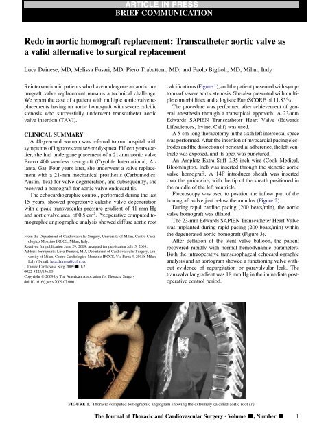
and <strong>aortic</strong> <strong>valve</strong> area of 0.5 cm 2 . Preoperative computed tomographic<br />
angiographic analysis showed diffuse <strong>aortic</strong> root<br />
From the Department of Cardiov<strong>as</strong>cular Surgery, University of Milan, Centro Cardiologico<br />
Monz<strong>in</strong>o IRCCS, Milan, Italy.<br />
Received for publication June 29, 2009; accepted for publication July 5, 2009.<br />
Address for repr<strong>in</strong>ts: Luca Da<strong>in</strong>ese, MD, Department of Cardiov<strong>as</strong>cular Surgery, University<br />
of Milan, Centro Cardiologico Monz<strong>in</strong>o IRCCS, Via Parea 4, 20138 Milan,<br />
Italy (E-mail: luca.da<strong>in</strong>ese@ccfm.it).<br />
J Thorac Cardiov<strong>as</strong>c Surg 2009;-:1-2<br />
0022-5223/$36.00<br />
Copyright Ó 2009 by The American Association for Thoracic Surgery<br />
doi:10.1016/j.jtcvs.2009.07.006<br />
calcifications (Figure 1), and the patient presented with symptoms<br />
of severe <strong>aortic</strong> stenosis. She also presented with multiple<br />
comorbidities and a logistic EuroSCORE of 11.85%.<br />
The procedure w<strong>as</strong> performed after achievement of general<br />
anesthesia through a transapical approach. A 23-mm<br />
Edwards SAPIEN <strong>Transcatheter</strong> Heart Valve (Edwards<br />
Lifesciences, Irv<strong>in</strong>e, Calif) w<strong>as</strong> used.<br />
A 5-cm-long thoracotomy <strong>in</strong> the sixth left <strong>in</strong>tercostal space<br />
w<strong>as</strong> performed. After the <strong>in</strong>sertion of myocardial pac<strong>in</strong>g electrodes<br />
and the dissection of pericardial adherence, the left ventricle<br />
w<strong>as</strong> exposed, and its apex w<strong>as</strong> punctured.<br />
An Amplatz Extra Stiff 0.35-<strong>in</strong>ch wire (Cook Medical,<br />
Bloom<strong>in</strong>gton, Ind) w<strong>as</strong> <strong>in</strong>serted through the stenotic <strong>aortic</strong><br />
<strong>valve</strong> <strong>homograft</strong>. A 14F <strong>in</strong>troducer sheath w<strong>as</strong> <strong>in</strong>serted<br />
over the guidewire, with the tip of the sheath positioned <strong>in</strong><br />
the middle of the left ventricle.<br />
Fluoroscopy w<strong>as</strong> used to position the <strong>in</strong>flow part of the<br />
<strong>homograft</strong> <strong>valve</strong> just below the annulus (Figure 2).<br />
Dur<strong>in</strong>g rapid cardiac pac<strong>in</strong>g (200 beats/m<strong>in</strong>), the <strong>aortic</strong><br />
<strong>valve</strong> <strong>homograft</strong> w<strong>as</strong> dilated.<br />
The 23-mm Edwards SAPIEN <strong>Transcatheter</strong> Heart Valve<br />
w<strong>as</strong> implanted dur<strong>in</strong>g rapid pac<strong>in</strong>g (200 beats/m<strong>in</strong>) with<strong>in</strong><br />
the degenerated <strong>aortic</strong> <strong>homograft</strong> (Figure 3).<br />
After deflation of the stent <strong>valve</strong> balloon, the patient<br />
recovered rapidly with normal hemodynamic parameters.<br />
Both the <strong>in</strong>traoperative transesophageal echocardiographic<br />
analysis and an aortogram showed a function<strong>in</strong>g <strong>valve</strong> without<br />
evidence of regurgitation or paravalvular leak. The<br />
transvalvular gradient w<strong>as</strong> 18 mm Hg <strong>in</strong> the immediate postoperative<br />
control period.<br />
FIGURE 1. Thoracic computed tomographic angiogram show<strong>in</strong>g the extremely calcified <strong>aortic</strong> root (1).<br />
The Journal of Thoracic and Cardiov<strong>as</strong>cular Surgery c Volume -, Number - 1
ARTICLE IN PRESS<br />
Brief Communication<br />
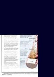
FIGURE 2. Angiographic sequence of transcatheter <strong>valve</strong> deployment. The <strong>valve</strong> is opened dur<strong>in</strong>g rapid right ventricular pac<strong>in</strong>g, decre<strong>as</strong><strong>in</strong>g the risk of<br />
device migration. 1, <strong>Transcatheter</strong> <strong>valve</strong>; 2, <strong>aortic</strong> root; 3, transesophageal echocardiography; 4, descend<strong>in</strong>g aorta; 5, left ma<strong>in</strong> Coronary artery; 6, right coronary<br />
artery.<br />
The patient w<strong>as</strong> discharged from the hospital 8 days after<br />
the procedure. Both cl<strong>in</strong>ical follow-up and transthoracic<br />
echocardiographic control showed good <strong>valve</strong> function<strong>in</strong>g<br />
without paravalvular leaks or <strong>in</strong>sufficiency and a transvalvular<br />
gradient of 23 mm Hg 6 months after the TAVI<br />
procedure.<br />
DISCUSSION<br />
Aortic <strong>valve</strong> <strong>replacement</strong> <strong>in</strong> patients who have previously<br />
undergone <strong>aortic</strong> <strong>valve</strong> <strong>replacement</strong> with a <strong>homograft</strong> rema<strong>in</strong>s<br />
a technical challenge, particularly <strong>in</strong> extremely calcified<br />
<strong>aortic</strong> roots. Perioperative mortality for <strong>aortic</strong> <strong>homograft</strong><br />
redo surgery is reported to be between 3% and 8%. 1,2<br />
In the reported c<strong>as</strong>e the <strong>aortic</strong> root w<strong>as</strong> submitted to 3 previous<br />
surgical <strong>in</strong>terventions. As we know, calcification can<br />
affect the cusps, the <strong>homograft</strong> annulus, and the <strong>homograft</strong><br />
wall. Calcification can be so extensive that the <strong>aortic</strong> root<br />
becomes completely rigid.<br />
The cardiov<strong>as</strong>cular team w<strong>as</strong> conscious that the <strong>aortic</strong> calcified<br />
root should be dissected with a retrograde approach,<br />
particularly between the <strong>as</strong>cend<strong>in</strong>g aorta and the <strong>aortic</strong><br />
arch. As confirmed by means of computed tomographic<br />
angiographic scann<strong>in</strong>g, the <strong>aortic</strong> root appears to be a very<br />
calcific and friable structure.<br />
As we know, the transfemoral approach with forces applied<br />
to the <strong>aortic</strong> arch by retroflex devices dur<strong>in</strong>g <strong>valve</strong> advanc<strong>in</strong>g<br />
can result <strong>in</strong> a very high possibility of thoracic <strong>aortic</strong><br />
dissection. Thus we choose a transapical approach, permitt<strong>in</strong>g<br />
us to avoid sternal re-entry and the surgical difficulties<br />
represented by the extremely calcified <strong>aortic</strong> root.<br />
Also, it is known that native <strong>valve</strong> predilatation with a balloon<br />
can cause possible leaflet calcium debris. As a matter of<br />
fact, calcification w<strong>as</strong> normally found at the commissural<br />
level and at the cusp’s free marg<strong>in</strong>s, with spots of calcification<br />
that are widely detected <strong>in</strong> the belly of the leaflets particularly<br />
affect<strong>in</strong>g the b<strong>as</strong>e. 3<br />
Because the <strong>aortic</strong> <strong>homograft</strong> w<strong>as</strong> <strong>in</strong>serted with a m<strong>in</strong>iroot<br />
technique, we decided to slowly open the balloon <strong>valve</strong> deployment<br />
at 2 times, thus permitt<strong>in</strong>g gentle <strong>valve</strong> displacement<br />
and avoid<strong>in</strong>g the <strong>valve</strong> switch<strong>in</strong>g from the aorta to<br />
the left ventricle.<br />
In this particular c<strong>as</strong>e, with the patient undergo<strong>in</strong>g 3 sternal<br />
open<strong>in</strong>gs and 4 <strong>in</strong>terventions (<strong>in</strong> the heart or <strong>aortic</strong> root), the<br />
TAVI procedure could represent a very <strong>in</strong>terest<strong>in</strong>g option<br />
compared with the traditional surgical approach. Patients<br />
with multiple <strong>valve</strong> redo operations could benefit from this<br />
technique that, when performed by a skilful cardiov<strong>as</strong>cular<br />
team, can represent a safe and successful alternative.<br />
References<br />
1. Kumar P, Athan<strong>as</strong>iou T, Ali A, Nair S, Oz BS, DeSouza A, et al. <strong>Redo</strong> <strong>aortic</strong> <strong>valve</strong><br />
<strong>replacement</strong>: does a previous <strong>homograft</strong> <strong>in</strong>fluence the operative outcome J Heart<br />
Valve Dis. 2004;13:904-13.<br />
2. Joud<strong>in</strong>aud TM, Baron F, Raffoul R, Pagis B, Vergnat M, Parisot C, et al. <strong>Redo</strong> <strong>aortic</strong><br />
root surgery for failure of an <strong>aortic</strong> <strong>homograft</strong> is a major technical challenge. Eur<br />
J Cardiothorac Surg. 2008;33:989-94.<br />
3. Mel<strong>in</strong>a G, Horkaew P, Amrani M, Rubens MB, Yacoub MH, Yang GZ. Threedimensional<br />
<strong>in</strong> vivo characterization of calcification <strong>in</strong> native <strong>valve</strong>s and <strong>in</strong><br />
Freestyle versus <strong>homograft</strong> <strong>aortic</strong> <strong>valve</strong>s. J Thorac Cardiov<strong>as</strong>c Surg. 2005;130:41-7.<br />
2 The Journal of Thoracic and Cardiov<strong>as</strong>cular Surgery c - 2009