Functions of AP1 (Fos/Jun) in bone development
Functions of AP1 (Fos/Jun) in bone development Functions of AP1 (Fos/Jun) in bone development
ii40 REPORT Functions of AP1 (Fos/Jun) in bone development E F Wagner ............................................................................................................................. Genetically modified mice and cells have provided important insights into the biological functions of the dimeric transcription factor complex AP1, in particular into its role in skeletal development. Data obtained from knockout mice revealed that some components, such as c-Fos are key regulators of bone cell differentiation, whereas others, like c-Jun, JunB and Fra-1 are essential in embryonic and/or postnatal development. Apart from identifying the specific roles of AP1 proteins in developmental processes, researchers are beginning to obtain a better molecular understanding of their cell-context dependent functions, their downstream target genes and how they regulate bone cell proliferation, differentiation, and apoptosis. A great variety of dimers composed of members of the Fos, Jun, and ATF families of proteins constitute the transcription factor complex AP1 (for review see references 12 ). While the Fos proteins (c-Fos, FosB, Fra-1, Fra-2) can only heterodimerise with members of the Jun family, the Jun proteins (c-Jun, JunB, JunD) can both homodimerise and heterodimerise with Fos members to form transcriptionally active complexes. Some members of the ATF and CREB families of proteins are also part of AP1 complexes. AP1 converts extracellular signals into changes in the expression of specific target genes, which harbour AP1 binding site(s) in their promoter or enhancer regions. The activity of AP1 is modulated by interactions with other transcriptional regulators and is further controlled by upstream kinases that link AP1 to various signal transduction pathways. AP1 has been implicated in a large variety of biological processes including cell differentiation, proliferation, apoptosis, and www.annrheumdis.com Downloaded from ard.bmj.com on November 5, 2012 - Published by group.bmj.com Table 1 Analysis of genetically modified fos and jun mice Ann Rheum Dis 2002;61(Suppl II):ii40–ii42 oncogenic transformation. Here I will summarise the current understanding of AP1 functions in bone development, which was largely obtained from the analysis of genetically modified mice and cells in which specific AP1 genes have been ectopically expressed, inactivated, mutated, or replaced by each other. Most important insights regarding the specific functions of AP1 proteins in mouse development were obtained from loss of function experiments using ES cell technology. A summary of the phenotypes of mice harbouring genetic modifications of the different fos and jun genes is given in table 1. These analyses showed that AP1 proteins have specific functions during embryogenesis and organogenesis. With regard to embryonic development, the Fos and Jun proteins can be grouped into two categories: some such as c-Fos, FosB, and JunD are dispensable, whereas others like c-Jun, JunB and Fra-1 are essential for embryonic development. In the following I will discuss the specific phenotypes of knock out mice in particular of c-Fos and Fra-1 as well as studies in transgenic mice expressing individual AP1 components with an emphasis on the effects observed on bone development. AP1 IN BONE DEVELOPMENT The growth and the maintenance of the skeleton depends on the coordinated function of osteoblasts and osteoclasts, the two principal cell types of bone tissue. 34 Osteoblasts, which derive from mesenchymal progenitors, produce the extracellular matrix of the bone that later undergoes mineralisation. In contrast, osteoclasts belong to the monocyte/macrophage lineage and reduce bone mass by resorbing the mineralised extracellular matrix. Recent studies have shown that AP1 components, mainly members of the Fos family such as c-Fos and Fra-1, have important functions in both of these cell types. Knockout Phenotypes Affected organs and cell types c-Fos osteopetrosis bone, osteoclasts FosB nurturing defect brain, hypothalamus Fra-1 embryonic lethality (E9.5) extra-embryonic tissue, placenta Fra-2 lethal at birth (unpublished) c-Jun embryonic lethality (E12.5) liver, hepatoblasts and heart outflow tract JunB embryonic lethality (E8–10) extra-embryonic tissue, placenta JunD male sterility testis, spermatogenesis Transgene Promoters Phenotypes Affected organs and cell types c-Fos H2k b osteosarcoma bone, osteoblasts FosB H2k b none ΔFosB TCRβ impaired T cell differentiation thymus, immature thymocytes NSE osteosclerosis bone, osteoblasts Fra-1 H2k b osteosclerosis bone, osteoblasts Fra-2 CMV occular malformations eye, anterior eye structures c-Jun H2k b none JunB ubiquitin C none CD4 enhanced Th2 maturation thymus / CD4 thymocytes JunD not reported For details and further references please see Jochum et al. 1
- Page 2 and 3: Downloaded from ard.bmj.com on Nove
- Page 4: References Email alerting service N
ii40<br />
REPORT<br />
<strong>Functions</strong> <strong>of</strong> <strong>AP1</strong> (<strong>Fos</strong>/<strong>Jun</strong>) <strong>in</strong> <strong>bone</strong> <strong>development</strong><br />
E F Wagner<br />
.............................................................................................................................<br />
Genetically modified mice and cells have provided important<br />
<strong>in</strong>sights <strong>in</strong>to the biological functions <strong>of</strong> the dimeric<br />
transcription factor complex <strong>AP1</strong>, <strong>in</strong> particular <strong>in</strong>to its role<br />
<strong>in</strong> skeletal <strong>development</strong>. Data obta<strong>in</strong>ed from knockout<br />
mice revealed that some components, such as c-<strong>Fos</strong> are<br />
key regulators <strong>of</strong> <strong>bone</strong> cell differentiation, whereas others,<br />
like c-<strong>Jun</strong>, <strong>Jun</strong>B and Fra-1 are essential <strong>in</strong> embryonic<br />
and/or postnatal <strong>development</strong>. Apart from identify<strong>in</strong>g the<br />
specific roles <strong>of</strong> <strong>AP1</strong> prote<strong>in</strong>s <strong>in</strong> <strong>development</strong>al processes,<br />
researchers are beg<strong>in</strong>n<strong>in</strong>g to obta<strong>in</strong> a better molecular<br />
understand<strong>in</strong>g <strong>of</strong> their cell-context dependent functions,<br />
their downstream target genes and how they regulate<br />
<strong>bone</strong> cell proliferation, differentiation, and apoptosis.<br />
A great<br />
variety <strong>of</strong> dimers composed <strong>of</strong> members <strong>of</strong> the <strong>Fos</strong>,<br />
<strong>Jun</strong>, and ATF families <strong>of</strong> prote<strong>in</strong>s constitute the<br />
transcription factor complex <strong>AP1</strong> (for review see<br />
references 12 ). While the <strong>Fos</strong> prote<strong>in</strong>s (c-<strong>Fos</strong>, <strong>Fos</strong>B, Fra-1,<br />
Fra-2) can only heterodimerise with members <strong>of</strong> the <strong>Jun</strong><br />
family, the <strong>Jun</strong> prote<strong>in</strong>s (c-<strong>Jun</strong>, <strong>Jun</strong>B, <strong>Jun</strong>D) can both<br />
homodimerise and heterodimerise with <strong>Fos</strong> members to form<br />
transcriptionally active complexes. Some members <strong>of</strong> the ATF<br />
and CREB families <strong>of</strong> prote<strong>in</strong>s are also part <strong>of</strong> <strong>AP1</strong> complexes.<br />
<strong>AP1</strong> converts extracellular signals <strong>in</strong>to changes <strong>in</strong> the expression<br />
<strong>of</strong> specific target genes, which harbour <strong>AP1</strong> b<strong>in</strong>d<strong>in</strong>g<br />
site(s) <strong>in</strong> their promoter or enhancer regions. The activity <strong>of</strong><br />
<strong>AP1</strong> is modulated by <strong>in</strong>teractions with other transcriptional<br />
regulators and is further controlled by upstream k<strong>in</strong>ases that<br />
l<strong>in</strong>k <strong>AP1</strong> to various signal transduction pathways. <strong>AP1</strong> has<br />
been implicated <strong>in</strong> a large variety <strong>of</strong> biological processes<br />
<strong>in</strong>clud<strong>in</strong>g cell differentiation, proliferation, apoptosis, and<br />
www.annrheumdis.com<br />
Downloaded from<br />
ard.bmj.com on November 5, 2012 - Published by group.bmj.com<br />
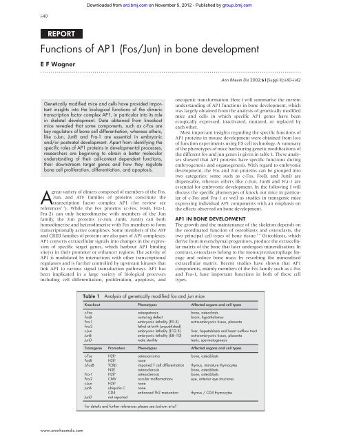
Table 1 Analysis <strong>of</strong> genetically modified fos and jun mice<br />
Ann Rheum Dis 2002;61(Suppl II):ii40–ii42<br />
oncogenic transformation. Here I will summarise the current<br />
understand<strong>in</strong>g <strong>of</strong> <strong>AP1</strong> functions <strong>in</strong> <strong>bone</strong> <strong>development</strong>, which<br />
was largely obta<strong>in</strong>ed from the analysis <strong>of</strong> genetically modified<br />
mice and cells <strong>in</strong> which specific <strong>AP1</strong> genes have been<br />
ectopically expressed, <strong>in</strong>activated, mutated, or replaced by<br />
each other.<br />
Most important <strong>in</strong>sights regard<strong>in</strong>g the specific functions <strong>of</strong><br />
<strong>AP1</strong> prote<strong>in</strong>s <strong>in</strong> mouse <strong>development</strong> were obta<strong>in</strong>ed from loss<br />
<strong>of</strong> function experiments us<strong>in</strong>g ES cell technology. A summary<br />
<strong>of</strong> the phenotypes <strong>of</strong> mice harbour<strong>in</strong>g genetic modifications <strong>of</strong><br />
the different fos and jun genes is given <strong>in</strong> table 1. These analyses<br />
showed that <strong>AP1</strong> prote<strong>in</strong>s have specific functions dur<strong>in</strong>g<br />
embryogenesis and organogenesis. With regard to embryonic<br />
<strong>development</strong>, the <strong>Fos</strong> and <strong>Jun</strong> prote<strong>in</strong>s can be grouped <strong>in</strong>to<br />
two categories: some such as c-<strong>Fos</strong>, <strong>Fos</strong>B, and <strong>Jun</strong>D are<br />
dispensable, whereas others like c-<strong>Jun</strong>, <strong>Jun</strong>B and Fra-1 are<br />
essential for embryonic <strong>development</strong>. In the follow<strong>in</strong>g I will<br />
discuss the specific phenotypes <strong>of</strong> knock out mice <strong>in</strong> particular<br />
<strong>of</strong> c-<strong>Fos</strong> and Fra-1 as well as studies <strong>in</strong> transgenic mice<br />
express<strong>in</strong>g <strong>in</strong>dividual <strong>AP1</strong> components with an emphasis on<br />
the effects observed on <strong>bone</strong> <strong>development</strong>.<br />
<strong>AP1</strong> IN BONE DEVELOPMENT<br />
The growth and the ma<strong>in</strong>tenance <strong>of</strong> the skeleton depends on<br />
the coord<strong>in</strong>ated function <strong>of</strong> osteoblasts and osteoclasts, the<br />
two pr<strong>in</strong>cipal cell types <strong>of</strong> <strong>bone</strong> tissue. 34 Osteoblasts, which<br />
derive from mesenchymal progenitors, produce the extracellular<br />
matrix <strong>of</strong> the <strong>bone</strong> that later undergoes m<strong>in</strong>eralisation. In<br />
contrast, osteoclasts belong to the monocyte/macrophage l<strong>in</strong>eage<br />
and reduce <strong>bone</strong> mass by resorb<strong>in</strong>g the m<strong>in</strong>eralised<br />
extracellular matrix. Recent studies have shown that <strong>AP1</strong><br />
components, ma<strong>in</strong>ly members <strong>of</strong> the <strong>Fos</strong> family such as c-<strong>Fos</strong><br />
and Fra-1, have important functions <strong>in</strong> both <strong>of</strong> these cell<br />
types.<br />
Knockout Phenotypes Affected organs and cell types<br />
c-<strong>Fos</strong> osteopetrosis <strong>bone</strong>, osteoclasts<br />
<strong>Fos</strong>B nurtur<strong>in</strong>g defect bra<strong>in</strong>, hypothalamus<br />
Fra-1 embryonic lethality (E9.5) extra-embryonic tissue, placenta<br />
Fra-2 lethal at birth (unpublished)<br />
c-<strong>Jun</strong> embryonic lethality (E12.5) liver, hepatoblasts and heart outflow tract<br />
<strong>Jun</strong>B embryonic lethality (E8–10) extra-embryonic tissue, placenta<br />
<strong>Jun</strong>D male sterility testis, spermatogenesis<br />
Transgene Promoters Phenotypes Affected organs and cell types<br />
c-<strong>Fos</strong> H2k b osteosarcoma <strong>bone</strong>, osteoblasts<br />
<strong>Fos</strong>B H2k b none<br />
Δ<strong>Fos</strong>B TCRβ impaired T cell differentiation thymus, immature thymocytes<br />
NSE osteosclerosis <strong>bone</strong>, osteoblasts<br />
Fra-1 H2k b osteosclerosis <strong>bone</strong>, osteoblasts<br />
Fra-2 CMV occular malformations eye, anterior eye structures<br />
c-<strong>Jun</strong> H2k b none<br />
<strong>Jun</strong>B ubiquit<strong>in</strong> C none<br />
CD4 enhanced Th2 maturation thymus / CD4 thymocytes<br />
<strong>Jun</strong>D not reported<br />
For details and further references please see Jochum et al. 1
Downloaded from<br />
ard.bmj.com on November 5, 2012 - Published by group.bmj.com<br />
<strong>AP1</strong> <strong>in</strong> <strong>bone</strong> <strong>development</strong> ii41<br />
Osteoblasts and the role <strong>of</strong> c-<strong>Fos</strong> and Fra-1<br />
In osteoblasts, <strong>AP1</strong> activity can be <strong>in</strong>duced by transform<strong>in</strong>g<br />
growth factor β, parathyroid hormone and 1,25-dihydroxy<br />
vitam<strong>in</strong> D, which are potent regulators <strong>of</strong> osteoblast differentiation<br />
and proliferation. 4 The various members <strong>of</strong> the <strong>AP1</strong><br />
complex are differentially expressed dur<strong>in</strong>g osteoblast maturation<br />
<strong>in</strong> vitro 5 with all <strong>Fos</strong> and <strong>Jun</strong> prote<strong>in</strong>s <strong>in</strong>itially be<strong>in</strong>g<br />
highly expressed. Subsequently, dur<strong>in</strong>g the period <strong>of</strong> extracellular<br />
matrix production and m<strong>in</strong>eralisation, their levels<br />
decl<strong>in</strong>e, and Fra-2 and <strong>Jun</strong>D become the major components <strong>of</strong><br />
the <strong>AP1</strong> complex <strong>in</strong> fully differentiated osteoblasts. The<br />
pattern <strong>of</strong> c-<strong>Fos</strong> expression dur<strong>in</strong>g <strong>development</strong> suggests a<br />
critical role <strong>in</strong> endochondral ossification, 6 although the analysis<br />
<strong>of</strong> c-<strong>Fos</strong> deficient mice <strong>in</strong>dicates that c-<strong>Fos</strong> is dispensable<br />
for the differentiation <strong>of</strong> osteoblasts. 78<br />
When c-<strong>Fos</strong> was ectopically expressed <strong>in</strong> various cell types<br />
and <strong>in</strong> transgenic mice, specific effects on the skeleton were<br />
observed. Chimeric mice obta<strong>in</strong>ed from c-<strong>Fos</strong> overexpress<strong>in</strong>g<br />
embryonic stem cells develop chondrogenic 9 tumours imply<strong>in</strong>g<br />
a function <strong>of</strong> c-<strong>Fos</strong> <strong>in</strong> chondrogenesis <strong>in</strong> vivo. Surpris<strong>in</strong>gly,<br />
c-<strong>Fos</strong> overexpression <strong>in</strong> an <strong>in</strong> vitro model <strong>of</strong> chondrogenesis<br />
<strong>in</strong>hibits the differentiation <strong>of</strong> chondrocytes. 10 Ectopic c-<strong>Fos</strong><br />
expression from a ubiquitous promoter <strong>in</strong> transgenic mice has<br />
no noticeable effects on cell differentiation <strong>in</strong> most organs, but<br />
results <strong>in</strong> the specific transformation <strong>of</strong> osteoblasts lead<strong>in</strong>g to<br />
osteogenic sarcomas rem<strong>in</strong>iscent <strong>of</strong> human osteosarcomas. 11<br />
Importantly, transgenic mice overexpress<strong>in</strong>g Fra-1 <strong>in</strong> osteoblasts<br />
as well as <strong>in</strong> other cell types exhibit aga<strong>in</strong> a specific <strong>bone</strong><br />
phenotype. These mice show <strong>in</strong>creased <strong>bone</strong> formation and<br />
develop osteosclerosis <strong>of</strong> the entire skeleton. 12 This phenotype<br />
is attributable to a cell autonomous <strong>in</strong>crease <strong>in</strong> the number <strong>of</strong><br />
mature osteoblasts <strong>in</strong>dicat<strong>in</strong>g that Fra-1 <strong>in</strong>creases osteoblast<br />
differentiation. A similar osteosclerosis phenotype is observed<br />
<strong>in</strong> transgenic mice express<strong>in</strong>g Δ<strong>Fos</strong>B <strong>in</strong> osteoblasts. 13 These<br />
mice also show reduced adipogenesis, a phenotype that is not<br />
observed <strong>in</strong> Fra-1 transgenic mice.<br />
Given the osteoblastic phenotype <strong>of</strong> the Fra-1 transgenic<br />
mice it was <strong>of</strong> <strong>in</strong>terest to next ask whether the absence <strong>of</strong><br />
Fra-1 affects osteoblast or osteoclast differentiation. Inactivation<br />
<strong>of</strong> Fra-1, however, results <strong>in</strong> embryonic lethality around<br />
day 10 <strong>of</strong> <strong>development</strong> because <strong>of</strong> defects <strong>in</strong> the placenta<br />
thereby preclud<strong>in</strong>g the analysis on <strong>bone</strong> cell <strong>development</strong>. 14<br />
The labyr<strong>in</strong>th layer <strong>of</strong> mutant placentas is reduced <strong>in</strong> size and<br />
largely avascular suggest<strong>in</strong>g that the <strong>in</strong>vasion <strong>of</strong> allantoic vessels<br />
<strong>in</strong>to the chorionic plate is impaired <strong>in</strong> the absence <strong>of</strong><br />
Fra-1. The <strong>development</strong> <strong>of</strong> mutant fetuses can be rescued up to<br />
birth by provid<strong>in</strong>g wild type extra-embryonic tissues us<strong>in</strong>g<br />
tetraploid blastocyst <strong>in</strong>jection. 15 This suggests that Fra-1 is<br />
dispensable for the differentiation along most, if not all, cell<br />
l<strong>in</strong>eages <strong>in</strong> the fetus. 14 Moreover, the lethality <strong>of</strong> Fra-1<br />
deficient mice was fully rescued by the ectopic expression <strong>of</strong><br />
Fra-1 from transgenic mice 14 demonstrat<strong>in</strong>g that <strong>AP1</strong>/Fra-1<br />
activity dur<strong>in</strong>g <strong>development</strong> does not have to be tightly regulated.<br />
These rescued mice still developed osteosclerosis, which<br />
was <strong>in</strong>dist<strong>in</strong>guishable from the disease observed <strong>in</strong> the Fra-1<br />
transgenic mice. However, it cannot be excluded that Fra-1 has<br />
a critical role <strong>in</strong> osteoblast homeostasis dur<strong>in</strong>g postnatal life.<br />
We recently demonstrated us<strong>in</strong>g a conditional allele <strong>of</strong> fra-1<br />
that mice without Fra-1 can <strong>in</strong>deed be born and develop<br />
osteopenia, which has to be further characterised (Robert<br />
Eferl, unpublished data).<br />
Osteoclasts and the role <strong>of</strong> c-<strong>Fos</strong> and Fra-1<br />
Mice lack<strong>in</strong>g c-<strong>Fos</strong> are viable and fertile but lack osteoclasts and<br />
are therefore osteopetrotic. 78 This phenotype is stra<strong>in</strong> dependent,<br />
as on a 129/sv background much fewer pups are born and<br />
reach wean<strong>in</strong>g age, when compared with C57Bl/6 mutant mice<br />
(unpublished observations). Mutant mice also show abnormalities<br />
<strong>of</strong> the haematopoietic system <strong>in</strong>clud<strong>in</strong>g extramedullary<br />
haematopoiesis and lymphopenia, which are both secondary to<br />
the <strong>bone</strong> phenotype. 16 Although c-<strong>Fos</strong> is rapidly <strong>in</strong>duced <strong>in</strong> T<br />
cells upon activation and it regulates the transcription <strong>of</strong> various<br />
cytok<strong>in</strong>es <strong>in</strong>clud<strong>in</strong>g <strong>in</strong>terleuk<strong>in</strong> 2, the analysis <strong>of</strong> c-<strong>Fos</strong> deficient<br />
mice has shown that c-<strong>Fos</strong> is not required for the differentiation<br />
and activity <strong>of</strong> peripheral T cells. 17<br />
The lack <strong>of</strong> osteoclasts <strong>in</strong> c-<strong>Fos</strong> deficient mice is accompanied<br />
by <strong>in</strong>creased numbers <strong>of</strong> <strong>bone</strong> marrow macrophages,<br />
which is specific for this osteopetrotic mutant. The osteopetrosis<br />
is characterised by <strong>in</strong>creased <strong>bone</strong> mass attributable to<br />
reduced <strong>bone</strong> resorption. 7818 Recent data us<strong>in</strong>g retroviral gene<br />
transfer <strong>in</strong>to c-<strong>Fos</strong> mutant osteoclast precursors <strong>in</strong> vitro have<br />
shown that all <strong>Fos</strong> prote<strong>in</strong>s, most efficiently Fra-1, can<br />
complement for the absence <strong>of</strong> c-<strong>Fos</strong>. 19 Further structurefunction<br />
analysis also demonstrated that the major<br />
C-term<strong>in</strong>al transactivation doma<strong>in</strong>s <strong>of</strong> c-<strong>Fos</strong> and <strong>Fos</strong>B are dispensable<br />
for the rescue <strong>of</strong> osteoclast formation <strong>in</strong> vitro. Interest<strong>in</strong>gly,<br />
Fra-1, which lacks a transactivation doma<strong>in</strong>, has the<br />
highest rescue activity. Moreover, the osteoclast differentiation<br />
factor RANKL also known as ODF, OPGL, TRANCE (for review<br />
see Karsenty and Wagner 4 ) <strong>in</strong>duces besides c-<strong>Fos</strong> also Fra-1<br />
transcription <strong>in</strong> a c-<strong>Fos</strong> dependent manner, thereby establish<strong>in</strong>g<br />
a l<strong>in</strong>k between RANK signall<strong>in</strong>g and the expression <strong>of</strong> <strong>AP1</strong><br />
prote<strong>in</strong>s dur<strong>in</strong>g osteoclast differentiation 19 (see also below).<br />
The osteopetrotic phenotype can be partly cured <strong>in</strong> c-<strong>Fos</strong><br />
mutant mice by express<strong>in</strong>g a Fra-1 transgene, one target gene<br />
<strong>of</strong> c-<strong>Fos</strong> <strong>in</strong> the osteoclast l<strong>in</strong>eage. 19 Importantly, total restoration<br />
<strong>of</strong> osteoclast differentiation was achieved <strong>in</strong> knock <strong>in</strong><br />
mice generated by the <strong>in</strong>sertion <strong>of</strong> the fra-1 gene <strong>in</strong>to the c-fos<br />
locus. 20 Fra-1 was able to rescue c-<strong>Fos</strong> dependent functions, <strong>in</strong><br />
particular <strong>bone</strong> <strong>development</strong> and light <strong>in</strong>duced photoreceptor<br />
apoptosis. Interest<strong>in</strong>gly, the rescue <strong>of</strong> <strong>bone</strong> cell differentiation<br />
was gene-dose dependent and Fra-1 was unable to substitute<br />
for the expression <strong>of</strong> <strong>AP1</strong> target genes <strong>in</strong> fibroblasts. These<br />
results show that c-<strong>Fos</strong> and Fra-1 have ma<strong>in</strong>ta<strong>in</strong>ed functional<br />
equivalence dur<strong>in</strong>g vertebrate evolution and further suggest<br />
that functional diversity <strong>of</strong> <strong>Fos</strong> genes is a result <strong>of</strong> divergence<br />
<strong>of</strong> regulatory rather than prote<strong>in</strong> cod<strong>in</strong>g sequences.<br />
In addition to the effects on osteoblasts, ectopic Fra-1<br />
expression also <strong>in</strong>creases the differentiation <strong>of</strong> osteoclasts,<br />
both <strong>in</strong> progenitor cell l<strong>in</strong>es and primary osteoclast<br />
progenitors. 19 21 This strong osteoclastogenic effect is not<br />
apparent <strong>in</strong> Fra-1 transgenic mice <strong>in</strong> vivo. 12 Osteoclast<br />
differentiation on the other hand does not seem to require the<br />
presence <strong>of</strong> Fra-1, as the skeleton <strong>of</strong> rescued Fra-1 deficient<br />
fetuses conta<strong>in</strong>s functional osteoclasts at birth. 14 Moreover,<br />
recently established Fra-1 knockout mice us<strong>in</strong>g a conditional<br />
gene target<strong>in</strong>g approach demonstrate that a normal skeleton<br />
can be formed (Robert Eferl, unpublished data and see above)<br />
suggest<strong>in</strong>g that Fra-1 is dispensable for the formation and<br />
differentiation <strong>of</strong> the major <strong>bone</strong> cell types, the osteoblasts<br />
and the osteoclasts.<br />
The role <strong>of</strong> c-<strong>Fos</strong> <strong>in</strong> signall<strong>in</strong>g dur<strong>in</strong>g osteoclast<br />
differentiation<br />
To ma<strong>in</strong>ta<strong>in</strong> <strong>bone</strong> homeostasis the signall<strong>in</strong>g by the osteoclastogenic<br />
ligand RANKL, a member <strong>of</strong> the TNF family and an<br />
essential factor for osteoclast differentiation, has to be tightly<br />
controlled, as deregulated osteoclast activity leads to severe<br />
<strong>bone</strong> diseases such as osteoporosis. 4 In a genomic search for<br />
c-<strong>Fos</strong> target genes <strong>in</strong>duced by RANKL <strong>in</strong> osteoclast precursor<br />
cells us<strong>in</strong>g wild type and c-fos-/- cells, a group <strong>of</strong> mRNAs<br />
whose expression is dependent on <strong>in</strong>terferon α/β signall<strong>in</strong>g<br />
was found to be specifically <strong>in</strong>duced by c-<strong>Fos</strong>. The <strong>in</strong>duction<br />
was decreased <strong>in</strong> cells lack<strong>in</strong>g c-<strong>Fos</strong> and the down regulated<br />
genes <strong>in</strong>cluded <strong>in</strong>terferon responsive genes such as Ifit-3,<br />
Ifit-2, Mx-1, Scyb-10. and Irf-7 (unpublished and Takayanagi<br />
et al 22 ). Although the <strong>in</strong>tegral role <strong>of</strong> the <strong>in</strong>terferon α/β system<br />
<strong>in</strong> the regulation <strong>of</strong> immune responses has been extensively<br />
documented, it was unknown whether this system is l<strong>in</strong>ked to<br />
RANKL signall<strong>in</strong>g and <strong>bone</strong> metabolism. We found that<br />
www.annrheumdis.com
ii42 Wagner<br />
Figure 1 Signall<strong>in</strong>g <strong>in</strong> osteoclast progenitor cells and cross talk<br />
with the <strong>in</strong>terferon pathway. For details see also Karsenty and<br />
Wagner 4 ).<br />
RANKL directly <strong>in</strong>duced the <strong>in</strong>terferon β gene <strong>in</strong> a c-<strong>Fos</strong><br />
dependent manner <strong>in</strong> osteoclast precursor cells. However,<br />
<strong>in</strong>terferon β <strong>in</strong>hibits the differentiation <strong>of</strong> osteoclasts by <strong>in</strong>terfer<strong>in</strong>g<br />
with the RANKL <strong>in</strong>duced expression <strong>of</strong> c-<strong>Fos</strong>, which is<br />
essential for osteoclast formation. 22 The mechanism <strong>of</strong><br />
<strong>in</strong>terferon β gene <strong>in</strong>duction by c-<strong>Fos</strong> is dist<strong>in</strong>ct from that<br />
<strong>in</strong>duced by viruses and implies that an autoregulatory mechanism<br />
is operat<strong>in</strong>g <strong>in</strong> f<strong>in</strong>e tun<strong>in</strong>g the activity <strong>of</strong> osteoclasts.<br />
RANKL <strong>in</strong>duces <strong>in</strong> the same progenitor cell c-<strong>Fos</strong>, which <strong>in</strong><br />
turn <strong>in</strong>duces the expression <strong>of</strong> positive regulators such as<br />
Fra-1 as well as its own <strong>in</strong>hibitor <strong>in</strong>terferon β (fig 1). The <strong>in</strong>hibition<br />
<strong>of</strong> c-<strong>Fos</strong> by <strong>in</strong>terferon α/β signall<strong>in</strong>g occurs at the posttranscriptional<br />
level and the molecular mechanism has yet to<br />
be def<strong>in</strong>ed. The importance <strong>of</strong> this regulatory mechanism for<br />
<strong>bone</strong> homeostasis is supported by the observation that mice<br />
deficient <strong>in</strong> <strong>in</strong>terferon β signall<strong>in</strong>g exhibit severe <strong>bone</strong> loss<br />
accompanied by <strong>in</strong>creased osteoclastogenesis. 22 Moreover, we<br />
were able to show <strong>in</strong> a model <strong>of</strong> LPS <strong>in</strong>duced <strong>in</strong>flammatory<br />
<strong>bone</strong> destruction that the application <strong>of</strong> <strong>in</strong>terferon β had a<br />
therapeutic effect <strong>in</strong> this disease model. Therefore, this study<br />
may <strong>of</strong>fer new approaches to the treatment <strong>of</strong> <strong>bone</strong> diseases<br />
caused by excessive osteoclastogenesis, such as autoimmune<br />
arthritis and osteoporosis.<br />
CONCLUSIONS<br />
Through the extensive analysis <strong>of</strong> the <strong>in</strong>dividual <strong>AP1</strong> components<br />
us<strong>in</strong>g genetically modified mice and cells we are beg<strong>in</strong>n<strong>in</strong>g<br />
to unravel the specific functions <strong>of</strong> these prote<strong>in</strong>s <strong>in</strong><br />
<strong>development</strong> and/or <strong>in</strong> the adult organism. The <strong>in</strong>itial studies<br />
have largely focused on c-<strong>Fos</strong> and c-<strong>Jun</strong>, but have been<br />
extended to other family members. As the absence <strong>of</strong> c-<strong>Jun</strong>,<br />
<strong>Jun</strong>B and Fra-1 results <strong>in</strong> embryonic lethality, the analysis <strong>of</strong><br />
these prote<strong>in</strong>s dur<strong>in</strong>g later stages <strong>of</strong> <strong>development</strong> and <strong>in</strong> postnatal<br />
life were not possible without the <strong>development</strong> <strong>of</strong> new<br />
genetic tools. With the conditional <strong>in</strong>activation <strong>of</strong> <strong>AP1</strong> genes<br />
<strong>in</strong> a cell type specific and/or <strong>in</strong>ducible manner us<strong>in</strong>g the<br />
established Cre/loxP and tet regulated systems, novel functions<br />
are be<strong>in</strong>g assigned to the <strong>in</strong>dividual members <strong>of</strong> <strong>AP1</strong> (for<br />
example, Behrens et al 23 ). However, we still do not understand<br />
the enormous specificity <strong>of</strong> prote<strong>in</strong>s such as c-<strong>Fos</strong> and Fra-1<br />
for osteoblast and/or osteoclast function. Nor do we know the<br />
<strong>in</strong>dividual partners <strong>of</strong> these molecules responsible for their<br />
transcriptional activity and specificity. In addition, we only<br />
have a very vague picture about the important downstream<br />
genes controlled by these factors. The genomic analysis exemplified<br />
by the discovery <strong>of</strong> the <strong>in</strong>terferon β system as a c-<strong>Fos</strong><br />
www.annrheumdis.com<br />
Downloaded from<br />
ard.bmj.com on November 5, 2012 - Published by group.bmj.com<br />
regulated gene cascade illustrates the power <strong>of</strong> these<br />
approaches. F<strong>in</strong>ally, we will have to come back to classic<br />
biochemical and/or proteomic approaches to precisely def<strong>in</strong>e<br />
the molecular mechanisms how a transcription factor<br />
complex such as <strong>AP1</strong> <strong>in</strong>fluences the decision <strong>of</strong> a cell to divide,<br />
differentiate, or die.<br />
ACKNOWLEDGEMENTS<br />
The IMP is supported by Boehr<strong>in</strong>ger-Ingelheim and I thank PD for<br />
comments on the manuscript.<br />
.....................<br />
Author’s affiliations<br />
E F Wagner, Research Institute <strong>of</strong> Molecular Pathology (IMP), Dr<br />
Bohr-Gasse 7, A-1030 Vienna, Austria<br />
Correspondence to: Dr E F Wagner; wagner@nt.imp.univie.ac.at<br />
REFERENCES<br />
1 Jochum W, Passegue E, Wagner EF. AP-1 <strong>in</strong> mouse <strong>development</strong> and<br />
tumorigenesis. Oncogene 2001;20:2401–12.<br />
2 Shaulian E, Kar<strong>in</strong> M. AP-1 as a regulator <strong>of</strong> cell life and death. Nat Cell<br />
Biol 2002;4:p. E131–6.<br />
3 Wagner EF, Karsenty G. Genetic control <strong>of</strong> skeletal <strong>development</strong>. Curr<br />
Op<strong>in</strong> Genet Dev 2001;11:527–32.<br />
4 Karsenty G, Wagner EF. Reach<strong>in</strong>g a genetic and molecular<br />
understand<strong>in</strong>g <strong>of</strong> skeletal <strong>development</strong>. Dev Cell 2002;2:389–406.<br />
5 McCabe LR, Banerjee C, Kundu R, Harrison RJ, Dobner PR, Ste<strong>in</strong> LJ, et<br />
al. Developmental expression and activities <strong>of</strong> specific fos and jun<br />
prote<strong>in</strong>s are functionally related to osteoblast maturation: role <strong>of</strong> Fra- 2<br />
and <strong>Jun</strong> D dur<strong>in</strong>g differentiation. Endocr<strong>in</strong>ology 1996;137:4398–408.<br />
6 Sandberg M, Vuorio T, Hirvonen H, Alitalo K, Vuorio E. Enhanced<br />
expression <strong>of</strong> TGF-beta and c-fos mRNAs <strong>in</strong> the growth plates <strong>of</strong><br />
develop<strong>in</strong>g human long <strong>bone</strong>s. Development 1988;102:461–70.<br />
7 Johnson RS, Spiegelman BM, Papaioannou V. Pleiotropic effects <strong>of</strong> a<br />
null mutation <strong>in</strong> the c-fos proto-oncogene. Cell 1992;71:577–86.<br />
8 Wang ZQ, Ovitt C, Grigoriadis AE, Mohle-Ste<strong>in</strong>le<strong>in</strong> U, Ruther U,<br />
Wagner EF. Bone and hematopoietic defects <strong>in</strong> mice lack<strong>in</strong>g c-fos.<br />
Nature 1992;360:741–5.<br />
9 Wang ZQ, Grigoriadis AE, Mohle-Ste<strong>in</strong>le<strong>in</strong> U, Wagner EF. A novel<br />
target cell for c-fos-<strong>in</strong>duced oncogenesis: <strong>development</strong> <strong>of</strong> chondrogenic<br />
tumours <strong>in</strong> embryonic stem cell chimeras. EMBO J 1991;10:2437–50.<br />
10 Thomas DP, Sunters A, Gentry A, Grigoriadis AE. Inhibition <strong>of</strong><br />
chondrocyte differentiation <strong>in</strong> vitro by constitutive and <strong>in</strong>ducible<br />
overexpression <strong>of</strong> the c-fos proto-oncogene. J Cell Sci 2000;113:439–<br />
50.<br />
11 Grigoriadis AE, Schellander K, Wang ZQ, Wagner EF. Osteoblasts are<br />
target cells for transformation <strong>in</strong> c-fos transgenic mice. J Cell Biol<br />
1993;122:685–701.<br />
12 Jochum W, David JP, Elliott C, Wutz A, Plenk HJ, Matsuo K, et al.<br />
Increased <strong>bone</strong> formation and osteosclerosis <strong>in</strong> mice overexpress<strong>in</strong>g the<br />
transcription factor Fra-1. Nat Med 2000;6:985–90.<br />
13 Sabatakos G, Sims NA, Chen J, Aoki K, Kelz MB, Aml<strong>in</strong>g M, et al.<br />
Overexpression <strong>of</strong> delta <strong>Fos</strong>B transcription factor(s) <strong>in</strong>creases <strong>bone</strong><br />
formation and <strong>in</strong>hibits adipogenesis. Nat Med 2000;6:985–90.<br />
14 Schreiber M, Wang ZQ, Jochum W, Fetka I, Elliott C, Wagner EF.<br />
Placental vascularisation requires the AP-1 component fra1. Development<br />
2000;127:4937–48.<br />
15 Wang ZQ, Kiefer F, Urbanek P, Wagner EF. Generation <strong>of</strong> completely<br />
embryonic stem cell-derived mutant mice us<strong>in</strong>g tetraploid blastocyst<br />
<strong>in</strong>jection. Mech Dev 1997;62:137–45.<br />
16 Okada S, Wang ZQ, Grigoriadis AE, Wagner EF, von Ruden T. Mice<br />
lack<strong>in</strong>g c-fos have normal hematopoietic stem cells but exhibit altered<br />
B-cell differentiation due to an impaired <strong>bone</strong> marrow environment. Mol<br />
Cell Biol 1994;14:382–90.<br />
17 Ja<strong>in</strong> J, Nalefski EA, McCaffrey PG, Johnson RS, Spiegelman BM,<br />
Papaioannou V, et al. Normal peripheral T-cell function <strong>in</strong> c-<strong>Fos</strong>-deficient<br />
mice. Mol Cell Biol 1994;14: 1566–74.<br />
18 Grigoriadis AE, Wang ZQ, Cecch<strong>in</strong>i MG, H<strong>of</strong>stetter W, Felix R, Fleisch<br />
HA, et al. c-<strong>Fos</strong>: a key regulator <strong>of</strong> osteoclast-macrophage l<strong>in</strong>eage<br />
determ<strong>in</strong>ation and <strong>bone</strong> remodel<strong>in</strong>g. Science 1994;266:443–8.<br />
19 Matsuo K, Owens JM, Tonko M, Elliott C, Chambers TJ, Wagner EF.<br />
<strong>Fos</strong>l1 is a transcriptional target <strong>of</strong> c-<strong>Fos</strong> dur<strong>in</strong>g osteoclast differentiation.<br />
Nat Genet 2000;24:184–7.<br />
20 Fleischmann A, Hafezi F, Elliott C, Reme CE, Ruther U, Wagner EF.<br />
Fra-1 replaces c-<strong>Fos</strong>-dependent functions <strong>in</strong> mice. Genes Dev<br />
2000;14:2695–700.<br />
21 Owens JM, Matsuo K, Nicholson GC, Wagner EF, Chambers TJ. Fra-1<br />
potentiates osteoclastic differentiation <strong>in</strong> osteoclast-macrophage precursor<br />
cell l<strong>in</strong>es. J Cell Physiol 1999; 179:170–8.<br />
22 Takayanagi H, Kim S, Matsuo K, Suzuki H, Suzuki T, Sato K, et al.<br />
RANKL ma<strong>in</strong>ta<strong>in</strong>s <strong>bone</strong> homeostasis through c-<strong>Fos</strong>-dependent <strong>in</strong>duction <strong>of</strong><br />
<strong>in</strong>terferon-beta. Nature 2002;416:744–9.<br />
23 Behrens A, Sibilia M, David JP, Mohle-Ste<strong>in</strong>le<strong>in</strong> U, Tronche F, Schutz G,<br />
et al. Impaired postnatal hepatocyte proliferation and liver regeneration<br />
<strong>in</strong> mice lack<strong>in</strong>g c-jun <strong>in</strong> the liver. EMBO J 2002;21:1782–90.
References<br />
Email alert<strong>in</strong>g<br />
service<br />
Notes<br />
<strong>Functions</strong> <strong>of</strong> <strong>AP1</strong> (<strong>Fos</strong>/<strong>Jun</strong>) <strong>in</strong> <strong>bone</strong><br />
<strong>development</strong><br />
E F Wagner<br />
Ann Rheum Dis 2002 61: ii40-ii42<br />
doi: 10.1136/ard.61.suppl_2.ii40<br />
Updated <strong>in</strong>formation and services can be found at:<br />
http://ard.bmj.com/content/61/suppl_2/ii40.full.html<br />
These <strong>in</strong>clude:<br />
To request permissions go to:<br />
http://group.bmj.com/group/rights-licens<strong>in</strong>g/permissions<br />
To order repr<strong>in</strong>ts go to:<br />
http://journals.bmj.com/cgi/repr<strong>in</strong>tform<br />
To subscribe to BMJ go to:<br />
http://group.bmj.com/subscribe/<br />
Downloaded from<br />
ard.bmj.com on November 5, 2012 - Published by group.bmj.com<br />
This article cites 22 articles, 9 <strong>of</strong> which can be accessed free at:<br />
http://ard.bmj.com/content/61/suppl_2/ii40.full.html#ref-list-1<br />
Article cited <strong>in</strong>:<br />
http://ard.bmj.com/content/61/suppl_2/ii40.full.html#related-urls<br />
Receive free email alerts when new articles cite this article. Sign up <strong>in</strong> the<br />
box at the top right corner <strong>of</strong> the onl<strong>in</strong>e article.


