Booklet - Georg Eisner, Ophthalmologie
Booklet - Georg Eisner, Ophthalmologie Booklet - Georg Eisner, Ophthalmologie
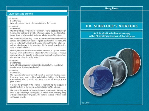
Georg Eisner Questions and answers Dr. Watson: Dear Sherlock, What is the clinical interest in the examination of the vitreous Dr. Sherlock: Dear Watson, The clinical interest of the vitreous lies in its property as a body cavity, which like any other body cavity provides information about the condition of adjoining tissue. In other words, the vitreous tells the story of the retina. Yet, in contrast to other body cavities, such as the anterior chamber which consists merely of fluid where invading cells can form free sediments, the vitreous contains a framework and invading cells must therefore follow predetermined pathways. At the same time, this framework may also be the cause of retinal pathology. In a way, the anatomical structures can be compared to a grammar of the language by which the vitreous tells its story. The knowledge of vitreous anatomy is thus the basis for a reliable prognosis in all conditions where vitreo-retinal interactions play a role. Dr. Watson: Dear Sherlock, What is the advantage in investigating the details of vitreous anatomy Aren’t vitreous structures just chaotic Dr. Sherlock: Dear Watson, The impression of chaos is merely the result of a restricted optical access. High-power preset lenses lead to a spatial picture that is heavily distorted, whereas three mirror contact lenses reveal only a small segment at any given moment. A correct interpretation of the distorted or fragmented pictures requires a sound knowledge of the general anatomical pattern of the vitreous. The vitreous framework can be revealed either by means of a slit lamp (as areas of light scattering), histologically (as stained structures) or by filling with coloured ink (as interspaces). The patterns revealed by these three methods do coincide. Dr. Sherlock’s Vitreous An Introduction to Biomicroscopy in the Clinical Examination of the Vitreous 05 | 2008
- Page 2 and 3: Biography of Georg Eisner Georg Eis
- Page 4: Content Disc 1 The Normal Anatomy o
<strong>Georg</strong> <strong>Eisner</strong><br />
Questions and answers<br />
Dr. Watson:<br />
Dear Sherlock,<br />
What is the clinical interest in the examination of the vitreous<br />
Dr. Sherlock:<br />
Dear Watson,<br />
The clinical interest of the vitreous lies in its property as a body cavity, which<br />
like any other body cavity provides information about the condition of adjoining<br />
tissue. In other words, the vitreous tells the story of the retina.<br />
Yet, in contrast to other body cavities, such as the anterior chamber which<br />
consists merely of fluid where invading cells can form free sediments, the<br />
vitreous contains a framework and invading cells must therefore follow predetermined<br />
pathways. At the same time, this framework may also be the<br />
cause of retinal pathology.<br />
In a way, the anatomical structures can be compared to a grammar of the<br />
language by which the vitreous tells its story. The knowledge of vitreous<br />
anatomy is thus the basis for a reliable prognosis in all conditions where<br />
vitreo-retinal interactions play a role.<br />
Dr. Watson:<br />
Dear Sherlock,<br />
What is the advantage in investigating the details of vitreous anatomy<br />
Aren’t vitreous structures just chaotic<br />
Dr. Sherlock:<br />
Dear Watson,<br />
The impression of chaos is merely the result of a restricted optical access.<br />
High-power preset lenses lead to a spatial picture that is heavily distorted,<br />
whereas three mirror contact lenses reveal only a small segment at any<br />
given moment.<br />
A correct interpretation of the distorted or fragmented pictures requires a<br />
sound knowledge of the general anatomical pattern of the vitreous.<br />
The vitreous framework can be revealed either by means of a slit lamp (as<br />
areas of light scattering), histologically (as stained structures) or by filling<br />
with coloured ink (as interspaces). The patterns revealed by these three<br />
methods do coincide.<br />
Dr. Sherlock’s Vitreous<br />
An Introduction to Biomicroscopy<br />
in the Clinical Examination of the Vitreous<br />
05 | 2008
Biography of <strong>Georg</strong> <strong>Eisner</strong><br />
<strong>Georg</strong> <strong>Eisner</strong> was born 1930 in Basel, Switzerland, to<br />
Willy <strong>Eisner</strong>, M.D. (Internal Medicine) and Irma <strong>Eisner</strong>-<br />
Guggenheim, M.D. (Ophthalmology). He is married to<br />
Susanne <strong>Eisner</strong>-Kartagener, has three children, Daniel,<br />
Miriam, Simone, and six grandchildren, Yael, Ruben,<br />
Nomi, Manuel, Marva and Miron.<br />
As a young man,<br />
<strong>Georg</strong> <strong>Eisner</strong> attended a humanities-oriented high school, the “Humanistisches<br />
Gymnasium” in Basel. He then worked as a carpenter in a kibbutz before attending<br />
medical school at the Universities of Basel and Paris. He received his<br />
M.D. degree in 1960.<br />
Later in life,<br />
<strong>Georg</strong> <strong>Eisner</strong> was being trained in ophthalmology with both Prof. F. Rintelen in<br />
Basel and with Prof. H. Goldmann and Prof. P. Niesel in Bern. As a full professor<br />
of ophthalmology, he held leading positions at the Bern University Eye Hospital<br />
(Inselspital), and as Dean of the Faculty of Medicine. In 1989, the medical students<br />
awarded Prof. <strong>Eisner</strong> the diploma “Teacher of the Year“. He retired from<br />
his academic positions in 1993.<br />
Prof. <strong>Eisner</strong>’s research interests are well illustrated by his principal publications:<br />
• Biomicroscopy of the Peripheral Fundus (Springer Verlag 1973)<br />
• Clinical Anatomy of the Vitreous (in Duane’s Biomedical Foundations in Ophthalmology,<br />
J.B.Lippincott 1989)<br />
• Introduction to the Biomicroscopy of the Eye (DVD-series, Haag-Streit, 2002)<br />
• Eye Surgery (Springer Verlag 1978/80, 1990)<br />
• Various scientific reports in the field of viscosurgery<br />
Since his retirement, Prof. <strong>Eisner</strong> has been enjoying family life, leisure, nature<br />
and art. He is the author of several introductions to contemporary art catalogues,<br />
as well as of many lectures and publications in a number of subjects<br />
such as:<br />
• physiological optics and art<br />
• color deficiency and painting<br />
• aniconism and the prohibition of pictorial presentation<br />
• migration of symbols<br />
• the art of gold casting in West Africa<br />
Acknowledgements<br />
This program is offered to the ophthalmic community gratuitously through<br />
the support of the following sponsors<br />
• Ambulante Augenchirurgie Zürich<br />
• Alcon Switzerland SA<br />
• Werner H. Spross-Stiftung Zürich<br />
• Swiss Ophthalmological Society SOG/SSO<br />
and by the author<br />
Special thanks<br />
This program would not have been possible without the help of<br />
• Prof. Elmar Messmer, Zürich, CH<br />
• Prof. Yves Robert, Zürich, CH<br />
• Prof. Jan Worst, Groningen, NL<br />
• Prof. Sebastian Wolf and collaborators, University Eye Clinic,Inselspital,<br />
Bern, CH<br />
• Birgit Stuber, Alcon Pharmaceuticals Ltd., Switzerland<br />
• Willy R. Hess, Adelheid Meyer, scientific illustrators<br />
• Dr. Peter Frey, Hans Holzherr, Giovanni Ferrieri, Béatrice Boog<br />
(Education and Media Unit, University of Bern)<br />
• Dr. Christoph Amstutz, Zürich, CH<br />
• Susanne <strong>Eisner</strong>-Kartagener, Bolligen, CH<br />
• The patients of the University Eye Hospital, Bern, who voluntarily agreed to<br />
undergo the cumbersome examination with the video slit lamp for the sole<br />
purpose of assisting in the training of future ophthalmologists<br />
Production<br />
• Education and Media Unit, Institute of Medical Education,<br />
University of Bern<br />
• Producer: Dr. Peter Frey, M.D<br />
• Graphical work: Hans Holzherr<br />
• Technical assistance: Giovanni Ferrieri<br />
• Layout of the printouts: Béatrice Boog
Illustrations in high resolution<br />
In addition to DVD-videos, the discs contain scientific illustrations in high<br />
resolution for printing or embedding in PowerPoint presentations.<br />
The additional material is accessible only when using the DVD as a normal<br />
data disc on a personal computer (PC or Macintosh).<br />
Artwork<br />
The use of artistic drawings as well as video-sequences combines the advantages<br />
of both imaging techniques: the continually moving scenery during slit<br />
lamp examination which the examiners must stabilize in their minds, and the<br />
comprehensive view of the artist.<br />
In addition, for posterity’s sake, we have saved a form of art doomed for<br />
oblivion: pictures drawn after nature by artists especially trained in biomicroscopic<br />
examination techniques. One series was drawn by Adelheid Meyer for<br />
the Goldmann collection that was part of the Swiss contribution to science<br />
at the World Exhibition in Bruxelles in 1958 (partially published in the “Rapport<br />
de la société française d’ophtalmologie 1958”, now at the University Eye<br />
Hospital, Inselspital, Bern). The other set was drawn by Willi Hess for various<br />
publications authored by <strong>Georg</strong> <strong>Eisner</strong> (now at the Institute for the History<br />
of Medicine, Bern). The graphical work was drawn by Hans Holzherr (with<br />
some parts especially designed for this program, while others made for<br />
the DVD program: <strong>Georg</strong> <strong>Eisner</strong>, Introduction to Biomicroscopy of the Eye,<br />
Haag-Streit AG, 2002).<br />
Copyright<br />
© Schweizerische Ophthalmologische Gesellschaft /<br />
Société suisse d’ophtalmologie / Società svizzera di oftalmologia<br />
Copying is permitted under the condition that no changes are made in any<br />
parts of the program and its presentation.<br />
Using parts of this program is permitted with acknowledgment of<br />
the source: “<strong>Eisner</strong> <strong>Georg</strong>: Dr. Sherlock’s Vitreous; DVD Supplement<br />
to the Goldmann-Lecture 2008 (© Schweizerische Ophthalmologische<br />
Gesellschaft, 9435 Heerbrugg, Hrsg.)”<br />
About this program<br />
“By <strong>Georg</strong>e!” cried the inspector. “How ever did you see that”<br />
“Because I looked for it.”<br />
(From Sherlock Holmes “The Adventure of the Dancing Men”)<br />
Sherlock Holmes (whose creator, Sir Arthur Conan Doyle, happened to be<br />
a trained ophthalmologist) has always been one of my favourite heroes. His<br />
example guided me whenever I taught students the art of observing minute<br />
signs that can be easily overlooked, the kind of signs that Sherlock would have<br />
pointed out to his rivals at Scotland Yard with a fine twinkle in his eye.<br />
I myself was taught this art by Hans Goldmann, my venerable teacher, who<br />
would insist on our looking out for discreet, concealed, and unexpected signs<br />
indicative of a correct diagnosis. The vitreous is indeed a treasure trove for<br />
such signs.<br />
In this program, Dr. Sherlock is a lady, in honor of my mother, Dr. Irma <strong>Eisner</strong>-<br />
Guggenheim, who was among the first women ophthalmologists in Switzerland.<br />
I also like to pay tribute to my teachers Hans Goldmann, Peter Niesel,<br />
and Franz Fankhauser, who taught me not only the art of ophthalmology as<br />
such, but also the pleasure of further promoting it. Passing on this heritage to<br />
future generations has been our main goal.<br />
“Introduction to Biomicroscopy in the Clinical Examination of the Vitreous”<br />
contributes to this goal in two ways. On the one hand, it contains a textbook<br />
offering a systematic approach to the anatomy and the pathology of the vitreous.<br />
On the other hand, it also includes a compilation of video-sessions of<br />
selected cases with Dr. Sherlock explaining vitreous signs indicative of posterior<br />
segment pathology. Links are provided to switch from one section to the<br />
other and thus to correlate actual cases with their theoretical background.<br />
<strong>Georg</strong> <strong>Eisner</strong><br />
Terminology<br />
Terminology in the field of the vitreous is often misleading because historical<br />
factors and local preferences have led to the use of identical terms for different<br />
anatomical structures. To avoid confusion, only descriptive terms are used<br />
in this program. The aim is not to replace established local usage, but merely<br />
to simplify the access to this program for users with different backgrounds.
Content Disc 1<br />
The Normal Anatomy of the Vitreous<br />
Schematic Description<br />
Slit Lamp Examination of Autopsy Specimens<br />
Development and Ageing<br />
Slit Lamp Examination of Living Eyes<br />
Content Disc 2<br />
PATHOLOGY OF THE VITREOUS<br />
Posterior Vitreous Detachments<br />
Invasion of the Vitreous Cavity by Foreign Substances<br />
Semiology of the Posterior Limiting Lamina (PLL)<br />
• Deformations of the PLL (“Omega-Folds”)<br />
• The Syndrome of the Pseudo-PLL<br />
• The Thick PLL<br />
• Deposits on the Posterior Limiting Lamina<br />
Semiology of the Vitreous Base<br />
• Tears at the Vitreous Base<br />
• Classification of Retinal Defects<br />
• Disruptive and Non-Disruptive Lesions<br />
• Inflammation at the Vitreous Base<br />
ADDITIONAL INFORMATION<br />
Optical and Structural Properties<br />
• Light Scattering and Mechanical Properties<br />
• Light scattering and Anatomical Properties<br />
• Transparency and Structural Properties<br />
The Origin of Vitreous Tracts and the Preretinal Lacunae<br />
Slitlamp Photography in Autopsy Eyes<br />
Technique of Examination<br />
FROM THE CASEBOOK OF DR. SHERLOK<br />
Tales of Vitreous Tracts, Laminae and Lines<br />
• Finding Ariadne’s Threads in the Cavity of the Vitreous<br />
• What is Wrong with Tracts Running the Wrong Way<br />
• One Lamina Too Many<br />
• Omega-Folds as Alpha-Signs of Imminent Danger<br />
• The Enigma of Horizontal Lines<br />
Stories of Vitreous Detachments and the Vitreous Base<br />
• Lessons from the Shape of the Emptying Bag<br />
• Atypical Patterns of Vitreous Detachments after a Punch<br />
• How Do Ora Tears Differ from all the other Tears<br />
• The Case of Straight Lines, No Folds, a Flap Tear, and No Retinal<br />
Detachment<br />
Stories of Invading Substances<br />
• What Sediments Tell about their Sites of Origin<br />
• A Case of Progression, Regression, and Recurrence of Inflammation<br />
• How Unusual Patterns of Hemorrhages Lead to Unusual Sources


