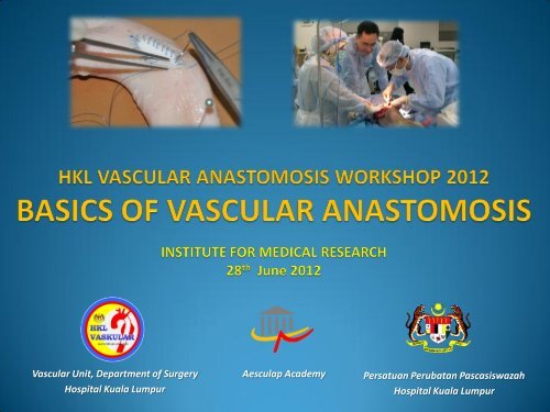
8th hkl vascular surgery workshop INSTITUTE OF MEDICAL ...
8th hkl vascular surgery workshop INSTITUTE OF MEDICAL ...
8th hkl vascular surgery workshop INSTITUTE OF MEDICAL ...

Create successful ePaper yourself
Turn your PDF publications into a flip-book with our unique Google optimized e-Paper software.
Vascular Unit, Department of Surgery<br />
Hospital Kuala Lumpur<br />
Aesculap Academy<br />
Persatuan Perubatan Pascasiswazah<br />
Hospital Kuala Lumpur
Introduction<br />
• Vascular reconstruction<br />
• No compromise<br />
• No second chance<br />
• No delays<br />
• No temporary measures<br />
• Immediate results –<br />
technical success
Objectives<br />
• Exposure to principles of <strong>vascular</strong> <strong>surgery</strong> & anastomotic<br />
techniques<br />
• Hands on experience on:<br />
• Handling of instruments<br />
• Handling of sutures, <strong>vascular</strong> grafts and blood vessels<br />
• Live dissection<br />
• Dissection of major blood vessels<br />
• Proximal and distal control<br />
• Perform live anastomosis<br />
• Assess technical success
Principles of <strong>vascular</strong> suturing<br />
• Appropriate sutures<br />
• Double ended, monofilament<br />
sutures<br />
• Appropriate size<br />
• Appropriate needle type, size,<br />
curve<br />
• Never handle suture-thread<br />
with instruments<br />
• Appropriate instruments<br />
• Enable better handling of<br />
sutures<br />
• Avoid damage to instruments
Principles of <strong>vascular</strong> suturing<br />
• In – out manner<br />
• On the arterial side<br />
• End-graft to side-artery anastomosis<br />
• Distal end<br />
• End-end anastomosis
Principles of <strong>vascular</strong> suturing<br />
• Hold needle at it’s mid body<br />
• Pierce the artery at 90 0 to vessel wall<br />
• Less force to pierce vessel – calcified wall
Principles of <strong>vascular</strong> suturing<br />
• Needle point of entry -<br />
perpendicular to vessel surface<br />
• Direction of pull of suture –<br />
perpendicular to vessel surface
Principles of <strong>vascular</strong> suturing<br />
• Minimise handling of the arterial intimal layer<br />
• Hold the arterial wall by the adventitia<br />
• Do not grip the whole thickness of the artery<br />
• Use of appropriate instruments – non traumatic forceps
Vessel Control<br />
• Never suture on pulsating vessels<br />
• Poor vision<br />
• Expansion of suture holes<br />
• Length of vessel dissected<br />
• Enough space for anastomosis<br />
• Adequate space for placement of clamps<br />
• Appropriate clamp according to vessels size
Vessel Control<br />
• Applying loops around the vessels
Vessel Control<br />
• Applying loops, followed by clamps
Clamping on calcified vessels<br />
• Ant-post clamping<br />
• Break in calcified plaque<br />
• Distal embolisation<br />
• Ineffective clamping/control<br />
• Side clamping
Terminology<br />
TOE<br />
HEEL
HKL Vascular Surgery Workshop<br />
28 June 2012, IMR<br />
Hands-on Practical Session : Grouping<br />
Dr Ngoo Kay Seong<br />
94 Hospital Angkatan Tentera<br />
Dr Patrick Chang Siong Junk<br />
Hospital Umum Sarawak<br />
Dr Stewart Santos Santos<br />
Chinese General Hospital & Medical Center<br />
Dr Tan Yee Ling<br />
Hospital Sultanah Aminah<br />
Dr Anthony Molina Manio<br />
Philippine Heart Center<br />
Dr Azhar bin Amir Hamzah<br />
Hospital Universiti Sains Malaysia<br />
Dr Jay Alan Enojas Junio<br />
Philippine Heart Center<br />
Dr Mohd Hisyam Sidek<br />
Hospital Kuala Terengganu<br />
Dr Prasopchai Kongsakphaisal<br />
Siriraj Hospital<br />
Dr Saiful Azli Zainuddin<br />
Hospital Kuala Terengganu<br />
Dr Nattawut Puangpunngam<br />
Siriraj Hospital<br />
Dr Ahmad Rafizi Hariz Ramly<br />
University Malaya Medical Centre
Workshop Layout<br />
• Bench-work<br />
10.00 am – 1-00 pm ( 3 hours )<br />
• Arteriotomy<br />
Transverse & Longitudinal<br />
• Arteriotomy closure<br />
Interrupted, Continuous & Patch<br />
• Basic anastomosis<br />
End to side & End to end<br />
Parachute & Tie down<br />
• Live Dissection<br />
2.00 pm – 5.00 pm ( 3 hours )<br />
• Dissection of vessels<br />
• Proximal and distal control<br />
• Anastomosis
Instruments<br />
• 1 Castroviejo micro needle holder<br />
• 1 DeBakey arterial clamp<br />
• 1 Potts 60 0 scissors<br />
• 1 DeBakey atraumatic dissecting tissue forceps<br />
• 1 Suture board<br />
• 1 Hegar-Mayo needle holder<br />
• 2 Halsted mosquito forceps (straight)<br />
• 2 Halsted mosquito forceps (curved)<br />
• 1 McIndoe dissecting forceps<br />
• 1 toothed tissue forceps<br />
• 1 Mayo scissors (straight)<br />
• 1 Mayo scissors (curved)<br />
• 1 Metzenbaum scissors<br />
• 1 Balfour self-retaining abdominal retractor<br />
• 2 scalpel handles (No.3 & 4)<br />
• Bovine carotids, grafts, <strong>vascular</strong> patch and sutures<br />
• Live “patients”
Castroviejo<br />
DeBakey clamps<br />
Pott’s scissors<br />
DeBakey atraumatic forceps
Arteriotomy<br />
• Stab incision with size 11<br />
scalpel blade<br />
• Extend incision with Pott’s<br />
scissors
Arteriotomy<br />
• Longitudinal<br />
• Most commonly practiced<br />
• Easily extended<br />
• Conversion into anastomosis<br />
• Stricture upon closure<br />
• Patch closure<br />
• Transverse<br />
• Minimise stricture<br />
• Limited application<br />
• Not suitable for bypass
Arteriotomy closure<br />
• Primary closure<br />
• Large, medium sized<br />
arteries (7 – 8 mm)<br />
• Interrupted<br />
• Continuous<br />
• Run suture towards you<br />
• Patch closure<br />
• Small sized arteries (
Patch closure<br />
• Tie down<br />
• Limited working space<br />
• Vein patch<br />
• Parachute<br />
• Ample working space<br />
• Start at corner away from you<br />
• Work towards yourself<br />
• Tie knot on the side
End - side anastomosis<br />
• Parachute<br />
• Start from the heel<br />
• 2 or 3 sutures on closer side<br />
• Then do side away from you<br />
• Don’t struggle doing it later<br />
• Inspect suture line from inside<br />
• Go around the toe<br />
• Tie knot on the side closer to<br />
you in the middle<br />
• Anchor (Tie-down)<br />
• Heel to toe<br />
• Tie knot on the side
End – side anastomosis<br />
• Completing anastomosis<br />
• Tie down on the side
End - end anastomosis<br />
• Straight<br />
• Large, good sized vessel<br />
• Anterior, then flip over<br />
• Interposition vein graft<br />
• Posterior, then anterior<br />
• Primary anastomosis<br />
• Out-in on proximal end<br />
• In-out on distal end
End to end anastomosis<br />
• Beveled<br />
• Small sized vessels<br />
• Toe – heel; heel – toe<br />
• Unequal diameters
Subject preparation<br />
• “Patient under”<br />
• Instrument dish<br />
• Diathermy return pad<br />
• Drape around proposed<br />
laporotomy site
Laparotomy<br />
• Midline incision<br />
• Rectus<br />
• Peritoneum<br />
• Abdominal self-retainers
Trouble shooting<br />
• Distended bladder
Dissection of the aorta
Dissection of the aorta<br />
IVC<br />
Aorta<br />
• Loop the aorta in preparation<br />
for clamping
Anatomy of canine aorta
Aortic cross-clamp<br />
• Systemic heparin – intra-arterial<br />
• 1,000 unit<br />
• Before clamping<br />
• Proximal and distal<br />
• Horizontal to occlude lumbar<br />
vessels<br />
• Arteriotomy – size 11 blade
Arteriotomy<br />
• Extend arteriotomy with Pott’s scisssors
End-side anastomosis<br />
• Placing sutures<br />
• Parachute the graft
Completion of anastomosis<br />
• Release clamps<br />
• Check anastomosis<br />
• Suture hole bleeding<br />
• Pulsating graft<br />
• Pulsating vessel (aorta)<br />
distal to anastomosis<br />
• Remember: No bleeding –<br />
• Sound anastomosis<br />
• Thrombosed graft
BENCHWORK EXERCISE<br />
• Arteriotomy – Simple closure, interrupted<br />
• Arteriotomy – Patch closure, continuous<br />
• End-Side – Parachute<br />
• End-Side – Tie down<br />
• End-End – Beveled, continuous<br />
LIVE DISECTION<br />
• Aortic dissection<br />
• Proximal & Distal clamps<br />
• End graft-side aorta – proximal anastomosis<br />
• End graft-side aorta – distal anastomosis
www.<strong>hkl</strong><strong>vascular</strong>.org