Neurological Examination, clinical cases and neuropsychological ...
Neurological Examination, clinical cases and neuropsychological ...
Neurological Examination, clinical cases and neuropsychological ...

Create successful ePaper yourself
Turn your PDF publications into a flip-book with our unique Google optimized e-Paper software.
23/07/54<br />
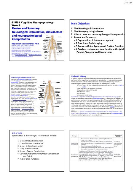
415703 Cognitive Neuropsychology<br />
Week 8:<br />
Review <strong>and</strong> Summary:<br />
<strong>Neurological</strong> <strong>Examination</strong>, <strong>clinical</strong> <strong>cases</strong><br />
<strong>and</strong> <strong>neuropsychological</strong><br />
interpretation<br />
etat Naiphinich Kotchabhakdi, Ph.D.<br />
Director, Salaya Stem Cell R & D Project,<br />
Research Center for Neuroscience,<br />
Institute of Molecular Biosciences,<br />
Mahidol University Salaya Campus,<br />
999 Phutthamonthol 4 Road, Salaya, Phutthamonthol,<br />
Nakornpathom 73170 Thail<strong>and</strong><br />
Email: scnkc@mahidol.ac.th or naiphinich@gmail.com<br />
Web: www.neuroscience.mahidol.ac.th<br />
Main Objectives:<br />
1. The <strong>Neurological</strong> <strong>Examination</strong><br />
2. The Neuropsychological tests<br />
3. Clinical <strong>cases</strong> <strong>and</strong> <strong>neuropsychological</strong> interpretation<br />
4. Review <strong>and</strong> Summary<br />
4.1 Organization of the nervous system<br />
4.2 Functional Brain Imaging<br />
4.3 Sensory–Motor Systems <strong>and</strong> Cortical Functions<br />
4.4 Cerebral cortexes <strong>and</strong> lobe functions: Occipital,<br />
Parietal, Temporal <strong>and</strong> Frontal lobes<br />
A neurological examination is the<br />
assessment of sensory neuron <strong>and</strong> motor responses,<br />
especially reflexes, to determine whether the<br />
nervous system is impaired. It can be used both as a<br />
screening tool <strong>and</strong> as an investigative tool, the<br />
former of which when examining the patient when<br />
there is no expected neurological deficit <strong>and</strong> the<br />
latter of which when examining a patient where you<br />
do expect to find abnormalities. If a problem is found<br />
either in an investigative or screening process then<br />
further tests can be carried out to focus on a<br />
particular aspect of the nervous system (such as<br />
lumbar punctures <strong>and</strong> blood tests).<br />
Generally a neurological examination is focused<br />
towards finding out if there are lesions in the central<br />
<strong>and</strong> peripheral nervous systems or whether there is<br />
another diffuse process which is troubling the<br />
patient. Once the patient has been thoroughly<br />
tested, it is then the role of the physician to<br />
determine whether or not these findings combine to<br />
form a recognizable medical syndrome such as<br />
Parkinson's disease or motor neurone disease.<br />
Finally, it is the role of the physician to find the<br />
etiological reasons for why such a problem has<br />
occurred, for example finding if the problem was due<br />
to inflammation or congenital.<br />
Patient’s History<br />
A patient's history is the most important part of a neurological examination <strong>and</strong> must be<br />
performed before any other procedures unless impossible (i.e. the patient is unconscious).<br />
Certain aspects of a patients history will become more important depending upon the<br />
complaint issued. Important factors to be taken in the medical history include:<br />
1. Time of onset, duration <strong>and</strong> associated symptoms (e.g. is the complaint chronic or<br />
acute)<br />
2. Age, gender <strong>and</strong> occupation of the patient<br />
3. H<strong>and</strong>edness (right or left h<strong>and</strong>ed)<br />
4. Past medical history<br />
5. Drug history<br />
6. Family <strong>and</strong> social history<br />
H<strong>and</strong>edness is important in establishing the area of the brain important for language (as<br />
almost all right‐h<strong>and</strong>ed people have a left hemisphere which is responsible for language). As<br />
patients answer questions, it is important to gain an idea of the complaint thoroughly <strong>and</strong><br />
underst<strong>and</strong> its time course. Underst<strong>and</strong>ing the patient's neurological state at the time of<br />
questioning is important, <strong>and</strong> an idea should be obtained of how competent the patient is<br />
with various tasks <strong>and</strong> their level of impairment in carrying out these tasks. The interval of a<br />
complaint is important as it can help aid the diagnosis. For example, vascular disorders occur<br />
very frequently over minutes <strong>and</strong> hours, whereas congenital disorders occur over a matter of<br />
years.<br />
Carrying out a 'general' examination is just as important as the neurological exam as it may<br />
lead to clues to the etiology of the complaint. This is shown by <strong>cases</strong> of cerebral metastases<br />
where the initial complaint was of a mass in the breast.<br />
List of tests<br />
Specific tests in a neurological examination include:<br />
1. Mental Status <strong>Examination</strong><br />
2. Cranial Nerves <strong>Examination</strong><br />
3. Motor System <strong>Examination</strong><br />
4. Deep tendon Reflexes<br />
5. Sensory System <strong>Examination</strong><br />
6. Cerebellum <strong>Examination</strong> (Motor Coordination<br />
<strong>and</strong> Gaits)<br />
7. Higher Brain Functions<br />
1
23/07/54<br />
Interpretation<br />
The results of the examination are taken together to anatomically identify the lesion. This may<br />
be diffuse (e.g. neuromuscular diseases, encephalopathy) or highly specific (e.g. abnormal<br />
sensation in one dermatome due to compression of a specific spinal nerve by a tumor deposit).<br />
A differential diagnosis may then be constructed that takes into account the patient's<br />
background (e.g. previous cancer, autoimmune diathesis) <strong>and</strong> present findings to include the<br />
most likely causes. <strong>Examination</strong>s are aimed at ruling out the most <strong>clinical</strong>ly significant causes<br />
(even if relatively rare, e.g. brain tumor in a patient with subtle word finding abnormalities but<br />
no increased intracranial pressure) <strong>and</strong> ruling in the most likely causes<br />
Romberg's test or the Romberg maneuver is a<br />
test used by doctors in a neurological examination, <strong>and</strong> also as a test<br />
for drunken driving. The exam is based on the premise that a person<br />
requires at least two of the three following senses to maintain<br />
balanced while st<strong>and</strong>ing:<br />
Proprioception (the ability to know one's body in space); Vestibular<br />
function (the ability to know one's head position in space); <strong>and</strong><br />
Vision (which can be used to monitor [<strong>and</strong> adjust for] changes in<br />
body position).<br />
A patient who has a problem with proprioception can still maintain<br />
balance by using vestibular function <strong>and</strong> vision. In the Romberg test,<br />
the patient is stood up <strong>and</strong> asked to close his eyes. A loss of balance<br />
is interpreted as a positive Romberg sign.<br />
The Romberg test is a test of the body's sense of positioning<br />
(proprioception), which requires healthy functioning of the dorsal<br />
columns of the spinal cord, [1] .<br />
The Romberg test is used to investigate the cause of loss of motor<br />
coordination (ataxia). A positive Romberg test suggests that the<br />
ataxia is sensory in nature, that is, depending on loss of<br />
proprioception. If a patient is ataxic <strong>and</strong> Romberg's test is not<br />
positive, it suggests that ataxia is cerebellar in nature, that is,<br />
depending on localized cerebellar dysfunction instead.<br />
It is used as an indicator for possible alcohol or drug impaired<br />
driving <strong>and</strong> neurological decompression sickness. [2][3] When used to<br />
test impaired driving, the test is performed with the subject<br />
estimating 30 seconds in his head. This is used to gauge the subject's<br />
internal clock <strong>and</strong> can be an indicator of stimulant or depressant<br />
use. The test was named after the German neurologist Moritz<br />
Heinrich Romberg [1] (1795‐1873), who also gave his name to Parry‐<br />
Romberg syndrome <strong>and</strong> Howship‐Romberg sign.<br />
Procedure for Romberg's test or the Romberg maneuver<br />
Ask the subject to st<strong>and</strong> erect with feet together <strong>and</strong> eyes closed. St<strong>and</strong> close by as a<br />
precaution in order to stop the person from falling over <strong>and</strong> hurting himself or herself.<br />
Watch the movement of the body in relation to a perpendicular object behind the<br />
subject (corner of the room, door, window etc). A positive sign is noted when a swaying,<br />
sometimes irregular swaying <strong>and</strong> even toppling over occurs. The essential feature is that<br />
the patient becomes more unsteady with eyes closed.<br />
The essential features of the test are as follows:<br />
1. the subject st<strong>and</strong>s with feet together, eyes open <strong>and</strong> h<strong>and</strong>s by the sides.<br />
2. the subject closes the eyes while the examiner observes for a full minute.<br />
Because the examiner is trying to elicit whether the patient falls when the eyes are<br />
closed, it is advisable to st<strong>and</strong> ready to catch the falling patient. For large subjects, a<br />
strong assistant is recommended.<br />
Romberg's test is positive if the patient sways or falls while the patient's eyes are closed.<br />
Patients with a positive result are said to demonstrate Romberg's sign or Rombergism.<br />
They can also be described as Romberg's positive. The basis of this test is that balance<br />
comes from the combination of several neurological systems, namely proprioception,<br />
vestibular input, <strong>and</strong> vision. If any two of these systems are working the person should be<br />
able to demonstrate a fair degree of balance. The key to the test is that vision is taken<br />
away by asking the patient to close their eyes. This leaves only two of the three systems<br />
remaining <strong>and</strong> if there is a vestibular disorder (labyrinthine) or a sensory disorder<br />
(proprioceptive dysfunction) the patient will become much more imbalanced.<br />
2
23/07/54<br />
Maintaining balance while st<strong>and</strong>ing in the stationary position relies on intact<br />
sensory pathways, sensorimotor integration centers <strong>and</strong> motor pathways.<br />
The main sensory inputs are:<br />
1. Joint position sense (proprioception), carried in the dorsal columns<br />
of the spinal cord;<br />
2. Vision<br />
3. Vestibular apparatus<br />
Crucially, the brain can obtain sufficient information to maintain balance if any<br />
two of the three systems are intact.<br />
Sensorimotor integration is carried out by the cerebellum <strong>and</strong> by the dorsal<br />
column‐medial lemniscus tract. The motor pathway is the corticospinal<br />
(pyramidal) tract <strong>and</strong> the medial <strong>and</strong> lateral vestibular tracts.<br />
The first stage of the test (st<strong>and</strong>ing with the eyes open), demonstrates that at<br />
least two of the three sensory pathways is intact, <strong>and</strong> that sensorimotor<br />
integration <strong>and</strong> the motor pathway are functioning.<br />
In the second stage, the visual pathway is removed by closing the eyes, known as<br />
a "sharpened Romberg". If the proprioceptive <strong>and</strong> vestibular pathways are intact,<br />
balance will be maintained. But if proprioception is defective, two of the sensory<br />
inputs will be absent <strong>and</strong> the patient will sway then fall.<br />
The sharpened Romberg does have an early learning effect that will plateau<br />
between the third <strong>and</strong> fourth attempts.<br />
Positive Romberg<br />
Romberg's test is positive in conditions causing sensory ataxia such as:<br />
Conditions affecting the dorsal columns of the spinal cord, such as tabes<br />
dorsalis (neurosyphilis), in which it was first described.<br />
Conditions affecting the sensory nerves (sensory peripheral neuropathies), such<br />
as chronic inflammatory demyelinating polyradiculoneuropathy (CIDP).<br />
Friedreich's Ataxia<br />
Romberg <strong>and</strong> cerebellar function<br />
Romberg's test is not a test of cerebellar function, as it is commonly<br />
misconstrued. Patients with cerebellar ataxia will, generally, be unable to<br />
balance even with the eyes open; [5] therefore, the test cannot proceed beyond<br />
the first step <strong>and</strong> no patient with cerebellar ataxia can correctly be described as<br />
Romberg's positive. Rather, Romberg's test is a test of the proprioception<br />
receptors <strong>and</strong> pathways function. A positive Romberg's test has been shown to<br />
be 90% sensitive for lumbar spinal stenosis. [<br />
<strong>Neurological</strong> <strong>Examination</strong> Videos<br />
And <strong>Neurological</strong> case examples<br />
http://library.med.utah.edu/neurologicexam/<strong>cases</strong>/html_case01/case01_history.html<br />
A case begins with a Case<br />
History in which<br />
preliminary information<br />
about the patient <strong>and</strong> any<br />
signs <strong>and</strong> symptoms are<br />
presented.<br />
The <strong>Neurological</strong><br />
<strong>Examination</strong> follows the<br />
Case History. You can choose<br />
the order <strong>and</strong> the parts of<br />
the neurological<br />
examination that you would<br />
like to view by clicking on<br />
the icon that represents that<br />
part of the exam.<br />
After completing the exam, you advance to listing your<br />
abnormal findings. You use the supplied Checklist of<br />
Findings <strong>and</strong> compare your choices with that of an<br />
expert's. You are now ready to begin the process of<br />
anatomical localization.<br />
You start to Localize the Level(s) of the Lesion by<br />
selecting from the level of the neuroaxis. Your<br />
choices include:<br />
1. Supratentorial<br />
2. Infratentorial<br />
3. Spinal Cord<br />
4. Peripheral Nerve System<br />
5. Multiple Levels<br />
From a list of the structures, you choose the brain<br />
structure(s) those you think are damaged for the case.<br />
Your choices are compared to an expert's, <strong>and</strong> the<br />
lesion is highlighted on the image.<br />
You have now arrived at an anatomical diagnosis, the<br />
first essential step in making a neurological diagnosis.<br />
Finally, in the Case Discussion, you review the case <strong>and</strong><br />
the thought processes used to reach the diagnosis.<br />
Neuroimaging studies, if available, are shown as part of<br />
the case discussion.<br />
3
23/07/54<br />
Case No. 01: The Upset Office Manager<br />
The patient is a 48‐year‐old woman who was in her usual state of<br />
good health when she experienced nausea <strong>and</strong> vomiting after being<br />
emotionally upset. After 3 hours of nausea <strong>and</strong> vomiting she had the<br />
sudden onset of numbness of her left arm which progressed to<br />
include her left leg <strong>and</strong> the left side of her face.<br />
She was taken to the Emergency Room. Upon arrival she complained<br />
that she had double vision especially when she looked to the right.<br />
When she covered her right eye, the most peripheral image (the<br />
ghost image) would disappear. She also noticed that when she looked<br />
in the mirror the right side of her face didn’t move.<br />
Over the next 2 months, the double vision resolved, but the rest of<br />
her complaints have persisted<br />
http://library.med.utah.edu/neurologicexam/<strong>cases</strong>/html_case01/case01_history.html<br />
4
23/07/54<br />
5
23/07/54<br />
This 48‐year‐old woman had the sudden onset <strong>and</strong> rapid progression of her symptoms,<br />
which is the temporal profile of an acute vascular event or a stroke. Risk factors for stroke<br />
include hypertension, cardiac disease, diabetes mellitus, smoking as well as coagulation <strong>and</strong><br />
autoimmune disorders. This patient had none of these risk factors.<br />
One of the first steps in localizing the occluded vessel in a stroke is to decide if the vessel is<br />
in the anterior or posterior arterial circulation of the brain. Most strokes occur in the<br />
anterior or carotid circulation because most of the brain’s blood supply is supplied by the<br />
carotids. Although strokes in the posterior or vertebrobasilar circulation are less common,<br />
this patient’s cranial nerve findings suggest an infarct in the brainstem <strong>and</strong> not the carotid<br />
territory.<br />
By history <strong>and</strong> examination there are 3 findings that indicate possible cranial nerve<br />
involvement. Her history of diplopia indicates a right 6th (abducens) cranial nerve deficit<br />
(remember the most peripheral image is the false image <strong>and</strong> covering the right eye<br />
eliminated this image) <strong>and</strong> by examination she has a right 7th (facial) cranial nerve deficit. To<br />
explain these two deficits one has to localize the lesion in the caudal pons in the area of the<br />
6th <strong>and</strong> 7th cranial nerve nuclei or the pathway of the nerves at this level.<br />
To explain the sensory findings on the left side of her face, we could postulate either a left<br />
spinal trigeminal tract lesion or it could be from a lesion affecting the axons of the 2nd order<br />
neurons which have crossed over to the right side of the pons <strong>and</strong> are ascending in the<br />
ventral trigeminothalamic tract. The left spinal trigeminal tract lesion hypothesis is<br />
unattractive, because it would mean a second lesion. In the pons, the ascending ventral<br />
trigeminothalamic tract is near the facial nucleus, so a lesion affecting this tract is the likely<br />
explanation.<br />
The last finding that we have to account for anatomically is the sensory deficit on<br />
the left side of the body. We first have to decide if the deficit is from a lesion in the<br />
DC‐ML system or the spinothalamic (ALS) system. For this patient, pain <strong>and</strong><br />
temperature are affected while vibratory, position sense, <strong>and</strong> discriminatory<br />
sensation are preserved, which indicates that the spinothalamic tract is involved<br />
but the DC‐ML system is spared. Recall that the axons from the second order<br />
neurons that form the spinothalamic tract cross immediately in the spinal cord <strong>and</strong><br />
ascend in the anterolateral spinal cord <strong>and</strong> the lateral part of the brainstem. In the<br />
pons, the spinothalamic tract, carrying pain <strong>and</strong> temperature for the left side of the<br />
body, is adjacent to the facial motor nucleus.<br />
So we could explain all the <strong>clinical</strong> findings for this case by a lesion in the<br />
mediolateral part of the right lower pons most likely caused by occlusion of one of<br />
the short circumferential branches of the basilar artery. It is not a paramedian<br />
lesion because the patient has no findings referable to the corticospinal tracts <strong>and</strong><br />
the medial lemniscus. It is also not a far lateral lesion because there are no rightsided<br />
spinal trigeminal tract, vestibular, or cerebellar findings.<br />
An MRI scan of the patient done 6 months after her stroke shows a small residual<br />
lesion in the area of the right abducens nucleus. This imaging finding doesn’t cover<br />
the entire anatomical area where we know there has to be disease, but it does<br />
support the hypothesis that there has been a small stroke in the area where we<br />
localized her lesion based on her <strong>clinical</strong> findings<br />
Review <strong>and</strong> Summary:<br />
6
23/07/54<br />
415703 Cognitive Neuropsychology<br />
Week 1:<br />
Introduction to Neuropsychology,<br />
<strong>and</strong> the Organization of the<br />
Nervous System.<br />
Naiphinich Kotchabhakdi, Ph.D.<br />
Director, Salaya Stem Cell R & D Project,<br />
Research Center for Neuroscience,<br />
Institute of Molecular Biosciences,<br />
Mahidol University Salaya Campus,<br />
999 Phutthamonthol 4 Road, Salaya, Phutthamonthol,<br />
Nakornpathom 73170 Thail<strong>and</strong><br />
Email: scnkc@mahidol.ac.th or naiphinich@gmail.com<br />
Web: www.neuroscience.mahidol.ac.th<br />
Main Objectives:<br />
1. What is Neuropsychology (for education)?<br />
2. What are <strong>neuropsychological</strong> disorders?<br />
3. New Approaches <strong>and</strong> tools in <strong>neuropsychological</strong><br />
disorders.<br />
4. What are <strong>neuropsychological</strong> py<br />
assessment?<br />
5. What is the Organization of the nervous system?<br />
6. What are the structure <strong>and</strong> functions of specific<br />
human brain areas?<br />
Neuropsychology (Brain <strong>and</strong> Mind)<br />
Neuropsychology studies the structure <strong>and</strong> function of the<br />
brain related to specific psychological (mental) processes<br />
<strong>and</strong> behaviors.<br />
The term neuropsychology has been applied to lesion studies in humans <strong>and</strong><br />
animals. It has also been applied to efforts to record electrical activity from<br />
individual cells (or groups of cells) in higher primates (including some studies<br />
of human patients). It is scientific in its approach <strong>and</strong> shares an information<br />
processing view of the mind with cognitive psychology <strong>and</strong> cognitive<br />
neuroscience.<br />
In practice neuropsychologists tend to work in <strong>clinical</strong> settings (involved<br />
in assessing or treating patients with <strong>neuropsychological</strong> problems –<br />
see <strong>clinical</strong> neuropsychology), forensic settings or industry (often as<br />
consultants where <strong>neuropsychological</strong> knowledge is applied to product design<br />
or in the management of pharmaceutical <strong>clinical</strong>-trials research for drugs that<br />
might have a potential impact on CNS functioning).<br />
Posner, M.I.& DiGirolamo,G.J.(2000<br />
2000) Cognitive Neuroscience:Origins<br />
<strong>and</strong> Promise,Psychological<br />
Psychological Bulletin, 126:6, 873‐889<br />
889<br />
From Wikipedia, the free encyclopedia<br />
Divisions of the Nervous System<br />
Cellular components of the nervous tissue<br />
7
23/07/54<br />
Spinal cord<br />
The spinal cord is a long, thin, tubular bundle of nervous tissue <strong>and</strong> support cells that<br />
extends from the brain (the medulla oblongata specifically). The brain <strong>and</strong> spinal cord<br />
together make up the central nervous system. The spinal cord begins at the Occipital bone<br />
<strong>and</strong> extends down to the space between the first <strong>and</strong> second lumbar vertebrae; itdoes<br />
not extend the entire length of the vertebral column.Itisaround45cm(18in)inmen<strong>and</strong><br />
around 43 cm (17 in) long in women. Also, the spinal cord has a varying width, ranging<br />
from 1/2 inch thick in the cervical <strong>and</strong> lumbar regions to 1/4 inch thick in the thoracic<br />
area. The enclosing bony vertebral column protects the relatively shorter spinal cord.<br />
The spinal cord functions primarily in the transmission of neural signals between the brain<br />
<strong>and</strong> the rest of the body but also contains ti neural circuits it thatt can independently d control<br />
numerous reflexes <strong>and</strong> central pattern generators. The spinal cord has three major<br />
functions:1.Serve<br />
as a conduit for motor information, which travels down the spinal cord.<br />
2.Serve<br />
as a conduit for sensory information, which travels up the spinal cord. 3. Serve as<br />
acenter<br />
for coordinating certain reflexes.<br />
The spinal cord is the main pathway for information connecting the brain <strong>and</strong> peripheral<br />
nervous system. The length of the spinal cord is much shorter than the length of the bony<br />
spinal column. The human spinal cord extends from the medulla oblongata <strong>and</strong> continues<br />
through the conus medullaris near the first lumbar vertebra, terminating in a fibrous<br />
extension known as the filum terminale<br />
Spinal cord<br />
Somatosensory system<br />
Dermatome<br />
8
23/07/54<br />
Autonomic nervous<br />
nervous system)<br />
system<br />
(ANS<br />
or<br />
visceral<br />
system) is the part of the peripheral nervous system that acts<br />
as a control system functioning largely below the level of consciousness, <strong>and</strong><br />
controls visceral functions. The ANS affects heart rate, digestion, respiration rate,<br />
salivation, perspiration, diameter of the pupils, micturition (urination), <strong>and</strong> sexual<br />
arousal. Whereas most of its actions are involuntary, some, such as breathing,<br />
work in t<strong>and</strong>em with the conscious mind.<br />
It is classically divided into two subsystems: the parasympathetic nervous system<br />
(PSNS) <strong>and</strong> sympathetic nervous system (SNS). Relatively recently, a third<br />
subsystem of neurons that have been named 'non‐adrenergic<br />
<strong>and</strong> non‐cholinergic'<br />
neurons (because they use nitric oxide as a neurotransmitter) have been<br />
described <strong>and</strong> found to be integral in autonomic function, particularly in the gut<br />
<strong>and</strong> the lungs.<br />
With regard to function, the ANS is usually divided into sensory (afferent) <strong>and</strong><br />
motor (efferent) subsystems. Within these systems, however, there are inhibitory<br />
<strong>and</strong> excitatory synapses between neurons.<br />
The enteric nervous system is sometimes considered part of the autonomic<br />
nervous system, <strong>and</strong> sometimes considered an independent system.<br />
Autonomic Nervous<br />
System (ANS) <strong>and</strong> autonomic reflexes<br />
Alongside the other two components of the autonomic nervous system, thesympathetic<br />
nervous system aids in the control of most of the body's internal organs. Stress—as in the<br />
flight‐or‐fight response—is thought to counteract the parasympathetic system, which<br />
generally works to promote maintenance of the body at rest. In truth, the functions of<br />
both the parasympathetic <strong>and</strong> sympathetic nervous systems are not so straightforward,<br />
but this is a useful rule of thumb.<br />
There are two kinds of neurons involved in the transmission of any signal through the<br />
sympathetic system; pre‐ <strong>and</strong> post‐ ganglionic. The shorter preganglionic neurons originate<br />
from the thoracolumbar region of the spinal cord (levels T1 ‐ L2, specifically) <strong>and</strong> travel to<br />
a ganglion, often one of the paravertebral ganglia, where they synapse with a<br />
postganglionic neuron. From there, the long postganglionic neurons extend across most of<br />
the body.<br />
At the synapses within the ganglia, preganglionic neurons release acetylcholine, a<br />
neurotransmitter that activates nicotinic acetylcholine receptors on postganglionic<br />
neurons. In response to this stimulus postganglionic neurons ‐ with two important<br />
exceptions ‐ release norepinephrine, which activates adrenergic receptors on the<br />
peripheral target tissues. The activation of target tissue receptors causes the effects<br />
associated with the sympathetic system.<br />
The two exceptions mentioned above are postganglionic neurons innervating sweat<br />
gl<strong>and</strong>s—which release acetylcholine for the activation of muscarinic receptors ‐ <strong>and</strong> the<br />
adrenal medulla. The adrenal medulla develops in t<strong>and</strong>em with the sympathetic nervous<br />
system, <strong>and</strong> acts as a modified sympathetic ganglion: synapses occur between pre‐ <strong>and</strong><br />
post‐ ganglionic neurons within it, but the post ganglionic neurons do not leave the<br />
medulla; instead they directly release norepinephrine <strong>and</strong> epinephrine into the blood.<br />
Sympathetic <strong>and</strong> parasympathetic systems<br />
Sympathetic nervous system<br />
Parasympathetic nervous system<br />
9
23/07/54<br />
Control of blood<br />
vessels<br />
Control of<br />
pupil<br />
Brainstem (or brain stem) is the posterior part of the brain,<br />
adjoining <strong>and</strong> structurally continuous with the spinal cord. The brain stem<br />
providesthemainmotor<strong>and</strong>sensoryinnervationtotheface<strong>and</strong>neckviathe<br />
cranial nerves. Though small, this is an extremely important part of the brain as<br />
the nerve connections of the motor <strong>and</strong> sensory systems from the main part of<br />
the brain to the rest of the body pass through the brain stem. This includes the<br />
corticospinal tract (motor), the posterior column‐medial lemniscus pathway (fine<br />
touch, vibration sensation <strong>and</strong> proprioception)<strong>and</strong>thespinothalamic tract (pain,<br />
temperature, itch <strong>and</strong> crude touch). The brain stem also plays an important role<br />
in the regulation of cardiac <strong>and</strong> respiratory function. It also regulates the central<br />
nervoussystem,<strong>and</strong>ispivotalinmaintaining consciousness <strong>and</strong> regulating the<br />
sleep cycle.<br />
Brainstem is made up of 1. medulla oblongata (myelencephalon), 2. pons (part of<br />
metencephalon), 3. midbrain (mesencephalon), <strong>and</strong> 4. diencephalon<br />
Mid‐sagittal<br />
view of the<br />
adult human brain<br />
There are three main functions of the brain stem:<br />
1. The first is its role in conduction. That is, all information relayed from the body<br />
to the cerebrum <strong>and</strong> cerebellum <strong>and</strong> vice versa, must traverse the brain stem.<br />
The ascending pathways coming from the body to the brain are the sensory<br />
pathways, <strong>and</strong> include the spinothalamic tract for pain <strong>and</strong> temperature<br />
sensation <strong>and</strong> the dorsal column, fasciculus gracilis, <strong>and</strong> cuneatus for touch,<br />
proprioception, <strong>and</strong> pressure sensation (both of the body). (The facial sensations<br />
have simiar pathways, <strong>and</strong> will travel in the spinothalamic tract <strong>and</strong> the medial<br />
lemniscus also). Descending tracts are upper motor neurons destined to synapse<br />
on lower motor neurons in the ventral horn <strong>and</strong> intermediate horn of the spinal<br />
cord. In addition, there are upper motor neurons that originate in the brain<br />
stem's vestibular, red, tectal, <strong>and</strong> reticular nuclei, which also descend <strong>and</strong> synapse<br />
in the spinal cord.<br />
2. The cranial nerves 3‐12 emerge from the brain stem.<br />
3. The brain stem has integrative functions (it is involved in cardiovascular system<br />
control, respiratory control, pain sensitivity control, alertness, awareness, <strong>and</strong><br />
consciousness). Thus, brain stem damage is a very serious <strong>and</strong> often lifethreatening<br />
problem.<br />
Brainstem <strong>and</strong> cerebellum<br />
Ventral<br />
view<br />
10
23/07/54<br />
12 Cranial nerves:<br />
1. Olfactory<br />
2. Optic<br />
3. Oculomotor<br />
4. Trochlear<br />
5. Trigeminal<br />
6. Abducens<br />
7. Facial<br />
8. Auditory <strong>and</strong><br />
Vestibular<br />
9. Glossopharyngeal<br />
10. Vagus<br />
11. Spinal Accessory<br />
12. Hypoglossal<br />
Control of respiration<br />
Cardiovascular controls<br />
Long tracts of Sensory <strong>and</strong> Motor<br />
Systems<br />
1. Pathways in spinal cord <strong>and</strong> brainstem<br />
2. Functions<br />
3. Clinical correlates, signs <strong>and</strong> symptoms<br />
11
23/07/54<br />
Cerebellum<br />
Vestibular<br />
<strong>and</strong><br />
Cerebellum<br />
The cerebellum (Latin for little brain) isaregionofthe<br />
brain that plays an important role in motor control.Itisalsoinvolved<br />
in some cognitive functions such as attention <strong>and</strong> language, <strong>and</strong><br />
probably in some emotional functions such as regulating fear <strong>and</strong><br />
pleasure responses. Its movement‐related functions are the most<br />
clearly understood, however. The cerebellum does not initiate<br />
movement, but it contributes to coordination, precision, <strong>and</strong><br />
accurate timing. It receives input from sensory systems <strong>and</strong> from<br />
other parts of the brain <strong>and</strong> spinal cord, <strong>and</strong> integrates these inputs<br />
to fine tune motor activity. Because of this fine‐tuning function,<br />
damage to the cerebellum does not cause paralysis, but instead<br />
produces disorders in fine movement, equilibrium, posture, <strong>and</strong><br />
motor learning<br />
In addition to its direct role in motor control <strong>and</strong> coordination, the<br />
cerebellum also is necessary for several types of motor learning, the<br />
most notable one being learning to adjust<br />
to changes<br />
in<br />
sensorimotor relationships.<br />
The Human Cerebellum:<br />
Cerebellar Functions (Classical Functions):<br />
Co‐ordination of Movements<br />
Stabilizing of Vestibulo‐ocular Reflex (VOR)<br />
Patterned Skilled Movements<br />
Coordination of Eye Movements<br />
Vermis Lobule VI‐‐‐‐Saccadic Eye Movements<br />
Flocculus‐‐‐‐ Visual Tracking<br />
Equilibrium, Gait <strong>and</strong> Postural Controls<br />
Controls of Autonomic Functions (Cardiovascular)<br />
Immune Functions (?)<br />
12
23/07/54<br />
The Human Cerebellum:<br />
Clinical Correlates on the Human Cerebellum:<br />
Flocculo‐Nodular Lobe:<br />
Disturbed Equilibrium or Balance<br />
Tendency to fall on the side of the lesion<br />
Extensor hypotonia<br />
“Reeling or Drunken” ”Gi Gait or Posture<br />
Deviation Nystagmus<br />
1. Eye in mid‐line Position:<br />
fine nystagmus, quick phase toward lesion side<br />
2. Eye fixed 10 –30 degree away from the lesion side:<br />
No nystagmus<br />
3. Eye fixed beyond 30 degree away from the lesion side<br />
nystagmus with quick phase away from lesion side<br />
4. Eye shift beyond midline toward the lesion side<br />
gross nystagnus, quick phase toward lesion side<br />
The Human Cerebellum:<br />
Clinical Correlates on the Human Cerebellum:<br />
Anterior Lobe & Vermis:<br />
Hypotonia (Reduced muscle tone)<br />
Hyporeflexia<br />
Ataxia (Incoordination of Movements)<br />
Asynergia, Dysynergia (lack of synergy)<br />
Intention or Action Tremor (Atelokinesia)<br />
Asthenia (Weakness of muscular strength)<br />
Rebounded Phenomenon<br />
Cerebellar Hemisphere & Dentate Nucleus:<br />
Dysmetria (Pass pointing)<br />
Dysdiadochokinesia or Adiadochokinesia<br />
(can not perform rapid alternating movements)<br />
Disturbed Voluntary Skilled movements<br />
Disturbed Speech (Drunken Speech)<br />
13
23/07/54<br />
Dysmetria of thought<br />
The Principles of Motor Controls of Movements:<br />
1. The central nervous system (CNS) has to choose the right group<br />
of muscles by selecting specific pathways.<br />
2. The CNS must give the right amount of excitatory or inhibitory<br />
inputs (“Comm<strong>and</strong>”) to specific motoneuron pools<br />
3. The excitatory <strong>and</strong> inhibitory comm<strong>and</strong>s must be regulated<br />
“Spatially” <strong>and</strong> “Temporally”.<br />
4. The CNS must regulate the following parameters:<br />
‐ force<br />
‐displacement (distance)<br />
‐ velocity, acceleration or deceleration<br />
Pyramidal System:<br />
Cortico‐spinal tracts<br />
UMN Lesions (Pyramidal Syndrome)<br />
A. Paralyze movements in hemiplegic,<br />
quadriplegic, or paraplegic distribution, not<br />
individual muscles<br />
B. Atrophy of disuse only (late <strong>and</strong> slight)<br />
C. Hyperactive MSRs Clonus<br />
D. Clasp‐knife spasticity<br />
E. Absent abdominal <strong>and</strong> cremasteric reflexes<br />
F. Extensor toe sign (Babinski sign)<br />
14
23/07/54<br />
Plantar Flexion<br />
Dorsi‐ Flexion<br />
LMN Lesions<br />
A. Paralyze individual muscles or sets of muscles<br />
in root or peripheral nerve distribution<br />
B. Atrophy of denervation (early <strong>and</strong> severe<br />
C. Fasciculations <strong>and</strong> fibrillations<br />
D. Hypoactive or absent MSRs Hypotonia<br />
UMN = upper motoneuron; LMN = lower<br />
motoneuron; MSRs = muscle stretch reflexes<br />
Basal ganglion <strong>and</strong><br />
Extrapyramidal<br />
System<br />
Disease of the basal ganglion:<br />
Parkinson’s disease<br />
Chorea <strong>and</strong> Huntington’s Chorea<br />
Athetosis <strong>and</strong> Athetoid<br />
Sydenham’s Chorea<br />
Hemibalism .. Lesion of subthalamic nucleus<br />
Dystonia, Torticolis,<br />
Wilson’s disease (Copper)<br />
Kern icterus (Bilirubin stain)<br />
Parkinson’s disease:<br />
Akinesia or Hypokiesia<br />
cog wheel rigidity<br />
Resting Tremor<br />
Degeneration of Dopaminergic neurons in<br />
Pars Compacta of the Substantia Nigra<br />
Motor<br />
control<br />
system<br />
Motor<br />
homonculus<br />
(Maps)<br />
15
23/07/54<br />
Control of<br />
body<br />
movements,<br />
visceral<br />
organs,<br />
behavior<br />
<strong>and</strong><br />
emotion<br />
Hypothalamus<br />
The Hypothalamus is a portion of the brain that contains a number of small<br />
nuclei with a variety of functions. One of the most important functions of the<br />
hypothalamus is to link the nervous system to the endocrine system via the<br />
pituitary gl<strong>and</strong> (hypophysis).<br />
The hypothalamus is located below the thalamus, just above the brain stem. In<br />
the terminology of neuroanatomy, itformstheventral part of the diencephalon.<br />
All vertebrate brains contain a hypothalamus. In humans, it is roughly the size of<br />
an almond.<br />
The hypothalamus is responsible for certain metabolic processes <strong>and</strong> other<br />
activities of the autonomic nervous system. It synthesizes <strong>and</strong> secretes certain<br />
neurohormones, often called hypothalamic‐releasing hormones, <strong>and</strong> these in<br />
turn stimulate or inhibit the secretion of pituitary hormones. The hypothalamus<br />
controls body temperature, hunger, thirst,fatigue,sleep,<strong>and</strong>circadian cycles.<br />
Controls of body<br />
temperature<br />
Controls of food<br />
intake <strong>and</strong> body<br />
weight<br />
Endocrine <strong>and</strong><br />
Hormonal controls<br />
Controls of<br />
kidney functions<br />
16
23/07/54<br />
Limbic<br />
System<br />
Controls of<br />
emotion <strong>and</strong><br />
motivation<br />
The thalamus is a midline paired symmetrical<br />
structure within the brains of vertebrates, including<br />
humans. It is situated between the cerebral cortex <strong>and</strong><br />
midbrain, both in terms of location <strong>and</strong> neurological<br />
connections. Its function includes relaying sensation,<br />
spatial sense, <strong>and</strong> motor signals to the cerebral cortex,<br />
along with the regulation of consciousness, sleep, <strong>and</strong><br />
alertness. The thalamus surrounds the third ventricle. It is<br />
the main product of the embryonic diencephalon<br />
Thalamus<br />
<strong>and</strong><br />
Cerebral<br />
cortex<br />
The thalamus has multiple functions. It is generally believed to act as a relay between a<br />
variety of subcortical areas <strong>and</strong> the cerebral cortex. In particular, every sensory system (with the<br />
exception of the olfactory system) includes a thalamic nucleus that receives sensory signals <strong>and</strong> sends<br />
them to the associated primary cortical area. For the visual system, for example, inputs from the retina<br />
are sent to the lateral geniculate nucleus ofthethalamus,whichinturnprojectstotheprimary visual<br />
cortex (area V1) in the occipital lobe. The thalamus is believed to both process sensory information as<br />
well as relaying it—each of the primary sensory relay areas receives strong "back projections" from<br />
the cerebral cortex. Similarly the medial geniculate nucleus acts as a key auditory relay between the<br />
inferior colliculus of the midbrain <strong>and</strong> the primary auditory cortex,<strong>and</strong>theventral posterior nucleus is<br />
a key somatosensory relay, which sends touch <strong>and</strong> proprioceptive information to the primary<br />
somatosensory cortex.<br />
The thalamus also plays an important role in regulating states of sleep <strong>and</strong> wakefulness. [4] Thalamic<br />
nuclei have strong reciprocal connections with the cerebral cortex, forming thalamo‐cortico‐thalamic<br />
thalamic<br />
circuits that are believed to be involved with consciousness. The thalamus plays a major role in<br />
regulating arousal, the level of awareness, <strong>and</strong> activity. Damage to the thalamus can lead to<br />
permanent coma.<br />
Many different functions are linked to various regions of the thalamus. This is the case for many of the<br />
sensory systems (except for the olfactory system), such as the auditory, somatic, visceral, gustatory<br />
<strong>and</strong> visual systems where localized lesions provoke specific sensory deficits. A major role of the<br />
thalamus is devoted to "motor" systems. This has been <strong>and</strong> continues to be a subject of interest for<br />
investigators. VIm, the relay of cerebellar afferences, is the target of stereotactians particularly for the<br />
improvement of tremor. The role of the thalamus in the more anterior pallidal <strong>and</strong> nigral territories in<br />
the basal ganglia system disturbances is recognized but still poorly understood. The contribution of the<br />
thalamus to vestibular or to tectal functions is almost ignored. The thalamus has been thought of as a<br />
"relay" that simply forwards signals to the cerebral cortex. Newer research suggests that thalamic<br />
function is more selective<br />
Cerebral cortex in different areas<br />
The cerebral cortex is a sheet of neural tissue that is outermost to the<br />
cerebrum of the mammalian brain. It plays a key role in memory, attention,<br />
perceptual awareness, thought, language, <strong>and</strong>consciousness. It is constituted of<br />
up to six horizontal layers, each of which has a different composition in terms of<br />
neurons <strong>and</strong> connectivity. The human cerebral cortex is 2–4 mm (0.08–<br />
0.16 inches) thick.<br />
In preserved brains, it has a gray color, hence the name "gray matter". In contrast<br />
to gray matter that is formed from neurons <strong>and</strong> their unmyelinated fibers, the<br />
white matter below them is formed predominantly by myelinated axons<br />
interconnecting neurons in different regions of the cerebral cortex with each<br />
other <strong>and</strong> neurons in other parts of the central nervous system.<br />
The surface of the cerebral cortex is folded in large mammals, such that more<br />
than two‐thirds of it in the human brain is buried in the grooves, called "sulci".<br />
The phylogenetically most recent part of the cerebral cortex, the neocortex (also<br />
called isocortex), is differentiated into six horizontal layers; themoreancientpart<br />
of the cerebral cortex, the hippocampus (also called archicortex), has at most<br />
three cellular layers, <strong>and</strong> is divided into subfields. Neurons in various layers<br />
connect vertically to form small microcircuits, called columns. Different<br />
neocortical architectonic fields are distinguished upon variations in the thickness<br />
of these layers, their predominant cell type <strong>and</strong> other factors such as<br />
neurochemical markers.<br />
17
23/07/54<br />
Functional brain mapping<br />
แผนที<br />
่การทํางานของสมอง (Brain Mapping<br />
Brain Mapping)<br />
Broadmann’ s Areas<br />
Broadmann’s area #<br />
Brodmann’s area is a region of the cerebral cortex defined based on<br />
its cytoarchitectonics, or organization of cells<br />
Brodmann areas were originally defined <strong>and</strong> numbered by the German neurologist<br />
Korbinian Brodmann basedonthecytoarchitecture organisation of neurons he<br />
observed in the cerebral cortex using the Nissl stain. Brodmann published his maps<br />
of cortical areas in humans, monkeys, <strong>and</strong> other species in 1909, along with many<br />
other findings <strong>and</strong> observations regarding the general cell types <strong>and</strong> laminar<br />
organization of the mammalian cortex. (The same Brodmann area number in<br />
different species does not necessarily indicate homologous areas.)<br />
A more detailed <strong>and</strong> verifiable cortical map have since been published by Constantin von<br />
Economo <strong>and</strong> Georg N. Koskinas which greatly improves the quality of the cytoarchitectonic<br />
classifications.<br />
Many of the areas Brodmann defined based solely on their neuronal organization have since<br />
been correlated closely to diverse cortical functions. For example, Brodmann areas 1, 2 <strong>and</strong><br />
3aretheprimary somatosensory cortex; area4istheprimary motor cortex; area17isthe<br />
primary visual cortex; <strong>and</strong> areas 41 <strong>and</strong> 42 correspond closely to primary auditory cortex.<br />
Higher order functions of the association cortical areas are also consistently localized to the<br />
same Brodmann areas by neurophysiological, functional imaging, <strong>and</strong> other methods (e.g.,<br />
the consistent localization of Broca's speech <strong>and</strong> language area to the left Brodmann areas<br />
44 <strong>and</strong> 45). However, functional imaging can only identify the approximate localization of<br />
brain activations in terms of Brodmann areas since their actual boundaries in any individual<br />
brain requires its histological examination.<br />
18
่<br />
23/07/54<br />
Brodmann areas for human & non‐human primates<br />
Brodmann’s areas 3D<br />
map: Lateral Surface<br />
map: Medial Surface<br />
Brodmann areas for human & non‐human primates<br />
Paul MacLean<br />
M.D.<br />
Paul MacLean’s<br />
Triune Brain<br />
The Reptilian Brain : Core brainstem<br />
The Paleomammalian Brain : the limbic system<br />
The Neomammalian Brain : neocortex <strong>and</strong> neocerebellum<br />
สมองส่วนแรก คือ สมองของสัตว์เลื้อยคลาน (Reptilian Brain)<br />
เป็นสมองที่มนุษย์เราได้รับมรดกตกทอดมาจากสัตว์เลื้อยคลานยุคดึกดําบรรพ์ อยู ่ภายใต้<br />
อิทธิพลของพันธุกรรม 90 – 95 % และเจริญเติบโตในระหว่างที่อยู ่ในครรภ์มารดาเป็น<br />
ส่วนใหญ่ เมื่อเกิดมาแล้วสิ่งแวดล้อมมีอิทธิพลต่อสมองส่วนนี้น้อยมาก มันจะถูกปัจจัย<br />
ทางพันธุกรรมกําหนดมาเลยว่าเป็นสมองคน หรือสมองสัตว์และมีโครงสร้างและการ<br />
ทํางานอย่างไร สมองส่วนนี้ควบคุมการทํางานของอวัยวะต่างในร่างกายโดยอัตโนมัติ<br />
และพฤติกรรมที่เป็นสัญชาติญาณของสิ่งมีชีวิตที่มีมาโดยกําเนิดโดยการกําหนดของ<br />
พันธุกรรม ได้มรดกโดยตรงมาจากพ่อแม่ พ่อแม่เป็นอย่างไรลูกจะได้มรดกตกทอดมาเป็น<br />
อย่างนั้นเลย อยางนนเลย Reptilian Brain มีลักษณะเป็นแกนอย่ตอนในสดของสมองเป็นส่วนของ<br />
มลกษณะเปนแกนอยูตอนในสุดของสมองเปนสวนของ<br />
ก้านสมองและสมองตอนกลาง สมองส่วนที่หนึ่งนี้ เป็นสมองส่วนที่ทําให้มนุษย์มีสัญชาติ<br />
ญาณของการอยู ่รอด การกิน การขับถ่าย การสืบพันธ์ เริ่มสร้างขึ้นตั้งแต่ขณะที่ทารกอยู ่ใน<br />
ครรภ์มารดา ในวันที่คลอดนั้นสมองส่วนนี้สามารถทํางานได้ราว 99 % และเติบโตสมบูรณ์<br />
พร้อมทํางานเต็มที่ในช่วงขวบปีแรก ถ้าสมองส่วนแรกนี้ไม่สามารถทํางานได้ดีทารกก็ไม่<br />
อาจมีชีวิตอยู ่รอดได้ เพราะมันไปควบคุมการเต้นของหัวใจ การหายใจ ระบบขับถ่าย การ<br />
กินการอยู ่ การตื่น การนอนหลับทุกอย่างหมดเลย ในช่วงสองขวบปีแรก พ่อแม่ และผู ้เลี้ยง<br />
ดูเด็กจะสอนเด็กให้สามารถควบคุมร่างกาย ควบคุมการกินอยู ่ ควบคุมการขับถ่าย และ<br />
สร้างนิสัยต่างๆที่เหมาะสมกับการอยู ่รอดในสังคม<br />
สมองส่วนที ่สอง คือ สมองสัตว์เลี้ยงลูกด้วยนมยุคโบราณ<br />
(Paleomammalian Brain หรือ Limbic System) เป็นสมองส่วนที่<br />
มนุษย์เราได้รับมรดกตกทอดมาจากสัตว์เลี้ยงลูกด้วยนมยุคโบราณ สมองส่วนนี้จะเริ่ม<br />
สร้างและเจริญเติบโตเมื่อทารกอยู ่ในครรภ์มารดาได้ราว ๆ หกเดือน Limbic<br />
Systemจะมีลักษณะคล้ายวงแหวนที่หุ ้มรอบๆสมองส่วนแรกซึ่งมีลักษณะเป็นแกน<br />
เอาไว้ หน้าที่ของสมองส่วนนี้ก็คือ ทําให้ทารกเกิดความจําเกี่ยวกับเหตุการณ์และสถานที่<br />
(Episodic or Spatiotemporal Memory) โดยเฉพาะความจําที่เกี่ยวกับ<br />
ใบหน้าแม่ จํากลิ่นแม่ได้ ทําให้มนุษย์รู ้จักตัวเอง (“Self”) และพัฒนาให้มีความรู ้สึก<br />
(Feeling)และการแสดงออกทางอารมณ์ต่าง ๆ มันจะเป็นตัวที่ทําให้ทารกร้องไห้โยเย<br />
เรียกร้องความสนใจ แสดงอารมณ์ความรู ้สึกเวลา ดีใจ-เสียใจ ชอบ-ไม่ชอบ พอใจ-ไม่<br />
พอใจ สมองส่วนที่สองนี้ทําให้มนุษย์เราแตกต่างจากสัตว์เลื้อยคลาน เช่น จิ้งจก กิ้งก่า<br />
เต่า ซึ่งมีเพียงแค่สัญชาติญาณแต่ปราศจากความรู ้สึก และอารมณ์ อย่างไรก็ตามตอนที่<br />
ทารกคลอดออกมาสมองส่วนนี้ เพิ่งสร้างเสร็จไปเพียง 50 % เท่านั้น มันจะเจริญเติบโต<br />
ต่อไปโดยเฉพาะในช่วงสี่ขวบปีแรกของชีวิต<br />
19
้<br />
23/07/54<br />
สมองส่วนที<br />
่สองจะได้รับอิทธิพลจากพันธุกรรมประมาณ 50 % ส่วนอีก 50 % ที่<br />
เหลือนั้นพัฒนาตามสภาพแวดล้อม ประสบการณ์และการเรียนรู ้โดยเฉพาะช่วงตั้งแต่<br />
แรกเกิด ขวบปีแรกจนถึงปฐมวัย (0 – 8 ปี ) สมองส่วนนี้สําคัญมากตรงที่ เป็น<br />
ตัวกําหนด พื้นอารมณ์ (Temperament) ควบคุมการแสดงออกของอารมณ์ให้<br />
เหมาะกับเหตุการณ์ และสถานการณ์ ซึ่งเป็นรากฐานของบุคลิกภาพของปัจเจกคน<br />
(Individual Personality)ที่ทําให้เราทุกคนแตกต่างกัน การที่เด็กจะเติบโตเป็นคน<br />
ที่ฉลาดทางอารมณ์ ทฉลาดทางอารมณ (Emotional Intelligence) มีมนษยสัมพันธ์ดีหรือไม่ขึ้นอย่<br />
มมนุษยสมพนธดหรอไมขนอยู<br />
กับการเลี้ยงดูในช่วงปฐมวัย และการพัฒนาของสมองส่วนนี้ เป็นสําคัญ<br />
สมองส่วนที ่สาม คือ สมองของสัตว์เลี้ยงลูกด้วยนมยุคใหม่ และเปลือกหุ ้ม<br />
สมองใหม่ (Neo‐Mammalianหรือ Neo‐Cortex Brain) คือ สมองที่พบได้<br />
เฉพาะในสัตว์ชั้นสูงที่มีเปลือกหุ ้มสมองใหญ่เท่านั้น เช่น มนุษย์ ปลาโลมาและสัตว์<br />
ประเภทวานร ลิง (Primates)เป็นต้น สมองส่วนที่สามนี้จะมีลักษณะคล้ายเปลือกหุ ้ม<br />
สมอง หุ ้มสมองส่วนที่หนึ่งและส่วนที่สองเอาไว้ ตอนที่ทารกคลอดออกมาใหม่ ๆ สมอง<br />
ส่วนนี้ยังไม่พัฒนามากเลย มันจะเริ่มก่อร่างสร้างตัวและเจริญเติบโตอย่างรวดเร็วมาก<br />
ในช่วงสามปีแรกของชีวิต จนกระทั่งเมื่อเด็กอายุได้หกขวบจึงเจริญเติบโตราว 80 %<br />
ตอนเกาขวบจะเตบโตราว ้ ิ 90 % และจะเจรญเตบโตเรอยตอไปกระทงอายุ ิ ิ ื่ ่ ั่ 25 ปี สมอง<br />
ส่วนที่สามจะได้รับอิทธิพลจากพันธุกรรมน้อยมาก แทบจะเรียกได้ว่าพันธุกรรมควบคุม<br />
มัน 10-20 % เท่านั้น เพราะมันมาเจริญเติบโตหลังคลอด พัฒนาการของสมองส่วนนี้<br />
จึงได้รับอิทธิพลมาจากสิ ่งแวดล้อมเป็ นส่วนใหญ่ และต้องการการกระตุ ้นจาก<br />
สิ ่งแวดล้อมให้สามารถพัฒนาได้เต็มที ่ตามศักยภาพที ่มีมากับตัวของเด็ก<br />
สมองส่วนที ่สาม มีความยืดหยุ ่นค่อนข้างมาก มีบทบาทเปรียบได้กับหน้าต่าง<br />
ของโอกาส (Windows of opportunities)ที่จะส่งเสริมให้เด็กฉลาดโดยการ<br />
กระตุ ้นการรับรู ้ และกิจกรรมต่างๆจากประสบการณ์การเรียนรู ้ต่างๆ การได้รับอาหารที่<br />
มีครบทุกหมู ่อาหารในปริมาณที่เหมาะสม และคุณภาพที่ดีจําเป็นมากต่อการ<br />
เจริญเติบโตของสมองส่วนนี้ การสัมผัสและการกระตุ ้นประสาทสัมผัสต่างๆอย่าง<br />
เหมาะสมเป็นความจําเป็นอย่างยิ่งที่จะทําให้สมองส่วนนี้พัฒนาก้าวหน้า และสามารถ<br />
เรียนร้ประสบการณ์ต่างๆ เรยนรูประสบการณตางๆ ททาใหอยางเตมท ที่ทําให้อย่างเต็มที่ เพราะฉะนน เพราะฉะนั้น เรองการเลยงดูเดกในชวง<br />
เรื่องการเลี้ยงดเด็กในช่วง<br />
สามขวบปีแรกจึงเป็นเรื่องสําคัญมาก เพราะในช่วงนี้สมองส่วนนี้จะเจริญเติบโตจากที่<br />
ไม่มีอะไรมากเลย คือ ประมาณ 25% ของผู ้ใหญ่ตอนแรกเกิด จนกระทั่งเติบโตได้ถึง 80<br />
% ตอนอายุ 3 ขวบปีแรก สมองส่วนนี้ทําให้เด็กสามารถเรียนรู ้ สร้างโลกทัศน์ของการ<br />
รับรู ้ และความเข้าใจเกี่ยวกับจักรวาลรอบตัว มีทักษะต่างๆในการเคลื่อนไหว เรียนรู<br />
ภาษาที่ใช้ในการสื่อสาร ทั้งภาษาพูด ภาษาเขียน การคํานวณ การคิดหาเหตุผล<br />
คณิตศาสตร์ และตรรกวิทยา (Logic thinking) รวมทั้งการเรียนรู ้วิชาการต่างๆ และ<br />
จินตนาการทางศิลปะ<br />
ในสมองส่วนที ่สําคัญที ่สุด ในด้านการพัฒนาสมอง คือ สมอง<br />
ส่วนหน้า (Frontal lobe) ที่อยู ่ด้านหลังหน้าผากของมนุษย์ หรือ สมองส่วนปรี<br />
ฟรอนตัล (Prefrontal Cortex) เป็นสมองส่วนที่อยู ่ในสมองส่วนที่สาม สาเหตุที่ทํา<br />
ให้สมองส่วนนี้มีความสําคัญมาก เพราะมันมีหน้าที่ความสําคัญเปรียบได้กับเป็น “นาย<br />
ของสมอง” (Chief Executive Officer หรือCEO ของสมองทั้งหมด) เพราะเป็น<br />
สมองส่วนที่เกิดทีหลังสุด ในช่วงสองขวบปีแรกเพิ่งเริ่มสร้างเท่านั้นเอง ทําหน้าที่เชื่อมโยง<br />
กับสมองทีทีสร้างก่อนมาทังหมด ี่ ี่ ่ ั้ สมองส่วนนีจะได้รับเส้นประสาทมาจากสมองส่วนต่างๆ<br />
ี้ ้ ั ้ ่ ่<br />
เมื่อเจริญเติบโตเต็มที่ในช่วงที่ย่างเข้าสู ่วัยรุ ่น จะเป็นส่วนที่ควบคุมร่างกายและจิตใจ<br />
ทั้งหมด ทําให้เราเหมือนมีจิตใจเป็นหนึ่งเดียว มีเจ้านายคนเดียวสั่งงาน สังเกตดูจะเห็นว่า<br />
ช่วงวัยเด็กเล็ก เด็ก ๆ จะวิ่งเล่นตามประสา สะเปะสะปะไปตามสิ่งเร้า สิ่งกระตุ ้น เหมือน<br />
ไม่มีการควบคุมการสั่งงาน แต่พอเราโตขึ้นชีวิตเริ่มมีการวางแผน สมองส่วนนี้นี่เองที่จะ<br />
คอยควบคุมกําหนดให้มนุษย์มีการวางแผนงานล่วงหน้า มีความรับผิดชอบ มีสมาธิ<br />
ปรีฟรอนตัล<br />
Prefrontal<br />
ภาพสมองคนแสดง สมองสามระบบ (Triune brain) สมองระบบแรก Reptilian brain ควบคุมสมดุลของการมีชีวิต<br />
และการอยู ่รอด (Homeostasis <strong>and</strong> survival) อยู ่ในบริเวณก้านของสมอง และสมองส่วนลึกที่อยู ่ใจกลางภายใน<br />
ของสมอง ระบบที่สอง ส่วนของสมองลิมบิค (Limbic brain structures) หุ ้มห่อสมองระบบแรกที่อยู ่ภายใน ซึ่งทํา<br />
หน้าที่เกี่ยวกับพัฒนาการของอารมณ์ ความสัมพันธ์และสังคมกับคนอื่นๆ และกับจิตใจกับความประพฤติของตัวเราเอง ระบบ<br />
ที่สาม นีโอคอร์เท็กซ์ (Neocortex) เป็นส่วนเปลือกที่หุ ้มห่อภายนอกของสมองใหญ่ ทั้ง Cerebrum <strong>and</strong><br />
cerebellum ควบคุมการรับรู ้ การเรียนรู ้ และทักษะความชํานาญ และความเฉลียวฉลาด รวมทั้งบริเวณ ปรีฟรอนตัล<br />
(Prefrontal) ที่เป็น นายหรือ CEO ของสมอง<br />
20
23/07/54<br />
ทีมงานวิจัยของมหาวิทยาลัยไอโอวานําโดยประสาทแพทย์ชื่อ ดร.อันโตนิ<br />
โอ ดามาสซิโอ (Dr. Antonio Damassio) และภรรยา ดร.ฮันนา ดามาสซิโอ (Dr.<br />
Hanna Damassio) ได้ทําการวิจัยติดตามเด็กเล็กที่เมื่ออายุประมาณขวบหรือขวบ<br />
ครึ่งเคยได้รับบาดเจ็บจากอุบัติเหตุ เช่น หกล้มไปข้างหน้า แล้วศีรษะส่วนหน้าผาก<br />
ฟาดพื้น ทําให้สมองบริเวณนั้นเกิดอาการชํ้า ทีมงานวิจัยติดตามเด็กกลุ ่มนี้ไป<br />
จนกระทั่งวัยรุ่นแล้วพบว่า เด็กกลุ ่มนี้จะมีอาการทางประสาท ที่จิตแพทย์เรียกว่า<br />
สมองส่วนหน้าพิการ (Frontal lobe syndrome) คือ เด็กที่สมองส่วนหน้าทํางาน<br />
ไม่สมบูรณ์ ทําให้ประสบปัญหาเรื่องการเรียน และพฤติกรรมแม้ว่าบางคนจะมีไอ<br />
คิว (IQ) สูงก็ตาม เนืองจากมีสมาธิสัน ื่ ิ ้ (Attention Deficit หรือ AD)) ไม่สามารถ<br />
ควบคุมตัวเองให้สงบนิ่ง ที่จะทําอะไรนิ่งๆ อยู ่กับที่นาน ๆ ได้พอ ไม่มีการวางแผนที่<br />
ดี ขาดความรับผิดชอบ และมีปัญหาในการเรียน และการเข้าสมาคมกับคนอื่นๆ<br />
เด็กวัยรุ่นที่มาจากครอบครัวที่ดีแต่ตัวเด็กกลับมีพฤติกรรมไม่เหมาะสม และเป็น<br />
อันธพาลชอบต่อต้านกฎระเบียบต่างๆ ต่อต้านสังคม และบางครั้งชอบใช้ความ<br />
ก้าวร้าวและพฤติกรรมรุนแรง นั้น เมื่อศึกษาลึกลงไป จะพบว่ามีสาเหตุเกี่ยวกับ<br />
ความพิการของสมองส่วนนี้เข้ามาเกี่ยวข้องได้เสมอ ดังนั้น จึงควรดูแลป้ องกัน<br />
ระมัดระวังไม่ให้ศีรษะส่วนนี้ของเด็กทารกได้รับบาดเจ็บ<br />
Prefrontal lobe syndrome<br />
• Personality changes<br />
• Deficits in strategic planning<br />
• Perseveration<br />
• Release of primitive reflexes<br />
• Abulia = general slowing of the intellectual<br />
faculties i.e. apathetic, slow speech etc.<br />
Corpus<br />
callosum<br />
415703 Cognitive Neuropsychology<br />
Week 2:<br />
How neurons communicate, effects of<br />
drugs on the brain, <strong>and</strong> functional brain<br />
imaging<br />
Naiphinich Kotchabhakdi, Ph.D.<br />
Director, Salaya Stem Cell R & D Project,<br />
Research Center for Neuroscience,<br />
Institute of Molecular Biosciences,<br />
Mahidol University Salaya Campus,<br />
999 Phutthamonthol 4 Road, Salaya, Phutthamonthol,<br />
Nakornpathom 73170 Thail<strong>and</strong><br />
Email: scnkc@mahidol.ac.th or naiphinich@gmail.com<br />
Web: www.neuroscience.mahidol.ac.th<br />
Main Objectives:<br />
1. How neurons communicate?<br />
2. Concepts of chemical neurotransmissions <strong>and</strong> various<br />
neurotransmitters <strong>and</strong> neuromodulators?<br />
3. Effects of various chemicals <strong>and</strong> drugs on the brain<br />
(Nervous System).<br />
4. Concepts of functional localization in the brain <strong>and</strong><br />
brain mapping.<br />
5. Brain Imaging <strong>and</strong> Functional Brain Imaging.<br />
21
23/07/54<br />
22
23/07/54<br />
Axon terminal<br />
Synaptic cleft<br />
Excitation:<br />
EPSP<br />
Inhibition:<br />
IPSP<br />
สารสื<br />
่อประสาท (Neurotransmitters ) ที<br />
่สําคัญในสมอง<br />
Biogenic amines Amino acids Neuropeptides<br />
‐Acetyl Choline ‐Glutamic acid ‐Enkephalins<br />
‐Norepinephrine ‐Aspartic acid ‐Endorphins<br />
‐Dopamine ‐Glycine ‐Dynorphins<br />
‐Serotonin ‐GABA ‐Substance P<br />
‐Histamine Polyamines ‐VIP<br />
‐Epinephrine ‐Taurine ‐Somatostatin<br />
‐CCK<br />
Diagram of cholinergic nerve terminal<br />
with prototype drugs <strong>and</strong> chemicals<br />
23
23/07/54<br />
Neurotransmitters in Somatic <strong>and</strong><br />
Autonomic nervous system<br />
Cholinergic pathways in human brain<br />
Ach: Acetyl choline<br />
NE: Norepinephrine<br />
E: Epinephrine<br />
Glutamate<br />
GABA: Gamma amino<br />
butyric acid<br />
Dopamine<br />
VTA:<br />
Ventral<br />
tegment<br />
al area<br />
Dopamine pathway in<br />
the human brain<br />
24
23/07/54<br />
Central Noradrenergic<br />
nerve terminal<br />
Locus coeruleus<br />
Cocaine <strong>and</strong><br />
other local<br />
anesthetics<br />
25
23/07/54<br />
ลักษณะของยาและสารเสพติด<br />
‐Psychotomimetics, <strong>and</strong> some are Psychedelics<br />
‐Euphoria, Ecstasy etc.<br />
‐Reward <strong>and</strong> Reinforcement<br />
‐Tolerance<br />
‐Withdrawal syndromes <strong>and</strong> Dysphoria yp<br />
‐Physical, psychological <strong>and</strong><br />
behavioural dependence<br />
‐Craving<br />
‐Compulsive drug seeking behaviour<br />
‐Relapse<br />
Brain Reward System<br />
Serotonin<br />
5‐hydroxy tryptamine<br />
Raphe nucleus<br />
26
23/07/54<br />
415703 Cognitive Neuropsychology<br />
Week 3:<br />
Sensory ‐motor <strong>and</strong> cortical<br />
organization<br />
Naiphinich Kotchabhakdi, Ph.D.<br />
Director, Salaya Stem Cell R & D Project,<br />
Research Center for Neuroscience,<br />
Institute of Molecular Biosciences,<br />
Mahidol University Salaya Campus,<br />
999 Phutthamonthol 4 Road, Salaya, Phutthamonthol,<br />
Nakornpathom 73170 Thail<strong>and</strong><br />
Email: scnkc@mahidol.ac.th or naiphinich@gmail.com<br />
Web: www.neuroscience.mahidol.ac.th<br />
Main Objectives:<br />
1. Sensory ‐ motor <strong>and</strong> cortical organization?<br />
2. The sensory systems?<br />
3. The Reticular formation, Sensory‐Motor Integration<br />
for states of Consciousness, Waking, Sleep <strong>and</strong><br />
Dream.<br />
4. The Motor System, Movements <strong>and</strong> Motor Controls<br />
5. The Cerebral Cortex, <strong>and</strong> Cortical Columnar<br />
Organization, Concepts of functional localization <strong>and</strong><br />
representation in the brain <strong>and</strong> brain mapping.<br />
6. Brain Imaging <strong>and</strong> Functional Brain Imaging.<br />
sensory system is a part of the nervous system<br />
responsible for processing sensory information.<br />
A sensory system consists of sensory receptors, neural pathways,<br />
<strong>and</strong> parts of the brain involved in sensory perception. Commonly<br />
recognized sensory systems are those for vision, hearing, somatic<br />
sensation (touch), taste <strong>and</strong> olfaction (smell). In short, senses are<br />
transducers from the physical world to the realm of the mind.<br />
The receptive field is the specific part of the world to which a<br />
receptor organ <strong>and</strong> receptor cells respond. For instance, the part<br />
of the world an eye can see, is its receptive field; the light that<br />
each rod or cone can see, is its receptive field. Receptive fields<br />
have been identified for the visual system, auditory system <strong>and</strong><br />
somatosensory system, so far.<br />
Somatosensory system<br />
Dermatome<br />
Reticular<br />
Formation<br />
Ascending<br />
Reticular<br />
Activating<br />
System (ARAS)<br />
Regulation of<br />
States of<br />
Consciousness,<br />
e.g. waking, Sleep,<br />
Dream;<br />
attention;<br />
sensory‐motor<br />
integration<br />
Reticular formation is a part of the brain that is involved in actions such as<br />
awaking/sleeping cycle, <strong>and</strong> filtering incoming stimuli to discriminate irrelevant background stimuli. It is<br />
essential for governing some of the basic functions of higher organisms, <strong>and</strong> is one of the<br />
phylogenetically oldest portions of the brain. The reticular formation consists of more than 100 small<br />
neural networks, with varied functions including the following:<br />
1. Somatic motor control ‐ Some motor neurons send their axons to the reticular formation nuclei,<br />
giving rise to the reticulospinal tracts of the spinal cord. These tracts function in maintaining tone,<br />
balance, <strong>and</strong> posture‐‐especially during body movements. The reticular formation also relays eye <strong>and</strong><br />
ear signals to the cerebellum so that the cerebellum can integrate visual, auditory, <strong>and</strong> vestibular stimuli<br />
in motor coordination. Other motor nuclei include gaze centers, which enable the eyes to track <strong>and</strong><br />
fixate objects, <strong>and</strong> central pattern generators, which produce rhythmic signals to the muscles of<br />
breathing <strong>and</strong> swallowing.<br />
2. Cardiovascular control ‐ The reticular formation includes the cardiac <strong>and</strong> vasomotor centers of the<br />
medulla oblongata.<br />
3. Pain modulation ‐ The reticular formation is one means by which pain signals from the lower body<br />
reach the cerebral cortex. It is also the origin of the descending analgesic pathways. Thenervefibersin<br />
these pathways act in the spinal cord to block the transmission of some pain signals to the brain.<br />
4. Sleep <strong>and</strong> consciousness ‐ The reticular formation has projections to the thalamus <strong>and</strong> cerebral<br />
cortex that allow it to exert some control over which sensory signals reach the cerebrum <strong>and</strong> come to<br />
our conscious attention. It plays a central role in states of consciousness like alertness <strong>and</strong> sleep. Injury<br />
to the reticular formation can result in irreversible coma.<br />
5. Habituation ‐ This is a process in which the brain learns to ignore repetitive, meaningless stimuli while<br />
remaining sensitive to others. A good example of this is when a person can sleep through loud traffic in<br />
a large city, but is awakened promptly due to the sound of an alarm or crying baby. Reticular formation<br />
nuclei that modulate activity of the cerebral cortex are called the reticular activating system or<br />
extrathalamic control modulatory system.<br />
27
23/07/54<br />
The Human Reticular Formation:<br />
Reticular Functions:<br />
‐ Integration of Sensory <strong>and</strong> motor functions<br />
‐ Control of states of consciousness<br />
Waking, Sleep, Dream, Altered States<br />
‐ Control of behavioural states<br />
‐ Arousal, Attention, Orienting, Habituation<br />
‐ Sensory filtering<br />
(“The Cocktail Party Effect”)<br />
‐ Motor functions:<br />
Postural Controls<br />
Eye movements: Gaze, Saccade, REM<br />
‐ Autonomic controls:<br />
respiration, heart rates, blood pressure<br />
ARAS:<br />
Ascending<br />
Reticular<br />
Activating<br />
System<br />
415703 Cognitive Neuropsychology<br />
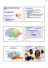
Week 4:<br />
The occipital lobes<br />
Naiphinich Kotchabhakdi, Ph.D.<br />
Director, Salaya Stem Cell R & D Project,<br />
Research Center for Neuroscience,<br />
Institute of Molecular Biosciences,<br />
Mahidol University Salaya Campus,<br />
999 Phutthamonthol 4 Road, Salaya, Phutthamonthol,<br />
Nakornpathom 73170 Thail<strong>and</strong><br />
Email: scnkc@mahidol.ac.th or naiphinich@gmail.com<br />
Web: www.neuroscience.mahidol.ac.th<br />
Main Objectives:<br />
1. The occipital lobes <strong>and</strong> their functions<br />
2. The Visual System<br />
3. Visual Perception <strong>and</strong> Visual Cortical<br />
Organization<br />
4. The two Stream Hypothesis: The Dorsal stream,<br />
“Where or How” <strong>and</strong> The Ventral Stream,<br />
“What”<br />
5. Neuropsychology of the Occipital lobes.<br />
28
23/07/54<br />
Occipital lobe<br />
Vision processing<br />
The occipital lobe is the visual processing center of the<br />
mammalian brain containing most of the anatomical region of the<br />
visual cortex.<br />
The primary visual cortex is Brodmann area 17, commonly called V1<br />
(visual one). Human V1 is located on the medial side of the occipital<br />
lobe within the calcarine sulcus; the full extent of V1 often<br />
continues onto the posterior pole of the occipital lobe. V1 is often<br />
also called striate cortex because it can be identified by a large<br />
stripe ti of myelin, the Striaof Gennari. Visually driven di regions<br />
outside V1 are called extrastriate cortex. There are many<br />
extrastriate regions, <strong>and</strong> these are specialized for different visual<br />
tasks, such as visuospatial processing, color discrimination <strong>and</strong><br />
motion perception. The name derives from the overlying occipital<br />
bone, which is named from the Latin oc‐ + caput, "back of the<br />
head".<br />
Functions of the Occipital lobes:<br />
Significant functional aspects of the occipital lobe is that it contains the primary<br />
visual cortex <strong>and</strong> is the part of the brain where dreams come from.<br />
Retinal sensors convey stimuli through the optic tracts to the lateral geniculate<br />
bodies, where optic radiations continue to the visual cortex. Each visual cortex<br />
receives raw sensory information from the outside half of the retina on the same<br />
side of the head <strong>and</strong> from the inside half of the retina on the other side of the<br />
head. The cuneus (Brodmann's area 17) receives visual information from the<br />
contralateral superior retina representing the inferior visual field. The lingula<br />
receives information from the contralateral inferior retina representing the<br />
superior visual field. The retinal inputs pass through a "way station" in the lateral<br />
geniculate nucleus of the thalamus before projecting to the cortex. Cells on the<br />
posterior aspect of the occipital lobes' gray matter are arranged as a spatial map<br />
of the retinal field. Functional neuroimaging reveals similar patterns of response<br />
in cortical tissue of the lobes when the retinal fields are exposed to a strong<br />
pattern.<br />
If one occipital lobe is damaged, the result can be homonomous vision loss from<br />
similarly positioned "field cuts" in each eye. Occipital lesions can cause visual<br />
hallucinations. Lesions in the parietal‐temporal‐occipital association area are<br />
associated with color agnosia, movement agnosia, <strong>and</strong> agraphia.<br />
The occipital lobe is divided into several functional visual<br />
areas. Each visual area contains a full map of the visual<br />
world. Although there are no anatomical markers<br />
distinguishing these areas (except for the prominent<br />
striations in the striate cortex), physiologists have used<br />
electrode recordings to divide the cortex into different<br />
functional regions.<br />
The first functional area is the primary visual cortex. It<br />
contains a low‐level description of the local orientation,<br />
spatial‐frequency <strong>and</strong> color properties within small<br />
receptive fields. Primary visual cortex projects to the<br />
occipital areas of the ventral stream (visual area V2 <strong>and</strong><br />
visual area V4), <strong>and</strong> the occipital areas of the dorsal<br />
stream—visual area V3, visual area MT (V5), <strong>and</strong> the<br />
dorsomedial area (DM).<br />
29
23/07/54<br />
The visual system is the part of the central nervous<br />
system which enables organisms to process visual detail, as well as<br />
enabling several non‐image forming photoresponse functions. It<br />
interprets information from visible light to build a representation<br />
of the surrounding world. The visual system accomplishes a<br />
number of complex tasks, including the reception of light <strong>and</strong> the<br />
formation of monocular representations; the construction of a<br />
binocular perception from a pair of two dimensional projections;<br />
the identification <strong>and</strong> categorization of visual objects; assessing<br />
distances to <strong>and</strong> between objects; <strong>and</strong> guiding body movements in<br />
relation to visual objects. The psychological manifestation of visual<br />
information is known as visual perception, a lack of which is called<br />
blindness. Non‐image forming visual functions, independent of<br />
visual perception, include the pupillary light reflex (PLR) <strong>and</strong><br />
circadian photoentrainment.<br />
The visual system includes<br />
the eyes, the connecting<br />
pathways through to the<br />
visual cortex <strong>and</strong> other parts<br />
of the brain. The illustration<br />
shows the mammalian<br />
system.<br />
Controls of eye<br />
movements<br />
Oculomotor (CN3)<br />
Trochlear (CN4)<br />
Abducens (CN6)<br />
PPRF<br />
PRF<br />
Frontal eye field<br />
(Area #8)<br />
NEUROPSYCHIATRY 177<br />
30
23/07/54<br />
Primary visual cortex (V1)<br />
The primary visual cortex is the best studied visual area in the brain. In all<br />
mammals studied, it is located in the posterior pole of the occipital cortex (the<br />
occipital cortex is responsible for processing visual stimuli). It is the simplest,<br />
earliest cortical visual area. It is highly specialized for processing information<br />
about static <strong>and</strong> moving objects <strong>and</strong> is excellent in pattern recognition.<br />
The functionally defined primary visual cortex is approximately equivalent to the<br />
anatomically defined striate cortex. The name "striate cortex" is derived from the<br />
stria of Gennari, a distinctive stripe visible to the naked eye that represents<br />
myelinated aonsfrom axons the lateral geniculate body terminating in layer 4 of the<br />
gray matter.<br />
The primary visual cortex is divided into six functionally distinct layers, labeled 1<br />
through 6. Layer 4, which receives most visual input from the lateral geniculate<br />
nucleus (LGN), is further divided into 4 layers, labelled 4A, 4B, 4Cα, <strong>and</strong> 4Cβ.<br />
Sublamina 4Cα receives most magnocellular input from the LGN, while layer 4Cβ<br />
receives input from parvocellular pathways.<br />
The average number of neurons in the adult human primary visual cortex, in each<br />
hemisphere, has been estimated at around 140 million (Leuba & Kraftsik,<br />
Anatomy <strong>and</strong> Embryology, 1994).<br />
V1 has a very well‐defined map of the spatial information in vision. For example, in humans<br />
the upper bank of the calcarine sulcus responds strongly to the lower half of visual field (below the center), <strong>and</strong> the lower bank of the<br />
calcarine to the upper half of visual field. Conceptually, this retinotopic mapping is a transformation of the visual image from retina to<br />
V1. The correspondence between a given location in V1 <strong>and</strong> in the subjective visual field is very precise: even the blind spots are<br />
mapped into V1. Evolutionarily, this correspondence is very basic <strong>and</strong> found in most animals that possess a V1. In human <strong>and</strong> animals<br />
with a fovea in the retina, a large portion of V1 is mapped to the small, central portion of visual field, a phenomenon known as cortical<br />
magnification. Perhaps for the purpose of accurate spatial encoding, neurons in V1 have the smallest receptive field size of any visual<br />
cortex microscopic regions.<br />
The tuning properties of V1 neurons (what the neurons respond to) differ greatly over time. Early in<br />
time (40<br />
ms <strong>and</strong> further) individual V1 neurons have strong tuning to a small set of stimuli. That is, the<br />
neuronal responses can discriminate small changes in visual orientations, spatial frequencies <strong>and</strong> colors. Furthermore, individual V1<br />
neurons in human <strong>and</strong> animals with binocular vision have ocular dominance, namely tuning to one of the two eyes. In V1, <strong>and</strong> primary<br />
sensory cortex in general, neurons with similar tuning properties tend to cluster together as cortical columns. David Hubel <strong>and</strong> Torsten<br />
Wiesel proposed the classic ice‐cube cube organization model of cortical columns for two tuning properties: ocular dominance <strong>and</strong><br />
orientation. However, this model cannot accommodate the color, spatial frequency <strong>and</strong> many other features to which neurons are<br />
tuned. The exact organization of all these cortical columns within V1 remains a hot topic of current research.<br />
Current consensus seems to be that early responses of V1 neurons consists of tiled sets of selective spatiotemporal filters. In the spatial<br />
domain, the functioning of V1 can be thought of as similar to many spatially local, complex Fourier transforms, or more accurately,<br />
Gabor transforms. Theoretically, these filters together can carry out neuronal processing of spatial frequency, orientation, motion,<br />
direction, speed (thus temporal frequency), <strong>and</strong> many other spatiotemporal features. Experiments of neurons substantiate these<br />
theories, but also raise new questions.<br />
Later in time (after 100 ms) neurons in V1 are also sensitive to the more global organisation of the scene (Lamme<br />
& Roelfsema, 2000).<br />
These response properties probably stem from recurrent processing (the influence of higher‐tier cortical areas on lower‐tier cortical<br />
areas) <strong>and</strong> lateral connections from pyramidal neurons (Hupe<br />
et al. 1998). While feedforward connections are mainly driving, feedback<br />
connections are mostly modulatory in their effects (Angelucci<br />
et al., 2003; Hupe et al., 2001). Evidence shows that feedback originating<br />
in higher level areas such as V4, IT or MT, with bigger <strong>and</strong> more complex receptive fields, can modify <strong>and</strong> shape V1 responses,<br />
accounting for contextual or extra‐classical receptive field effects (Guo<br />
et al., 2007; Harrison et al., 2007; Huang et al., 2007; Sillito et al.,<br />
2006).<br />
The visual information relayed to V1 is not coded in terms of spatial (or optical) imagery, but rather as<br />
the local contrast. As an example, for an image comprising half side black <strong>and</strong> half side white, the divide line between black <strong>and</strong><br />
white has strongest local contrast <strong>and</strong> is encoded, while few neurons code the brightness information (black or white per se). As<br />
information is further relayed to subsequent visual areas, it is coded as increasingly non‐local frequency/phase signals. Importantly, at<br />
these early stages of cortical visual processing, spatial location of visual information is well preserved amid the local contra<br />
trast encoding.<br />
Visual area V2, also called<br />
, also called prestriate cortex, is the second major area in the visual<br />
cortex, <strong>and</strong> the first region within the visual association area. It receives strong feedforward connections<br />
from V1 (direct <strong>and</strong> via the pulvinar) <strong>and</strong> sends strong connections to V3, V4, <strong>and</strong> V5. It also sends<br />
strong feedback connections to V1.<br />
Anatomically, V2 is split into four quadrants, a dorsal <strong>and</strong> ventral representation in the left <strong>and</strong> the<br />
right hemispheres. Together these four regions provide a complete map of the visual world.<br />
Functionally, V2 has many properties in common with V1. Cells are tuned to simple properties such as<br />
orientation, spatial frequency, <strong>and</strong> color. The responses of many V2 neurons are also modulated by<br />
more complex properties, such as the orientation of illusory contours <strong>and</strong> whether the stimulus is<br />
part of the figure or the ground (Qiu <strong>and</strong> von der Heydt, 2005).<br />
Recent research has shown that V2 cells show a small amount of attentional modulation (more than V1,<br />
less than V4), are tuned for moderately complex patterns, <strong>and</strong> may be driven by multiple orientations at<br />
different subregions within a single receptive field.<br />
It is argued that the entire ventral visual‐to‐hippocampal stream is important for visual memory. This<br />
theory, unlike the dominant one, predicts that object‐recognition memory (ORM) alterations could<br />
result from the manipulation in V2, an area that is highly interconnected within the ventral stream of<br />
visual cortices. In the monkey brain, this area receives strong feedforward connections from the primary<br />
visual cortex (V1) <strong>and</strong> sends strong projections to other secondary visual cortices (V3, V4, <strong>and</strong> V5) .<br />
Most of the neurons of this area are tuned to simple visual characteristics such as orientation, spatial<br />
frequency, size, color, <strong>and</strong> shape <strong>and</strong> V2 cells also respond to various complex shape characteristics,<br />
such as the orientation of illusory contours <strong>and</strong> whether the stimulus is part of the figure or the<br />
ground . Anatomical studies implicate layer 3 of area V2 in visual‐information processing. In contrast to<br />
layer 3, layer 6 of the visual cortex is composed of many types of neurons, <strong>and</strong> their response to visual<br />
stimuli is more complex.<br />
In a recent study, the Layer 6 cells of the V2 cortex were found to play a very important role in the<br />
storage of Object Recognition Memory as well as the conversion of short‐term object memories into<br />
long‐term memories.<br />
Third visual complex, including area V3<br />
The term third visual complex refers to the region of cortex located immediately in front of V2, which<br />
includes the region named visual area V3 in humans. The "complex" nomenclature is justified by the<br />
fact that some controversy still exists regarding the exact extent of area V3, with some researchers<br />
proposing that the cortex located in front of V2 may include two or three functional subdivisions. For<br />
example, David Van Essen <strong>and</strong> others (1986) have proposed that the existence of a "dorsal V3" in the<br />
upper part of the cerebral hemisphere, which is distinct from the "ventral V3" (or ventral posterior<br />
area, VP) located in the lower part of the brain. Dorsal <strong>and</strong> ventral V3 have distinct connections with<br />
other parts of the brain, appear different in sections stained with a variety of methods, <strong>and</strong> contain<br />
neurons that respond to different combinations of visual stimulus (for example, colour‐selective<br />
neurons are more common in the ventral V3). Additional subdivisions, including V3A <strong>and</strong> V3B have<br />
also been reported in humans. These subdivisions are located near dorsal V3, but do not adjoin V2.<br />
Dorsal V3 is normally considered to be part of the dorsal stream, receiving inputs from V2 <strong>and</strong> from<br />
the primary visual area <strong>and</strong> projecting to the posterior parietal cortex. It may be anatomically located<br />
in Brodmann area 19. Recent work with fMRI has suggested that area V3/V3A may play a role in the<br />
processing of global motion Other studies prefer to consider dorsal V3 as part of a larger area, named<br />
the dorsomedial area (DM), which contains a representation of the entire visual field. Neurons in area<br />
DM respond to coherent motion of large patterns covering extensive portions of the visual field (Lui<br />
<strong>and</strong> collaborators, 2006).<br />
Ventral V3 (VP), has much weaker connections from the primary visual area, <strong>and</strong> stronger connections<br />
with the inferior temporal cortex. While earlier studies proposed that VP only contained a<br />
representation of the upper part of the visual field (above the point of fixation), more recent work<br />
indicates that this area is more extensive than previously appreciated, <strong>and</strong> like other visual areas it<br />
may contain a complete visual representation. The revised, more extensive VP is referred to as the<br />
ventrolateral posterior area (VLP) by Rosa <strong>and</strong> Tweedale.<br />
Visual area V4 is one of the visual areas in the extrastriate visual cortex. It is located anterior to<br />
V2 <strong>and</strong> posterior to posterior inferotemporal area (PIT). It comprises at least four regions (left <strong>and</strong> right<br />
V4d, left <strong>and</strong> right V4v), <strong>and</strong> some groups report that it contains rostral <strong>and</strong> caudal subdivisions as well.<br />
It is unknown what the human homologue of V4 is, <strong>and</strong> this issue is currently the subject of much<br />
scrutiny.<br />
V4 is the third cortical area in the ventral stream, receiving strong feedforward input from V2 <strong>and</strong><br />
sending strong connections to the PIT. It also receives direct inputs from V1, especially for central<br />
space. In addition, it has weaker connections to V5 <strong>and</strong> dorsal prelunate gyrus (DP).<br />
V4 is the first area in the ventral stream to show strong attentional modulation. Most studies indicate<br />
that selective attention can change firing rates in V4 by about 20%. A seminal paper by Moran <strong>and</strong><br />
Desimone characterizing these effects was the first paper to find attention effects anywhere in the<br />
visual cortex.<br />
Like V1, V4 is tuned for orientation, spatial frequency, <strong>and</strong> color. Unlike V1, V4 is tuned for object<br />
features of intermediate complexity, like simple geometric shapes, although no one has developed a<br />
full parametric description of the tuning space for V4. Visual area V4 is not tuned for complex objects<br />
such as faces, as areas in the inferotemporal cortex are.<br />
The firing properties of V4 were first described by Semir Zeki in the late 1970s, who also named the<br />
area. Before that, V4 was known by its anatomical description, the prelunate gyrus. Originally, Zeki<br />
argued that the purpose of V4 was to process color information. Work in the early 1980s proved<br />
that V4 was as directly involved in form recognition as earlier cortical areas. This research supported<br />
the Two Streams hypothesis, first presented by Ungerleider <strong>and</strong> Mishkin in 1982.<br />
Recent work has shown that V4 exhibits long‐term plasticity, encodes stimulus salience, is gated by<br />
signals coming from the frontal eye fields <strong>and</strong> shows changes in the spatial profile of its receptive fields<br />
with attention.<br />
31
23/07/54<br />
V5/MT Visual area V5, V also known as visual area MT (middle<br />
temporal), is a region of extrastriate visual cortex that is thought to play a major<br />
role in the perception of motion, the integration of local motion signals into<br />
global percepts <strong>and</strong> the guidance of some eye movements<br />
MT is connected to a wide array of cortical <strong>and</strong> subcortical brain areas. Its inputs<br />
include the visual cortical areas V1, V2, <strong>and</strong> dorsal V3 (dorsomedial area), the<br />
koniocellular regions of the LGN, <strong>and</strong> the inferior pulvinar. The pattern of<br />
projections to MT changes somewhat between the representations of the foveal<br />
<strong>and</strong> peripheral visual fields, with the latter receiving inputs from areas located in<br />
the midline ecortex <strong>and</strong> retrosplenial ospe region<br />
A st<strong>and</strong>ard view is that V1 provides the "most important" input to MT.<br />
Nonetheless, several studies have demonstrated that neurons in MT are capable<br />
of responding to visual information, often in a direction‐selective manner, even<br />
after V1 has been destroyed or inactivated. Moreover, research by Semir Zeki<br />
<strong>and</strong> collaborators has suggested that certain types of visual information may<br />
reach MT before it even reaches V1.<br />
MT sends its major outputs to areas located in the cortex immediately<br />
surrounding it, including areas FST, MST <strong>and</strong> V4t (middle temporal crescent).<br />
Other projections of MT target the eye movement‐related areas of the frontal<br />
<strong>and</strong> parietal lobes (frontal eye field <strong>and</strong> lateral intraparietal area).<br />
Function of V5/MT<br />
The first studies of the electrophysiological properties of neurons in MT showed that a<br />
large portion of the cells were tuned to the speed <strong>and</strong> direction of moving visual stimuli<br />
These results suggested that MT played a significant role in the processing of<br />
visual motion.<br />
Lesion studies have also supported the role of MT in motion perception <strong>and</strong> eye<br />
movements <strong>and</strong> <strong>neuropsychological</strong> studies of a patient who could not see motion, seeing<br />
the world in a series of static "frames" instead, suggested that MT in the primate is<br />
homologous to V5 in the human.<br />
However, since neurons in V1 are also tuned to the direction <strong>and</strong> speed of motion, these<br />
early results left open the question of precisely what MT could do that V1 could not. Much<br />
work has been carried out on this region as it appears to integrate local visual motion<br />
signals into the global motion of complex objects ] For example, lesion to the V5 lead to<br />
deficits in perceiving motion <strong>and</strong> processing of complex stimuli. It contains many neurons<br />
selective for the motion of complex visual features (line ends, corners). Microstimulation of<br />
a neuron located in the V5 affects the perception of motion. For example, if one finds a<br />
neuron with preference for upward motion, <strong>and</strong> then we use an electrode to stimulate it,<br />
the monkey becomes more likely to report 'upward' motion.<br />
There is still much controversy over the exact form of the computations carried out in area<br />
MT <strong>and</strong> some research suggests that feature motion is in fact already available at lower<br />
levels of the visual system such as V1<br />
MT was shown to be organized in direction columns.<br />
NEUROPSYCHIATRY 190<br />
Organization of V1 <strong>and</strong> V2.<br />
A. Subregions in V1 (area 17)<br />
<strong>and</strong> V2 (area 18). This section<br />
from the occipital lobe of a<br />
squirrel<br />
monkey at the border of areas<br />
17 <strong>and</strong> 18 was reacted with<br />
cytochrome oxidase. The<br />
cytochrome<br />
oxidase stains the blobs in V1<br />
<strong>and</strong> the thick <strong>and</strong> thin stripes<br />
in V2. (Courtesy of M.<br />
Livingstone.)<br />
B. Connections between V1<br />
<strong>and</strong> V2. The blobs in V1<br />
connect primarily to the thin<br />
stripes in V2, while<br />
the interblobs in V1 connect to<br />
interstripes in V2. Layer 4B<br />
projects to the thick stripes in<br />
V2 <strong>and</strong> to<br />
the middle temporal area<br />
(MT). Both thin <strong>and</strong><br />
interstripes project to V4.<br />
Thick stripes in V2 also<br />
project to MT.<br />
Callosal Disconnection<br />
Syndromes<br />
NEUROPSYCHIATRY 192<br />
32
23/07/54<br />
415703 Cognitive Neuropsychology<br />
Week 5:<br />
The Parietal lobes<br />
Naiphinich Kotchabhakdi, Ph.D.<br />
Director, Salaya Stem Cell R & D Project,<br />
Research Center for Neuroscience,<br />
Institute of Molecular Biosciences,<br />
Mahidol University Salaya Campus,<br />
999 Phutthamonthol 4 Road, Salaya, Phutthamonthol,<br />
Nakornpathom 73170 Thail<strong>and</strong><br />
Email: scnkc@mahidol.ac.th or naiphinich@gmail.com<br />
Web: www.neuroscience.mahidol.ac.th<br />
NEUROPSYCHIATRY 193<br />
Main Objectives:<br />
1. The Parietal lobes <strong>and</strong> their functions<br />
2. The Somatosensory System<br />
3. Somatosensory Perception <strong>and</strong> Somatosensory<br />
Cortical Organization<br />
4. The two Stream Hypothesis: The Dorsal stream,<br />
“Where or How” <strong>and</strong> The Ventral Stream,<br />
“What”<br />
5. Neuropsychology of the Parietal lobes.<br />
The parietal lobe is a part of the Brain positioned<br />
above (superior to) the occipital lobe <strong>and</strong> behind<br />
(posterior to) the frontal lobe.<br />
The parietal lobe integrates sensory information from<br />
different modalities, particularly determining spatial<br />
sense <strong>and</strong> navigation. For example, it comprises<br />
somatosensory cortex <strong>and</strong> the dorsal stream of the visual<br />
system. This enables regions of the parietal cortex to map<br />
objects perceived visually into body coordinate positions.<br />
The name derives from the overlying parietal bone,<br />
which is named from the Latin pariet‐, wall.<br />
33
23/07/54<br />
The parietal lobe is defined by four anatomical boundaries: the central<br />
sulcus separates the parietal lobe from the frontal lobe; the parieto‐occipital<br />
sulcus separates the parietal <strong>and</strong> occipital lobes; the lateral sulcus (sylvian<br />
fissure) is the most lateral boundary separating it from the temporal lobe;<br />
<strong>and</strong> the medial longitudinal fissure divides the two hemispheres.<br />
Immediately posterior to the central sulcus, <strong>and</strong> the most anterior part of<br />
the parietal lobe, is the postcentral gyrus (Brodmann area 3), the secondary<br />
somatosensory cortical area. Dividing this <strong>and</strong> the posterior parietal cortex<br />
is the postcentral sulcus.<br />
The posterior parietal cortex can be subdivided into the superior parietal<br />
lobule (Brodmann areas 5 + 7) <strong>and</strong> the inferior parietal lobule (39 + 40),<br />
separated by the intraparietal sulcus (IPS). The intraparietal sulcus <strong>and</strong><br />
adjacent gyri are essential in guidance of limb <strong>and</strong> eye movement, <strong>and</strong><br />
based on cytoarchitectural <strong>and</strong> functional differences is further divided into<br />
medial (MIP), lateral (LIP), ventral (VIP), <strong>and</strong> anterior (AIP) areas<br />
Parietal lobe Function<br />
The parietal lobe plays important roles in integrating sensory information from various<br />
parts of the body, knowledge of numbers <strong>and</strong> their relations, <strong>and</strong> in the manipulation of<br />
objects. Portions of the parietal lobe are involved with visuospatial processing. Although<br />
multisensory in nature, the posterior parietal cortex is often referred to by vision scientists<br />
as the dorsal stream of vision (as opposed to the ventral stream in the temporal lobe). This<br />
dorsal stream has been called both the 'where' stream (as in spatial vision) <strong>and</strong> the 'how'<br />
stream (as in vision for action).<br />
Various studies in the 1990s found that different regions of the posterior parietal cortex in<br />
Macaques represent different parts of space.<br />
► The lateral intraparietal (LIP) contains a map of neurons (retinotopically‐coded when<br />
the eyes are fixed) representing the saliency of spatial locations, <strong>and</strong> attention to these<br />
spatial locations. It can be used by the oculomotor system for targeting eye movements,<br />
when appropriate.<br />
►The ventral intraparietal (VIP) area receives input from a number of senses (visual,<br />
somatosensory, auditory, <strong>and</strong> vestibular). Neurons with tactile receptive fields represented<br />
space in a head‐centered reference frame. The cells with visual receptive fields also fire<br />
with head‐centered reference frames but possibly also with eye‐centered coordinates<br />
► The medial intraparietal (MIP) area neurons encode the location of a reach target in<br />
nose‐centered coordinates.<br />
► The anterior intraparietal (AIP) area contains neurons responsive to shape, size, <strong>and</strong><br />
orientation of objects to be grasped as well as for manipulation of h<strong>and</strong>s themselves, both<br />
to viewed <strong>and</strong> remembered stimuli.<br />
The somatosensory system is a diverse sensory system comprising the<br />
receptors <strong>and</strong> processing centres to produce the sensory modalities such as touch,<br />
temperature, proprioception (body position), <strong>and</strong> nociception (pain). The sensory<br />
receptors cover the skin <strong>and</strong> epithelia, skeletal muscles, bones <strong>and</strong> joints, internal organs,<br />
<strong>and</strong> the cardiovascular system.<br />
While touch (also, more formally, tactition; adjectival form: "tactile" or "somatosensory")<br />
is considered one of the five traditional senses, the impression of touch is formed from<br />
several modalities. In medicine, the colloquial term touch is usually replaced with somatic<br />
senses to better reflect the variety of mechanisms involved.<br />
The system reacts to diverse stimuli using different receptors: thermoreceptors,<br />
nociceptors, mechanoreceptors <strong>and</strong> chemoreceptors. Transmission of information from<br />
the receptors passes via sensory nerves through htracts in the spinal cord <strong>and</strong> into the<br />
brain. Processing primarily occurs in the primary somatosensory area in the parietal lobe<br />
of the cerebral cortex.<br />
The cortical homunculus was devised by Wilder Penfield.<br />
At its simplest, the system works when activity in a sensory neuron is triggered by a<br />
specific stimulus such as heat; this signal eventually passes to an area in the brain<br />
uniquely attributed to that area on the body—this allows the processed stimulus to be felt<br />
at the correct location. The point‐to‐point mapping of the body surfaces in the brain is<br />
called a homunculus <strong>and</strong> is essential in the creation of a body image. This brain‐surface<br />
("cortical") map is not immutable, however. Dramatic shifts can occur in response to<br />
stroke or injury.<br />
Somatosensory system<br />
Dermatome<br />
Somatic sensory<br />
areas of the cortex.<br />
34
23/07/54<br />
The neural architecture of<br />
the somatosensory system.<br />
Postcentral gyrus<br />
The lateral postcentral gyrus is bounded<br />
by:<br />
medial longitudinal fissure medially (to the<br />
middle)<br />
central sulcus rostrally (in front)<br />
postcentral sulcus caudally (in back)<br />
lateral sulcus inferiorly (underneath)<br />
It is the location of primary<br />
somatosensory cortex, the main<br />
sensory receptive area for the sense of<br />
touch. Like other sensory areas, there is<br />
a map of sensory space called a<br />
homunculus in this location. For the<br />
primary somatosensory cortex, this is<br />
called the sensory homunculus.<br />
A somatotopic map of<br />
the body surface onto<br />
primary somatosensory<br />
cortex.<br />
Somatosensory<br />
homunculus<br />
Brodmann areas 3, 1 <strong>and</strong> 2 comprise the primary somatosensory cortex of the human brain<br />
(or S1). Because Brodmann sliced the brain somewhat obliquely, he encountered area 1 first; however, from rostral to<br />
caudal the Brodmann designations are 3, 1 <strong>and</strong> 2, respectively.<br />
Brodmann area 3 is subdivided into area 3a <strong>and</strong> 3b. Where BA 1 occupies the apex of the postcentral gyrus, the rostral<br />
border of BA 3a is in the nadir of the Central sulcus, <strong>and</strong> is caudally followed by BA 3b, then BA 1, with BA 2 following<br />
<strong>and</strong> ending in the nadir of the postcentral sulcus. BA 3b is now conceived as the primary somatosensory cortex because<br />
1) it receives dense inputs from the NP nucleus of the thalamus; 2) its neurons are highly responsive to somatosensory<br />
stimuli, but not other stimuli; 3) lesions here impair somatic sensation; <strong>and</strong> 4) electrical stimulation evokes somatic<br />
sensory experience. BA 3a also receives dense input from the thalamus; however, this area is concerned with<br />
proprioception.<br />
Areas 1 <strong>and</strong> 2 receive dense inputs from BA 3b. The projection from 3b to 1 primarily relays texture information; the<br />
projection to area 2 emphasizes size <strong>and</strong> shape. Lesions confined to these areas produce predictable dysfunction in<br />
texture, size, <strong>and</strong> shape discrimination.<br />
Somatosensory cortex, like other neocortex, is layered. Like other sensory cortex (i.e. visual <strong>and</strong> auditory) the thalamic<br />
inputs project into layer IV, which in turn project into other layers. Also like other sensory cortices, S1 neurons are<br />
grouped together with similar inputs <strong>and</strong> responses into vertical columns that extend across cortical layers (e.g. As<br />
shown by Vernon Mountcastle, into alternating layers of slowly adapting <strong>and</strong> rapidly adapting neurons; or spatial<br />
segmentation of the vibrissae on mouse/rat cerebral cortex).<br />
This area of cortex, as shown by Wilder Penfield <strong>and</strong> others, is organized somatotopically, having the pattern of a<br />
homunculus. That is, the legs <strong>and</strong> trunk fold over the midline; the arms <strong>and</strong> h<strong>and</strong>s are along the middle of the area<br />
shown here; <strong>and</strong> the face is near the bottom of the figure. While it is not well‐shown here, the lips <strong>and</strong> h<strong>and</strong>s are<br />
enlarged on a proper homunculus, since a larger number of neurons in the cerebral cortex are devoted to processing<br />
information from these areas.<br />
The positions of Brodmann area's 3, 1 <strong>and</strong> 2 are ‐ from the nadir of the central sulcus towards the apex of the<br />
postcentral gyrus ‐ 3a, 3b, 1 <strong>and</strong> 2 respectfully.<br />
These areas contain cells that project to the secondary somatosensory cortex.<br />
The human secondary somatosensory cortex (S2) is a region<br />
of cerebral cortex lying mostly on the parietal operculum.<br />
Region S2 was first described by Adrian in 1940, who found that feeling in cats' feet was not only<br />
represented in the previously described primary somatosensory cortex (S1) but also in a second<br />
region adjacent to S1. In 1954, Penfield <strong>and</strong> Jasper evoked somatosensory sensations in human<br />
patients during neurosurgery using electrical stimulation in the lateral sulcus, which lies adjacent to<br />
S1, <strong>and</strong> their findings were confirmed in 1979 by Woolsey et al. using evoked potentials <strong>and</strong> electrical<br />
stimulation.<br />
Functional neuroimaging studies have found S2 activation in response to light touch, pain, visceral<br />
sensation, <strong>and</strong> tactile attention.<br />
In monkeys, apes <strong>and</strong> hominids region S2 is divided into several "areas". The area adjoining the<br />
primary somatosensory cortex is called the parietal ventral area (PV). Adjacent to PV, but towards the<br />
posterior of the parietal operculum, is area S2 ‐ which must not be confused with region S2 (which<br />
designates the entire secondary somatosensory cortex, of which area S2 is a part). Deeper in the<br />
lateral sulcus, bordering areas PV <strong>and</strong> S2, lies the ventral somatosensory area (VS). In humans, the<br />
secondary somatosensory cortex includes parts of Brodmann areas 40 <strong>and</strong> 43.<br />
Areas PV <strong>and</strong> S2 both map the body surface. Functional neuroimaging in humans has revealed that in<br />
areas PV <strong>and</strong> S2 the face is represented nearest the entrance to the lateral sulcus, <strong>and</strong> the h<strong>and</strong>s <strong>and</strong><br />
feet deeper in the fissure, nearer the border with VS. Individual neurons in PV <strong>and</strong> S2 receive input<br />
from wide areas of the body surface (they have large "receptive fields"), <strong>and</strong> respond readily to<br />
stimuli such as wiping a sponge over a large area of skin.<br />
Areas S2 in the left <strong>and</strong> right hemispheres are densely interconnected, <strong>and</strong> stimulation on one side of<br />
the body will activate area S2 in both hemispheres. Area S2 is interconnected with Brodmann area<br />
(BA) 1 <strong>and</strong> densely so with BA 3b, <strong>and</strong> projects to PV, BA 7b, insular cortex, amygdala <strong>and</strong><br />
hippocampus. PV connects densely with BA 5 <strong>and</strong> the premotor cortex.<br />
S2 is colored green <strong>and</strong> the insular cortex brown in the top right image<br />
(coronal section) of the human brain. S1 is green in the top left, <strong>and</strong> the<br />
supplementary somatosensory area is green in the bottom left.<br />
35
23/07/54<br />
• Brodmann area 5 is one of<br />
Brodmann's cytologically<br />
defined regions of the<br />
brain. It is involved in<br />
somatosensory processing<br />
<strong>and</strong> association.<br />
• Brodmann area 5 is part of<br />
the parietal cortex in the<br />
human brain. It is situated<br />
immediately posterior to<br />
the primary somatosensory<br />
areas (Brodmann areas 3, 1,<br />
<strong>and</strong> 2), <strong>and</strong> anterior to<br />
Brodmann area 7.<br />
• Brodmann area 7 is one of Brodmann's<br />
cytologically defined regions of the brain. It is<br />
involved in locating objects in space. It serves<br />
as a point of convergence between vision<br />
<strong>and</strong> proprioception to determine where<br />
objects are in relation to parts of the body.<br />
• Brodmann area 7 is part of the parietal<br />
cortex in the human brain. Situated posterior<br />
to the primary somatosensory cortex<br />
(Brodmann areas 3, 1 <strong>and</strong> 2), <strong>and</strong> superior to<br />
visual cortices (Brodmann areas 17, 18 <strong>and</strong><br />
19), this region is believed to play a role in<br />
visuo‐motor coordination (e.g., in reaching to<br />
grasp an object).<br />
Astereognosis is the inability to identify an object<br />
by touch without visual input. It is a form of tactile<br />
agnosia in which an individual is unable to identify<br />
objects by h<strong>and</strong>ling them, despite intact sensation .<br />
With the absence of vision (i.e. eyes closed), an<br />
individual with astereognosis is unable to identify what<br />
is placed din their h<strong>and</strong> . As opposed to agnosia, when<br />
the object is observed visually, one should be able to<br />
successfully identify the object.<br />
Astereognosis is associated with lesions of the parietal<br />
lobe or dorsal column or parieto‐temporo‐occipital lobe<br />
(posterior association areas) of either the right or left<br />
hemisphere of the cerebral cortex<br />
Brodmann area 39, or BA39, is part of the<br />
parietal cortex in the human brain. BA39 encompasses<br />
the angular gyrus, lying near to the junction of<br />
temporal, occipital <strong>and</strong> parietal lobes.<br />
This area is also known as angular area 39 (H). It<br />
corresponds to the angular gyrus surrounding the<br />
caudal tip of the superior temporal sulcus. Dorsally it is<br />
bounded approximately by the intraparietal sulcus.<br />
Cytoarchitecturally it is bounded rostrally by the<br />
supramarginal area 40 (H), dorsally <strong>and</strong> caudally by the<br />
peristriate area 19, <strong>and</strong> ventrally by the<br />
occipitotemporal area 37 (H) (Brodmann‐1909).<br />
Damage to Brodmann area 39 plays a role in semantic<br />
aphasia. It was regarded by Alex<strong>and</strong>er Luria as a part of<br />
the temporo‐parieto‐occipital area, which includes<br />
Brodmann area 40, Brodmann area 19, <strong>and</strong> Brodmann<br />
area 37.<br />
Brodmann area 40, or BA40, is part of the parietal<br />
cortex in the human brain. The inferior part of BA40 is<br />
in the area of the supramarginal gyrus, which lies at the<br />
posterior end of the lateral fissure, in the inferior<br />
lateral part of the parietal lobe.<br />
It is bounded approximately by the intraparietal sulcus,<br />
the inferior postcentral sulcus, the posterior subcentral<br />
sulcus <strong>and</strong> the lateral sulcus. Cytoarchitecturally it is<br />
bounded caudally by the angular area 39 (H), rostrally<br />
<strong>and</strong> dorsally by the caudal postcentral area 2, <strong>and</strong><br />
ventrally by the subcentral area 43 <strong>and</strong> the superior<br />
temporal area 22 (Brodmann‐1909).<br />
Cytoarchitectonically defined subregions of rostral<br />
BA40/the supramarginal gyrus are PF, PFcm, PFm,<br />
PFop, <strong>and</strong> PFt. Area PF is the homologue to macaque<br />
area PF, part of the mirror neuron system, <strong>and</strong> active in<br />
humans during imitation.<br />
The supramarginal gyrus part of Brodmann area 40 is<br />
the region in the inferior parietal lobe that is involved<br />
in reading both in regards to meaning <strong>and</strong> phonology.<br />
More recent fMRI studies have shown that humans have similar functional regions in <strong>and</strong><br />
around the intraparietal sulcus <strong>and</strong> parietal‐occipital junction. The human 'parietal eye<br />
fields' <strong>and</strong> 'parietal reach region', equivalent to LIP <strong>and</strong> MIP in the monkey, also appear to<br />
be organized in gaze‐centered coordinates so that their goal‐related activity is 'remapped'<br />
when the eyes move. Both the left <strong>and</strong> right parietal systems play a determining role in self<br />
transcendence, the personality trait measuring predisposition to spirituality<br />
This lobe is divided into two hemispheres‐ left <strong>and</strong> right.<br />
The left hemisphere plays a more prominent role for right h<strong>and</strong>ers <strong>and</strong> is involved in<br />
symbolic functions in language <strong>and</strong> mathematics.<br />
Meanwhile, the right hemisphere plays a more prominent role for left h<strong>and</strong>ers <strong>and</strong> is<br />
specialised to carry out images <strong>and</strong> underst<strong>and</strong>ing of maps i.e. spatial relationships.<br />
Damage to the right hemisphere of this lobe results in the loss of imagery,<br />
visualization of spatial relationships <strong>and</strong> neglect of left side space <strong>and</strong> left side of<br />
the body. Even drawing may be neglected from the left side.<br />
Damage to the left hemisphere of this lobe will result in problems in<br />
mathematics, long reading, writing <strong>and</strong> underst<strong>and</strong>ing symbols.<br />
The parietal association cortex enables individuals to read, write, <strong>and</strong> solve<br />
mathematical problems. The sensory inputs from the right side go to the left<br />
side <strong>and</strong> vice‐versa.<br />
36
23/07/54<br />
Clinical significance<br />
Lesions affecting the primary somatosensory cortex<br />
produce characteristic symptoms including:<br />
agraphesthesia, astereognosia, loss of vibration,<br />
proprioception <strong>and</strong> fine touch (because the third‐order<br />
neuron of the medial‐lemniscal pathway cannot<br />
synapse in the cortex). It can also produce hemineglect,<br />
if it affects the non‐dominant hemisphere.<br />
It could also reduce nociception, thermoception <strong>and</strong><br />
crude touch, but since information from the<br />
spinothalamic tract is interpreted mainly by other areas<br />
of the brain (insular cortex <strong>and</strong> cingulate gyrus), it is not<br />
as relevant as the other symptoms.<br />
Analgesia, this is the difficulty perceiving <strong>and</strong> processing<br />
pain; thought to underpin some forms of self injury. [<br />
Tactile agnosia Impaired ability to recognize or identify<br />
objects by touch alone.<br />
Agraphesthesia is a disorder of directional cutaneous<br />
kinesthesia or a disorientation in cutaneous space. It is a<br />
difficulty recognizing a written number or letter traced on<br />
the palm of one's h<strong>and</strong> after parietal damage<br />
Astereognosis or Somatosensory agnosia is connected<br />
to tactile sense ‐ that is, touch. Patient finds it difficult to<br />
recognize objects by touch based on its texture, size <strong>and</strong><br />
weight. However, they may be able to describe it<br />
verbally or recognize same kind of objects from pictures<br />
or draw pictures of them. Thought to be connected to<br />
lesions or damage in somatosensory cortex.<br />
Hemispatial neglect<br />
neglect, also called<br />
hemiagnosia, hemineglect, unilateral<br />
neglect, spatial neglect, unilateral visual<br />
inattention, hemi‐inattention or neglect<br />
syndrome is a <strong>neuropsychological</strong><br />
condition in which, after damage to one<br />
hemisphere of the brain, a deficit in<br />
attention to <strong>and</strong> awareness of one side<br />
of space is observed. It is defined by the<br />
inability for a person to process <strong>and</strong><br />
perceive stimuli on one side of the body<br />
or environment that is not due to a lack<br />
of sensation. Hemispatial neglect is very<br />
commonly contralateral to the damaged<br />
hemisphere, but instances of ipsilesional<br />
neglect (on the same side as the lesion)<br />
have been reported<br />
Hemispatial neglect is most<br />
frequently associated with a<br />
lesion of the right parietal<br />
lobe<br />
An example of a<br />
neglect syndrome.<br />
Self-portraits by an<br />
artist after damage to<br />
his right posterior<br />
parietal cortex.<br />
Gerstmann's syndrome is associated with lesion to the<br />
dominant (usually left) parietal lobe.<br />
Balint's syndrome is associated with bilateral lesions.<br />
The syndrome of hemispatial neglect is usually<br />
associated with large deficits of attention of the non‐<br />
dominant hemisphere.<br />
Optic ataxia is associated with difficulties reaching<br />
toward objects in the visual field opposite to the side of<br />
the parietal damage. Some aspects of optic ataxia have<br />
been explained in terms of the functional organization.<br />
37
23/07/54<br />
Gerstmann syndrome is a neurological disorder that is characterized by<br />
a constellation of symptoms that suggests the presence of a lesion in a particular area of<br />
the brain.<br />
Gerstmann syndrome is characterized by four primary symptoms:<br />
►Dysgraphia/agraphia: deficiency in the ability to write<br />
► Dyscalculia/acalculia: difficulty in learning or comprehending mathematics<br />
► Finger agnosia: inability to distinguish the fingers on the h<strong>and</strong><br />
► Left‐right disorientation<br />
Causes<br />
This disorder is often associated with brain lesions in the dominant (usually left)<br />
hemisphere including the angular <strong>and</strong> supramarginal gyri near the temporal <strong>and</strong> parietal<br />
lobe junction. There is significant debate in the scientific literature as to whether<br />
Gerstmann Syndrome truly represents a unified, theoretically motivated syndrome. Thus<br />
its diagnostic utility has been questioned by neurologists <strong>and</strong> neuropsychologists alike.<br />
The angular gyrus is generally involved in translating visual patterns of letter <strong>and</strong> words<br />
into meaningful information, such as is done while reading.<br />
In adults<br />
In adults, the syndrome may occur after a stroke or in association with damage to the<br />
parietal lobe. In addition to exhibiting the above symptoms, many adults also experience<br />
aphasia, which is a difficulty in expressing oneself when speaking, in underst<strong>and</strong>ing<br />
speech, or in reading <strong>and</strong> writing.<br />
In children<br />
There are few reports of the syndrome, sometimes called<br />
Developmental Gerstmann syndrome, in children. The<br />
cause is not known. Most <strong>cases</strong> are identified when children reach<br />
school age, a time when they are challenged with writing <strong>and</strong> math<br />
exercises. Generally, children with the disorder exhibit poor<br />
h<strong>and</strong>writing <strong>and</strong> spelling skills, <strong>and</strong> difficulty with math functions,<br />
including adding, subtracting, ti multiplying, l i <strong>and</strong> dividing. idi An inability<br />
to differentiate right from left <strong>and</strong> to discriminate among individual<br />
fingers may also be apparent.<br />
In addition to the four primary symptoms, many children also suffer<br />
from constructional apraxia, an inability to copy simple drawings.<br />
Frequently, there is also an impairment in reading. Children with a<br />
high level of intellectual functioning as well as those with brain<br />
damage may be affected with the disorder.<br />
Bálint's syndrome is an uncommon <strong>and</strong> incompletely understood triad<br />
of severe <strong>neuropsychological</strong> impairments involving space representation<br />
(visuospatial processing). Its three major components are<br />
1) Simultanagnosia, i.e., the inability to perceive the visual field as a whole,<br />
2) Ocular apraxia, a deficit of visual scanning, <strong>and</strong> Apraxia—inability to carry out<br />
familiar movements when asked to do so<br />
3) Optic ataxia, an impairment of pointing <strong>and</strong> reaching under visual guidance.<br />
The syndrome was named in 1909 for the Austro‐Hungarian neurologist Rezső Bálint who<br />
had been the first to identify it. Since it represents impairment of both visual <strong>and</strong><br />
language functions, it is a significant disability that can affect the patient's safety even in<br />
one's own home environment, <strong>and</strong> can render the person incapable of maintaining<br />
employment. Lack of awareness of this syndrome may lead to a misdiagnosis <strong>and</strong><br />
resulting inappropriate or inadequate treatment. Therefore, clinicians should be familiar<br />
with Bálint's syndrome <strong>and</strong> its various etiologies.<br />
Balint's syndrome occurs most often with an acute onset as a consequence of<br />
multiple bilateral strokes. The most frequent cause of complete Balint's syndrome is<br />
said by some to be sudden <strong>and</strong> severe hypotension, resulting in bilateral<br />
borderzone infarction in the occipito‐parietal region. More rarely, <strong>cases</strong> of<br />
progressive Balint's syndrome have been found in degenerative disorders such as<br />
Alzheimer's disease or certain other traumatic brain injuries at the border of the<br />
parietal <strong>and</strong> the occipital lobes of the brain.<br />
Acalculia<br />
7/23/2011 NEUROPSYCHIATRY 226<br />
Body Schema Disturbance<br />
415703 Cognitive Neuropsychology<br />
Week 6:<br />
The Temporal lobes<br />
7/23/2011 NEUROPSYCHIATRY 227<br />
Naiphinich Kotchabhakdi, Ph.D.<br />
Naiphinich Kotchabhakdi, Ph.D.<br />
Director, Salaya Stem Cell R & D Project,<br />
Research Center for Neuroscience,<br />
Institute of Molecular Biosciences,<br />
Mahidol University Salaya Campus,<br />
999 Phutthamonthol 4 Road, Salaya, Phutthamonthol,<br />
Nakornpathom 73170 Thail<strong>and</strong><br />
Email: scnkc@mahidol.ac.th or naiphinich@gmail.com<br />
Web: www.neuroscience.mahidol.ac.th<br />
38
23/07/54<br />
Main Objectives:<br />
1. The temporal lobes <strong>and</strong> their functions<br />
2. The Auditory System<br />
3. Auditory Perception <strong>and</strong> Auditory Cortical<br />
Organization<br />
4. The two Stream Hypothesis: The Dorsal stream,<br />
“Where or How” <strong>and</strong> The Ventral Stream,<br />
“What”<br />
5. Deep brain structures in Temporal lobe, e.g.,<br />
Limbic brain structures <strong>and</strong> their functions<br />
6. Neuropsychology of the Temporal lobes.<br />
The temporal lobe is a region of the cerebral cortex that is located beneath<br />
the Sylvian fissure on both cerebral hemispheres of the mammalian brain.<br />
The temporal lobe is involved in auditory perception <strong>and</strong> is home to the primary auditory<br />
cortex. It is also important for the processing of semantics in both speech <strong>and</strong> vision. The<br />
temporal lobe contains the hippocampus <strong>and</strong> plays a key role in the formation of long‐term<br />
memory.<br />
The superior temporal gyrus includes an area (within the Sylvian fissure) where auditory signals from<br />
the cochlea (relayed via several subcortical nuclei) first reach the cerebral cortex. This part of the cortex<br />
(primary auditory cortex) is involved in hearing. Adjacent areas in the superior, posterior <strong>and</strong> lateral<br />
parts of the temporal lobes are involved in high‐level auditory processing. In humans this includes<br />
speech, for which the left temporal lobe in particular seems to be specialized. Wernicke's area, which<br />
spans the region between temporal <strong>and</strong> parietal lobes, plays a key role (in t<strong>and</strong>em with Broca's area,<br />
which is in the frontal lobe). The functions of the left temporal lobe are not limited to low‐level<br />
perception but extend to comprehension, naming, verbal memory <strong>and</strong> other language functions.<br />
The underside (ventral) part of the temporal cortices appear to be involved in high‐level visual<br />
processing of complex stimuli such as faces (fusiform gyrus) <strong>and</strong> scenes (parahippocampal gyrus).<br />
Anterior parts of this ventral stream for visual processing are involved in object perception <strong>and</strong><br />
recognition.<br />
The medial temporal lobes (near the Sagittal plane that divides left <strong>and</strong> right cerebral hemispheres) are<br />
thought to be involved in episodic/declarative memory. Deep inside the medial temporal lobes lie the<br />
hippocampi, which are essential for memory function ‐ particularly the transference from short to long<br />
term memory <strong>and</strong> control of spatial memory <strong>and</strong> behavior. Damage to this area typically results in<br />
anterograde amnesia.<br />
The superior temporal gyrus is one<br />
of three (sometimes two) gyri in the temporal<br />
lobe of the human brain, which is located<br />
laterally to the head, situated somewhat above<br />
the external ear.<br />
The superior temporal gyrus is bounded by:<br />
the lateral sulcus above;<br />
the superior temporal sulcus below;<br />
an imaginary line drawn from the preoccipital<br />
notch to the lateral sulcus posteriorly.<br />
The superior temporal gyrus contains several<br />
important structures of the brain, including:<br />
Brodmann areas 41 <strong>and</strong> 42, marking the<br />
location of the primary auditory cortex, the<br />
cortical region responsible for the sensation of<br />
sound<br />
Wernicke's area, Brodmann 22p, an important<br />
region for the processing of speech so that it<br />
can be understood as language.<br />
Brain mechanisms<br />
for auditory<br />
functions<br />
(Hearing)<br />
7/23/2011 NEUROPSYCHIATRY 232<br />
Auditory Perception or Hearing<br />
(or audition; "auditory" or "aural") is the ability<br />
to perceive sound by detecting vibrations<br />
through an organ such as the ear. It is one of<br />
the traditional five senses. The inability to hear<br />
is called deafness.<br />
In humans <strong>and</strong> other vertebrates, hearing is<br />
performed primarily by the auditory system:<br />
vibrations are detected by the ear <strong>and</strong><br />
transduced into nerve impulses that are<br />
perceived by the brain (primarily in the<br />
temporal lobe). Like touch, audition requires<br />
sensitivity to the movement of molecules in<br />
the world outside the organism. Both hearing<br />
<strong>and</strong> touch are types of mechanosensation.<br />
The primary auditory<br />
cortex<br />
cortex is the region of the brain<br />
that is responsible for the<br />
processing of auditory (sound)<br />
information. Corresponding<br />
roughly with Brodmann areas 41<br />
<strong>and</strong> 42, it is located on the<br />
temporal lobe, <strong>and</strong> performs the<br />
basics of hearing—pitch <strong>and</strong><br />
volume. Besides receiving input<br />
from the ear <strong>and</strong> lower centers of<br />
the brain, the primary auditory<br />
cortex also transmits signals back<br />
to these areas.<br />
39
23/07/54<br />
Function of Primary auditory cortex<br />
As with other primary sensory cortical areas, auditory sensations reach perception only if<br />
received <strong>and</strong> processed by a cortical area. Evidence for this comes from lesion studies in<br />
human patients who have sustained damage to cortical areas through tumors or strokes, or<br />
from animal experiments in which cortical areas were deactivated by cooling or locally<br />
applied drug treatment. Damage to the Primary Auditory Cortex in humans leads to a loss of<br />
any awareness of sound, but an ability to react reflexively to sounds remains as there is a<br />
great deal of subcortical processing in the auditory brainstem <strong>and</strong> midbrain.<br />
Neurons in the auditory cortex are organized according to the frequency of sound to which<br />
they respond best. Neurons at one end of the auditory cortex respond best to low<br />
frequencies; neurons at the other respond best to high frequencies.<br />
There are multiple auditory areas (much like the multiple areas in the visual cortex), which<br />
can be distinguished anatomically <strong>and</strong> on the basis that they contain a complete "frequency<br />
map." The purpose of this frequency map (known as a tonotopic map) is unknown, <strong>and</strong><br />
is likely to reflect the fact that the cochlea is arranged according to sound frequency. The<br />
auditory cortex is involved in tasks such as identifying <strong>and</strong> segregating auditory "objects" <strong>and</strong><br />
identifying the location of a sound in space.<br />
Human brain scans have indicated that a peripheral bit of this brain region is active when<br />
trying to identify musical pitch. Individual cells consistently get excited by sounds at<br />
specific frequencies, or multiples of that frequency.<br />
The auditory cortex is an important yet ambiguous part of the hearing process. When the<br />
sound pulses pass into the cortex the specifics of what exactly takes place are unclear.<br />
Distinguished scientist <strong>and</strong> musician James Beament puts it into perspective when he<br />
writes, “The cortex is so complex that the most we may ever hope for is to underst<strong>and</strong> it<br />
in principle, since the evidence we already have suggests that no two cortices work in<br />
precisely the same way."<br />
In hearing process, multiple sounds are being absorbed simultaneously. The role of the<br />
auditory system is to decide which components form the sound link. Many have surmised<br />
that this linking is based on location of sounds; however, there are numerous distortions<br />
i<br />
of sound when reflected off different mediums, which makes this thinking unlikely.<br />
Instead, the auditory cortex forms groupings based on other more of the reliable,<br />
fundamentals. In music for example, this would include harmony, timing, <strong>and</strong> pitch.<br />
The primary auditory cortex lies in the posterior half of the superior temporal gyrus <strong>and</strong><br />
also dives into the lateral sulcus as the transverse temporal gyri (also called<br />
Heschl's gyri).<br />
The primary auditory cortex is located in the temporal lobe. There are additional areas of<br />
the human cerebral cortex that are involved in processing sound, in the frontal <strong>and</strong><br />
parietal lobes.<br />
Brodmann area 41 is also known as the anterior transverse temporal area 41 (H).<br />
It is a subdivision of the cytoarchitecturally‐defined temporal region of cerebral cortex,<br />
occupying the anterior transverse temporal gyrus (H) in the bank of the lateral sulcus on<br />
the dorsal surface of the temporal lobe. Brodmann area 41 is bounded medially by the<br />
parainsular area 52 (H) <strong>and</strong> laterally by the posterior transverse temporal area 42 (H).<br />
Brodmann area 42 is also known as the posterior transverse temporal area 42 (H). It is a<br />
subdivision of the cytoarchitecturally‐defined temporal region of cerebral cortex, located in<br />
the bank of the lateral sulcus on the dorsal surface of the temporal lobe. Brodmann area<br />
42 is bounded medially by the anterior transverse temporal area 41 (H) <strong>and</strong> laterally by the<br />
superior temporal area 22.<br />
The primary auditory cortex is tonotopically organized, which means that<br />
neighboring cells in the cortex respond to neighboring frequencies. This is a<br />
fascinating function which has been preserved throughout most of the audition<br />
circuit. This area of the brain is thought to identify the fundamental elements of<br />
music, such as pitch <strong>and</strong> loudness. This makes sense, as this is the area which<br />
receives direct input from the medial geniculate nucleus of the thalamus. The<br />
secondary auditory cortex has been indicated in the processing of<br />
“harmonic, melodic <strong>and</strong> rhythmic patterns.” The tertiary auditory cortex<br />
supposedly integrates everything into the overall experience of music<br />
7/23/2011 NEUROPSYCHIATRY 238<br />
Broca's area is a region of the hominid brain<br />
with functions linked to speech production.<br />
The production of language has been linked to the<br />
Broca’s area since Pierre Paul Broca reported<br />
impairments in two patients. They had lost the<br />
ability to speak after injury to the posterior inferior<br />
frontal gyrus of the brain. Since then, the<br />
approximate region he identified has become<br />
known as Broca’s area, <strong>and</strong> the deficit in language<br />
production as Broca’s aphasia. Broca’s area is now<br />
typically defined in terms of the pars opercularis<br />
<strong>and</strong> pars triangularis of the inferior frontal gyrus,<br />
represented in Brodmann’s<br />
cytoarchitectonic map<br />
as areas 44 <strong>and</strong> 45. Studies of chronic aphasia have<br />
implicated an essential role of Broca’s area in<br />
various speech <strong>and</strong> language functions. Further,<br />
functional MRI studies have also identified<br />
activation patterns in Broca’s area associated with<br />
various language tasks. However, slow destruction<br />
of the Broca's area by brain tumors can leave<br />
speech relatively intact suggesting its functions can<br />
shift to nearby areas in the brain.<br />
7/23/2011 NEUROPSYCHIATRY 240<br />
40
23/07/54<br />
Two Streams hypothesis:<br />
As visual information passes forward through the visual hierarchy, the complexity<br />
of the neural representations increase. Whereas a V1 neuron may respond<br />
selectively to a line segment of a particular orientation in a particular retinotopic<br />
location, neurons in the lateral occipital complex respond selectively to complete<br />
object (e.g., a figure drawing), <strong>and</strong> neurons in visual association cortex may<br />
respond selectively to human faces, or to a particular object.<br />
Along with this increasing complexity of neural representation may come a level<br />
of specialization of processing into two distinct pathways: the dorsal stream <strong>and</strong><br />
the ventral stream (the Two Streams hypothesis, first proposed by Ungerleider<br />
<strong>and</strong> Mishkin in 1982).<br />
The dorsal stream, , commonly referred to as the "where" stream, is involved in<br />
spatial attention (covert <strong>and</strong> overt), <strong>and</strong> communicates with regions that control<br />
eye movements <strong>and</strong> h<strong>and</strong> movements. More recently, this area has been called<br />
the "how" stream to emphasize its role in guiding behaviors to spatial locations.<br />
The ventral stream, commonly referred as the "what" stream, is involved in the<br />
recognition, identification <strong>and</strong> categorization of visual stimuli.<br />
However, there is still much debate about the degree of specialization within<br />
these two pathways, since they are in fact heavily interconnected<br />
The dorsal<br />
stream (Parietal<br />
lobe) for<br />
“Where”<br />
NEUROPSYCHIATRY 243 NEUROPSYCHIATRY 244<br />
The ventral stream is associated with object recognition <strong>and</strong> form<br />
representation. It has strong connections to the medial temporal lobe (which stores longterm<br />
memories), the limbic system (which controls emotions), <strong>and</strong> the dorsal stream<br />
(which deals with object locations <strong>and</strong> motion).<br />
The ventral stream gets its main input from the parvocellular (as opposed to<br />
magnocellular) layer of the lateral geniculate nucleus of the thalamus. These neurons<br />
project to V1 sublayers 4Cβ, 4A, 3B <strong>and</strong> 2/3a successively. From there, the ventral<br />
pathway goes through V2 <strong>and</strong> V4 to areas of the inferior temporal lobe: PIT (posterior<br />
inferotemporal), CIT (central inferotemporal), <strong>and</strong> AIT (anterior inferotemporal). Each<br />
visual area contains a full representation of visual space. That is, it contains neurons<br />
whose receptive fields together represent the entire visual field. Visual information<br />
enters the ventral stream through hthe primary visual cortex <strong>and</strong> travels through hthe rest<br />
of the areas in sequence.<br />
Moving along the stream from V1 to AIT, receptive fields increase their size, latency, <strong>and</strong><br />
the complexity of their tuning.<br />
All the areas in the ventral stream are influenced by extraretinal factors in addition to the<br />
nature of the stimulus in their receptive field. These factors include attention, working<br />
memory, <strong>and</strong> stimulus salience. Thus the ventral stream does not merely provide a<br />
description of the elements in the visual world—it also plays a crucial role in judging the<br />
significance of these elements.<br />
NEUROPSYCHIATRY 246<br />
41
23/07/54<br />
Brain mechanisms<br />
for olfaction<br />
(Smell), the nose<br />
brain or<br />
“Rhinencephalon”<br />
<strong>and</strong> associated<br />
limbic structures<br />
7/23/2011 NEUROPSYCHIATRY 248<br />
The limbic system (or Paleomammalian brain) is a set of brain structures<br />
including the hippocampus, amygdala, anterior thalamic nuclei, septum, limbic<br />
cortex <strong>and</strong> fornix, which seemingly support a variety of functions including<br />
emotion, behavior, long term memory, <strong>and</strong> olfaction.<br />
The limbic system operates by influencing the endocrine system <strong>and</strong> the<br />
autonomic nervous system. It is highly interconnected with the nucleus<br />
accumbens, the brain's pleasure center, which plays a role in sexual arousal <strong>and</strong><br />
the "high" derived from certain recreational drugs. These responses are heavily<br />
modulated by dopaminergic projections from the limbic system. In 1954, Olds<br />
<strong>and</strong> Milner found dthat t rats with metal tlelectrodes implanted dinto their nucleus<br />
accumbens as well as their septal nuclei repeatedly pressed a lever activating<br />
this region, <strong>and</strong> did so in preference to eating <strong>and</strong> drinking, eventually dying of<br />
exhaustion.<br />
The limbic system is also tightly connected to the prefrontal cortex. Some<br />
scientists contend that this connection is related to the pleasure obtained from<br />
solving problems. To cure severe emotional disorders, this connection was<br />
sometimes surgically severed, a procedure of psychosurgery, called a prefrontal<br />
lobotomy (this is actually a misnomer). Patients who underwent this procedure<br />
often became passive <strong>and</strong> lacked all motivation.<br />
The limbic system is the set of brain structures that forms the inner border of the cortex. The cortical<br />
components generally have fewer layers than the classical 6‐layered neocortex, <strong>and</strong> are usually classified as allocortex<br />
or archicortex.<br />
The limbic system includes many structures in the cerebral pre‐cortex <strong>and</strong> sub‐cortex of the brain. The term has been<br />
used within psychiatry <strong>and</strong> neurology, although its exact role <strong>and</strong> definition have been revised considerably since the<br />
term was introduced.<br />
The following structures are, or have been considered to be, part of the limbic system:<br />
Hippocampus <strong>and</strong> associated structures:<br />
Hippocampus: Required for the formation of long‐term memories <strong>and</strong> implicated in maintenance of<br />
cognitive maps for navigation.<br />
Amygdala:Involved in signaling the cortex of motivationally significant stimuli such as those related<br />
to reward <strong>and</strong> fear in addition to social functions such as mating.<br />
Fornix: carries signals from the hippocampus to the mammillary bodies <strong>and</strong> septal nuclei.<br />
Mammillary body:Important for the formation of memory;<br />
Septal nuclei: Located anterior to the interventricular septum, the septal nuclei provide critical<br />
interconnections<br />
Limbic lobe<br />
Parahippocampal gyrus: Plays a role in the formation of spatial memory<br />
Cingulate gyrus: Autonomic functions regulating heart rate, blood pressure <strong>and</strong> cognitive <strong>and</strong><br />
attentional processing<br />
Dentate gyrus: thought to contribute to new memories <strong>and</strong> to regulate happiness.<br />
In addition, these structures are sometimes also considered to be part of the limbic system:<br />
Entorhinal cortex: Important memory <strong>and</strong> associative components.<br />
Piriform cortex: The function of which relates to the olfactory system.<br />
Fornicate gyrus: Region encompassing the cingulate, hippocampus, <strong>and</strong> parahippocampal gyrus<br />
Nucleus accumbens: Involved in reward, pleasure, <strong>and</strong> addiction<br />
Orbitofrontal cortex: Required for decision making.<br />
Clinical Correlates of Limbic System:<br />
Amygdala ,,,,,, Fear, Anxiety, Aggressive, Violence, Rage<br />
Hippocampus… Episodic Memories<br />
Cingulate Gyrus …. Instinctive Behaviours, Parenting,<br />
Social bonding, Moral reasoning,<br />
Delayed alternating tasks<br />
Septal Nucleus … Docility,<br />
Hypothalamus ……ANS, Endocrine, Drive, Motivation<br />
Mammillary Body….. Memory retrieval, recall<br />
7/23/2011 NEUROPSYCHIATRY 251<br />
42
23/07/54<br />
Clinical Correlates of Limbic System:<br />
Kluver-Bucy Syndrome:<br />
Fearlessness. Hyperphagia, Hypersexuality,<br />
Psychic Blindness<br />
Korsakoff’s Psychosis: Confabulation<br />
Amnesia: (Retrograde & Anterograde Amnesia)<br />
Temporal Lobe Epilepsy:<br />
Stress, Post-traumatic Stress Disorders<br />
Anxiety, Fear & Phobia<br />
Panic Attacks, Emotional Depression<br />
Obsessive- Compulsive Disorders<br />
Abnormal Aggressive & Violence Behaviours<br />
Paranoid, Delusion, Schizophrenia<br />
7/23/2011 NEUROPSYCHIATRY 254<br />
What is Klüver-Bucy Syndrome?<br />
Klüver‐Bucy syndrome is a rare behavioral impairment<br />
that is associated with damage to both of the anterior<br />
temporal lobes of the brain. It causes individuals to put<br />
objects in their mouths <strong>and</strong> engage in inappropriate<br />
sexual behavior. Other symptoms may include visual<br />
agnosia (inability to visually recognize objects), loss of<br />
normal fear <strong>and</strong> anger responses, memory loss,<br />
distractibility, seizures, <strong>and</strong> dementia. The disorder may<br />
be associated with herpes encephalitis <strong>and</strong> trauma,<br />
which can result in brain damage<br />
Klüver-Bucy syndrome is a behavioral disorder that occurs<br />
when both the right <strong>and</strong> left medial temporal lobes of the brain<br />
malfunction. The amygdala has been a particularly implicated brain<br />
region in the pathogenesis of this syndrome<br />
The syndrome is named for Heinrich Klüver <strong>and</strong> Paul Bucy, who removed the<br />
temporal lobe bilaterally in rhesus monkeys in an attempt to determine its function.<br />
This caused the monkeys to develop visual agnosia, emotional changes, altered<br />
sexual behavior, <strong>and</strong> oral tendencies.<br />
Though the monkeys could see, they were unable to recognize even previously<br />
familiar objects, or their use. They would examine their world with their mouths<br />
instead of their eyes ("oral tendencies") <strong>and</strong> developed a desire to explore<br />
everything ("hypermetamorphosis").<br />
Their overt sexual behavior increased dramatically ("hypersexualism"), <strong>and</strong> the<br />
monkeys indulged in indiscriminate sexual behavior including masturbation,<br />
heterosexual acts <strong>and</strong> homosexual acts.<br />
Emotionally, the monkeys became dulled, <strong>and</strong> their facial expressions <strong>and</strong><br />
vocalizations became far less expressive. They were also less fearful of things that<br />
would have instinctively panicked them in their natural state, such as humans or<br />
snakes. Even after being attacked by a snake, they would willingly approach it again.<br />
This aspect of change was termed “Placidity”.<br />
In humans: (Klüver-Bucy(<br />
syndrome)<br />
People with lesions in their temporal lobes (a bilateral<br />
lesion) show similar behaviors. They may display oral or<br />
tactile exploratory behavior (socially inappropriate licking<br />
or touching); hypersexuality; bulimia; memory disorders;<br />
flattened emotions; <strong>and</strong> an inability to recognize objects or<br />
inability to recognize faces.<br />
The full syndrome rarely, if ever, develops in humans.<br />
However, parts of it are often noted in patients with<br />
extensive bilateral temporal damage caused by herpes or<br />
other encephalitis, dementias of degenerative (Alzheimer's<br />
disease, Pick's Disease) or post‐traumatic etiologies or<br />
cerebrovascular disease.<br />
The fusiform gyrus is part of the temporal<br />
lobe in Brodmann Area 37. It is also known as the<br />
(discontinuous) occipitotemporal gyrus. [1] Other sources have<br />
the fusiform gyrus above the occipitotemporal gyrus <strong>and</strong><br />
underneath the parahippocampal gyrus. [2]<br />
Function<br />
There is still some dispute over the functionalities of this area,<br />
but there is relative consensus on the following:<br />
►Processing of color information<br />
► Face <strong>and</strong> body recognition ( Fusiform face area)<br />
► Word recognition<br />
► Number recognition<br />
► Within‐category identification<br />
Some researchers think that the fusiform gyrus may be related<br />
to the disorder known as prosopagnosia, or face blindness.<br />
Research has also shown that the fusiform face area, the area<br />
within the fusiform gyrus, is heavily involved in face perception<br />
but only to any generic within‐category identification which is<br />
shown to be one of the functions of the fusiform gyrus.<br />
Fusiform gyrus has also been involved in the perception of<br />
emotions in facial stimuli.<br />
Recent research has seen activation of the fusiform gyrus<br />
during subjective grapheme‐color perception in people with<br />
synaesthesia<br />
Medial surface of left cerebral hemisphere.<br />
(Fusiform gyrus visible near bottom)<br />
43
23/07/54<br />
The fusiform face area (FFA) is a part of the human visual system which might be specialized for facial<br />
recognition, although there is some evidence that it also processes categorical information about other objects,<br />
particularly familiar ones.<br />
Localization<br />
The FFA is located in the ventral stream on the ventral surface of the temporal lobe on the fusiform gyrus. It is<br />
adjacent to the parahippocampal place area <strong>and</strong> near the putative extrastriate body area. It is in a slightly different<br />
place for each human <strong>and</strong> displays some lateralization, usually being larger in the right hemisphere.<br />
The FFA was discovered <strong>and</strong> continues to be investigated in humans using positron emission tomography (PET) <strong>and</strong><br />
functional magnetic resonance imaging (fMRI) studies. Usually, a participant views images of faces, objects, places,<br />
bodies, scrambled faces, scrambled objects, scrambled places <strong>and</strong> scrambled bodies. This is called a functional<br />
localizer. Comparing the neural response between faces <strong>and</strong> scrambled faces will reveal areas that are faceresponsive,<br />
while comparing cortical activation between faces <strong>and</strong> objects will reveal areas that are face‐selective.<br />
Functional role<br />
The human FFA was first described by Justine Sergent in 1992 <strong>and</strong> more recently by Nancy Kanwisher in 1997 who<br />
proposed that the existence of the FFA is evidence for domain specificity in the visual system. More recently, it has<br />
been suggested that the FFA processes more than just faces. Some groups, including Isabel Gauthier <strong>and</strong> others,<br />
maintain that the FFA is an area for recognizing fine distinctions between well‐known objects. Gauthier et al. tested<br />
both car <strong>and</strong> bird experts, <strong>and</strong> found some activation in the FFA when car experts were identifying cars <strong>and</strong> when bird<br />
experts were identifying birds. A recent paper by Kalanit Grill‐Spector et al. also suggests that processing in the FFA is<br />
not exclusive to faces, although an erratum was later published which brought to light some errors.The debate about<br />
the functional role of the FFA is ongoing.<br />
A 2009 magnetoencephalography study found that objects incidentally perceived as faces, an example of pareidolia,<br />
evoke an early (165 ms) activation in the FFA, at a time <strong>and</strong> location similar to that evoked by faces, whereas other<br />
common objects do not evoke such activation. This activation is similar to a slightly earlier peak at 130 ms seen for<br />
images of real faces. The authors suggest that face perception evoked by face‐like objects is a relatively early process,<br />
<strong>and</strong> not a late cognitive reinterpretation phenomenon.<br />
Fusiform Face Area (FFA)<br />
N & NJ Kotchabhakdi 2008 260<br />
Prosopagnosia (Greek: "prosopon" = "face", "agnosia" = "inabilty to<br />
recognise/identify familiar people or objects") is a disorder of face perception<br />
where the ability to recognize faces is impaired, while the ability to recognize<br />
other objects may be relatively intact. The term originally referred to a condition<br />
following acute brain damage, but a congenital form of the disorder has been<br />
proposed, which may be inherited by about 2.5% of the population. The specific<br />
brain area usually associated with prosopagnosia is the fusiform gyrus.<br />
Few successful therapies have so far been developed for affected people,<br />
although individuals often learn to use 'piecemeal' or 'feature by feature'<br />
recognition strategies. This may involve secondary clues such as clothing, gait,<br />
hair color, body shape, <strong>and</strong> voice. Because the face seems to function as an<br />
important identifying feature in memory, it can also be difficult for people with<br />
this condition to keep track of information about people, <strong>and</strong> socialize normally<br />
with others.<br />
Some also use the term prosophenosia, which refers to the inability to<br />
recognize faces following extensive damage of both occipital <strong>and</strong> temporal lobes<br />
415703 Cognitive Neuropsychology<br />
Week 6:<br />
The Frontal lobes<br />
Naiphinich Kotchabhakdi, Ph.D.<br />
Director, Salaya Stem Cell R & D Project,<br />
Research Center for Neuroscience,<br />
Institute of Molecular Biosciences,<br />
Mahidol University Salaya Campus,<br />
999 Phutthamonthol 4 Road, Salaya, Phutthamonthol,<br />
Nakornpathom 73170 Thail<strong>and</strong><br />
Email: scnkc@mahidol.ac.th or naiphinich@gmail.com<br />
Web: www.neuroscience.mahidol.ac.th<br />
Main Objectives:<br />
1. The Frontal lobes <strong>and</strong> their functions<br />
2. The Motor System<br />
3. Motor Cortical Organization in the Frontal Lobe<br />
4. The Prefrontal cortex<br />
5. The Frontal lobes <strong>and</strong> higher or executive brain<br />
functions<br />
6. Deep brain structures in Frontal lobe, e.g.,<br />
Limbic brain structures <strong>and</strong> their functions<br />
7. Neuropsychology of the Frontal lobes <strong>and</strong><br />
executive brain function disorders.<br />
44
23/07/54<br />
The Frontal lobe is an area in the brain of humans <strong>and</strong> other<br />
mammals, located at the front of each cerebral hemisphere <strong>and</strong> positioned<br />
anterior to (in front of) the parietal lobes <strong>and</strong> superior <strong>and</strong> anterior to the<br />
temporal lobes (i.e. directly behind the forehead or "temple"). It is<br />
separated from the parietal lobe by the post‐central gyrus primary motor<br />
cortex, which controls voluntary movements of specific body parts<br />
associated with the precentral gyrus posteriorly, inferiorly by lateral<br />
sulcus[slyvian] which separates it from the temporal lobe, superiorly by the<br />
superior margin of the hemisphere <strong>and</strong> anteriorly by the frontal pole.<br />
The frontal lobe contains most of the dopamine‐sensitive neurons in the<br />
cerebral cortex. The dopamine system is associated with reward, attention,<br />
short‐term term memory tasks, planning, <strong>and</strong> drive. Dopamine tends to limit <strong>and</strong><br />
select sensory information arriving from the thalamus to the fore‐brain. A<br />
report from the National Institute of Mental Health says a gene variant that<br />
reduces dopamine activity in the prefrontal cortex is related to poorer<br />
performance <strong>and</strong> inefficient functioning of that brain region during working<br />
memory tasks, <strong>and</strong> to slightly increased risk for schizophrenia.<br />
Frontal lobe Anatomy<br />
On the lateral surface of the human brain, the central sulcus separates the frontal lobe from<br />
the parietal lobe. The lateral sulcus separates the frontal lobe from the temporal lobe.<br />
The frontal lobe can be divided into a lateral, polar, orbital (above the orbit; also called basal<br />
or ventral), <strong>and</strong> medial part. Each of these parts consists of particular gyri:<br />
∆ Lateral part: Precentral gyrus, lateral part of the superior frontal gyrus, middle frontal<br />
gyrus, inferior frontal gyrus.<br />
∆ Polar part: Transverse frontopolar gyri, frontomarginal gyrus.<br />
∆ Orbital part: Lateral orbital gyrus, anterior orbital gyrus, posterior orbital gyrus, medial<br />
orbital gyrus, gyrus rectus.<br />
∆ Medial part: Medial part of the superior frontal gyrus, cingulate gyrus.<br />
The gyri are separated by sulci. E.g., the precentral gyrus is in front of the central sulcus, <strong>and</strong><br />
behind the precentral sulcus. The superior <strong>and</strong> middle frontal gyri are divided by the<br />
superior frontal sulcus. The middle <strong>and</strong> inferior frontal gyri are divided by the inferior frontal<br />
sulcus.<br />
In humans, the frontal lobe reaches full maturity around only after the 20s, marking the<br />
cognitive maturity associated with adulthood.<br />
Dr. Arthur Toga, a UCLA professor of neurology, found increased myelin in the frontal lobe<br />
white matter of young adults compared to that of teens. A typical onset of schizophrenia in<br />
early adult years correlates with poorly myelinated <strong>and</strong> thus inefficient connections<br />
between cells in the fore‐brain<br />
Pyramidal motor system:<br />
Corticospinal tracts<br />
tracts is a collection of<br />
axons that travel between the cerebral cortex<br />
of the brain <strong>and</strong> the spinal cord.<br />
The corticospinal tract mostly contains motor axons.<br />
It actually consists of two separate tracts in the<br />
spinal cord: the lateral corticospinal tract <strong>and</strong> the<br />
anterior corticospinal tract.<br />
An underst<strong>and</strong>ing of these tracts leads to an<br />
underst<strong>and</strong>ing of why for the most part, one side of<br />
the body is controlled by the opposite side of the<br />
brain.<br />
The corticobulbar tract is also considered to be a<br />
pyramidal tract, though it carries signals to motor<br />
neurons of the cranial nerve nuclei, rather than the<br />
spinal cord.<br />
The neurons of the corticospinal tracts are referred<br />
to as pyramidal neurons. The name comes from the<br />
shape of the corticospinal tracts, which somewhat<br />
resemble pyramids as they pass through the<br />
medulla.<br />
The corticospinal tract is concerned specifically with<br />
discrete voluntary skilled movements, especially of<br />
the distal parts of the limbs. (Sometimes called<br />
"fractionated" movements)<br />
The motor pathway<br />
The corticospinal tract originates from pyramidal cells in layer V of the<br />
cerebral cortex.<br />
About half of its fibres arise from the primary motor cortex. Other<br />
contributions come from the supplementary motor area, premotor<br />
cortex, somatosensory cortex, parietal lobe, <strong>and</strong> cingulate gyrus. The<br />
average fiber diameter is in the region of 10μm; around 3% of fibres are<br />
extra‐large (20μm) <strong>and</strong> arise from Betz cells, mostly in the leg area of the<br />
primary motor cortex.<br />
Upper motor neurons<br />
The neuronal cell bodies in the motor cortex, together with their axons<br />
that travel down through the brain stem <strong>and</strong> spinal cord are commonly<br />
referred to as upper motor neurons. It should be noted however, that<br />
they do not project to muscles, <strong>and</strong> thus the term 'motor neuron' is<br />
somewhat misleading.<br />
Anatomy of the motor cortex<br />
The motor cortex can be divided into four main parts:<br />
∆ the primary motor cortex (or M1, Broadman area #4),<br />
responsible for generating the neural impulses controlling<br />
execution of movement<br />
<strong>and</strong> the secondary motor cortices, including<br />
∆ the posterior parietal cortex, responsible for transforming<br />
visual information into motor comm<strong>and</strong>s<br />
∆ the premotor cortex, (Broadman areas #6, 8, 9,10)<br />
responsible for motor guidance of movement <strong>and</strong> control of<br />
proximal <strong>and</strong> trunk muscles of the body<br />
∆ <strong>and</strong> the supplementary motor area (or SMA), responsible<br />
for planning <strong>and</strong> coordination of complex movements such<br />
as those requiring two h<strong>and</strong>s.<br />
Other brain regions outside the cortex are also of great<br />
importance to motor function, most notably the cerebellum <strong>and</strong><br />
subcortical motor nuclei.<br />
45
23/07/54<br />
The extrapyramidal system is a neural network located in the<br />
brain that is part of the motor system involved in the coordination of movement.<br />
The system is called "extrapyramidal" to distinguish it from the tracts of the motor<br />
cortex that reach their targets by traveling through the "pyramids" of the medulla.<br />
The pyramidal pathways (corticospinal <strong>and</strong> some corticobulbar tracts) may directly<br />
innervate motor neurons of the spinal cord or brainstem (anterior (ventral) horn<br />
cells or certain cranial nerve nuclei), whereas the extrapyramidal system centers<br />
around the modulation <strong>and</strong> regulation (indirect control) of anterior (ventral) horn<br />
cells.<br />
Extrapyramidal tracts are chiefly found in the reticular formation of the pons <strong>and</strong><br />
medulla, <strong>and</strong> target neurons in the spinal cord involved in reflexes, locomotion,<br />
complex movements, <strong>and</strong> postural control. These tracts are in turn modulated by<br />
various parts of the central nervous system, including the nigrostriatal pathway,<br />
the basal ganglia, the cerebellum, the vestibular nuclei, <strong>and</strong> different sensory<br />
areas of the cerebral cortex. All of these regulatory components can be considered<br />
part of the extrapyramidal system, in that they modulate motor activity without<br />
directly innervating motor neurons.<br />
Corticospinal tract damage<br />
Damage to the descending motor pathways anywhere<br />
along the trajectory from the cerebral cortex to the lower<br />
end of the spinal cord gives rise to a set of symptoms<br />
called the "upper motor neuron syndrome". A few days<br />
after the injury to the upper motor neurons a pattern of<br />
motor signs <strong>and</strong> symptoms appears, including spasticity,<br />
the decreased vigor (<strong>and</strong> increased threshold) of<br />
superficial reflexes, a loss of the ability to perform fine<br />
movements, <strong>and</strong> an extensor plantar response known as<br />
the Babinski sign. [<br />
The frontal eye fields (FEF) is a region located in the premotor cortex,<br />
which is part of the frontal cortex of the primate brain.<br />
Function<br />
The cortical area called frontal eye fields (FEF) plays an important role in the control of<br />
visual attention <strong>and</strong> eye movements. Electrical stimulation in the FEF elicits saccadic eye<br />
movements. The FEF have a topographic structure <strong>and</strong> represents saccade targets in<br />
retinotopic coordinates.<br />
The frontal eye field is reported to be activated during the initiation of eye movements,<br />
such as voluntary saccades <strong>and</strong> pursuit eye movements. There is also evidence that it<br />
plays a role in purely sensory processing <strong>and</strong> that it belongs to a “fast brain” system<br />
through a superior colliculus – medial dorsal nucleus –FEF ascending pathway.In<br />
humans, its earliest activations in regard to visual stimuli occur at 45 ms with activations<br />
related to changes in visual stimuli within 45–60 ms (these are comparable with<br />
response times in the primary visual cortex). This fast brain pathway also provides<br />
auditory input at even shorter times starting at 24 ms <strong>and</strong> being affected by auditory<br />
characteristics at 30–60 ms. The FEF constitutes together with the supplementary eye<br />
fields (SEF), the intraparietal sulcus (IPS) <strong>and</strong> the superior colliculus (SC) one of the most<br />
important brain areas involved in the generation <strong>and</strong> control of eye movements,<br />
particularly in the direction contralateral to the frontal eye fields' location.<br />
46
23/07/54<br />
Brodmann area 8, or BA8, is part of the<br />
frontal cortex in the human brain. Situated just<br />
anterior to the premotor cortex (BA6), it includes the<br />
frontal eye fields (so‐named because they are<br />
believed to play an important role in the control of<br />
eye movements). Damage to this area, by stroke,<br />
trauma or infection, causes tonic deviation of the<br />
eyes towards the side of the injury. This finding<br />
occurs during the first few hours of an acute event<br />
such as cerebrovascular infarct (stroke) or<br />
hemorrhage (bleeding).<br />
Distinctive features (Brodmann‐1905): compared to Brodmann area 6‐<br />
1909, area 8 has a diffuse but clearly present internal granular layer (IV);<br />
sublayer 3b of the external pyramidal layer (III) has densely distributed<br />
medium sized pyramidal cells; the internal pyramidal layer (V) has larger<br />
ganglion cells densely distributed with some granule cells interspersed; the<br />
external granular layer (II) is denser <strong>and</strong> broader; cell layers are more<br />
distinct; the abundance of cells is somewhat greater.<br />
Other Functions<br />
The area is involved in the management of uncertainty. A functional<br />
magnetic resonance imaging study demonstrated that brodmann area 8<br />
activation occurs when test subjects experience uncertainty, <strong>and</strong> that with<br />
increasing uncertainty there is increasing activation.<br />
An alternative interpretation is that this activation in frontal cortex encodes<br />
hope, a higher‐order expectation positively correlated with uncertainty.<br />
Brain: Brodmann area 8<br />
The limbic system is also tightly connected to the<br />
prefrontal cortex.<br />
Some scientists contend that this connection is<br />
related to the pleasure obtained from solving problems. To<br />
cure severe emotional disorders, this connection was<br />
sometimes surgically severed, a procedure of<br />
psychosurgery, called a prefrontal lobotomy. Patients who<br />
underwent this procedure often became passive <strong>and</strong><br />
lacked all motivation.<br />
There is circumstantial evidence that the limbic<br />
system also provides a custodial function for the<br />
maintenance of a healthy conscious state of mind.<br />
Brodmann’s areas 3D<br />
map: Lateral Surface<br />
map: Medial Surface<br />
Brodmann areas for human & non‐human primates<br />
47
23/07/54<br />
Brodmann area 44, , or BA44<br />
44, is part of the frontal<br />
cortex in the human brain. Situated just anterior to premotor<br />
cortex (BA6) <strong>and</strong> on the lateral surface, inferior to BA9.<br />
This area is also known as pars opercularis (of the inferior<br />
frontal gyrus), <strong>and</strong> it refers to a subdivision of the<br />
cytoarchitecturally defined frontal region of cerebral cortex. In<br />
the human it corresponds approximately to the opercular part<br />
of inferior frontal gyrus (H). Thus, it is bounded caudally by the<br />
inferior precentral sulcus (H) <strong>and</strong> rostrally by the anterior<br />
ascending limb of lateral sulcus (H). It surrounds the diagonal<br />
sulcus (H). In the depth of the lateral sulcus it borders on the<br />
insula. Cytoarchitectonically it is bounded caudally <strong>and</strong> dorsally<br />
by the agranular frontal area 6, dorsally by the granular frontal<br />
area 9 <strong>and</strong> rostrally by the triangular area 45 (Brodmann‐1909).<br />
Together with left‐hemisphere BA 45, the left hemisphere . BA<br />
44 comprises Broca's area a region involved in semantic tasks.<br />
Some data suggest that BA44 is more involved in phonological<br />
<strong>and</strong> syntactic processing. Some recent findings also suggest the<br />
implication of this region in music perception. In 95.5% of righth<strong>and</strong>ers<br />
<strong>and</strong> 61.4% of left‐h<strong>and</strong>ers, therefore about 90% of the<br />
<strong>clinical</strong> population, speech is lateralised in the left hemisphere.<br />
Brain: Brodmann area 44<br />
Brodmann area 45 (BA45),<br />
is part of the frontal<br />
cortex in the human brain. Situated on the lateral surface,<br />
inferior to BA9 <strong>and</strong> adjacent to BA46. This area is also known as<br />
pars triangular (of the inferior frontal gyrus). In the human, it<br />
occupies the triangular part of inferior frontal gyrus (H) <strong>and</strong>, surrounding<br />
the anterior horizontal limb of lateral sulcus (H), a portion of the orbital<br />
part of inferior frontal gyrus (H). Bounded caudally by the anterior<br />
ascending limb of lateral sulcus (H), it borders on the insula in the depth<br />
of the lateral sulcus. Cytoarchitectonically it is bounded caudally by the<br />
opercular area 44 (BA44), rostrodorsally by the middle frontal area 46<br />
(BA46) <strong>and</strong> ventrally by the orbital area 47 (BA47) (Brodmann‐1909).<br />
Together with BA 44 it comprises Broca's area, a region which is active in<br />
semantic tasks, such as semantic decision tasks (determining whether a<br />
word represents an abstract or a concrete entity) <strong>and</strong> generation tasks<br />
(generating a verb associated with a noun).<br />
The precise role of BA45 in semantic tasks remains controversial. For<br />
some researchers, its role would be to subserve semantic retrieval or<br />
semantic working memory processes. Under this view, BA44 <strong>and</strong> BA45<br />
would together guide recovery of semantic information <strong>and</strong> evaluate the<br />
recovered information with regards to the criterion appropriate to a<br />
given context. A slightly modified account of this view is that activation<br />
of BA45 is needed only under controlled semantic retrieval, when strong<br />
stimulus‐stimulus associations are absent. For other researchers, BA45's<br />
role is not restricted to semantics per se, but to all activities which<br />
require task‐relevant representations from among competing<br />
representations.<br />
Brain: Brodmann area 45<br />
Brodmann area 47, , or BA47<br />
47, is part of<br />
the frontal cortex in the human brain. Curving from the<br />
lateral surface of the frontal lobe into the ventral<br />
(orbital) frontal cortex. It is below areas BA10 <strong>and</strong> BA45,<br />
<strong>and</strong> beside BA11.<br />
This area is also known as orbital area 47. In the human,<br />
on the orbital surface it surrounds the caudal portion of<br />
the orbital sulcus (H) from which it extends laterally into<br />
the orbital part of inferior frontal gyrus (H).<br />
Cytoarchitectonically yoac eco cayit is bounded caudally by the<br />
triangular area 45, medially by the prefrontal area 11 of<br />
Brodmann‐1909, <strong>and</strong> rostrally by the frontopolar area 10<br />
(Brodmann‐1909).<br />
It incorporates the region that Brodmann identified as<br />
"Area 12" in the monkey, <strong>and</strong> therefore, following the<br />
suggestion of Michael Petrides, some contemporary<br />
neuroscientists refer to the region as "BA47/12."<br />
BA47 has been implicated in the processing of syntax<br />
in spoken <strong>and</strong> signed languages, <strong>and</strong> more recently in<br />
musical syntax.<br />
Brain: Brodmann area 47<br />
Prefrontal<br />
cortex<br />
Brodmann area 9, , or BA9, is part of the<br />
frontal cortex in the human brain. It<br />
contributes to the dorsolateral prefrontal<br />
cortex.<br />
Brodmann area 9 refers to a cytoarchitecturally defined<br />
portion of the frontal lobe of the guenon (Old world<br />
monkeys). Brodmann‐1909 regarded it on the whole as<br />
topographically <strong>and</strong> cytoarchitecturally homologous to the<br />
granular frontal area 9 <strong>and</strong> frontopolar area 10 in the<br />
human. Distinctive features (Brodmann‐1905): unlike<br />
Brodmann area 6‐1909, area 9 has a distinct internal<br />
granular layer (IV); unlike Brodmann area 6 or Brodmann<br />
area 8‐1909 its internal pyramdal layer (V) is divisible into<br />
two sublayers, an outer layer 5a of densely distributed<br />
medium sized ganglion cells that partially merges with<br />
layer IV, <strong>and</strong> an inner, clearer, cell‐poor layer 5b; the<br />
pyramidal cells of sublayer 3b of the external pyramidal<br />
layer (III) are smaller <strong>and</strong> sparser in distribution; the<br />
external granular layer (II) is narrow, with small numbers<br />
of sparsely distributed granule cells.<br />
Brain: Brodmann area 9<br />
The dorsolateral prefrontal cortex (DL‐PFC or DLPFC),<br />
according to a more restricted definition, is roughly equivalent to Brodmann areas 9 <strong>and</strong> 46.<br />
According to a broader definition DL‐PFC consists of the lateral portions of Brodmann areas<br />
9 – 12, of areas 45, 46, <strong>and</strong> the superior part of area 47.These regions mainly receive their<br />
blood supply from the middle cerebral artery. With respect to neurotransmitter systems,<br />
there is evidence that dopamine plays a particularly important role in DL‐PFC.<br />
DL‐PFC is connected to the orbitofrontal cortex, <strong>and</strong> to a variety of brain areas, which<br />
include the thalamus, parts of the basal ganglia (the dorsal caudate nucleus), the<br />
hippocampus, <strong>and</strong> primary <strong>and</strong> secondary association areas of neocortex, including<br />
posterior temporal, parietal, <strong>and</strong> occipital areas.<br />
DL‐PFC is the last area, 45th, to develop myelinate in the human cerebrum<br />
DL‐PFC serves as the highest h cortical area responsible for motor planning, organization, i <strong>and</strong><br />
regulation. It plays an important role in the integration of sensory <strong>and</strong> mnemonic<br />
information <strong>and</strong> the regulation of intellectual function <strong>and</strong> action. It is also involved in<br />
working memory. However, DL‐PFC is not exclusively responsible for the executive<br />
functions. All complex mental activity requires the additional cortical <strong>and</strong> subcortical<br />
circuits with which the DL‐PFC is connected.<br />
Damage to the DL‐PFC can result in the dysexecutive syndrome, [4] which leads to problems<br />
with affect, social judgement, executive memory, abstract thinking <strong>and</strong> intentionality. [<br />
Lucid dream states<br />
More recent research has found a connection between the DL‐PFC <strong>and</strong> lucid dream states<br />
in which executive function is retained<br />
48
23/07/54<br />
Brodmann area 10, or BA10<br />
is the<br />
frontopolar part of the frontal cortex in the human brain.<br />
BA10 was originally defined in terms of microscopic<br />
cytoarchitecturic traits in autopsy brains; modern functional<br />
imaging research cannot directly identify these boundaries<br />
<strong>and</strong> the terms anterior prefrontal, rostral prefrontal cortex<br />
<strong>and</strong> frontopolar prefrontal cortex are used to refer to the<br />
area in the most anterior part of the frontal cortex that<br />
approximates to or principally covers BA10.<br />
BA10 is the largest cytoarchitectonic area in the human<br />
brain. It has been described as "one of the least well<br />
understood dregions of the human brain". Present research<br />
suggests that it is involved in strategic processes in memory<br />
retrieval <strong>and</strong> executive function. During human evolution,<br />
the functions in this area resulted in its expansion relative to<br />
the rest of the brain.<br />
Although this region is extensive in humans, its function is poorly understood.<br />
Koechlin & Hyafil have proposed that processing of 'cognitive branching' is the<br />
core function of the frontopolar cortex. Cognitive branching enables a<br />
previously running task to be maintained in a pending state for subsequent<br />
retrieval <strong>and</strong> execution upon completion of the ongoing one. Many of our<br />
complex behaviors <strong>and</strong> mental activities require simultaneous engagement of<br />
multiple tasks, <strong>and</strong> they suggest the anterior prefrontal cortex may perform a<br />
domain‐general function in these scheduling operations. However, other<br />
hypotheses have also been proffered, such as those by Burgess et al.<br />
Brain: Brodmann area 10<br />
Brodmann area 11 is one of Brodmann's<br />
cytologically defined regions of the brain. It is<br />
involved in planning, reasoning, <strong>and</strong> decision<br />
making.<br />
Brodmann area 11, or BA11, is part of the frontal cortex in the<br />
human brain. BA11 covers the medial part of the ventral surface of<br />
the frontal lobe.<br />
Prefrontal area 11 of Brodmann‐1909 is a subdivision of the frontal<br />
lobe in the human defined on the basis of cytoarchitecture. Defined<br />
<strong>and</strong> illustrated in Brodmann‐1909, it included the areas<br />
subsequently illustrated in Brodmann‐10 as prefrontal area 11 <strong>and</strong><br />
rostral area 12.<br />
prefrontal area 11 is a subdivision of the cytoarchitecturally defined<br />
frontal region of cerebral cortex of the human. As illustrated in<br />
Brodmann‐10, It constitutes most of the orbital gyri, gyrus rectus<br />
<strong>and</strong> the most rostral portion of the superior frontal gyrus. It is<br />
bounded medially by the inferior rostral sulcus (H) <strong>and</strong> laterally<br />
approximately by the frontomarginal sulcus (H). Cytoarchitecturally<br />
it is bounded on the rostral <strong>and</strong> lateral aspects of the hemisphere<br />
by the frontopolar area 10, the orbital area 47, <strong>and</strong> the triangular<br />
area 45; on the medial surface it is bounded dorsally by the rostral<br />
area 12 <strong>and</strong> caudally by the subgenual area 25. In an earlier map,<br />
the area labeled 11, i.e., prefrontal area 11 of Brodmann‐1909, was<br />
larger; it included the area now designated rostral area 12.<br />
Brain: Brodmann area 11<br />
Brodmann area 46, or BA46<br />
46, is part of the frontal cortex<br />
in the human brain. It is between BA10 <strong>and</strong> BA45.<br />
BA46 is known as middle frontal area 46. In the human brain it<br />
occupies approximately the middle third of the middle frontal<br />
gyrus <strong>and</strong> the most rostral portion of the inferior frontal gyrus.<br />
Brodmann area 46 roughly corresponds with the dorsolateral<br />
prefrontal cortex (DLPFC), although the borders of area 46 are<br />
based on cytoarchitecture rather than function. The DLPFC also<br />
encompasses part of granular frontal area 9, directly adjacent on<br />
the dorsal surface of the cortex.<br />
Cytoarchitecturally, BA46 is bounded dorsally by the granular frontal area<br />
9, rostroventrally by the frontopolar area 10 <strong>and</strong> caudally by the<br />
triangular area 45 (Brodmann‐1909). There is some discrepancy between<br />
the extent of BA8 (Brodmann‐1905) <strong>and</strong> the same area as described by<br />
Walker (1940)<br />
The DLPFC plays a role in sustaining attention <strong>and</strong> working<br />
memory. Lesions to the DLPFC impair short‐term memory <strong>and</strong><br />
cause difficulty inhibiting responses. Lesions may also eliminate<br />
much of the ability to make judgements about what's relevant<br />
<strong>and</strong> what's not as well as causing problems in organization.<br />
The DLPFC has recently been found to be involved in exhibiting<br />
self‐control. The dorsolateral prefrontal cortex, which is one of the few<br />
areas deactivated during REM sleep. Neuroscientist J. Allan Hobson has<br />
hypothesized that activation of the dorsolateral prefrontal cortex produce<br />
lucid dreams.<br />
Brain: Brodmann area 46<br />
The Brodmann area 32<br />
32, also known in the<br />
human brain as the dorsal anterior cingulate area<br />
32, refers to a subdivision of the cytoarchitecturally<br />
defined cingulate region of cerebral cortex. In the<br />
human it forms an outer arc around the anterior<br />
cingulate gyrus. The cingulate sulcus defines<br />
approximately its inner boundary <strong>and</strong> the superior<br />
rostral sulcus (H) its ventral boundary; rostrally it<br />
extends almost to the margin of the frontal lobe.<br />
Cytoarchitecturally it is bounded internally by the<br />
ventral anterior cingulate area 24, externally by<br />
medial margins of the agranular frontal area 6,<br />
intermediate frontal area 8, granular frontal area 9,<br />
frontopolar area 10, <strong>and</strong> prefrontal area 11‐1909.<br />
(Brodmann19‐09).<br />
Dorsal region of anterior cingulate gyrus is<br />
associated with rational thought processes, most<br />
notably active during the Stroop task.<br />
Brain: Brodmann area 32<br />
Stroop effect is a<br />
demonstration of the reaction time of a<br />
task. When the name of a color (e.g.,<br />
"blue," "green," or "red") is printed in a<br />
color not denoted by the name (e.g., the<br />
word "red" printed in blue ink instead of<br />
red ink), naming the color of the word<br />
takes longer <strong>and</strong> is more prone to errors<br />
than when the color of the ink matches the<br />
name of the color. The effect is named<br />
after John Ridley Stroop who first<br />
published the effect in English in 1935. The<br />
effect had previously been published in<br />
Germany in 1929. The original paper has<br />
been one of the most cited papers in the<br />
history of experimental psychology, leading<br />
to more than 700 replications. The effect<br />
has been used to create a psychological<br />
test (Stroop<br />
Test) that is widely used in<br />
<strong>clinical</strong> practice <strong>and</strong> investigation.<br />
This test is considered to measure selective<br />
attention, cognitive flexibility <strong>and</strong> processing<br />
speed, <strong>and</strong> it is used as a tool in the<br />
evaluation of executive functions. An<br />
increased interference effect is found in<br />
disorders such as brain damage, dementias<br />
<strong>and</strong> other neurodegenerative diseases,<br />
attention‐deficit hyperactivity disorder, or a<br />
variety of mental disorders such as<br />
schizophrenia, addictions, <strong>and</strong> depression<br />
The anterior cingulate cortex has been related<br />
to the processing of the Stroop effect<br />
49
23/07/54<br />
Brodmann area 24 is part of the anterior<br />
cingulate in the human brain.<br />
In the human this area is known as ventral anterior<br />
cingulate area 24, <strong>and</strong> it refers to a subdivision of the<br />
cytoarchitecturally defined cingulate cortex region of<br />
cerebral cortex (area cingularis anterior ventralis). It<br />
occupies most of the anterior cingulate gyrus in an arc<br />
around the genu of corpus callosum. Its outer border<br />
corresponds approximately to the cingulate sulcus.<br />
Cytoarchitecturally it is bounded internally by the<br />
pregenual area 33, externally by the dorsal anterior<br />
cingulate area 32, <strong>and</strong> caudally by the ventral<br />
posterior cingulate area 23 <strong>and</strong> the dorsal posterior<br />
cingulate area 31.<br />
Francis Crick, one of the discoverers of DNA, listed<br />
area 24 as the seat of free will because of its<br />
centrality in abulia <strong>and</strong> amotivational syndromes.<br />
Brain: Brodmann area 24<br />
Figure 1 from Experiment 2 of the original description of the Stroop<br />
Effect (1935). 1 is the time that it takes to name the color of the dots<br />
while 2 is the time that it takes to say the color when there is a conflict<br />
with the written word<br />
Aboulia or Abulia (from the Greek "αβουλία", meaning<br />
"non‐will"), in neurology, refers to a lack of will or initiative<br />
<strong>and</strong> is one of the Disorders of Diminished Motivation or<br />
DDM. Aboulia falls in the middle of the spectrum of<br />
diminished motivation, with apathy being less extreme <strong>and</strong><br />
akinetic mutism being more extreme than aboulia. A<br />
patient with aboulia is unable to act or make decisions<br />
independently. d It may range in severity from subtle bl to<br />
overwhelming. It is also known as Blocq's disease (which<br />
also refers to abasia <strong>and</strong> astasia‐abasia). Abulia was<br />
originally considered to be a disorder of the will. [<br />
Aboulia has been known to clinicians since 1838. However, in the time since its<br />
inception, the definition of aboulia has been subjected to many different forms, some<br />
even contradictory with previous ones. Aboulia has been described as a loss of drive,<br />
expression, loss of behavior <strong>and</strong> speech output, slowing <strong>and</strong> prolonged speech latency,<br />
<strong>and</strong> reduction of spontaneous thought content <strong>and</strong> initiative. The <strong>clinical</strong> features most<br />
commonly associated with aboulia are:<br />
1. Difficulty in initiating <strong>and</strong> sustaining purposeful movements<br />
2. Lack of spontaneous movement<br />
3. Reduced spontaneous movement<br />
4. Increased response‐time to queries<br />
5. Passivity<br />
6. Reduced emotional responsiveness <strong>and</strong> spontaneity<br />
7. Reduced social interactions<br />
8. Reduced interest in usual pastimes<br />
Especially in patients with progressive dementia, it may affect feeding. Patients may<br />
continue to chew or hold food in their mouths for hours without swallowing it. The<br />
behavior may be most evident after these patients have eaten part of their meals<br />
<strong>and</strong> no longer have strong appetites.<br />
Amotivational syndrome is a psychological condition associated<br />
with diminished inspiration to participate in social situations <strong>and</strong> activities,<br />
with lapses in apathy caused by an external event, situation, substance (or<br />
lack of), relationship, or other cause.<br />
While some have claimed that chronic use of cannabis causes amotivational<br />
syndrome in some users, empirical studies suggest that there is no such thing<br />
as "amotivational syndrome", per se, but that chronic cannabis intoxication<br />
can lead to apathy <strong>and</strong> amotivation. From a World Health Organization report:<br />
The evidence for an "amotivational syndrome" among adults consists largely<br />
of case histories i <strong>and</strong> observational reports (e.g. Kl Kolansky<strong>and</strong> Moore, 1971;<br />
Millman <strong>and</strong> Sbriglio, 1986). The small number of controlled field <strong>and</strong><br />
laboratory studies have not found compelling evidence for such a syndrome<br />
(Dornbush, 1974; Negrete, 1983; Hollister, 1986)... (I)t is doubtful that<br />
cannabis use produces a well defined amotivational syndrome. It may be more<br />
parsimonious to regard the symptoms of impaired motivation as symptoms of<br />
chronic cannabis intoxication rather than inventing a new psychiatric<br />
syndrome.<br />
Apathy (also called impassivity or<br />
perfunctoriness) is a state of<br />
indifference, or the suppression of emotions<br />
such as concern, excitement, motivation <strong>and</strong><br />
passion. An apathetic individual has an<br />
absence of interest in or concern about<br />
emotional, social, spiritual, philosophical or<br />
physical life.<br />
They may lack a sense of purpose or<br />
meaning in their life. He or she may also<br />
exhibit hb insensibility bl or sluggishness. The<br />
opposite of apathy is flow. In positive<br />
psychology, apathy is described as a result of<br />
the individual feeling they have much more<br />
than the level of skill required to confront a<br />
challenge. It may also be a result of<br />
perceiving no challenge at all (e.g. the<br />
challenge is irrelevant to them, or conversely,<br />
they have learned helplessness).<br />
50
23/07/54<br />
Brodmann area 25 (BA25) is an area in the<br />
cerebral cortex of the brain <strong>and</strong> delineated based on<br />
its cytoarchitectonic characteristics, also called the<br />
subgenual area, area subgenualis or subgenual<br />
cingulate. It is the 25th "Brodmann area" defined by<br />
Korbinian Brodmann (thus its name). BA25 is located in the<br />
cingulate region as a narrow b<strong>and</strong> in the caudal portion of<br />
the subcallosal area adjacent to the paraterminal gyrus. The<br />
posterior parolfactory sulcus separates the paraterminal<br />
gyrus from BA25. Rostrally it is bound by the prefrontal area<br />
11 of Brodmann. This region is extremely rich in serotonin<br />
transporters <strong>and</strong> is considered as a governor for a vast<br />
network involving areas like hypothalamus <strong>and</strong> brain stem,<br />
which influences changes in appetite <strong>and</strong> sleep; the<br />
amygdala <strong>and</strong> insula, which affect the mood <strong>and</strong> anxiety; the<br />
hippocampus, which plays an important role in memory<br />
formation; <strong>and</strong> some parts of the frontal cortex responsible<br />
for self‐esteem.<br />
One study has noted that BA25 is metabolically overactive in treatmentresistant<br />
depression <strong>and</strong> has found that chronic deep brain stimulation in the<br />
white matter adjacent to the area is a successful treatment for some patients.<br />
A different study found that metabolic hyperactivity in this area is associated<br />
with poor therapeutic response of persons with Major Depressive Disorder to<br />
cognitive‐behavioral therapy <strong>and</strong> venlafaxine<br />
Brain: Brodmann area 25<br />
Brodmann area 33, , also known as<br />
pregenual area 33, is a subdivision<br />
of the cytoarchitecturally defined<br />
cingulate region of cerebral cortex. It<br />
is a narrow b<strong>and</strong> located in the<br />
anterior cingulate gyrus adjacent to<br />
the supracallosal gyrus in the depth<br />
of the callosal sulcus.<br />
Cytoarchitecturally it is bounded by<br />
the ventral anterior cingulate area<br />
24 <strong>and</strong> the supracallosal gyrus<br />
(Brodmann‐1909).<br />
Brain: Brodmann area 33<br />
Blood supply<br />
Branches of the middle cerebral artery provide most of<br />
the arterial blood supply for the primary motor cortex.<br />
The medial aspect (leg areas) is supplied by branches of<br />
the anterior cerebral artery.<br />
Neural input from the thalamus<br />
The primary motor cortex receive thalamic input from<br />
the Ventral lateral nucleus of the Thalamus.<br />
Pathology<br />
Lesions of the precentral gyrus result in paralysis of the<br />
contralateral side of the body (facial palsy, arm‐/leg<br />
monoparesis, hemiparesis) ‐ see upper motor neuron.<br />
51
23/07/54<br />
52