the tweed profile - The Charles H. Tweed International Foundation
the tweed profile - The Charles H. Tweed International Foundation
the tweed profile - The Charles H. Tweed International Foundation

Create successful ePaper yourself
Turn your PDF publications into a flip-book with our unique Google optimized e-Paper software.
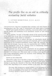
<strong>the</strong> final result. Communication with <strong>the</strong> parent and/or <strong>the</strong><br />
patient (if an adult) is paramount in order to impart an understanding<br />
of <strong>the</strong> process of treatment. We must keep in mind<br />
that some patients need extractions and some do not.<br />
If we apply <strong>the</strong>se principles in our daily practice, we should<br />
be able to maintain and/or improve facials es<strong>the</strong>tics. <strong>The</strong><br />
following case studies illustrate <strong>the</strong> importance of accurate<br />
diagnosis and treatment planning.<br />
CASE #1<br />
Patient SK’s chief complaint was her “crooked teeth” and<br />
not being able to “get my front teeth toge<strong>the</strong>r”. A review<br />
of her records showed a total discrepancy of approximately<br />
15mm, which included 5mm of crowding, and 10 mm of<br />
cephalometric discrepancy. Skeletally she had an open bite<br />
tendency as well (Figure 6). With a history of previous orthodontic<br />
treatment and <strong>the</strong> obvious relapse due to <strong>the</strong> large<br />
discrepancy, an extraction treatment plan was chosen. <strong>The</strong><br />
results (Figure 7) show nice soft tissue <strong>profile</strong> improvement<br />
into <strong>the</strong> es<strong>the</strong>tic zone due to <strong>the</strong> alignment and retraction of<br />
<strong>the</strong> anterior teeth.<br />
CASE #2<br />
An African-American female presented to Dr. John Bilodeau<br />
as a transfer patient with partial appliances in place.<br />
<strong>The</strong> diagnostic records revealed a dentoalveolar protrusion<br />
with edentulous spaces in both arches (Figure 8). After discussing<br />
<strong>the</strong> problem and her desires for soft tissue <strong>profile</strong><br />
improvement, she was retreated with <strong>the</strong> extraction of three<br />
premolars. Mini-anchors were used for absolute anchorage.<br />
<strong>The</strong> protrusion was reduced and all edentulous spaces were<br />
closed. <strong>The</strong> facial change was evident due to <strong>the</strong> reduction<br />
of <strong>the</strong> protrusion (Figure 9).<br />
Figure 8. Mulu Pretreatment<br />
Figure 6. SK Pretreatment<br />
Figure 9. Mulu Posttreatment<br />
Figure 7. SK Posttreatment<br />
CASE # 3<br />
Patient CW’s chief complaint was her “crooked teeth”. Her<br />
diagnostic records revealed 6mm of TSAL and about 6mm<br />
of protrusion (Figure 10). Due to <strong>the</strong> low mandibular plane<br />
angle, <strong>the</strong> crowding and <strong>the</strong> protrusion and an already pleasing<br />
<strong>profile</strong>, a non-extraction treatment plan utilizing interproximal<br />
reduction was selected. <strong>The</strong> result reveals good<br />
tooth and dental arch alignment, a slight reduction in <strong>the</strong><br />
dental protrusion and no change in <strong>the</strong> <strong>profile</strong> (Figure 11).<br />
5