the tweed profile - The Charles H. Tweed International Foundation
the tweed profile - The Charles H. Tweed International Foundation
the tweed profile - The Charles H. Tweed International Foundation

You also want an ePaper? Increase the reach of your titles
YUMPU automatically turns print PDFs into web optimized ePapers that Google loves.
and mandibular intermolar widths decreased 1.7 and<br />
2.1 mm, respectively. Whereas maxillary widths<br />
remained unchanged over <strong>the</strong> postretention period,<br />
mandibular intercanine width decreased 1.4 mm.<br />
Mandibular incisor irregularity was reduced 5.3 mm<br />
during treatment and increased only 0.7 mm during<br />
<strong>the</strong> postretention period. Based on this study, <strong>the</strong><br />
following conclusions can be drawn; satisfactory longterm<br />
results can be achieved for most Class I, four<br />
premolar extraction patients for whom evidence based<br />
treatment objectives including minimal alteration<br />
of <strong>the</strong> mandibular arch form and <strong>the</strong> retraction and<br />
uprighting or maintenance of mandibular incisors in<br />
<strong>the</strong>ir original position have been met.<br />
An article by Vaden et al. quantified changes in<br />
tooth relationships in a series of cases (N=36) at<br />
6 years and again at 15 years after treatment. All<br />
patients were treated with <strong>the</strong> extraction of first<br />
premolars, second premolars, or a combination of<br />
first and second premolars after a careful differential<br />
diagnosis. All patients were treated in adolescence<br />
by one clinician with an edgewise appliance. During<br />
treatment, maxillary and mandibular intercanine<br />
width was expanded slightly, more in <strong>the</strong> mandible<br />
than <strong>the</strong> maxilla. After treatment, most of <strong>the</strong> modest<br />
expansion in <strong>the</strong> maxilla remained, but half <strong>the</strong><br />
canine expansion in <strong>the</strong> mandible was lost (Fig. 1).<br />
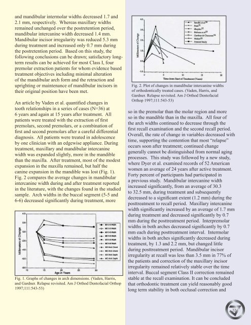
Fig. 2 compares <strong>the</strong> average changes in mandibular<br />
intercanine width during and after treatment reported<br />
in <strong>the</strong> literature, with <strong>the</strong> changes found in <strong>the</strong> studied<br />
sample. Arch widths in <strong>the</strong> buccal segment (5-5 and<br />
6-6) decreased significantly during treatment, more<br />
Fig. 1. Graphs of changes in arch dimensions. (Vaden, Harris,<br />
and Gardner. Relapse revisited. Am J Orthod Dentofacial Orthop<br />
1997;111:543-53)<br />
Fig. 2. Plot of changes in mandibular intercanine widths<br />
of orthodontically treated cases. (Vaden, Harris, and<br />
Gardner. Relapse revisited. Am J Orthod Dentofacial<br />
Orthop 1997;111:543-53)<br />
so in <strong>the</strong> premolar than <strong>the</strong> molar region and more<br />
so in <strong>the</strong> mandible than in <strong>the</strong> maxilla. All four of<br />
<strong>the</strong> arch widths continued to decrease through <strong>the</strong><br />
first recall examination and <strong>the</strong> second recall period.<br />
Overall, <strong>the</strong> rate of change in variables decreased with<br />
time, supporting <strong>the</strong> contention that most “relapse”<br />
occurs soon after treatment; continued change<br />
generally cannot be distinguished from normal aging<br />
processes. This study was followed by a new study,<br />
where Dyer et al. examined records of 52 American<br />
women an average of 24 years after active treatment.<br />
Forty percent of participants had participated in<br />
a previous study. Mandibular intercanine width<br />
increased significantly, from an average of 30.3<br />
to 32.5 mm, during treatment and subsequently<br />
decreased to a significant extent (1.2 mm) during <strong>the</strong><br />
posttreatment to recall period. Maxillary intercanine<br />
width significantly increased by an average of 1.7 mm<br />
during treatment and decreased significantly by 0.7<br />
mm during <strong>the</strong> posttreatment period. Interpremolar<br />
widths in both arches decreased significantly by 0.7<br />
mm each during posttreatment interval. Intermolar<br />
widths in both arches significantly decreased during<br />
treatment, by 1.3 and 2.2 mm, but changed little<br />
during posttreatment period. Mandibular incisor<br />
irregularity at recall was less than 3.5 mm in 77% of<br />
<strong>the</strong> patients and correction of <strong>the</strong> maxillary incisor<br />
irregularity remained relatively stable over <strong>the</strong> time<br />
interval. Buccal segment Class II correction remained<br />
stable at <strong>the</strong> recall examination. It can be concluded<br />
that orthodontic treatment can yield reasonably good<br />
long term stability in both occlusal correction and<br />
22