

THE KELLER BUNIONECTOMY - The Podiatry Institute
THE KELLER BUNIONECTOMY - The Podiatry Institute
THE KELLER BUNIONECTOMY - The Podiatry Institute

Create successful ePaper yourself
Turn your PDF publications into a flip-book with our unique Google optimized e-Paper software.
<strong>THE</strong> <strong>KELLER</strong> <strong>BUNIONECTOMY</strong><br />
lohn M. Stutz, D.P.M.,<br />
lngrid Kruse-Edelman, D.P.M.<br />
Donald R. Green, D.P.M.<br />
William Keller produced two articles, the first appearing in<br />
1904 and the second in 1912, describing a bunionectomy<br />
which essentially resected the medial eminence of the first<br />
metatarsal head and the base of the proximal phalanx for<br />
treatment of hallux valgus deformity. Due to the ensuing<br />
popularity, this procedure became known as the Keller procedure.<br />
William J. Keller was not the first to describe the<br />
procedure. Reidel as early as 1886 and Davies-Colley in<br />
1887 had previously described similar procedures.<br />
Keller's original technique utilized a 2" incision. <strong>The</strong><br />
medial exostosis was removed by the use of a rongure. <strong>The</strong><br />
base of the proximal phalanx was freed dorsally, medially,<br />
laterally, and plantarly preserving the periosteum and then<br />
removed using a Cigley saw. <strong>The</strong> preserved periosteum/<br />
capsular tissue was used to cover the exposed surface of<br />
bone.<br />
<strong>The</strong> Keller procedure has retained a great deal of its<br />
original popularity over the last 80 years. Accordingly, there<br />
are strong proponents and strong critics of this technique. lt<br />
is primarily used today in the United States as a joint<br />
destructive type procedure for the correction of a painful,<br />
dislocated, or arthritic joint. In 1 959, DuVries indicated that<br />
patients "with disability (following the Keller) represent a<br />
larger group than from any other operation employed for<br />
alleviation of this deformity." However, Wrighton indicated<br />
that "those who perform the Keller operation do not need to<br />
report their results, because they know them to be good."<br />
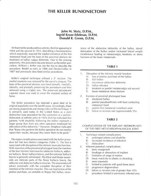
<strong>The</strong> major complications associated with the Keller procedure<br />
fall into two distinct categories (Table 1). <strong>The</strong> first is<br />
associated with disruption of the intrinsic muscular function.<br />
With resection of the proximal phalangeal base the insertion<br />
of the four intrinsic foot muscles (abductor hallucis, adductor<br />
hallucis, flexor hallucis brevis, and the extensor hallucis<br />
brevis) is generally eliminated. <strong>The</strong> tibial and fibular sesamoids<br />
are intricate parts of the flexor hallucis brevis, the<br />
adductor and abductor hallucis muscles. <strong>The</strong> sesamoids will<br />
retract proximally with the complete disruption of their<br />
insertion of the intrinsic muscles. This can lead to loss of the<br />
plantar purchase, an unstable or floppy flail hallux, recur-<br />
rence of the abduction deformity of the hallux, dorsal<br />
dislocation of the hallux and/or increased lateral weight<br />
transference leading to metatarsalgia, keratosis, or stress<br />
fractures of the lesser metatarsals.<br />
TABLE 1<br />
1 . Disruption of the intrinsic muscle function<br />
A) Ioss of plantar purchase of the hallux<br />
B) flail hallux<br />
C) recurrence abduction deformity<br />
D) hallux elevatus<br />
E) keratosis or painful metatarsalgia sub second<br />
F) lesser metatarsal stress fracture<br />
2. Excision of proximal phalangeal base<br />
A) shortened hallux<br />
B) painful pseudoarthrosis with base contacting<br />
metatarsal head<br />
C) painful first metatarsal cuneiform joint<br />
D) failure or revision rate (approximately 5%)<br />
1.<br />
2.<br />
TABLE 2<br />
COMPLICATIONS OF <strong>THE</strong> IMPLANT ARTHROPLASTY<br />
OF <strong>THE</strong> FIRST METATARSOPHALANCEAL JOINT<br />
Technique related complications<br />
A) prolonged edema and disability<br />
B) increased incidence of infection<br />
C) dislocation<br />
lnherent potential complications<br />
A) bony overgrowth<br />
B) breakage or absorption of implant<br />
C) limitation of range of motion<br />
D) tissue reactivity to silastic or shrarding<br />
E) more traumatic<br />
F) limited to patients with good bone stock<br />
C) progressivedegeneration<br />
H) failure or revision rate of greater than 10%<br />
l) procedure limited in previously infected area<br />
152
<strong>The</strong> second set of complications can be due to the amount<br />
of bone resected from the base of the proximal phalanx.<br />
Excessive osseous resection at this level will lead to a<br />
significantly shortened hallux which will be cosmetically<br />
unacceptable. <strong>The</strong>re will always be some shortening of the<br />
hallux, but both Hardie and Clapman, and Canley have<br />
indicated that the large majority of patients have a great toe<br />
that is longer than the second. lnadequate resection can lead<br />
to painful pseudoarthrosis especially if no interposition of<br />
soft tissue is utilized to cover the head or the base of the<br />
proximal phalangeal stump.<br />
However, with all these possible complications, Ganley<br />
reports a failure or revision rate to be only 5%. <strong>The</strong> advantages<br />
of the procedure advocated by its proponents include<br />
a patient group that is generally hrppy with the overall<br />
results of the surgery. <strong>The</strong> painful first metatarsophalangeal<br />
joint symptoms essentially resolve. <strong>The</strong> loss of the intrinsics<br />
and decreased purchase power has not been significantly<br />
problematic in the patient population that is many times<br />
apropu I s ive preoperative I y. I nc reased I atera I transference of<br />
weight bearing is a complication that can occur in almost<br />
any bunion procedure and needs to be controlled by biomechanical<br />
means. <strong>The</strong> flail toe, or loss of intrinsic function<br />
may not lead to dorsal dislocation or recurrence, if the<br />
extensor hallucis longustendon is lengthened and the extensor<br />
hallucis brevis is resected. <strong>The</strong> medial capsulartissue can<br />
be reinforced with tendon or fascial graft according to<br />
Canley, or reattached to the stump of the proximal phalanx<br />
via drill holes according to McClamry. Postoperative splintage<br />
of the deformity is utilized for an adequate period of<br />
time (a minimum of 6 weeks) to help maintain the correction.<br />
<strong>The</strong> Ioss of the intrinsic muscle function may be necessary for<br />
pain relief, for correction due to the longstanding deformity,<br />
and due to any coexistant limitation of dorsiflexion.<br />
ln the early 1970's, silastic implant replacements were<br />
introduced in an attemptto eliminate some of the complications<br />
of the Keller procedure. <strong>The</strong>re were hemi-implants,<br />
angulated hemi-implants, and total implants. <strong>The</strong>se primarily<br />
were utilized in an attempt to maintain length and<br />
preserve some degree of purchase power and function of the<br />
hallux. lnitially, they were thought to be abrle to resist<br />
deformity or re-deforming forces. It quickly became known<br />
that the silastic implants were primarily spacers and could<br />
not really resist deforming forces for any length of time. ln<br />
addition, the implants themselves created an additional set<br />
of complications (Table 2).<br />
<strong>The</strong>re have been a number of modifications of the Keller<br />
operation in an attempt to avoid some of the potential<br />
complications (Table 3). A variety of contradictory reports<br />
exist regarding the transfixation of the first metatarsophalangeal<br />
joint to maintain separation or a space.<br />
<strong>The</strong>re are a variety of ways to interpose soft tissue, most of<br />
which are effective. However, adequate bone must be<br />
resected and the raw bone or cartilage must be completely<br />
covered with the soft tissue of choice for the best results.<br />
Canley stressed the need for lengthening the extensor hallucis<br />
longus tendon to prevent dorsal dislocation or recurrent<br />
abduction deformity when the insertion of the intrinsic<br />
muscles had been eliminated. McGlamry stressed the need<br />
for re-establishing intrinsic muscle activity to the hallux and<br />
recommended drill holes plantarly to reattach the intersesamoid<br />
ligament, thereby re-establishing the force of the<br />
flexor hallucis brevis. He also emphasized re-attaching the<br />
medial capsularflaptothe stump ofthe proximal phalanx via<br />
drill holes. Canley on the other hand reinforced the medial<br />
structures with a tendon graft from the extensor hallucis<br />
brevis. <strong>The</strong> long flexor tendon of the hallux has also been<br />
reattached to the stump of the proximal phalanx to give more<br />
propulsive power to the hallux. Fusion of the first metatarsophalangeal<br />
joint Ieads to the ultimate stability. However, it<br />
also leads to lack of motion and will often cause difficulties<br />
in women with different heel heights and in men if they are<br />
very active in sports, or have a job that requires bending and<br />
sq uatti ng.<br />
Finally, the synthetic joint implants have gained a great<br />
deal of popularity in the last 20 years. However, now we are<br />
seeing some of the complications of Iong term use and a<br />
higher incidence of revision or failure being necessary. ln<br />
some European quarters, it is recommended that the implants<br />
be removed after 2 years in an attempt to avoid later<br />
complications, yet the temporary use prevents severe shortening<br />
which may occur if the implant were not used at all.<br />
TABLE 3<br />
1. Distraction of the ioint by internal fixation (K-wire,<br />
staple)<br />
2. lnterposing capsular tissue in a variety of manners<br />
3. Lengthening of the extensor hallucis longus tendon<br />
4. Re-anastamosis of the sesamoidal ligament<br />
5. Re-attachment of the intrinsic muscles to the proximal<br />
phalangeal stump<br />
6. Tenodesis of the flexor hallucis longus to the remaining<br />
proximal phalanx<br />
7. Fusion of the first metatarsophalangeal joint<br />
8. Spacing via synthetic joint implant<br />
9. Reinforcing the medial capsular tissue by a variety of<br />
means<br />
153
ln 19BB a review of the Keller bunionectomy was undertaken<br />
at Hillside Hospital. This was a retrospective study<br />
done to help us become more aware of the results one can<br />
expect from the Keller procedure. Between 1984 and I 986<br />
approximately 3 6 Kel ler arth roplasties were performed i n 3 1<br />
patients. A complete preoperative evaluation includ ing range<br />
of motion studies were performed. <strong>The</strong>re were 29 females<br />
and 2 males with an average age of 69 years, ranging from<br />
42 to 84. <strong>The</strong>se figures include 9 patients with first metatarsophalangeal<br />
joint implants (2 hemi-implants and 7 totals).<br />
Of these 31 patients 3 were deceased, 5 had moved from the<br />
area and 9 did not respond to several written invitations, nor<br />
could they be reached by telephone. <strong>The</strong> remaining '14<br />
patients agreed to bre seen for evaluation of the surgical<br />
results. <strong>The</strong>y represented a total of 16 Keller procedures.<br />
Prospective data on these patients included preoperative<br />
symptoms, clinical appearance, quantitative and qualitative<br />
ranges of motion and associated Iesions. Roentgenographic<br />
measu rements (intermetatarsal angle, hallux abductus angle,<br />
relative metatarsal length, first metatarsal declination, tibial<br />
sesamoid position) were also available. As some individuals<br />
did not completely fulfill the criteria for the study, the series<br />
proved to be smaller than originally anticipated.<br />
<strong>The</strong> follow up examination was performed on average at<br />
38 months and ranged lrom 29 to 52 months after the initial<br />
surgery. <strong>The</strong> operations were performed by a total of 7<br />
podiatric surgeons. <strong>The</strong> typical procedure based upon operative<br />
reports included I ) dorsal medial incision, 2) T or U<br />
capsulotomy, 3) resection of 1/3 to 112 the proximal pha-<br />
Ianx, 4) capsu lar closure over the metatarsal head, 5) lengthening<br />
of extensor hallucis longus, 6) no reattachment of the<br />
flexors, 7) Lawrence total implant in 5 of the 16 feet evaluated.<br />
<strong>The</strong> patients were evaluated objectively and subjectively<br />
utilizing standard forms. <strong>The</strong>y were given a questionnaire to<br />
rate their cosmetic result, present activity Ievel, amount of<br />
discomfort, and their rating of the overall result. <strong>The</strong> independent<br />
evaluator (JS) performed a biomechanical range of<br />
motion testing which included resting position, assisted dorsiflexion,<br />
unassisted dorsiflexion, and assisted plantar flexion.<br />
He also reviewed the quality of motion, tracking or tract<br />
bound crepitus, and symptoms with range of motion. <strong>The</strong><br />
objectiveform involved measuringthe length and alignment<br />
of the hallux, the scar, toe purchase, activity level of the individual,<br />
and their discomfort.<br />
RESULTS<br />
<strong>The</strong> total range of motion of the first metatarsophalangeal<br />
joints decreased postoperatively from an average of 66<br />
degrees to 52 degrees with an average loss of dorsiflexion of<br />
7 degrees. <strong>The</strong> hallux abductus angle was reduced an<br />
average of 20 degrees. <strong>The</strong> tibial sesamoid position was<br />
usually improved by unit.<br />
<strong>The</strong> remaining stump of the proximal phalanx was in much<br />
closer proximity to the first metatarsal head when implants<br />
were not used. <strong>The</strong> distance between phalanx and the<br />
metatarsal measured without an implant averaged 3.3 mm,<br />
whereas it was approximately 10 mm. when implants were<br />
utilized. Substantial shortening of the hallux was seen in<br />
73% of those cases where an implant was not used, but in<br />
none of the cases where implant arthroplasty was performed.<br />
Rotational or angulation abnormalities of the hallux<br />
were observed in 70'k of the cases.<br />
Radiographically the intermetatarsal angle decreased an<br />
average of 2.9 degrees (excluding the 1 case where a base<br />
wedge osteotomy was performed). Only one patient complained<br />
of a painful scar postoperatively. However, three<br />
patients were found to have one of the following: hypertrophic<br />
scar, discoloration of the scar, and numbness along the<br />
incision site. Despite these findings, each of the patients was<br />
satisfied or extremely satisfied with their surgical outcome.<br />
Hallux purchase was found to be good to fair in 60% of the<br />
cases. In 6 feet, or 40o/" of the cases, the hallux did not<br />
purchase the ground on full weight bearing and the result<br />
was rated as poor.<br />
Regardless of the inadequacies perceived by the observer,<br />
overall patient satisfaction with the surgery was 84%. Only<br />
2 patients were either dissatisfied or extremely dissatisfied<br />
with their surgical result. Both these patients were among the<br />
th ree that experienced serious postoperative compl ications.<br />
Patient number 3 had a cerebrovascular accident postoperatively<br />
from which he failed to recover and is now nonambulatory.<br />
Patient number 7 developed a hallux malleus<br />
bilaterally resulting in severe discomfort and pain caused by<br />
irritation from the shoes. Patient number 14 developed<br />
osteomyelitis postoperatively requiring a second surgery to<br />
debride additional portions of the proximal phalanx. This<br />
resulted in excessive scarring and fibrosis with resultant<br />
Iimitation of motion.<br />
Of the 5 total Lawrence implants, none of them showed<br />
significant shortening. <strong>The</strong>re was one that demonstrated a<br />
recurrence of the hallux abducto valgus. Three of the 5 were<br />
either extremely satisfied or satisfied. Of particular note,<br />
purchase power was good in 1, fair in 3 and poor in 1 . First<br />
metatarsophalangeal joint range of motion increased in 3 of<br />
the 5 total implant patients, whereas without the implants<br />
the first metatarsophalangeal joint range of motion generally<br />
decreased. None had a painful range of motion.<br />
154
SIGNIFICANT TINDINGS<br />
1 . Only 3 of the 16 procedures resulted in increased<br />
motion of the first metatarsophalangeal joint and these<br />
3 were total Lawrence implants.<br />
2. Mal position of the hallux was a very common finding<br />
(70%).<br />
3. Purchase of the hallux was frequently decreased or lost<br />
in 73"h.<br />
4. Despite the results patient satisfaction remained high at<br />
84%.<br />
5. Hallux extensus or hallux malleus from overpowering<br />
the extensor hallucis longus muscle has been reported in<br />
the literature as one of the mostcommon complications.<br />
ln our series only 1 patient (bilateral procedures) developed<br />
th is postoperative deform ity. lt appears that Can Iey<br />
is correct when he recommends routine lengthening of<br />
the extensor hallucis Iongus in the Keller procedure to<br />
prevent th is disabl ing compl ication.<br />
CONCLUSION<br />
Patient satisfaction with the Keller procedure was quite<br />
high at B4o/o. Favorable results in the patients' eyes must<br />
therefore be based on criteria other than recurrence or<br />
rotational or angulation deformities (70o/o), decreased range<br />
of motion (B1o/"),and reduced hallux purchase power (73"/r).<br />
<strong>The</strong> high patient satisfaction rate seems to be because of the<br />
relief o{ pain and ability to wear "normal" foot gear. All but<br />
2 patients had relief of pain postoperatively and all patients<br />
stated they were able to return to regular shoes after their<br />
surgeries.<br />
Take home message:<br />
<strong>The</strong> Keller arthroplasty is a joint destructive procedure<br />
designed primarily for relief of pain at the first metatarsophalangeal<br />
joint. Patient selection and preoperative patient<br />
expectations generally will lead to a very satisfactory result.<br />
<strong>The</strong> procedure should be used in the elderly with degenerative<br />
joint changes and impaired function of the first metatarsophalangeal<br />
joint. If utilized for hallux limitus it must bre<br />
noted that generally in the long term, range of motion will<br />
not be increased although painful motion may be eliminated.<br />
<strong>The</strong>refore, the use of the Keller procedure in a<br />
younger hallux limitus or rigidus patient must be done with<br />
caution. <strong>The</strong> patient and the surgeon should be aware that<br />
angulation, rotation, and shortening are common following<br />
this procedure. Furthermore, some loss of purchase power is<br />
to be expected. However, none of these complications<br />
seemed to interfere with the patients ability to wear regular<br />
shoes or perform their daily activities. Suturing the short or<br />
long flexors to the proximai phalangeal stump may increase<br />
the purchase power of the hallux postoperatively. This was<br />
not done routinely in our series. However caution should be<br />
given to reattaching the short flexors in the face of limited<br />
dorsal range of motion. <strong>The</strong> Keller procedure has passed the<br />
test of time when the appropriate patient and realistic<br />
expectations are identified preoperatively.<br />
Bibliography<br />
1 . DuVries HL Surgery of the Foot, C.V. Mosby, St. Louis<br />
1 9s9.<br />
2. Fuson SM: Modifications of the Keller Operation for<br />
l ncreased F u nctional Capacity, J F oot S u rg, 2l :29 2-29 6,<br />
1982.<br />
-)- Cerbert J, Mercado OA, Sokoloff TH: <strong>The</strong> Surgical<br />
Treatment of Hallux Abductovalgus and Allied Deform i-<br />
ties, Podiatric Medicine and Surgery, A Monograph<br />
Series, pg. 53-58.<br />
4. Kalish SR, McClamry ED: <strong>The</strong> Modified Keller Hallux<br />
Valgus Repair Utilizing Silastic lmplants, J Am <strong>Podiatry</strong><br />
Assoc 64:7 61 -773, 1 97 4.<br />
Mcclamry ED (ed): Reconstructive Surgery of the Foot<br />
and Leg, New York lntercontinental Medical Book Corp.<br />
1974, pg.39-56.<br />
6. Keller WL: <strong>The</strong> Surgical Treatment of Bunions and<br />
Hallux Valgus, NY Sfafe Med J, 8O:741-742, 1904.<br />
7. Keller WL: Further Observations on the Surgical Treatment<br />
of Hallux Valgus and Bunion, NY State Med J,<br />
95:696-698,1912.<br />
o. Canley l, Lynch F, Darrigan R: Keller Bunionectomy<br />
with Fascia and Tendon Cralt, J Am Podiatr Med Assoc,<br />
76:1986.<br />
9. LaPorta CA: Keller lmplant Procedure, Arch Podiat Med<br />
Foot Surg,2:1974.<br />
10. McClamry ED (ed): Comprehensive Textbook of Foot<br />
Surgery, Williams and Wilkins, Baltimore, Maryland,<br />
1987 , pg. 756-807.<br />
11. Swanson AB: lmplant Arthroplasty for the Creat Toe,<br />
Clin Orthop, Vol. 85, pg. 75-81 ,1972.<br />
12. VanOrr JV, O'Keefe R, Pikscher I: First Metatarsophalangeal<br />
Joint Implant Arthroplasty, in Comprehensive<br />
Textbook of Foot Surgery, Williams and Wilkins, Baltimore,<br />
Maryl and, 1987, p9.756-807.<br />
13. Wrighton JD: Ten Year Review of Keller's Operation,<br />
Cl i n Orthop, 89 :207 -21 4, 1 97 2.<br />
155


