Video assisted thoracoscopy in thoracic injury - Acta Bio Medica ...
Video assisted thoracoscopy in thoracic injury - Acta Bio Medica ... Video assisted thoracoscopy in thoracic injury - Acta Bio Medica ...
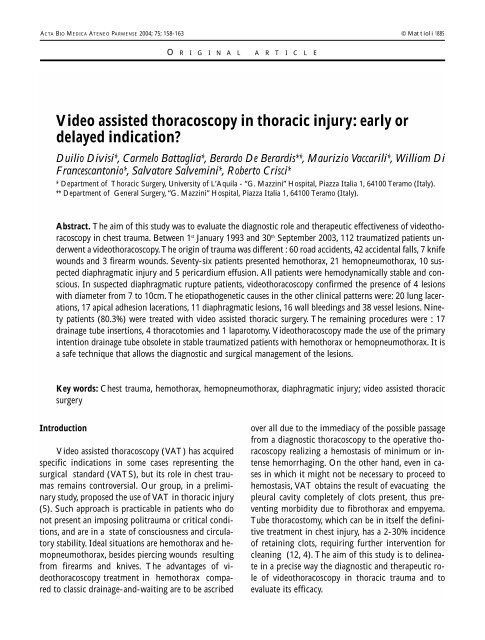
ACTA BIO MEDICA ATENEO PARMENSE 2004; 75; 158-163 © Mattioli 1885 O R I G I N A L A R T I C L E Video assisted thoracoscopy in thoracic injury: early or delayed indication? Duilio Divisi*, Carmelo Battaglia*, Berardo De Berardis**, Maurizio Vaccarili*, William Di Francescantonio*, Salvatore Salvemini*, Roberto Crisci* * Department of Thoracic Surgery, University of L’Aquila - “G. Mazzini” Hospital, Piazza Italia 1, 64100 Teramo (Italy). ** Department of General Surgery, “G. Mazzini” Hospital, Piazza Italia 1, 64100 Teramo (Italy). Abstract. The aim of this study was to evaluate the diagnostic role and therapeutic effectiveness of videothoracoscopy in chest trauma. Between 1 st January 1993 and 30 th September 2003, 112 traumatized patients underwent a videothoracoscopy. The origin of trauma was different : 60 road accidents, 42 accidental falls, 7 knife wounds and 3 firearm wounds. Seventy-six patients presented hemothorax, 21 hemopneumothorax, 10 suspected diaphragmatic injury and 5 pericardium effusion. All patients were hemodynamically stable and conscious. In suspected diaphragmatic rupture patients, videothoracoscopy confirmed the presence of 4 lesions with diameter from 7 to 10cm. The etiopathogenetic causes in the other clinical patterns were: 20 lung lacerations, 17 apical adhesion lacerations, 11 diaphragmatic lesions, 16 wall bleedings and 38 vessel lesions. Ninety patients (80.3%) were treated with video assisted thoracic surgery. The remaining procedures were : 17 drainage tube insertions, 4 thoracotomies and 1 laparotomy. Videothoracoscopy made the use of the primary intention drainage tube obsolete in stable traumatized patients with hemothorax or hemopneumothorax. It is a safe technique that allows the diagnostic and surgical management of the lesions. Key words: Chest trauma, hemothorax, hemopneumothorax, diaphragmatic injury; video assisted thoracic surgery Introduction Video assisted thoracoscopy (VAT) has acquired specific indications in some cases representing the surgical standard (VATS), but its role in chest traumas remains controversial. Our group, in a preliminary study, proposed the use of VAT in thoracic injury (5). Such approach is practicable in patients who do not present an imposing politrauma or critical conditions, and are in a state of consciousness and circulatory stability. Ideal situations are hemothorax and hemopneumothorax, besides piercing wounds resulting from firearms and knives. The advantages of videothoracoscopy treatment in hemothorax compared to classic drainage-and-waiting are to be ascribed over all due to the immediacy of the possible passage from a diagnostic thoracoscopy to the operative thoracoscopy realizing a hemostasis of minimum or intense hemorrhaging. On the other hand, even in cases in which it might not be necessary to proceed to hemostasis, VAT obtains the result of evacuating the pleural cavity completely of clots present, thus preventing morbidity due to fibrothorax and empyema. Tube thoracostomy, which can be in itself the definitive treatment in chest injury, has a 2-30% incidence of retaining clots, requiring further intervention for cleaning (12, 4). The aim of this study is to delineate in a precise way the diagnostic and therapeutic role of videothoracoscopy in thoracic trauma and to evaluate its efficacy.
- Page 2 and 3: Video assisted thoracoscopy in thor
- Page 4 and 5: Video assisted thoracoscopy in thor
- Page 6: Video assisted thoracoscopy in thor
ACTA BIO MEDICA ATENEO PARMENSE 2004; 75; 158-163 © Mattioli 1885<br />
O R I G I N A L A R T I C L E<br />
<strong>Video</strong> <strong>assisted</strong> <strong>thoracoscopy</strong> <strong>in</strong> <strong>thoracic</strong> <strong>in</strong>jury: early or<br />
delayed <strong>in</strong>dication?<br />
Duilio Divisi*, Carmelo Battaglia*, Berardo De Berardis**, Maurizio Vaccarili*, William Di<br />
Francescantonio*, Salvatore Salvem<strong>in</strong>i*, Roberto Crisci*<br />
* Department of Thoracic Surgery, University of L’Aquila - “G. Mazz<strong>in</strong>i” Hospital, Piazza Italia 1, 64100 Teramo (Italy).<br />
** Department of General Surgery, “G. Mazz<strong>in</strong>i” Hospital, Piazza Italia 1, 64100 Teramo (Italy).<br />
Abstract. The aim of this study was to evaluate the diagnostic role and therapeutic effectiveness of video<strong>thoracoscopy</strong><br />
<strong>in</strong> chest trauma. Between 1 st January 1993 and 30 th September 2003, 112 traumatized patients underwent<br />
a video<strong>thoracoscopy</strong>. The orig<strong>in</strong> of trauma was different : 60 road accidents, 42 accidental falls, 7 knife<br />
wounds and 3 firearm wounds. Seventy-six patients presented hemothorax, 21 hemopneumothorax, 10 suspected<br />
diaphragmatic <strong>in</strong>jury and 5 pericardium effusion. All patients were hemodynamically stable and conscious.<br />
In suspected diaphragmatic rupture patients, video<strong>thoracoscopy</strong> confirmed the presence of 4 lesions<br />
with diameter from 7 to 10cm. The etiopathogenetic causes <strong>in</strong> the other cl<strong>in</strong>ical patterns were: 20 lung lacerations,<br />
17 apical adhesion lacerations, 11 diaphragmatic lesions, 16 wall bleed<strong>in</strong>gs and 38 vessel lesions. N<strong>in</strong>ety<br />
patients (80.3%) were treated with video <strong>assisted</strong> <strong>thoracic</strong> surgery. The rema<strong>in</strong><strong>in</strong>g procedures were : 17<br />
dra<strong>in</strong>age tube <strong>in</strong>sertions, 4 thoracotomies and 1 laparotomy. <strong>Video</strong><strong>thoracoscopy</strong> made the use of the primary<br />
<strong>in</strong>tention dra<strong>in</strong>age tube obsolete <strong>in</strong> stable traumatized patients with hemothorax or hemopneumothorax. It is<br />
a safe technique that allows the diagnostic and surgical management of the lesions.<br />
Key words: Chest trauma, hemothorax, hemopneumothorax, diaphragmatic <strong>in</strong>jury; video <strong>assisted</strong> <strong>thoracic</strong><br />
surgery<br />
Introduction<br />
<strong>Video</strong> <strong>assisted</strong> <strong>thoracoscopy</strong> (VAT) has acquired<br />
specific <strong>in</strong>dications <strong>in</strong> some cases represent<strong>in</strong>g the<br />
surgical standard (VATS), but its role <strong>in</strong> chest traumas<br />
rema<strong>in</strong>s controversial. Our group, <strong>in</strong> a prelim<strong>in</strong>ary<br />
study, proposed the use of VAT <strong>in</strong> <strong>thoracic</strong> <strong>in</strong>jury<br />
(5). Such approach is practicable <strong>in</strong> patients who do<br />
not present an impos<strong>in</strong>g politrauma or critical conditions,<br />
and are <strong>in</strong> a state of consciousness and circulatory<br />
stability. Ideal situations are hemothorax and hemopneumothorax,<br />
besides pierc<strong>in</strong>g wounds result<strong>in</strong>g<br />
from firearms and knives. The advantages of video<strong>thoracoscopy</strong><br />
treatment <strong>in</strong> hemothorax compared<br />
to classic dra<strong>in</strong>age-and-wait<strong>in</strong>g are to be ascribed<br />
over all due to the immediacy of the possible passage<br />
from a diagnostic <strong>thoracoscopy</strong> to the operative <strong>thoracoscopy</strong><br />
realiz<strong>in</strong>g a hemostasis of m<strong>in</strong>imum or <strong>in</strong>tense<br />
hemorrhag<strong>in</strong>g. On the other hand, even <strong>in</strong> cases<br />
<strong>in</strong> which it might not be necessary to proceed to<br />
hemostasis, VAT obta<strong>in</strong>s the result of evacuat<strong>in</strong>g the<br />
pleural cavity completely of clots present, thus prevent<strong>in</strong>g<br />
morbidity due to fibrothorax and empyema.<br />
Tube thoracostomy, which can be <strong>in</strong> itself the def<strong>in</strong>itive<br />
treatment <strong>in</strong> chest <strong>in</strong>jury, has a 2-30% <strong>in</strong>cidence<br />
of reta<strong>in</strong><strong>in</strong>g clots, requir<strong>in</strong>g further <strong>in</strong>tervention for<br />
clean<strong>in</strong>g (12, 4). The aim of this study is to del<strong>in</strong>eate<br />
<strong>in</strong> a precise way the diagnostic and therapeutic role<br />
of video<strong>thoracoscopy</strong> <strong>in</strong> <strong>thoracic</strong> trauma and to<br />
evaluate its efficacy.
<strong>Video</strong> <strong>assisted</strong> <strong>thoracoscopy</strong> <strong>in</strong> <strong>thoracic</strong> <strong>in</strong>jury<br />
159<br />
Patients and Methods<br />
From 1 st January 1993 to 30 th September 2003<br />
112 traumatized patients underwent a video<strong>thoracoscopy</strong>,<br />
75 males (67%) and 37 females (33%) with an<br />
average age of 55 ± 1 year (range: 16 - 75 years).<br />
Trauma orig<strong>in</strong> varied: 60 road accidents (53.6%), 42<br />
accidental falls (37.5%), 7 knife wounds (6.3%) and 3<br />
firearm wounds (2.6%). Cl<strong>in</strong>ical patterns were: 76 hemothorax<br />
(67.8%), 21 hemopneumothorax (18.7%),<br />
10 suspected diaphragmatic <strong>in</strong>juries (9%) and 5 pericardium<br />
effusions (4.5%). All patients underwent<br />
preoperative test<strong>in</strong>g <strong>in</strong>clud<strong>in</strong>g a general evaluation<br />
(hemochrome with leukocyte formula and electrolytes,<br />
liver function), blood gas analysis, electrocardiogram,<br />
lung radiography, CT of the thorax and other<br />
diagnostic procedures that allow visualization of the<br />
associated lesions. Patients were hemodynamically<br />
stable, conscious and with blood gas analysis compatible<br />
with monopulmonary ventilation. Time between<br />
trauma and VAT was 18 ± 1 hours.<br />
<strong>Video</strong><strong>thoracoscopy</strong> technique<br />
Selective <strong>in</strong>tubation was performed under general<br />
anaesthesia. Cardio-respiratory parameters were carefully<br />
monitored. The patients were placed <strong>in</strong> the standard<br />
thoracotomic position, <strong>in</strong> case thoracotomy became<br />
necessary. An 8 mm trocar was positioned <strong>in</strong> the<br />
sixth or seventh <strong>in</strong>tercostal space <strong>in</strong> the midaxillary l<strong>in</strong>e,<br />
to provide passage for 0° optical. The pleural cavity<br />
along with other organs (chest wall, hemidiaphragm,<br />
lung, pericardium) was <strong>in</strong>spected for damage. <strong>Video</strong><br />
<strong>assisted</strong> <strong>thoracic</strong> surgery required a one or two further<br />
trocars, to facilitate dissection and repair of the lesion.<br />
Haematic effusion was aspirated and blood clots were<br />
removed after break<strong>in</strong>g up. When no <strong>in</strong>jury was<br />
found, we placed a dra<strong>in</strong>age tube 32 Ch through the<br />
camera access. Only the diaphragmatic lacerations<br />
with a diameter superior to 3 cm were treated by thoracotomy<br />
or laparotomy. All patients were extubated<br />
<strong>in</strong> immediate postoperative time.<br />
Statistical analysis<br />
Two analysis were carried out. The first evaluation<br />
<strong>in</strong>cluded the time between trauma and VATS, the<br />
morbidity and the length of hospital stay <strong>in</strong> 21 hemopneumothorax,<br />
<strong>in</strong> which the parenchymal laceration<br />
was treated by a stapl<strong>in</strong>g device with or without polytetrafluoroethylene<br />
(PTFE). Statistical analysis was<br />
performed with SPSS (W<strong>in</strong>dows release 6.1) and the<br />
statistical significance was estimated by Student t test<br />
for unpaired data. Data were expressed as mean ±<br />
standard deviation and 95% Confidence Interval (CI)<br />
. Difference was considered significant if the p values<br />
were less than 0.05 level. The second evaluation <strong>in</strong>cluded<br />
the time between trauma and VATS and the<br />
length of hospital stay <strong>in</strong> the same pathology. All p values<br />
less than 0.05 were considered to <strong>in</strong>dicate significance,<br />
whereas the correlation coefficient (r) was<br />
analyzed us<strong>in</strong>g a correlation analysis.<br />
Results<br />
No operative or perioperative deaths were observed.<br />
Fifteen patients required a blood transfusion. In<br />
suspected diaphragmatic <strong>in</strong>jury patients, video<strong>thoracoscopy</strong><br />
allowed discovery of 2 lesions <strong>in</strong> the left side<br />
(7 cm and 8 cm) and 2 lesions <strong>in</strong> the right side (8 cm<br />
and 10 cm), treated by thoracotomy <strong>in</strong> the VIII <strong>in</strong>tercostal<br />
space and separate suture stitches. Six VAT resulted<br />
negative after careful exploration of the pleural<br />
cavity and the procedure ended with thoracostomy tube<br />
application. In the patients affected by a firearm<br />
wound, VAT revealed: 1) a double laceration of the<br />
diaphragm; 2) a 2 cm diaphragmatic lesion; 3) a lung<br />
laceration. Treatment required respectively: 1) the position<strong>in</strong>g<br />
of a dra<strong>in</strong>age tube <strong>in</strong> the thorax and a conversion<br />
<strong>in</strong>to laparotomy <strong>in</strong> order to repair the diaphragm<br />
and to explore the abdom<strong>in</strong>al organs more easily;<br />
2) repair of the diaphragm <strong>in</strong> video<strong>thoracoscopy</strong><br />
with separate stitches; 3) the carry<strong>in</strong>g out of a wedgeresection<br />
of parenchyma <strong>in</strong> VATS with stapler device.<br />
N<strong>in</strong>e microlesions of the diaphragm (6 <strong>in</strong> the left side<br />
and 3 <strong>in</strong> the right side), with 2-3cm diameter, due to<br />
a knife wound (4 cases) and fractured rib stumps (5<br />
cases) were sutured <strong>in</strong> VATS with separate stitches.<br />
Eleven patients showed wall bleed<strong>in</strong>g follow<strong>in</strong>g rib<br />
fractures; we carried out a coagulation of the pleural<br />
surface and <strong>in</strong>sertion of dra<strong>in</strong>age tube. In the remai-
160 D. Divisi, C. Battaglia, B. De Berardis, et al.<br />
Table 1. VATS procedures <strong>in</strong> 54 hemothorax patients<br />
Causes<br />
Number<br />
Intercostal artery lesions 25<br />
Apical adhesions lacerations 19<br />
Diaphragmatic vessel lesions 10<br />
n<strong>in</strong>g 54 hemothorax patients VAT was converted <strong>in</strong><br />
VATS (Table 1), allow<strong>in</strong>g the lesions repair. <strong>Video</strong><strong>thoracoscopy</strong><br />
exploration <strong>in</strong> hemopneumothorax<br />
suggested diagnosis of parenchymal lacerations ow<strong>in</strong>g<br />
to a knife wound <strong>in</strong> 3 patients, a firearm wound <strong>in</strong> 1<br />
patient, a burst lesion <strong>in</strong> 7 patients and a rib fracture<br />
<strong>in</strong> 10 patients. Hemostasis and aerostasis were achieved<br />
<strong>in</strong> VATS by Endo-GIA 30, with or without polytetrafluoroethylene<br />
(PTFE) strips to re<strong>in</strong>force the<br />
stitches. In this group we registered cl<strong>in</strong>ical complications<br />
only <strong>in</strong> patients treated videothoracoscopically<br />
12 hours after trauma (r = 0.931; p < 0.001; Table 2).<br />
F<strong>in</strong>ally, 5 pericardium effusions l<strong>in</strong>ked to pericardium<br />
vessel lesions were endoscopically treated by pericardiac<br />
fenestration (Figure 1). The mean length of hospital<br />
stay <strong>in</strong> 90 patients (80.3%), treated with VATS,<br />
was 4 ± 1 days (range: 3 - 23 days).<br />
Table 2. Correlation between tim<strong>in</strong>g of VATS and morbidity and hospitalization <strong>in</strong> 21 hemopneumothorax patients<br />
Year and sex Re<strong>in</strong>force lung staple l<strong>in</strong>es Time between trauma and VATS Morbidity Hospital stay P value<br />
41; M PTFE 3 ± 0.51 hours / 3 ± 1 days < 0.04<br />
35; M PTFE 5 ± 0.34 hours / 4 ± 2 days < 0.01<br />
28; M PTFE 26 ± 0.12 hours Pneumonia 10 ± 1 days < 0.002<br />
54; F / 10 ± 0.47 hours / 4 ± 1 days < 0.01<br />
47; M / 48 ± 0.13 hours Pulmonary microembolism 23 ± 3 days < 0.003<br />
23; F / 14 ± 0.26 hours Atelectasis 6 ± 2 days < 0.004<br />
16; F PTFE 12 ± 0.57 hours / 5 ± 3 days < 0.01<br />
53; M PTFE 9 ± 0.30 hours / 4 ± 2 days < 0.02<br />
42; M / 31 ± 0.19 hours Pneumonia 16 ± 4 days < 0.005<br />
53; F / 11 ± 0.58 hours / 4 ± 2 days < 0.01<br />
32; M PTFE 25 ± 0.35 hours Pneumonia 9 ± 4 days < 0.001<br />
30; M / 21 ± 0.18 hours Atelectasis 12 ± 1 days < 0.002<br />
61; M / 22 ± 0.39 hours Atelectasis 17 ± 1 days < 0.003<br />
33; F / 6 ± 0.10 hours / 5 ± 1 days < 0.05<br />
66; M PTFE 7 ± 0.23 hours / 4 ± 1 days < 0.01<br />
39; F / 9 ± 0.15 hours / 3 ± 1 days < 0.001<br />
44; F / 26 ± 0.41 hours Pneumonia 15 ± 2 days < 0.006<br />
71; M / 6 ± 0.34 hours / 6 ± 1 days < 0.004<br />
69; M / 8 ± 0.53 hours / 4 ± 1 days < 0.02<br />
52; M / 3 ± 0.15 hours / 3 ± 1 days < 0.05<br />
41; F / 5 ± 0.20 hours / 3 ± 1 days < 0.03<br />
r= 0.931; p
<strong>Video</strong> <strong>assisted</strong> <strong>thoracoscopy</strong> <strong>in</strong> <strong>thoracic</strong> <strong>in</strong>jury<br />
161<br />
Figure 1. <strong>Video</strong> Assisted Thoracoscopy Procedure Algorithm for Thoracic Injury (BGA = Blood Gas Analysis)<br />
Discussion<br />
Our study showed that video<strong>thoracoscopy</strong> permits<br />
a rapid diagnosis and a less <strong>in</strong>vasive, safer and easier<br />
treatment <strong>in</strong> chest trauma. In fact, it allows an immediate<br />
evaluation of causes of bleed<strong>in</strong>g and associated<br />
damage, very good hemorrhag<strong>in</strong>g control with<br />
anaemic prevention lead<strong>in</strong>g to a better therapeutic solution<br />
<strong>in</strong> the choice of VATS, thoracotomy or laparotomy.<br />
<strong>Video</strong><strong>thoracoscopy</strong> led to a swift and complete<br />
functions recovery <strong>in</strong> the patient prevent<strong>in</strong>g fibrothorax,<br />
empyema or delayed rupture of the diaphragm.<br />
The shorter duration of dra<strong>in</strong>age tube, a significant<br />
reduction <strong>in</strong> hospital stay (4 ± 1 days <strong>in</strong> our study for<br />
400 euros x day) and of hospital costs, among which<br />
<strong>in</strong>expensive treatment <strong>in</strong> VATS (466.9 – 854.16; Table<br />
3), are shown <strong>in</strong> our experience. Eddy et al (6) highlighted<br />
empyema <strong>in</strong> 5% of patients requir<strong>in</strong>g urgent<br />
tube thoracostomy, due to <strong>in</strong>complete dra<strong>in</strong>age of the<br />
pleural space and prolonged tube <strong>in</strong>sertion. Villavicencio<br />
et al (16), analys<strong>in</strong>g the <strong>thoracoscopy</strong> results <strong>in</strong><br />
trauma, detected a 98% of diagnostic accuracy <strong>in</strong><br />
diaphragmatic <strong>in</strong>juries (DI), a total evacuation of hemothorax<br />
and empyema <strong>in</strong> 90% and 86% of cases respectively<br />
and chest tube bleed<strong>in</strong>g control <strong>in</strong> 82.5%.<br />
The improvement of endoscopic materials permitted<br />
an exact dissection and exhaustive exploration of the<br />
pleural cavity, to which lower postoperative pa<strong>in</strong> and<br />
excellent aesthetic audit<strong>in</strong>g may be added; a variety of<br />
pathologic situations of the chest, exclusively treated<br />
with thoracotomy was managed <strong>in</strong> VATS (3). This technique<br />
can be applied only <strong>in</strong> conscious, hemodynamically<br />
stable patients with a blood gas analysis compatible<br />
with monolateral exclusion; cl<strong>in</strong>ical and/or radiologic<br />
suspicion of cardiac or big blood vessels lesions,<br />
follow<strong>in</strong>g a mediast<strong>in</strong>al widen<strong>in</strong>g superior to<br />
8cm, or a tracheo-bronchial laceration must <strong>in</strong>duce a<br />
thoracotomic or sternotomic approach <strong>in</strong> primary <strong>in</strong>tention.<br />
In our study VAT permitted a complete clean<strong>in</strong>g<br />
of the pleural cavity <strong>in</strong> 91 patients affected by he-<br />
Table 3. Cost of room surgery <strong>in</strong> case of conversion from VAT<br />
<strong>in</strong> VATS<br />
Materials<br />
Trocar<br />
Endo-GIA 30<br />
Recharge of Stapler<br />
Propophol 10 mg/ml<br />
Silicone Dra<strong>in</strong>age Tube<br />
PTFE<br />
Endo-Clips<br />
Mean Costs<br />
125 euros<br />
129 euros<br />
89 euros<br />
23 euros<br />
13.9 euros<br />
121.36 euros x 2 pieces<br />
180 euros
162 D. Divisi, C. Battaglia, B. De Berardis, et al.<br />
mothorax, pericardium effusion and suspected diaphragmatic<br />
rupture. This method represented the only<br />
practice performed <strong>in</strong> 17 patients (18.6%) without<br />
source of haematic loss; the thoracostomy tube was<br />
positioned under direct visualization. The percentage<br />
of conversion of VAT <strong>in</strong>to VATS was 75.8% (69 patients),<br />
<strong>in</strong>to thoracotomy 4.4% (4 patients) and <strong>in</strong>to<br />
laparotomy 1.1% (1 patient). Abolhoda et al (1), <strong>in</strong> a<br />
series of 16 patients, registered 4 conversions <strong>in</strong> thoracotomy<br />
(25%) due to <strong>in</strong>efficient monolateral ventilation<br />
or an important <strong>in</strong>flammatory pleural reaction; 12<br />
patients (75%) were successfully treated with video<strong>thoracoscopy</strong>.<br />
Although the Authors believe that<br />
evacuation of blood clots <strong>in</strong> hemothorax is easy until 7<br />
post-<strong>in</strong>jury days, we believe that the ideal tim<strong>in</strong>g for<br />
video<strong>thoracoscopy</strong> is with<strong>in</strong> 12 hours follow<strong>in</strong>g trauma<br />
(18 ± 1 hours altogether <strong>in</strong> our experience ) <strong>in</strong> order<br />
to facilitate thorax <strong>in</strong>spection and <strong>in</strong>jury treatment<br />
thus avoid<strong>in</strong>g formation of <strong>in</strong>veterate lesions and postoperative<br />
morbidity. In fact, <strong>in</strong> 21 hemopneumothorax<br />
we observed 4 pneumonias, 3 atelectasis and 1 pulmonary<br />
microembolism <strong>in</strong> patients who underwent<br />
VATS at 12 hours from trauma; these complications<br />
were positively solved <strong>in</strong> all cases but prolonged hospitalization.<br />
Meyer et al (11) analysed 39 hemothorax<br />
patients treated with 36 F tube thoracostomy <strong>in</strong> primary<br />
<strong>in</strong>tention, who showed persistent hemothorax or<br />
hemopneumothorax with<strong>in</strong> 72 hours from the <strong>in</strong>itial<br />
dra<strong>in</strong>age placement. Subsequently, these patients underwent<br />
a second tube <strong>in</strong>sertion or VATS. In a VATS<br />
group the Authors observed a reduction <strong>in</strong> duration of<br />
dra<strong>in</strong>age tube (2.53 ± 1.36 vs 4.50 ± 2.83 days), <strong>in</strong> hospital<br />
stay (3.60 ± 1.64 vs 7.21 ± 5.30) and <strong>in</strong> hospital<br />
costs ($ 7,689 ± 3,278 vs $ 13,273 ± 8,158). Liu et al<br />
(9), <strong>in</strong> 56 hemothorax or posthemothorax complication<br />
patients with penetrat<strong>in</strong>g (23 cases) and blunt (33<br />
cases) <strong>in</strong>juries, applied VATS successfully <strong>in</strong> 50 patients<br />
(89.2%) without morbidity; 6 patients (10.8%)<br />
affected by cardiovascular lesions (4 cases) and chest<br />
wall lacerations (2 cases) were not treated with VATS.<br />
These studies clarified that early <strong>in</strong>spection of the<br />
pleural cavity <strong>in</strong> video<strong>thoracoscopy</strong> and direct treatment<br />
of <strong>in</strong>juries reduced short-term and long-term<br />
complications of chest trauma. Heniford et al (8) had<br />
used VATS <strong>in</strong> 19 patients (76%) <strong>in</strong> order to evacuate<br />
reta<strong>in</strong>ed hemothorax and thoracotomy <strong>in</strong> 4 patients<br />
(16%); 2 patients (8%) needed further strategy to<br />
dra<strong>in</strong> collection. The unsuccess of VATS was l<strong>in</strong>ked to<br />
the time between <strong>in</strong>jury and <strong>in</strong>tervention (14.5 days;<br />
successful 4.5 days) and the nature of collection (hemothorax<br />
vs empyema).<br />
The use of <strong>thoracoscopy</strong> <strong>in</strong> diagnosis and treatment<br />
of patients with penetrat<strong>in</strong>g chest trauma has<br />
been described by Branco (2) s<strong>in</strong>ce 1946. Oakes et al<br />
(13) highlighted that <strong>thoracoscopy</strong> <strong>in</strong> penetrat<strong>in</strong>g<br />
chest <strong>in</strong>jury reduces the need for thoracotomy. Uribe<br />
et al (15), evaluat<strong>in</strong>g by video<strong>thoracoscopy</strong> 28 patients<br />
with thoraco-abdom<strong>in</strong>al penetrat<strong>in</strong>g trauma,<br />
thought that this technique provides excellent identification<br />
of diaphragmatic <strong>in</strong>jury and evacuation of<br />
blood clots from the pleural cavity. Mart<strong>in</strong>ez et al (10)<br />
referred to VATS 52 penetrat<strong>in</strong>g thoraco-abdom<strong>in</strong>al<br />
trauma patients without <strong>in</strong>dications for urgent surgery;<br />
40 (76.9%) of them were cl<strong>in</strong>ically asymptomatic.<br />
The Authors diagnosed 35 diaphragmatic <strong>in</strong>juries<br />
(67.3%), easily repaired by <strong>thoracoscopy</strong>. The diagnostic<br />
accuracy of chest radiography and computed tomography<br />
<strong>in</strong> diaphragmatic <strong>in</strong>juries is <strong>in</strong>ferior to 50%<br />
(14). We believe that, <strong>in</strong> selected penetrat<strong>in</strong>g wound<br />
patients, the use of video<strong>thoracoscopy</strong> can represent a<br />
ma<strong>in</strong> legal role besides precisely def<strong>in</strong><strong>in</strong>g the trajectory<br />
of bullet or knife wounds. Freeman et al (7), <strong>in</strong> a<br />
retrospective review of 171 patients undergo<strong>in</strong>g VATS<br />
after penetrat<strong>in</strong>g chest trauma, established five <strong>in</strong>dependent<br />
predictors of diaphragmatic <strong>in</strong>juries (abnormal<br />
chest radiograph, entrance wound <strong>in</strong>ferior to nipple<br />
l<strong>in</strong>e, <strong>in</strong>traabdom<strong>in</strong>al <strong>in</strong>juries, right-sided entrance<br />
wound and high-velocity mechanism). They advised<br />
video<strong>thoracoscopy</strong> only <strong>in</strong> patients <strong>in</strong> whom two or<br />
more <strong>in</strong>dependent predictors of DI were identified.<br />
We concluded that early video<strong>thoracoscopy</strong> is<br />
safe and the fastest method for diagnosis and surgical<br />
management <strong>in</strong> chest trauma. This technique makes<br />
the use of primary <strong>in</strong>tention dra<strong>in</strong>age tube obsolete <strong>in</strong><br />
stable patients with hemothorax and hemopneumothorax.<br />
A diaphragmatic <strong>in</strong>jury with a diameter superior<br />
to 3cm is the only therapeutic limit.
<strong>Video</strong> <strong>assisted</strong> <strong>thoracoscopy</strong> <strong>in</strong> <strong>thoracic</strong> <strong>in</strong>jury<br />
163<br />
References<br />
1. Abolhoda A, Liv<strong>in</strong>gston DH, Donahoo JS, Allen K. Diagnostic<br />
and therapeutic video <strong>assisted</strong> <strong>thoracic</strong> surgery<br />
(VATS) follow<strong>in</strong>g chest trauma. Eur J Cardio-thorac Surg<br />
1997; 12: 356.<br />
2. Branco JMC. Thoracoscopy as a method of exploration <strong>in</strong><br />
penetrat<strong>in</strong>g <strong>in</strong>juries of the chest. Dis Chest 1946; 12: 330.<br />
3. Carrillo EH, Richardson JD. Thoracoscopy <strong>in</strong> the management<br />
of hemothorax and reta<strong>in</strong>ed blood after trauma. Curr<br />
Op<strong>in</strong> Pulm Med 1998; 4: 243.<br />
4. Coselli JS, Mattox KL, Beall AC Jr. Reevaluation of early<br />
evacuation of clotted hemothorax. Am J Surg 1984; 148: 786.<br />
5. Crisci R. <strong>Video</strong><strong>thoracoscopy</strong> <strong>in</strong> chest trauma. Endosurgery<br />
1998; 6: 93.<br />
6. Eddy AC, Luna GK, Copass M. Empyema thoracis <strong>in</strong> patients<br />
undergo<strong>in</strong>g emergent closed tube thoracostomy for<br />
<strong>thoracic</strong> trauma. Am J Surg 1989; 157: 494.<br />
7. Freeman RK, Al-Dossari G, Hutcheson KA, et al. Indications<br />
for us<strong>in</strong>g video-<strong>assisted</strong> thoracoscopic surgery to diagnose<br />
diaphragmatic <strong>in</strong>juries after penetrat<strong>in</strong>g chest trauma.<br />
Ann Thorac Surg 2001; 72: 342.<br />
8. Heniford BT, Carrillo EH, Spa<strong>in</strong> DA, Sosa JL, Fulton RL,<br />
Richardson JD. The role of video-<strong>assisted</strong> <strong>thoracoscopy</strong> <strong>in</strong><br />
the management of reta<strong>in</strong>ed <strong>thoracic</strong> collections after trauma.<br />
Ann Thorac Surg 1997; 63: 940.<br />
9. Liu DW, Liu HP, L<strong>in</strong> PJ, Chang CH. <strong>Video</strong>-<strong>assisted</strong> <strong>thoracic</strong><br />
surgery <strong>in</strong> treatment of chest trauma. J trauma 1997; 42:<br />
670.<br />
10. Mart<strong>in</strong>ez M, Briz JE, Carillo EH. <strong>Video</strong>-<strong>thoracoscopy</strong> expedites<br />
the diagnosis and treatment of penetrat<strong>in</strong>g diaphragmatic<br />
<strong>in</strong>juries. Surg Endosc 2001; 15: 28.<br />
11. Meyer DM, Jessen ME, Wait MA, Estrera AS. Early evacuation<br />
of traumatic reta<strong>in</strong>ed hemothoraces us<strong>in</strong>g <strong>thoracoscopy</strong>:<br />
a prospective, randomized trial. Ann Thorac Surg<br />
1997; 64: 1396.<br />
12. Milfeld DJ, Mattox KL, Beall AC Jr. Early evacuation of<br />
clotted hemothorax. Am J Surg 1978; 136: 686.<br />
13. Oakes DD, Sherck JP, Brodsky JB, Mark JBD. Therapeutic<br />
<strong>thoracoscopy</strong>. J Thorac Cardiovasc Surg 1984; 87: 269.<br />
14. Toombs BD, Sandler CM, Lester RG. Computer tomography<br />
of chest trauma. Radiology 1981; 150: 733.<br />
15. Uribe RA, Pachon CE, Frame SB, Enderson BL, Escobar<br />
F, Garcia GA. A prospective evaluation of <strong>thoracoscopy</strong> for<br />
the diagnosis of penetrat<strong>in</strong>g thoracoabdom<strong>in</strong>al trauma. J<br />
Trauma 1994; 37: 650.<br />
16. Villavicencio RT, Aucar JA, Wall MJ Jr. Analysis of <strong>thoracoscopy</strong><br />
<strong>in</strong> trauma. Surg Endosc 1999; 13: 3.<br />
Received: 23 January 2004<br />
Accepted <strong>in</strong> orig<strong>in</strong>al form: 13 December 2004<br />
Correspondence: Dr. Duilio Divisi<br />
Circonvallazione Ragusa 39<br />
64100 Teramo (Italy)<br />
Tel: +39 0861 429482<br />
Fax +39 861 211626<br />
E-mail address: duilio.divisi@virgilio.it


