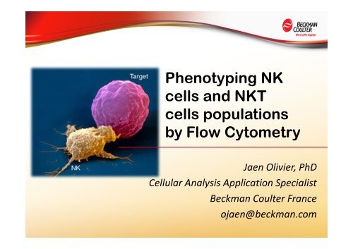
Phenotyping NK cells and NKT cells populations by Flow Cytometry
Phenotyping NK cells and NKT cells populations by Flow Cytometry
Phenotyping NK cells and NKT cells populations by Flow Cytometry

Create successful ePaper yourself
Turn your PDF publications into a flip-book with our unique Google optimized e-Paper software.
<strong>Phenotyping</strong> <strong>NK</strong><br />
<strong>cells</strong> <strong>and</strong> <strong>NK</strong>T<br />
<strong>cells</strong> <strong>populations</strong><br />
<strong>by</strong> <strong>Flow</strong> <strong>Cytometry</strong><br />
Jaen Olivier, PhD<br />
Cellular Analysis Application Specialist<br />
Beckman Coulter France<br />
ojaen@beckman.com
1.Introduction : <strong>NK</strong> <strong>cells</strong><br />
• Natural killer <strong>cells</strong> were discovered in 1975 (1)<br />
• <strong>NK</strong> express the NCAM-1 molecule, which clusterises as CD56 (2)<br />
• In blood, we identify at least two mains <strong>populations</strong> of <strong>NK</strong> <strong>cells</strong>, the CD56 dim <strong>and</strong> the<br />
CD56bright (2)<br />
• Another classical marker of <strong>NK</strong> <strong>cells</strong>, is the FcγRIII also called CD16 (3)<br />
• CD16 is an activating receptor, which could generate the ADCC (Antibody Dependant<br />
Cell Cytoxicity) (4)<br />
• CD16 expression confers to <strong>NK</strong>, the capacity to recognize opsonized cell <strong>by</strong><br />
antibodies, <strong>and</strong> then direct killing of <strong>cells</strong> (4,5)<br />
• Although <strong>NK</strong> <strong>cells</strong> are armed for killing, they are not dangerous in steady state<br />
conditions<br />
• In fact, they express activating <strong>and</strong><br />
inhibitory receptors (6)<br />
• The resulting effect depends of the<br />
balance of engaged activating <strong>and</strong><br />
inhibitory receptors (7)
1.Introduction : <strong>NK</strong> <strong>cells</strong><br />
• Inhibitory receptors prevent host <strong>cells</strong> killing, some are called KIRs (Killing Inhibitory<br />
Receptors or CD158…) (8)<br />
• KIRs recognize HLA class I molecules that prevent killing of normal <strong>cells</strong>, <strong>by</strong> an ITIM<br />
transduction dependent pathway (8,9)<br />
• KIRs are not the only inhibitory receptors, <strong>NK</strong>G2A is an important inhibitory receptor<br />
that heterodimerizes with CD94 (10)<br />
• Those molecules recognizes HLA-E molecule, unlike the KIRs which classically<br />
recognize HLA-A, B <strong>and</strong> C molecules (8,10,11)<br />
• CD94 also heterodimerizes with <strong>NK</strong>G2D, which is an activating receptor when linking<br />
to MIC-A, MIC-B or ULBP (12)<br />
• The last are cell stress induced molecules (12)<br />
• In some cases, upregulation of <strong>NK</strong>G2D lig<strong>and</strong>s is sufficient to induce <strong>NK</strong> cell mediated<br />
lysis of tumor <strong>cells</strong> (13)<br />
• Expression evaluation of the principal KIR, <strong>NK</strong>G2A <strong>and</strong> <strong>NK</strong>G2D would be important to<br />
predict the potential effect of <strong>NK</strong> cell infusion as a way of cancer treatment (13,14)
1.Introduction : <strong>NK</strong> <strong>cells</strong><br />
• Natural Cytotoxicity Receptors (NCRs) are very important in <strong>NK</strong> cell activation (15)<br />
• The well known NCRs are <strong>NK</strong>p30 (CD337), <strong>NK</strong>p44 (CD336) <strong>and</strong> <strong>NK</strong>p46 (CD335) (15)<br />
• Even if <strong>NK</strong>p lig<strong>and</strong>s are not fully known, they associate with different activating coreceptor<br />
<strong>and</strong> transduce signaling that promote <strong>NK</strong> cell activation <strong>and</strong> lysis of cancer <strong>cells</strong><br />
(13,16,17)<br />
• Evaluation of their expression should be of interest in <strong>NK</strong> cell based or related therapy<br />
(13,14)<br />
• <strong>NK</strong> cell related therapy have been tried in several pathologies (13) , such as acute myeloid<br />
leukemia (18,19,21)<br />
• Several <strong>NK</strong> cell based therapy were successfull in AML (19,21)<br />
• This success could be, in part, explained <strong>by</strong> non<br />
KIRs engagement <strong>by</strong> HLA molecules (KIR HLA<br />
mismatch), <strong>and</strong> activating receptors triggering <strong>by</strong> cancer<br />
<strong>cells</strong> lig<strong>and</strong>s (14,18,19,20,22)<br />
• This provides a positive balance that generates <strong>NK</strong><br />
activation <strong>and</strong> killing of tumor <strong>cells</strong> called Graft Versus<br />
Leukemia (7,20,22)
1.Introduction : CD3+ CD56+ <strong>cells</strong><br />
• In blood, there is at least two lymphocytes <strong>populations</strong> that express CD56 as<br />
<strong>NK</strong> <strong>cells</strong><br />
• Those <strong>cells</strong> are CD3 + also, so this is a kind of <strong>cells</strong> that express <strong>NK</strong> marker<br />
<strong>and</strong> T <strong>cells</strong> marker, that’s why some call them <strong>NK</strong>T <strong>cells</strong><br />
• <strong>NK</strong>T <strong>cells</strong> are a heterogenous population (23)<br />
• Some describe two mains <strong>populations</strong> : type I <strong>and</strong> type II <strong>NK</strong>T <strong>cells</strong> (23,24)<br />
• The first are the i<strong>NK</strong>T <strong>cells</strong>, that express the invariant TCR Vα24-Jα18 which<br />
recognizes the glycolipid αGalCer (alpha Galactosyl Ceramide) (23,24)<br />
• The second are also CD1d restricted, but they express a range of TCRs<br />
• They could recognize glycolipid also (25)<br />
• Type I <strong>and</strong> Type II <strong>NK</strong>T <strong>cells</strong> are in charge of recognizing microbial lipid<br />
presented <strong>by</strong> the CD1d molecules (25,26)<br />
• It was described that i<strong>NK</strong>T could prevent NOD mice diabetes development (27)<br />
• Even there is some αGalCer injection assays (28,29), injection of i<strong>NK</strong>T is poorly<br />
done in human, nevertheless it seems to have anti-tumor effect<br />
(30)<br />
• The definition of i<strong>NK</strong>T <strong>cells</strong>, is for a lot of works only the reactivity toward<br />
CD1d-αGalCer tetramer<br />
(one exemple in human : ref 31)
1.Introduction : CD3+ CD56+ <strong>cells</strong><br />
• <strong>NK</strong>T <strong>cells</strong> are CD3+ CD56+, but they are not the only <strong>populations</strong> that co-express these two<br />
molecules in blood<br />
• Υδ T <strong>cells</strong> also share this co-expression<br />
• This population is a heterogenous population that is implicated in innate immune response to<br />
tumors <strong>and</strong> pathogens (32,33)<br />
• There is two mains <strong>populations</strong> of Vδ1 <strong>and</strong> Vδ2, in blood (32,33)<br />
• The majority of Υδ T <strong>cells</strong> seems to be Vγ9Vδ2 (80-90% of Υδ T <strong>cells</strong> in peripheral blood)<br />
• Υδ T <strong>cells</strong> share innate immune receptors as well as adaptive immune receptors<br />
• They express <strong>NK</strong>G2D, <strong>NK</strong>G2A, CD94, as <strong>NK</strong> Cells (32-34)<br />
• In contact of tumor <strong>cells</strong> or pathogens infected<br />
<strong>cells</strong>, Υδ T <strong>cells</strong> secrete pro-inflammatory cytokines,<br />
<strong>and</strong> could kill the transform target (32,33,36)<br />
• With their Υδ TCRs, they recognize non peptidic<br />
phophorylated antigens or lipids (35-36)<br />
• The most described Υδ T <strong>cells</strong> population is the<br />
Vγ9Vδ2, it recognizes some pyrophosphate derived<br />
products, that are essential for cell growth, membrane<br />
integrity of bacteria <strong>and</strong> mammalians <strong>cells</strong> (36-38)
1.Introduction : CD3+ CD56+ <strong>cells</strong><br />
• Even if immunological memory is a key mark of adaptative immunity, Υδ T <strong>cells</strong><br />
could be differentiated into naive, central memory, effector memory <strong>and</strong> CD45RA +<br />
effector memory <strong>cells</strong> (39)<br />
• This differentiation could be done with CD45RA, CD45R0, CD62L, CD27 or CD28<br />
(39)<br />
• Vγ9Vδ2 T <strong>cells</strong> are strongly activated <strong>by</strong> phospho-antigens from mycobacterium<br />
tuberculosis, whereas Vδ1 emerged signicantly in HIV <strong>and</strong> CMV infection (40)<br />
• It seems that Υδ T <strong>cells</strong> could secrete IL17, this last has a critical role in tumors<br />
rejection <strong>and</strong> pathogens rejection (41)<br />
• Goal : the aim was to make a 10 colors tubes panel, that<br />
permit phenotyping of <strong>NK</strong> <strong>cells</strong>, <strong>NK</strong>T <strong>cells</strong> <strong>and</strong> T <strong>cells</strong>, in<br />
order to access to inhibitory <strong>and</strong> activating molecules, as<br />
well as naive <strong>and</strong> memory molecules on each <strong>cells</strong> type
2.Material <strong>and</strong> method<br />
• Healthy donor fresh blood sample was directly stained with those antibodies<br />
• Pacific Blue : Anti CD57 or anti CD45RA<br />
• Krome Orange : anti CD45 or CD4<br />
• FITC : anti TCR Vα24 or anti CD45R0 or anti CD95 or anti TCR Υδ<br />
• PE : anti CD11b or anti Vα24 or anti <strong>NK</strong>p46 or anti CD28 or anti CD94 or anti TCR Υδ or anti <strong>NK</strong>G2D or<br />
anti <strong>NK</strong>G2A or anti CD158a,h or anti CD158b or anti CD158e1,e2 or anti CD158i or anti <strong>NK</strong>p30<br />
• ECD : anti CD62L or anti CD45R0 or anti <strong>NK</strong>p46<br />
• PC5.5 : CD56 or PC5 : anti <strong>NK</strong>p44<br />
• PC7 : anti CD3 or anti CD56<br />
• APC : anti CD38 or anti CD69 or anti CD158b1,b2,j or anti CD158 a,h or anti CD158a<br />
• APC-AlexaFluor 700 : anti CD16<br />
• APC-AlexaFluor 750 : anti CD8 or anti CD64<br />
• Incubation was performed at room temperature during 15 minutes<br />
• Red Blood lysis was realized with 1 ml of Versalyse per 100µl of blood<br />
• Samples were then washed once, 1 ml PBS was added <strong>and</strong> centrifuged @ 300g during 5 min<br />
• Pelets were resuspended with 300µl of PBS<br />
• Acquired on Gallios 3 lasers (Blue, Red, Violet)<br />
• Data were analyzed with Kaluza Software V1.2
3.Results : Gating strategy to identify <strong>NK</strong>/<strong>NK</strong>T<br />
Doublets <strong>and</strong> aggregats of <strong>cells</strong> were eliminated with a FSC Area vs FSC TOF signal<br />
FSC vs SSC Dot plot was created to take<br />
lymphocytes<br />
In some tubes, CD45 permits to eliminate<br />
debris, <strong>and</strong> to take all the leukocytes
3.Results : Gating strategy to identify <strong>NK</strong>/<strong>NK</strong>T<br />
OR<br />
Lympho Pur gate was created as lympho <strong>and</strong> NOT<br />
Mono<br />
Monocytes were gated based on CD45 or CD4 Krome<br />
Orange or CD64 APC AlexaFluor750<br />
CD3 vs CD56 Dot Plot allows the identification of CD56 +<br />
CD3 - <strong>NK</strong> <strong>cells</strong>, CD56 - CD3 + T lymphocytes, CD3 + CD56 +<br />
<strong>NK</strong>T <strong>and</strong> CD3 bright CD56 + probably γδ T <strong>cells</strong>
3.Results : <strong>NK</strong> <strong>populations</strong><br />
CD16 <strong>and</strong> CD8 expression on <strong>NK</strong> <strong>cells</strong> permit the identification of differents<br />
<strong>populations</strong> :<br />
CD56 bright CD16 Low , CD56 bright CD16 Int ,CD56 dim CD16 bright<br />
CD56 bright CD8 - ,CD56 bright CD8 + ,CD56 dim CD8 - ,CD56 bright CD8 int ,CD56 dim CD8 +
3.Results : <strong>NK</strong> Populations<br />
As described (42) , CD62L expression on <strong>NK</strong> <strong>cells</strong> permits to identify three mains<br />
<strong>populations</strong> :<br />
CD56 bright CD62L High , CD56 dim CD62L Int ,CD56 dim CD62L -<br />
With CD57, there is also 3 mains <strong>populations</strong> CD56 bright CD57 - ,CD56 dim CD57 - ,<br />
CD56 dim CD57 +
3.Results : <strong>NK</strong> Populations<br />
CD38 is an important signaling molecule for <strong>NK</strong> cell. Its engagement <strong>by</strong> anti CD38<br />
conducts to increase of <strong>NK</strong> cell cytotoxicity (43,44)<br />
Here, we identified that nearly all <strong>NK</strong> express CD38<br />
CD69 is one of the activations<br />
markers expressed <strong>by</strong> <strong>NK</strong> <strong>cells</strong> (45)<br />
Here, we identified very few<br />
Activated <strong>NK</strong> cell, the majority of<br />
<strong>NK</strong> <strong>cells</strong> were CD69-, so those <strong>NK</strong><br />
<strong>cells</strong> are Resting <strong>NK</strong> <strong>cells</strong><br />
Taken from Llera et al, J Biol Chem, 2001,276:7312-19
3.Results : NCR in <strong>NK</strong> cell population<br />
For NCR evaluation, we made a 9 colors tube mix with anti CD4 KO, anti TCR Vα24<br />
FITC, anti <strong>NK</strong>p30 PE, anti <strong>NK</strong>p46 ECD, anti <strong>NK</strong>p44 PC5, anti CD56 PC7, anti CD3<br />
APC, anti CD16 AA700 <strong>and</strong> anti CD8 AA750<br />
All <strong>NK</strong> <strong>cells</strong> were <strong>NK</strong>p + , interestingly<br />
the CD56 dim subset was the most<br />
<strong>NK</strong>p30 + , <strong>and</strong> the less <strong>NK</strong>p44 + .<br />
For the CD56 bright subset, it was the<br />
opposite situation.<br />
Nkp46 was dimly expressed on the<br />
majority of the <strong>NK</strong><br />
Functional studies might explained<br />
if these differences were relevant
3.Results : NCR in <strong>NK</strong> cell population<br />
A CD4 vs CD56 plot, gated on lymphocyte,<br />
informs about monocytes contamination in<br />
the <strong>NK</strong> gate
3.Results : <strong>NK</strong> Populations<br />
CD45RA <strong>and</strong> CD62L are well defined for T <strong>cells</strong> <strong>populations</strong> (46)<br />
Using this two molecules, we descriminated at least 4 differents sub<strong>populations</strong>
3.Results : <strong>NK</strong> Populations<br />
As described (47) , CD94 on <strong>NK</strong> population permits to identify 3 mains <strong>populations</strong><br />
Using this molecule couples with CD62L, we descriminated at least 3 differents<br />
sub<strong>populations</strong>
3.Results : <strong>NK</strong> Populations<br />
<strong>NK</strong>G2D, <strong>NK</strong>G2A are important stimulatory <strong>and</strong> inhibitory receptors that are co<br />
expressed with CD94 (10,12)<br />
All <strong>NK</strong> <strong>cells</strong> were <strong>NK</strong>G2D + , unlike with<br />
<strong>NK</strong>G2A<br />
There are two <strong>populations</strong> of <strong>NK</strong>G2A +<br />
Where as we identified only one<br />
<strong>NK</strong>G2A - population<br />
Those <strong>cells</strong> might be interesting in<br />
treatment cancer because they lack<br />
the inhibitory receptor for HLA E
3.Results : <strong>NK</strong> Populations<br />
KIRs evaluation is an important point to know which HLA alleles are recognized <strong>by</strong><br />
killer <strong>cells</strong> (8)<br />
Here we identified <strong>NK</strong> cell <strong>populations</strong><br />
which were CD158a + CD158a,h + ,<br />
CD158b1,b2,j + , CD158b + , CD158e1,e2 + ,<br />
CD158i +<br />
The donor could recognize the HLA-C<br />
group 1 <strong>and</strong> 2 alleles with<br />
CD158a,b1,b2,j,h,i KIRs<br />
He could also recognize HLA-B <strong>and</strong> A<br />
alleles with CD158e1,e2 KIRs (8)
3.Results : <strong>NK</strong>T Populations<br />
Looking @ TCR Vα24 with FITC or PE<br />
coupled antibodies, brings CD3+ CD56+<br />
<strong>cells</strong>, but CD3+ CD56- <strong>cells</strong> also<br />
The last aren’t type I <strong>NK</strong>T <strong>cells</strong>, even<br />
there is a lot of work which considers<br />
those <strong>cells</strong> as <strong>NK</strong>T (31)<br />
These suggests that there are T <strong>cells</strong><br />
that share the same Vα24 TCR, as type I<br />
<strong>NK</strong>T <strong>cells</strong><br />
Use of anti TCR Vβ11 could inform us if<br />
those CD3+ CD56- <strong>cells</strong> really share the<br />
same specificity as i<strong>NK</strong>T <strong>cells</strong>
3.Results : i<strong>NK</strong>T Populations<br />
<strong>NK</strong>T gate permits to really identify the invariant TCR Vα24 CD1d restricted <strong>NK</strong>T<br />
<strong>cells</strong><br />
Frequency is about 0,005-0,006% of the total blood <strong>cells</strong>, about 0,2% of total<br />
lymphocytes<br />
Because of this low frequency, we<br />
recommend to make live gate<br />
around lymphocytes, <strong>and</strong> save<br />
about 1 million<br />
In this way, it may permit to analyze<br />
about 2000 i<strong>NK</strong>T <strong>cells</strong><br />
PE coupled antibody offers the best<br />
resolution in order to catch i<strong>NK</strong>T<br />
<strong>cells</strong><br />
Because we chose to stop acquisition @ 100 000 total <strong>cells</strong>, it’s not very<br />
pertinent to analyze so few i<strong>NK</strong>T <strong>cells</strong>
3.Results : CD3 bright Populations<br />
We clearly identify CD3 bright which were also CD56 + <strong>cells</strong>, so those <strong>cells</strong> could<br />
also be called <strong>NK</strong>T <strong>cells</strong>.<br />
Since they express more CD3, it could be convenient to separate them from the<br />
others<br />
Taking All the T lymphocytes, allow<br />
us to make a wonderfull CD8 vs CD4<br />
Dots Plot, <strong>and</strong> identify single positive<br />
<strong>populations</strong> <strong>and</strong> a non insignificant<br />
double negative population<br />
The majority of the CD3 bright was<br />
double negative, but we clearly<br />
identify CD8 int population also<br />
Using the CD3 vs CD4 dot plot, we clearly<br />
catch the T CD3 bright population
3.Results : CD3 bright Populations<br />
The CD3 bright population was clearly CD16 + Using the CD3+ CD16+ gate, we found<br />
the same CD4/CD8 profile, with a little<br />
CD4 + contamination
3.Results : CD3 bright Populations<br />
Compare to <strong>NK</strong> <strong>cells</strong>, CD3 bright population was <strong>NK</strong>p30, <strong>NK</strong>p44 <strong>and</strong> <strong>NK</strong>p46<br />
– or low
3.Results : CD3 bright Populations<br />
CD3 bright population express Υδ T Cell Receptor<br />
Υδ T Cell could be<br />
divided in three<br />
sub<strong>populations</strong><br />
toward their CD16<br />
<strong>and</strong> CD57<br />
expression<br />
This population<br />
express the CD38<br />
<strong>and</strong> partially<br />
CD62L, but not the<br />
CD69
3.Results : CD3 bright Populations<br />
It has just been demontrated that CD3 bright population express Υδ T Cell Receptor<br />
But those <strong>cells</strong> were trully KIR + also<br />
They express all the <strong>NK</strong> KIR<br />
expressed<br />
That’s a very original point, that<br />
those Υδ T Cells express KIRs<br />
Since they express T <strong>cells</strong>, <strong>and</strong> <strong>NK</strong><br />
<strong>cells</strong> markers, this Υδ T Cells<br />
population could be called <strong>NK</strong>T <strong>cells</strong><br />
also
3.Results : CD3 bright Populations<br />
Gates colors :<br />
Υδ T <strong>cells</strong> were CD45RA<br />
<strong>and</strong> R0 neg or low compare<br />
to CD4 <strong>and</strong> CD8 T <strong>cells</strong><br />
<strong>populations</strong><br />
With CD62L marker, we<br />
could identify four<br />
differents <strong>populations</strong>
4.Overlaying results<br />
<strong>NK</strong> <strong>cells</strong> were CD56, CD16, Nkp30, <strong>NK</strong>p44, <strong>NK</strong>p46, <strong>NK</strong>G2D, CD38,<br />
CD94 + , partly CD62L, CD57, <strong>NK</strong>G2A, CD8 + <strong>and</strong> naturally CD3 -<br />
Υδ T <strong>cells</strong> were positive for KIRs, CD56 (dim) , CD3 (bright) , CD16,<br />
<strong>NK</strong>G2D <strong>and</strong> CD94, partly CD62L, CD38, CD57, <strong>NK</strong>G2A <strong>and</strong> CD8 <strong>and</strong><br />
negative for <strong>NK</strong>ps markers
5.Results : T <strong>cells</strong> naive <strong>and</strong> memory status
6.Conclusion<br />
• Our 10 colors combinations allow us to clearly identify <strong>NK</strong>, i<strong>NK</strong>T, T <strong>cells</strong> <strong>and</strong> T CD3 bright Υδ<br />
<strong>cells</strong><br />
• Those combinations, could be very usefull to sort all the <strong>populations</strong> identified here, <strong>and</strong><br />
studying them functionnally<br />
• Real i<strong>NK</strong>T must be confirmed with the use of anti TCR Vα24 + anti TCR Vβ11, <strong>and</strong> with a<br />
longer acquisition<br />
• The specificity of TCR Vα24 CD3 + CD56 - could be complemented with the use of anti TCR<br />
Vβ11<br />
• We clearly see all the <strong>NK</strong> <strong>cells</strong> markers, such as <strong>NK</strong>p, KIRs, <strong>NK</strong>G2D, <strong>NK</strong>G2A, CD57, CD94<br />
• Use of anti-CD62L in ECD, could provide a good way to analyze <strong>and</strong> study the 3 differents<br />
<strong>NK</strong> <strong>cells</strong> <strong>populations</strong>, that present different functions<br />
• KIR phenotyping could be easy <strong>and</strong> very important in KIR mismatch grafting of <strong>NK</strong> <strong>cells</strong><br />
• Υδ T <strong>cells</strong> really share TCR <strong>and</strong> KIR, sorting those <strong>cells</strong> would be of interest in order to<br />
realize functional study, or in order to make adoptive transfer in mice<br />
• This work could be a very usefull tool for laboratory working on <strong>NK</strong>, i<strong>NK</strong>T, Υδ <strong>cells</strong>, since it<br />
could provide pertinent antibodies/dyes combinations on Navios/Gallios <strong>Flow</strong>Cytometers
7.References<br />
1. Kiesslin R et al, Eur.J.Immunol,1975;5:112-117<br />
2. Caligiuri MA, Blood, 2008;112:461-469<br />
3. Hanna J et al, Trends Immunol, 2007;28:201-206<br />
4. Perussia B, Curr Top Microbiol Immunol, 1998;63-88<br />
5. Marquez ME et al, Cellular Immunology, 2010;264:86-92<br />
6. Vivier E et al, Nat Immunol, 2008;9:503-510<br />
7. Vivier E et al, Science, 2011;331:44-49<br />
8. Campbell KS et al, Immunology, 2011;132:315-325<br />
9. MacFarlane AW et al, Curr Top Microbiol Immunol, 2006; 32. Casetti R et al, Cell Mol Immunol, 2008;5:161-170<br />
298:23-57<br />
33. Meraviglia S et al, Clin Dev Immunol, 2011;2011:1-11<br />
10. Braud VM et al, Nature, 1998;391:795-799<br />
34. Rincon-Orozco B et al, J Immunol, 2005;175:2144-2151<br />
11. Lee N et al, Proc Natl Acad Sci USA, 1998;95:5199-5204 35. Tanaka Y et al, Proc Natl Acad Sci USA, 1994;91:8175-8179<br />
12. Robert A Eagle et al, Nat Rev Immunol, 2007;7:737-744 36. Gober HJ et al, J Exp Med, 2003;197:163-168<br />
13. Terme M et al, Nat Immunol, 2008;9:486-494<br />
37. Bonneville M et al, Microbes Infect, 2005;7:503-509<br />
14. Romagné F et al, F1000 Medicine Reports, 2011;3:1-9 38. Poupot M et al, Immunol Lett, 2004;95:129-138<br />
15. Moretta A et al, Annu Rev Immunol, 2001;19:197-223 39. Dieli F et al, J Exp Med, 2003;198:391-397<br />
16. Br<strong>and</strong>t CS et al, J Exp Med, 2009;206:1495-1503<br />
40. De Paoli P et al, Clin Exp Immunol, 1991;83:187-191<br />
17. Sanchez Correa B et al, Cancer Immunol Immunother,<br />
2011;60:1195-1205<br />
18. Brune M et al, Blood, 2006;108:88-96<br />
19. Curti et al, Blood, 2011;118:3273-3279<br />
20. Barret AJ et al, Clin Exp Immunol,2010;161:223-232<br />
21. Rubnitz JE et al, J Clin Oncol, 2010;28:955-959<br />
22. Langenkamp U et al, Haematologica,2009;94:1590-1594<br />
23. B<strong>and</strong>elac A et al, Annu Rev Immunol, 2007;25:297-336<br />
24. Godfrey DI et al, Nat Rev Immunol, 2004;4:231-237<br />
25. Pellicci DG et al, Nat Immunol,2011;12:827-834<br />
26. Kinjo Y et al, Nat Immunol,2011;12:966-974<br />
27. Novak J et al, Cytokine,2011;53:263-270<br />
28. Giaccone G et al, Clin Cancer Res,2002;8:3702:3709<br />
29. Nieda M et al, Blood, 2004;103:383-389<br />
30. Kunii N et al, Cancer Sci, 2009;100:1092-1098<br />
31. Sharma AA et al, J Immunol Methods, 2011;373:1-7<br />
41. Caccamo N et al, Blood, 2011;118:129-138<br />
42. Juelke K et al, Blood, 2010;116:1299-1307<br />
43. Mallone R et al, Int Immunol, 2001;13:397-409<br />
44. Sconocchia G et al, Blood, 1999;94:3864-3871<br />
45. Don’s Koi BV et al, J Immunol Methods, 2011;372:187-195<br />
46. De Rosa SC et al, Nature Med, 2001;7:245-248<br />
47. Jianhua Y et al, Blood, 2010;115:274-281