Image Capture - NCI Division of Cancer Treatment and Diagnosis ...
Image Capture - NCI Division of Cancer Treatment and Diagnosis ...
Image Capture - NCI Division of Cancer Treatment and Diagnosis ...

Create successful ePaper yourself
Turn your PDF publications into a flip-book with our unique Google optimized e-Paper software.
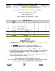
DCTD St<strong>and</strong>ard Operating Procedures (SOP)<br />
Title: <strong>Image</strong> <strong>Capture</strong> <strong>of</strong> Tumor Biopsy Slides from γH2AX Immun<strong>of</strong>luorescence Assay Page 9 <strong>of</strong> 25<br />
Doc. #: SOP340533 Revision: D Effective Date: 9/22/2013<br />
7.3.7 <strong>Capture</strong> Parameters: Left side <strong>of</strong> <strong>Capture</strong> Menu. Enter the information for each section<br />
<strong>of</strong> each slide as image capture is performed. This information is used to generate a<br />
unique image file name for each image. Example file name:<br />
WHY_ CTEP1234_0035_20101024_0T850MD_gH2AX_Predose_1678489_tumor<br />
biopsy_Sample_MP_1.tif<br />
Data Fields<br />
<strong>Image</strong> Directory<br />
<strong>Image</strong><br />
Directory:<br />
Project<br />
Use the default – C:\gH2AX qIFA\IpWin<br />
Source Files\Scripts\Projects\<br />
<strong>Capture</strong> Menu<br />
Investigator:<br />
Protocol:<br />
Slide<br />
Group ID:<br />
Patient ID:<br />
Dose:<br />
Dose Time:<br />
Bond Slide<br />
ID:<br />
Stain or<br />
Target<br />
Signal:<br />
Sample<br />
Unique<br />
Identifier:<br />
PHL<br />
Identifier:<br />
Tissue:<br />
Enter your initials<br />
“CTEP#_PatientID_Date (YYYYMM<br />
DD),” no hyphens<br />
(Names the Header Folder that is created in<br />
the <strong>Image</strong> Directory)<br />
Leave Blank<br />
Leave Blank<br />
Leave Blank<br />
Leave Blank<br />
Enter the Bond Slide ID Number as<br />
assigned by the Bond Processing Module<br />
(SOP340523)<br />
Enter “gH2AX”<br />
Type in either CalLow, CalMid, Cal<br />
High, Positive, Negative, Predose, or<br />
Postdose; no hyphens.<br />
Paraffin block number<br />
Select the type <strong>of</strong> tissue being imaged in<br />
the drop-down box (e.g., tumor biopsy)<br />
Click the appropriate checkbox<br />
<strong>Image</strong><br />
You are on<br />
<strong>Image</strong>...<br />
This will be automatically filled <strong>and</strong><br />
increment upwards; number will reset with<br />
each new Sample or Slide