Image Capture - NCI Division of Cancer Treatment and Diagnosis ...
Image Capture - NCI Division of Cancer Treatment and Diagnosis ...
Image Capture - NCI Division of Cancer Treatment and Diagnosis ...

Create successful ePaper yourself
Turn your PDF publications into a flip-book with our unique Google optimized e-Paper software.
DCTD St<strong>and</strong>ard Operating Procedures (SOP)<br />
Title: <strong>Image</strong> <strong>Capture</strong> <strong>of</strong> Tumor Biopsy Slides from γH2AX Immun<strong>of</strong>luorescence Assay Page 20 <strong>of</strong> 25<br />
Doc. #: SOP340533 Revision: D Effective Date: 9/22/2013<br />
APPENDIX 4: CALIBRATOR/CONTROL SLIDES AND CLINICAL SLIDES<br />
1. Calibrator/Control Slides<br />
A. Day-to-day variability in sample processing <strong>and</strong> staining is monitored by employing a set <strong>of</strong><br />
calibrator samples, <strong>and</strong> positive <strong>and</strong> negative controls. Two CalCon slides are required for each<br />
set <strong>of</strong> patient slides <strong>and</strong> are run in the first <strong>and</strong> last positions in each slide tray <strong>of</strong> a Bond-Max run.<br />
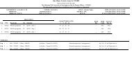
Sections & Tissue – CalCon slide<br />
Cal-Low (A)<br />
Xenograft (Vehicle-treated)<br />
Cal-Mid (B) Xenograft (<strong>Treatment</strong> A)<br />
Cal-High (C) Xenograft (<strong>Treatment</strong> B)<br />
Positive Control (D) Mouse testes<br />
Negative Control (E) Mouse jejunum<br />
B. Control Section Criteria<br />
a. The positive control specimen, mouse testes, should be intensely stained with the γH2AX<br />
Ab (> 10%). Staining should be restricted to spermatogonia, spermatocytes, <strong>and</strong><br />
spermatids; spermatozoa should be negative for γH2AX. Blood vessels, seminiferous<br />
tubules, <strong>and</strong> connective tissue should be negative for nuclear γH2AX staining.<br />
b. The negative control specimen, mouse jejunum, should be low/negative for γH2AX<br />
(≤ 1%). Minimal staining for γH2AX may be present in the crypt cells.<br />
C. γH2AX Calibrator Section Criteria<br />
a. Calibrator sections are generated from NBF-fixed, paraffin-embedded mouse xenograft<br />
tumor quadrants derived from vehicle <strong>and</strong> drug treated animals representing 3 different<br />
levels <strong>of</strong> γH2AX expression (Calibrators [Cal] -Low, -Mid, <strong>and</strong> -High). The calibrators<br />
serve as a visual reference st<strong>and</strong>ard for drug-effect on target.<br />
b. The specifications for γH2AX levels in the calibrators are determined by lot:<br />
Tissue<br />
Cal-Low (A)<br />
Cal-Mid (B)<br />
Cal-High (C)<br />
Specifications Provided by Lot<br />
Max %NAP threshold, set by lot<br />
Min %NAP threshold <strong>and</strong> min fold over Cal-Low, set<br />
by lot<br />
Min %NAP threshold <strong>and</strong> min fold over Cal-Low, set<br />
by lot<br />
c. If the γH2AX control specimens pass, but the calibrators fail, γH2AX can still be<br />
reported, but should not be considered quantitative.