Image Capture - NCI Division of Cancer Treatment and Diagnosis ...
Image Capture - NCI Division of Cancer Treatment and Diagnosis ...
Image Capture - NCI Division of Cancer Treatment and Diagnosis ...

You also want an ePaper? Increase the reach of your titles
YUMPU automatically turns print PDFs into web optimized ePapers that Google loves.
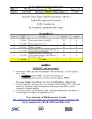
DCTD St<strong>and</strong>ard Operating Procedures (SOP)<br />
Title: <strong>Image</strong> <strong>Capture</strong> <strong>of</strong> Tumor Biopsy Slides from γH2AX Immun<strong>of</strong>luorescence Assay Page 10 <strong>of</strong> 25<br />
Doc. #: SOP340533 Revision: D Effective Date: 9/22/2013<br />
7.3.8 <strong>Capture</strong> Steps: Right side <strong>of</strong> <strong>Capture</strong> Menu:<br />
Identify phase-dense regions <strong>of</strong> the biopsy at 200x magnification under phase contrast.<br />
See Appendix 5, Sections 1 <strong>and</strong> 2 for field selection suggestions.<br />
Step 1 Change the microscope excitation filter to the DAPI filter (Chroma A4 filter, BP 360/40),<br />
click the Auto Exposure button, <strong>and</strong> adjust the focus on the sample to be imaged. Select<br />
areas on the slide with no necrosis or confounders <strong>and</strong> avoid edges <strong>of</strong> the tissue area to<br />
avoid edge-staining artifacts (for examples <strong>of</strong> phase-contrast <strong>and</strong> fluorescent images, see<br />
Appendix 5, Sections 2 <strong>and</strong> 3). Click Continue. Note: Keep the light source shutter<br />
closed on the microscope as much as possible to prevent bleaching <strong>of</strong> the Strp488 <strong>and</strong><br />
DAPI signals.<br />
Step 2 Adjust the exposure for the DAPI channel (in milliseconds [ms]) for optimal image<br />
capture. Begin with a test exposure time <strong>of</strong> 30-50 ms; click the Test Exposure button.<br />
The displayed image will represent what the downstream quantitation macro will identify<br />
as quantifiable nuclei (blue); overexposed nuclei (pink); or non-counted, underexposed<br />
nuclei (purple). Overexposure <strong>of</strong> the signal will have a “swelling” effect on the area <strong>of</strong><br />
the nuclei, giving a larger “false-positive” area. Underexposure will shrink the area,<br />
resulting in a weaker DAPI signal in stained nuclei. Sample images <strong>of</strong> over-, under-, <strong>and</strong><br />
correct exposures are in Appendix 6, Section 1.<br />
Increase or decrease the exposure time for the DAPI signal for optimal nuclei exposure<br />
within the image field (i.e., minimize the underexposed nuclei without swelling the<br />
nuclear area). Once the exposure is adjusted to give an accurate representation <strong>of</strong> the<br />
nuclei, click Continue, <strong>and</strong> the program will capture the DAPI image.<br />
Step 3 Change the microscope excitation filter to the Strp488 filter (FITC/fluorescein; Chroma<br />
L5 filter, BP 480/40). Click the Auto Exposure button (may need to click multiple times<br />
to increase the exposure enough to see some background aut<strong>of</strong>luorescence).<br />
A selection box will appear over a monochrome image in a capture window;<br />
alternatively, place a check by “Intensity Map,” <strong>and</strong> the monochrome image will be<br />
replaced by a color intensity map <strong>of</strong> the image. The selection box will be used to<br />
establish the Strp488 background aut<strong>of</strong>luorescence. Move the selection box to an area <strong>of</strong><br />
the image that shows the most intense background aut<strong>of</strong>luorescence signal. Within this<br />
region, ensure that there are no black areas (lacking signal), debris fluorescence, or<br />
γH2AX-specific signal. Once an area is selected, click Get Exposure.<br />
The γH2AX exposure is adjusted <strong>and</strong> captured by the macro automatically. This step<br />
may take a few moments depending on the amount <strong>of</strong> background aut<strong>of</strong>luorescence.<br />
The γH2AX/DAPI composite *.tif file will be saved to the Header Folder as entered in<br />
the <strong>Image</strong> Directory field <strong>of</strong> the <strong>Capture</strong> Menu. An example <strong>of</strong> a captured image is<br />
shown in Appendix 6, Section 2.<br />
Note: <strong>Image</strong>-Pro occasionally behaves erratically during image capture. For example, the<br />
selection box for aut<strong>of</strong>luorescence (Step 3 above) can jump across the capture window or the<br />
image capture process can take an inordinate amount <strong>of</strong> time. Be sure the correct version <strong>of</strong><br />
<strong>Image</strong>-Pro is being used <strong>and</strong> that the drivers for the camera are up to date. To update the drivers,<br />
follow the instructions in Appendix 2. With older computers, the speed <strong>and</strong> memory <strong>of</strong> the<br />
capture computer may need to be increased if the macro behaves erratically