Conservative Zirconia Bridge for Anterior Tooth Replacement
Conservative Zirconia Bridge for Anterior Tooth Replacement
Conservative Zirconia Bridge for Anterior Tooth Replacement

You also want an ePaper? Increase the reach of your titles
YUMPU automatically turns print PDFs into web optimized ePapers that Google loves.
2<br />
ce<br />
dentalCEtoday.com<br />
AESTHETICS<br />
Test 143.2<br />
<strong>Conservative</strong> <strong>Zirconia</strong> <strong>Bridge</strong> <strong>for</strong><br />
<strong>Anterior</strong> <strong>Tooth</strong> <strong>Replacement</strong><br />
Jack D. Griffin<br />
Jr, DMD<br />
<strong>Conservative</strong> replacement of lost or<br />
missing teeth is simplified with<br />
today’s restorative materials, and<br />
allows the clinician to create a “marriage”<br />
between aesthetics and durability. 1 Im -<br />
plants, full-contour bridges, and a variety of<br />
removable bridges must all be considered<br />
when planning any tooth replacement<br />
case. To achieve long-lasting success, a<br />
proper union of material selection, wellplanned<br />
tooth preparation, proper bridge<br />
design, dependable bonding, biologically<br />
acceptable soft-tissue treatment, tolerated<br />
occlusion, and accurate communication<br />
among the patient, dentist, and laboratory<br />
must all exist. 2,3<br />
Popular “metal-free” tooth colored indirect<br />
restorative materials include zirconia<br />
(no layering porcelain), zirconia (layering<br />
with porcelain), lituium disilicate ceramic,<br />
and leucite rein<strong>for</strong>med ceramic. <strong>Zirconia</strong><br />
has proven to be an excellent choice <strong>for</strong><br />
bridge substructures, but its bond to tooth<br />
structure has been minimal. However, with<br />
current primers that are shown to covalently<br />
bond to zirconia, conservative, metal-free<br />
options can be routinely considered.<br />
This article discusses the clinical applications<br />
and techniques <strong>for</strong> zirconia restorations.<br />
A case report is presented that<br />
describes a conservative anterior bridge<br />
using a new zirconia primer, adhesive<br />
bonding, and retentive preparations.<br />
ZIRCONIA AS A CONSERVATIVE BRIDGE<br />
SUBSTRATE<br />
Significant advances with indirect aesthetic<br />
materials the last few years have brought<br />
the profession higher levels of strength and<br />
aesthetics than ever be<strong>for</strong>e in bridge de -<br />
sign. 4,5 <strong>Zirconia</strong> has been widely used within<br />
the last few years as a bridge framework<br />
because of its nonmetalic color, fracture<br />
resistance with flexural tests over 1,000<br />
MPa, and excellent long-term clinical success.<br />
6,7 These CAD/CAM fabricated bridge<br />
frameworks have been primarily used <strong>for</strong><br />
full coverage restorations with dependable<br />
cosmetic results. 8,9<br />
Likewise, conservative zirconia-fixed<br />
partial dentures can be a minimally invasive<br />
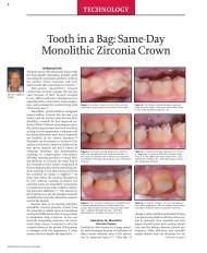
Figure 1. The patient had been concerned with<br />
darkening of the retained deciduous tooth “h” <strong>for</strong><br />
several years.<br />
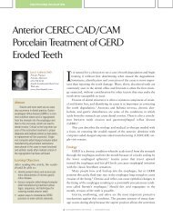
Figure 3. Extraction and replacement of tooth “h”<br />
was necessary after an increase in mobility and<br />
pain.<br />
alternative <strong>for</strong> anterior tooth replacement<br />
and have proven to be very successful; particularly<br />
if retentive preparations are done,<br />
there is a defined and limited path of insertion,<br />
if bonding is adequate, and if connectors<br />
are of sufficient thickness and height. 10<br />
A layering porcelain is added to enhance the<br />
aesthetics and has proven durable, particularly<br />
if occlusal stresses are minimal. 11,12<br />
Another advantage of zirconia is that it<br />
can be cemented, or in cases with less retention<br />
<strong>for</strong>m in the preparations, it can be<br />
bonded in place. <strong>Zirconia</strong> is an acid resistant,<br />
polycrystalline ceramic that does not<br />
contain amorphous silica, making it ineffective<br />
to traditional glass etching treatments<br />
such as hydrofluoric acid (HFl) followed by<br />
silane. 13,14 Bonding of zirconium based<br />
restorations cannot be done with the same<br />
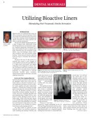
Figure 2. Examination revealed an otherwise<br />
healthy dentition with slight spaces and incisal<br />
wear. The patient wanted the space between the<br />
central incisors repaired if possible. Veneers and<br />
a full coverage bridge were ruled out.<br />
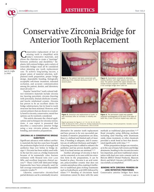
Figure 4. The permanent cuspid was palatally<br />
impacted mesioangularly at the level of the apex of<br />
teeth Nos. 9 and 10 and an implant was ruled out.<br />
Reprint permission <strong>for</strong> Figures 1, 2, 7, 9, 13, 14, 15, 18, 21.<br />
Griffin JD, Byound IS, Chen L, Brown DJ. Surface treatments <strong>for</strong> zirconia bonding: a clinical perspective. Canadian Journal<br />
of Restorative Dentistry and Prosthodontics. 2011;3:23-29.<br />
methods as traditional glass-porcelain. 15,16<br />
Bond strengths using differing methods<br />
including sand blasting with aluminum<br />
oxide, silane treatment, or other chemicals<br />
provided a weak bond at best that deteriorated<br />
significantly with time. 17-20<br />
When preparation designs are retentive,<br />
as in the case of many full crowns and bridge<br />
abutments, bonding to the zirconia becomes<br />
less important, and more traditional cementation<br />
with dual-cure resin cements such as<br />
BisCem (BISCO Dental Products), Maxcem<br />
(Kerr), and RelyX Unicem (3M ESPE) can be<br />
successfully accomplished.<br />
BONDING WITH ZIRCONIA PRIMERS ON<br />
LESS RETENTIVE PROSTHESES<br />
<strong>Conservative</strong> anterior bridges have been<br />
continued on page xx<br />
DENTALCETODAY.COM • NOVEMBER 2011
3<br />
AESTHETICS<br />
<strong>Conservative</strong> <strong>Zirconia</strong> <strong>Bridge</strong>...<br />
continued from page 00<br />
tried <strong>for</strong> many years with varying<br />
degrees of success and failure. An<br />
increase in adherence may make their<br />
use more attractive in future planning.<br />
Reliable adhesion, proper abutment<br />
preparation using boxes of a single<br />
insertion path, and group function<br />
or bicuspid disclusion can all be factors<br />
affecting predictable long-term<br />
success. 21,22<br />
Despite minimal chemical adhesion<br />
with zirconia using traditional<br />
bonding systems, micromechanical<br />
retention has been sufficient <strong>for</strong> complete<br />
coverage restorations. 23 Studies<br />
have been somewhat encouraging<br />
with some silanization techniques,<br />
but higher bond strengths <strong>for</strong> conservative<br />
restorations would be a significant<br />
advantage. 24,25 A zirconia<br />
primer, Z-Prime Plus (BISCO Dental<br />
Products) has been developed that has<br />
been shown to significantly increase<br />
the shear bond strength of 4 different<br />
light-cured and self-cured resin<br />
cements (BisCem [BISCO Dental<br />
Products], Unicem (3M ESPE), Bisfix<br />
SE (VOCO America), SmartCem 2<br />
[DENTSPLY Caulk]), according to<br />
research conducted at Bisco Inc. in<br />
2010. The author considers the use of<br />
primers on indirect substrates in<br />
retentive cases as an improved means<br />
to seal this interface and reduce water<br />
penetration between the cement and<br />
the zirconia undersurface.<br />
For restorations that have short<br />
clinical crowns, or where resistance<br />
<strong>for</strong>m is lacking, it is helpful to have a<br />
primer that will work on materials<br />
that are nonsilica based to increase<br />
retention of the restoration. 26,27 Ma -<br />
terials such as zirconia, alumina, and<br />
metal have no glass component, so traditional<br />
etching with HFl is not effective.<br />
The phosphate monomers <strong>for</strong>m<br />
the basis <strong>for</strong> chemical bonds between<br />
the zirconia and the primer, allowing<br />
<strong>for</strong> a cohesive bond to the resin<br />
cement. This reliable bond over time is<br />
critical, especially in a minimally<br />
retentive case with conservative<br />
bridge abutments, as in the case that<br />
follows. 28,19<br />
CASE REPORT: CONSERVATIVE<br />
REPLACEMENT OF TOOTH NO. 11<br />
Treatment Plan<br />
A 58-year-old woman had a retained<br />
deciduous tooth “h” with a mesioangular<br />
impacted tooth No. 11 extending<br />
under teeth No. 9 through No. 12<br />
(Figure 1). <strong>Tooth</strong> “h” had become<br />
mobile over the last year and reached<br />
the point of severe pain upon touch<br />
Figure 5. Extraction of “h” was uneventful<br />
and the mesial papilla was quite good, but<br />
there was slight blunting of the distal papilla.<br />
Figure 8. Granulation tissue was removed<br />
and a graft material placed in the extraction<br />
site. Be<strong>for</strong>e the temporary bridge was<br />
cemented, a collagen membrane was laid<br />
over the site to keep cement from moving<br />
toward the graft.<br />
Figure 11. Impressions and models were<br />
accompanied by photos and a CD<br />
containing all images taken be<strong>for</strong>e and<br />
during the procedure.<br />
with grade 3 mobility. The tooth was<br />
deemed hopeless and extraction was<br />
indicated (Figure 2). She had a 3-mm<br />
overjet and a one-mm deep bite with<br />
no centric contact on the lingual of<br />
the maxillary incisors. Her occlusion<br />
in lateral movements was in group<br />
function with the cusp tip of tooth<br />
No. 6, slightly worn with generalized<br />
enamel crazing. This occlusal scheme<br />
would provide a more suitable situation<br />
<strong>for</strong> a conservative bridge than<br />
someone with a more advanced deep<br />
bite, heavy cingulum contact, and evidence<br />
of parafunctional habits. The<br />
incisal aspect of tooth No. 9 had an<br />
asymptomatic fracture from trauma<br />
several years earlier.<br />
Surgical exposure of tooth No. 11<br />
with orthodontic ligation and traction<br />
was ruled out because of the<br />
unpredictability of <strong>for</strong>ced eruption in<br />
adults and the patient’s desire to avoid<br />
orthodontics. Because of the position<br />
Figure 6. After tooth preparation a temporary<br />
was made with a dual-cure composite<br />
in a matrix from the pre-op condition of the<br />
tooth.<br />
Figure 9. After 3 months of healing, the<br />
temporary was removed and a good ovate<br />
<strong>for</strong>m was present. The preparations were<br />
refined and impressions taken.<br />
Figure 12. The lab was instructed to remove<br />
a slight amount of stone (about 0.5 mm) in<br />
the pontic area so that the bridge would put<br />
slight pressure on the soft tissues <strong>for</strong><br />
support.<br />
of the impacted tooth, an oral surgeon<br />
felt that the extraction, bone grafting,<br />
and implant placement would be<br />
unpredictable.<br />
More extensive cosmetic/restorative<br />
treatment was ruled out <strong>for</strong> financial<br />
reasons and the patient’s request to<br />
not have teeth “ground on” (Figure 3).<br />
The accepted plan was <strong>for</strong> extraction of<br />
“h,” a conservative zirconia framework/porcelain<br />
bridge to replace tooth<br />
No. 11, use of composite on tooth No. 9<br />
and the mesial of tooth No. 10 to<br />
improve aesthetics, and on tooth No. 6<br />
to restore the cusp tip (Figure 4).<br />
Preparation of Tissues<br />
After administration of local anesthetic,<br />
the deciduous tooth “h” was extracted,<br />
granulation tissue was removed,<br />
and the abutment teeth were prepared<br />
using a medium grit tapered diamond<br />
(Figure 5). The lateral incisor was prepared<br />
with 1 to 1.5 mm reduction with<br />
Figure 7. Composite was placed on the<br />
tissue side, making a convex surface in all<br />
directions <strong>for</strong> tissue adaptation. The<br />
material was added and slight blanching of<br />
the tissues verified.<br />
Figure 10. Photos of the teeth to be<br />
matched were taken <strong>for</strong> the ceramist.<br />
Figure 13. The bridge framework of zirconia<br />
was covered with an add-on porcelain <strong>for</strong><br />
aesthetics.<br />
a rounded shoulder and 1.5 mm deep<br />
seating grooves placed on the mesial<br />
and distal aspects of the preparation.<br />
The box on the distal was 2 to 3 mm<br />
into the tooth and 3 to 4 mm in height<br />
from gingival to incisal.<br />
The composite on the bicuspid was<br />
removed and the lingual cusp reduced<br />
<strong>for</strong> about 2 mm of clearance in excursive<br />
movements. Interproximal box<br />
preparations with a width and height<br />
of about 3 mm were done to provide a<br />
dovetail with a definite path of insertion<br />
and to provide bulk <strong>for</strong> connectors.<br />
These boxes were made parallel<br />
with a limited path of insertion complimenting<br />
the preparation of the lateral<br />
incisor to decrease chances of displacement<br />
and to increase surface area<br />
<strong>for</strong> luting material bonding.<br />
A composite temporary restoration<br />
(Luxatemp [DMG America]) with<br />
an ovate type pontic was fabricated<br />
continued on page xx<br />
DENTALCETODAY.COM • NOVEMBER 2011
4<br />
AESTHETICS<br />
<strong>Conservative</strong> <strong>Zirconia</strong> <strong>Bridge</strong>...<br />
continued from page 00<br />
from a matrix made from the arch<br />
be<strong>for</strong>e the extraction was done<br />
(Figure 6). The tissue surface was<br />
smoothed and a small dome of composite<br />
was added to make a 100% convex<br />
surface to cover the extraction site<br />
and to provide slight pressure so that<br />
minimal blanching was seen when<br />
fully placed into position. This slight<br />
pressure is critical <strong>for</strong> papilla support<br />
and maintenance of aesthetic gingival<br />
contours (Figure 7). Support of the<br />
gingival tissues with good ovoid pontic<br />
<strong>for</strong>m and papilla support are critical<br />
in tooth replacement cases. Proper<br />
design is needed to maintain buccallingual<br />
width of the extraction site, to<br />
preserve the papilla with support by<br />
application of slight pressure, to<br />
encourage growth of soft tissue into<br />
an “ovoid” <strong>for</strong>m <strong>for</strong> proper emergence<br />
Figure 14. Materials needed <strong>for</strong> bonding a<br />
zirconia bridge include phosphoric acid<br />
etch, a dual-cure bonding agent, dual-cure<br />
resin cement, silane, and the zirconia<br />
primer.<br />
Figure 17. Composite bonding was per<strong>for</strong>med<br />
to close the diastema, correct the<br />
incisal chipping on tooth No. 9, to correct<br />
the size and position of tooth No. 10, and<br />
to add the incisal tip of tooth No. 6.<br />
Figure 20. The blending of the bridge with<br />
composite restorations gave a very natural<br />
result with very little occlusal change.<br />
profile of the final pontic, and to help<br />
stabilize the graft material.<br />
Pep Gen granular (DENTSPLY Cera -<br />
Med) was then placed in the extraction<br />
site to help minimize resorbtion<br />
(Figure 7). 29-31 A collagen membrane<br />
was placed over the graft material and<br />
the temporary was then cemented<br />
(TempBond Clear [Kerr]). It was fabricated<br />
to exert slight pressure on the gingiva<br />
with slight tissue blanching when<br />
in its final position (Figure 8). Final<br />
shaping was accomplished followed by<br />
polishing with disks.<br />
The patient returned <strong>for</strong> definitive<br />
tooth preparation and impressions 3<br />
months later. The healing of the soft<br />
tissue was good with acceptable adaptation<br />
to the temporary (Figure 9). The<br />
preparations were refined with a fine<br />
shamfer diamond with boxes and<br />
channel providing a single path of<br />
insertion. Vinyl polysiloxane impressions<br />
were then taken (Precision [Dis -<br />
Figure 15. After try in, the zirconia wings<br />
were cleaned in an ethyl alcohol bath,<br />
rinsed, silanated, and the primer applied.<br />
Figure 18. Preservation of the ridge with<br />
minor grafting, ovoid temporary fabrication,<br />
and an ovoid pontic with slight pressure on<br />
the gingiva allowed <strong>for</strong> a natural soft-tissue<br />
emergence.<br />
Figure 21. The result is an improvement in<br />
the smile with a natural blending of materials.<br />
The success of this conservative bridge<br />
will rely upon the preparation, dentin bonding,<br />
and adhesion to the bridge framework.<br />
cus Dental]) and sent to the lab with a<br />
full series of shade and character photos,<br />
all preoperative photos, bite registration,<br />
and opposing model. A key <strong>for</strong><br />
giving the ceramist the best chance of<br />
matching existing teeth is with good<br />
photography on nondesiccated teeth<br />
showing at least 2 different shade tabs<br />
on the same plane as the teeth being<br />
matched, a contrastor to provide a<br />
dark background <strong>for</strong> better character<br />
identification, and using a quality<br />
camera with good lighting control<br />
(Figure 10).<br />
Figure 16. The teeth preparations were<br />
etched <strong>for</strong> 15 seconds, rinsed thoroughly,<br />
suction dried, and several coats of dual-cure<br />
DBA were applied and air thinned.<br />
Figure 19. Soft-tissue tolerance at almost 2<br />
years was excellent, and the integrity of the<br />
materials has been acceptable.<br />
Figure 22. A final PA shows the position of<br />
the impacted cuspid and the bonded<br />
prosthesis; zirconia is radiopaque.<br />
A <strong>Zirconia</strong> Framework <strong>Bridge</strong><br />
The laboratory prescription was <strong>for</strong> a<br />
zirconia framework bridge with layering<br />
porcelain over an ovate pontic<br />
design (Figure 11). The lab technician<br />
was instructed to create an “ideal”<br />
ovate pontic site by removing stone to<br />
make a smooth, completely convex<br />
surface and a highly aesthetic emergence<br />
profile (Figure 12). A CAD/CAM<br />
zirconia framework was made (LAVA<br />
[3M ESPE]) and layering porcelain<br />
(Ceram Overlay Porcelain [3M ESPE])<br />
was added to the external surface <strong>for</strong><br />
customization (Figure 13).<br />
At the cementation appointment,<br />
the temporary was removed and the<br />
preparations were cleaned with a<br />
pumice and alcohol on a microbrush.<br />
The materials needed <strong>for</strong> cementation<br />
of zirconia are a dual-cure dentin<br />
bonding agent (All Bond 3 [BISCO<br />
Dental Products]), silane, zirconia<br />
primer (Z Prime Plus [BISCO Dental<br />
Products]), and a dual-cure composite<br />
cement (DuoLink [BISCO Dental<br />
Products]) (Figure 14). After verification<br />
of fit, the bridge was cleaned with<br />
ethyl alcohol and an unhydrolyzed 2-<br />
part silane agent (Bis-silane [BISCO<br />
Dental Products]) was applied. 32 A<br />
drop of zirconia primer (Z-Prime<br />
[BISCO Dental Products]) was placed<br />
on the internal surface of the porcelain<br />
abutments and dried after 60 seconds<br />
(Figure 15).<br />
The teeth were etched 20 seconds<br />
with phosphoric acid, rinsed, and left<br />
moist. The bonding agent was mixed<br />
and placed on both the teeth and<br />
bridge and air thinned. The luting<br />
cement (Duo-Link) was placed directly<br />
on the teeth and the bridge held<br />
into position with moderate digital<br />
pressure, cleaned, and cured with<br />
ultraviolet light (Figure 16). It is im -<br />
portant to note that both a dual-cure<br />
DBA and luting material were used<br />
because of the opacity and low light<br />
transmission of zirconia. Occlusion<br />
was checked, contact was minimal on<br />
the connectors, and group function<br />
was maintained.<br />
Composites were placed on teeth<br />
Nos. 6, 9, and 10 to correct cosmetic<br />
concerns of the patient and to restore<br />
the worn cusp tip on the cuspid.<br />
Removal of old composite material<br />
was done with a finishing diamond.<br />
An irregular finish line was created<br />
and the teeth were isolated with<br />
retractors (SeeMore [Discus Dental]),<br />
then etched with 37% phosphoric<br />
acid <strong>for</strong> 15 seconds. After thorough<br />
rinsing, several coats of a dentin bonding<br />
agent (All Bond 3 [BISCO Dental<br />
Products]) were applied and airthinned.<br />
Layers of dentin, enamel,<br />
and incisal opacity composites Re -<br />
namel [Cosmedent]) were placed and<br />
characterized with stain (Creative<br />
Color [Cosmedent]) (Figure 17).<br />
Shaping was completed with<br />
disks (SofLex [3M ESPE]) and polishing<br />
was per<strong>for</strong>med with rubber cups<br />
(FlexiDisk [Cosmedent]) (Figure 18). A<br />
continued on page xx<br />
DENTALCETODAY.COM • NOVEMBER 2011
5<br />
AESTHETICS<br />
<strong>Conservative</strong> <strong>Zirconia</strong> <strong>Bridge</strong>...<br />
continued from page 00<br />
clear vacuum-<strong>for</strong>med 2 mm hard/soft<br />
nocturnal bruxism splint was made<br />
(Erkodent [Glidewell Laboratories]),<br />
and the patient was encouraged to<br />
wear it nightly and daily when grinding/clenching<br />
was noticed.<br />
Follow-up and Evaluation<br />
The soft-tissue response at 16 months<br />
was excellent with good papilla support<br />
and a natural emergence profile.<br />
After almost 2 years, there have been<br />
no clinical problems and the patient is<br />
very happy with the results (Figure 19).<br />
She felt very com<strong>for</strong>table with “the fit”<br />
and stated she had been flossing under<br />
it almost every night and had been<br />
wearing the bruxism splint regularly.<br />
SUMMARY<br />
The demand <strong>for</strong> metal-free restorations<br />
coupled with the desire <strong>for</strong> conservation<br />
of tooth structure has put<br />
new demands on our profession.<br />
There is a symbiotic synergy among<br />
the great skills of our ceramists, the<br />
commitment to successful chemistry<br />
of our researchers and manufacturers,<br />
and the unwavering desire <strong>for</strong> happy<br />
patients and long lasting restorations<br />
by clinicians (Figure 20). With continually<br />
improving bonding materials<br />
and when tooth preparation and<br />
occlusion are well planned, conservative<br />
anterior bridges should be considered<br />
in many partially edentulous<br />
cases (Figure 21). This treatment op -<br />
tion was particularly appropriate in<br />
the case described in this article,<br />
where an impacted cuspid limited the<br />
choices of treatment and the desire <strong>for</strong><br />
conservative dentistry was maintained.(Figure<br />
22).✦<br />
Acknowledgement<br />
The author wishes to thank Erik<br />
Haupt of Haupt Dental Lab, Brea,<br />
Calif, <strong>for</strong> his excellent cosmetic work<br />
and understanding of the principles<br />
of smile design.<br />
References<br />
1. Rossmann JA, Cobb CM. Lasers in periodontal<br />
therapy. Periodontol 2000. 1995;9:150-164.<br />
2. Tipton PA. Aesthetic tooth alignment using<br />
etched porcelain restorations. Pract Proced<br />
Aesthet Dent. 2001;13:551-555.<br />
3. Dumfahrt H. Porcelain laminate veneers. A retrospective<br />
evaluation after 1 to 10 years of service:<br />
Part 1—Clinical procedure. Int J Prosthodont.<br />
1999;12:505-513.<br />
4. Christensen GJ. The ceramic crown dilemma. J<br />
Am Dent Assoc. 2010;141:1019-1022.<br />
5. Quinn GD, Studart AR, Hebert C, et al. Fatigue of<br />
zirconia and dental bridge geometry: Design<br />
implications. Dent Mater. 2010;26:1133-1136.<br />
6. Fischer H, Weber M, Marx R. Lifetime prediction<br />
of all-ceramic bridges by computational methods.<br />
J Dent Res. 2003;82:238-242.<br />
7. Quinn JB, Cheng D, Rusin R, et al. Fractographic<br />
analysis and material properties of a dental zirconia.<br />
Presented at: IADR/AADR/CADR 83rd<br />
General Session; March 9-12, 2005; Baltimore,<br />
MD.<br />
8. Burke FJ, Ali A, Palin WM. <strong>Zirconia</strong>-based allceramic<br />
crowns and bridges: three case reports.<br />
Dent Update. 2006;33:401-410.<br />
9. Kugel G, Perry RD, Aboushala A. Restoring anterior<br />
maxillary dentition using alumina- and zirconia-based<br />
CAD/CAM restorations. Compend<br />
Contin Educ Dent. 2003;24:569-576.<br />
10.Rosentritt M, Ries S, Kolbeck C, et al. Fracture<br />
characteristics of anterior resin-bonded zirconiafixed<br />
partial dentures. Clin Oral Investig.<br />
2009;13:453-457.<br />
11.Beuer F, Stimmelmayr M, Gernet W, et al.<br />
Prospective study of zirconia-based restorations:<br />
3-year clinical results. Quintessence Int.<br />
2010;41:631-637.<br />
12.Guess PC, Zavanelli RA, Silva NR, et al. Monolithic<br />
CAD/CAM lithium disilicate versus veneered Y-TZP<br />
crowns: comparison of failure modes and reliability<br />
after fatigue. Int J Prosthodont. 2010;23:434-442.<br />
13.Blatz MB, Sadan A, Kern M. Resin-ceramic bonding:<br />
a review of the literature. J Prosthet Dent.<br />
2003;89:268-274.<br />
14.Dérand P, Dérand T. Bond strength of luting cements<br />
to zirconium oxide ceramics. Int J Prosthodont.<br />
2000;13:131-135.<br />
15.Denry I, Kelly JR. State of the art of zirconia <strong>for</strong><br />
dental applications. Dent Mater. 2008;24:299-<br />
307.<br />
16.Conrad HJ, Seong WJ, Pesun IJ. Current ceramic<br />
materials and systems with clinical recommendations:<br />
a systematic review. J Prosthet Dent.<br />
2007;98:389-404.<br />
17.Ozcan M, Nijhuis H, Valandro LF. Effect of various<br />
surface conditioning methods on the adhesion of<br />
dual-cure resin cement with MDP functional<br />
monomer to zirconia after thermal aging. Dent<br />
Mater J. 2008;27:99-104.<br />
18.Wegner SM, Kern M. Long-term resin bond strength<br />
to zirconia ceramic. J Adhes Dent. 2000;2:139-<br />
147.<br />
19.Kern M, Barloi A, Yang B. Surface conditioning<br />
influences zirconia ceramic bonding. J Dent Res.<br />
2009;88:817-822.<br />
20.Ernst CP, Cohnen U, Stender E, et al. In vitro retentive<br />
strength of zirconium oxide ceramic crowns<br />
using different luting agents. J Prosthet Dent.<br />
2005;93:551-558.<br />
21.Quinn F, Gratton DR, McConnell RJ. The per<strong>for</strong>mance<br />
of conventional, fixed bridgework, retained<br />
by partial coverage crowns. J Ir Dent Assoc.<br />
1995;41:6-9.<br />
22.Foster LV. The relationship between failure and<br />
design in conventional bridgework from general<br />
dental practice. J Oral Rehabil. 1991;18:491-<br />
495.<br />
23.Kristallis TT, Phimmasone A. A zirconia-based<br />
long span restoration used in restoring anterior<br />
esthetics with minor orthodontic correction.<br />
Contemporary Esthetics. November 2006:2-7.<br />
24.Matinlinna JP, Lassila LV, Vallittu PK. Pilot evaluation<br />
of resin composite cement adhesion to zirconia<br />
using a novel silane system. Acta Odontol<br />
Scand. 2007;65:44-51.<br />
25.Atsu SS, Kilicarslan MA, Kucukesmen HC, et al.<br />
Effect of zirconium-oxide ceramic surface treatments<br />
on the bond strength to adhesive resin. J<br />
Prosthet Dent. 2006;95:430-436.<br />
26.Griffin J, Suh B, Liang C, et al. Surface treatments<br />
<strong>for</strong> zirconia bonding: A clinical perspective.<br />
Canadian Journal of Restorative Dentistry and<br />
Prosthodontics. 2010;3:23-29.<br />
27.Tanaka R, Fujishima A, Shibata Y, et al. Cooperation<br />
of phosphate monomer and silica modification on<br />
zirconia. J Dent Res. 2008;87:666-670.<br />
28.Yoshida K, Tsuo Y, Atsuta M. Bonding of dual-cured<br />
resin cement to zirconia ceramic using phosphate<br />
acid ester monomer and zirconate coupler. J Biomed<br />
Mater Res B Appl Biomater. 2006;77:28-33.<br />
29.Thompson DM, Rohrer MD, Prasad HS. Comparison<br />
of bone grafting materials in human extraction sockets:<br />
clinical, histologic, and histomorphometric evaluations.<br />
Implant Dent. 2006;15:89-96.<br />
30.Block MS, Jackson WC. Techniques <strong>for</strong> grafting<br />
the extraction site in preparation <strong>for</strong> dental im -<br />
plant placement. Atlas Oral Maxillofac Surg Clin<br />
North Am. 2006;14:1-25.<br />
31.Yukna RA, Krauser JT, Callan DP, et al. Thirty-six<br />
month follow-up of 25 patients treated with combination<br />
anorganic bovine-derived hydroxyapatite<br />
matrix (ABM)/cell-binding peptide (P-15) bone<br />
replacement grafts in human infrabony defects. I.<br />
Clinical findings. J Periodontol. 2002;73:123-128.<br />
32.Quintas AF. Predictable cementation of esthetic<br />
restorations: part II—selection criteria and guidelines<br />
<strong>for</strong> implementation. Pract Proced Aesthet<br />
Dent. 2007;19:7-9.<br />
Dr. Griffin has practiced in St. Louis county Mis -<br />
souri since 1988. In a very busy practice, he and<br />
his staff have consistently maintained a 50% to<br />
55% overhead by emphasizing cosmetics while<br />
doing all phases of general dentistry. He has<br />
been awarded by his peers Diplomate status<br />
with the American Board of Aesthetic Den tistry;<br />
holds accredited status with the Amer ican<br />
Academy of Cosmetic Dentistry; and has<br />
achieved Mastership in the AGD. He is the<br />
MasterTrack/CE chair <strong>for</strong> the Missouri AGD and<br />
is on the Council of Scien tific Affairs <strong>for</strong> the<br />
Greater St. Louis Dental Society. He considers it<br />
an honor to have taught many dentists how to<br />
improve their skills with digital photography, di -<br />
rect bonding techniques, efficiency with porcelain<br />
veneers, practice management, and CAD/CAM<br />
dentistry in lectures and scientific publication.<br />
He is grateful to be surrounded by those to help<br />
make the dental experience both re warding and<br />
enjoyable. He can be reached at esmilecenter@aol.com<br />
or visit eurekasmile.com.<br />
Disclosure: Dr. Griffin reports no disclosures.<br />
continued on page xx<br />
DENTALCETODAY.COM • NOVEMBER 2011
6<br />
AESTHETICS<br />
ce<br />
Test 143.2 Expiration date of this CE article is November 1, 2013.<br />
<strong>Conservative</strong> <strong>Zirconia</strong> <strong>Bridge</strong>...<br />
continued from page 00<br />
To submit Continuing Education answers, use the answer sheet on page xx.<br />
Circle the letter corresponding to the answer you believe is correct, detach the<br />
answer sheet from the magazine, and mail to Dentistry Today Department of<br />
Continuing Education. Or, use our easy online option at the Web site<br />
dentalcetoday.com. This article is available <strong>for</strong> 1 hour of CE credit.<br />
The following 8 questions were derived from the article <strong>Conservative</strong> <strong>Zirconia</strong><br />
<strong>Bridge</strong> <strong>for</strong> <strong>Anterior</strong> <strong>Tooth</strong> <strong>Replacement</strong> by Jack D. Griffin Jr, DMD, on pages xx<br />
through xx.<br />
Learning Objectives<br />
After reading this article, the individual will learn:<br />
l Clinical applications and techniques <strong>for</strong> zirconia restorations, and<br />
l A technique <strong>for</strong> a conservative anterior bridge using a new zirconia<br />
primer, adhesive bonding, and retentive preparations.<br />
1. Current popular metal-free indirect restorative materials include all of the following<br />
except:<br />
a. Full contour zirconia.<br />
b. <strong>Zirconia</strong> with layered porcelain.<br />
c. Captek.<br />
d. Lithium disilicate.<br />
2. An advantage of zirconia-based restorations is resistance to fracture<br />
because of flexural strength that is near:<br />
a. 100 MPa.<br />
b. 1000 MPa.<br />
c. 10,000 MPa.<br />
d. 50,000 MPa.<br />
3. Advantages of using zirconia include all of the following except:<br />
a. It has a nonmetalic color.<br />
b. It can have porcelain layered to its surface to increase aesthetics.<br />
c. It can be cemented or bonded into place.<br />
d. It can be porcelain etched and silanated like traditional ceramics.<br />
4. Treating the internal surface of zirconia by aluminum oxide sand blasting:<br />
a. Provides a weak bond at best and may provide little aid in retention.<br />
b. Is the preferred method <strong>for</strong> bonding zirconia.<br />
c. Gives reported bond strengths of around 45 MPa.<br />
d. Microetches the surface to provide a high level of mechanical retention.<br />
5. The reason zirconia doesn’t etch like porcelain is its lack of:<br />
a. Silica.<br />
b. Tin.<br />
c. Aluminum.<br />
d. Quartz.<br />
6. The following factors are critical to long-term success with conservative<br />
anterior bridges except:<br />
a. Ovoid pontic design.<br />
b. Adhesive luting.<br />
c. Proper tooth preparation with single path of insertion.<br />
d. Controlled occlusion in excursive movements.<br />
7. It is best to use a dual or self-cure luting material with zirconia because:<br />
a. Light transmission is accentuated by its high translucency.<br />
b. Lack of an air inhibited layer may decrease setting.<br />
c. Opacity of zirconium oxide may prevent adequate light penetration.<br />
d. Dual-cure materials exhibit more chance of post-cure color change.<br />
8. Important occlusal considerations <strong>for</strong> conservative anterior bridges, particularly<br />
cuspid replacement, is <strong>for</strong> the patient to have:<br />
a. A limited deep bite.<br />
b. Lack of evidence of parafunctional habits.<br />
c. Group function in lateral excursions.<br />
d. All of the above.<br />
Now available online at dentalcetoday.com. Simply log on and follow the easy<br />
directions. Immediate results with printable letter upon completion.<br />
DENTALCETODAY.COM • NOVEMBER 2011