Bone loss Factors that regulate osteoclast differentiation: an update
Bone loss Factors that regulate osteoclast differentiation: an update
Bone loss Factors that regulate osteoclast differentiation: an update

You also want an ePaper? Increase the reach of your titles
YUMPU automatically turns print PDFs into web optimized ePapers that Google loves.
http://arthritis-research.com/content/2/6/451<br />
Review<br />
<strong>Bone</strong> <strong>loss</strong><br />
<strong>Factors</strong> <strong>that</strong> <strong>regulate</strong> <strong>osteoclast</strong> <strong>differentiation</strong>: <strong>an</strong> <strong>update</strong><br />
Sophie Roux* <strong>an</strong>d Philippe Orcel †<br />
* † Lariboisière Hospital, Paris, <strong>an</strong>d *Bicêtre Hospital, Bicêtre, Fr<strong>an</strong>ce<br />
Received: 8 March 2000<br />
Revisions requested: 29 June 2000<br />
Revisions received: 7 August 2000<br />
Accepted: 14 August 2000<br />
Published: 6 September 2000<br />
Arthritis Res 2000, 2:451–456<br />
The electronic version of this article c<strong>an</strong> be found online at<br />
http://arthritis-research.com/content/2/6/451<br />
© Current Science Ltd (Print ISSN 1465-9905; Online ISSN 1465-9913)<br />
Abstract<br />
Osteoclast activation is a critical cellular process for pathological bone resorption, such as<br />
erosions in rheumatoid arthritis (RA) or generalized bone <strong>loss</strong>. Among m<strong>an</strong>y factors triggering<br />
excessive <strong>osteoclast</strong> activity, cytokines such as IL-1 or tumour necrosis factor (TNF)-α play a<br />
central role. New members of the TNF receptor lig<strong>an</strong>d family (namely receptor activator of<br />
nuclear factor-κB [RANK] <strong>an</strong>d RANK lig<strong>an</strong>d [RANKL]) have been discovered whose crossinteraction<br />
is m<strong>an</strong>datory for the <strong>differentiation</strong> of <strong>osteoclast</strong>s from hemopoietic precursors, in<br />
both physiological <strong>an</strong>d pathological situations. Osteoprotegerin, a decoy receptor which<br />
blocks this interaction, decreases <strong>osteoclast</strong> activity <strong>an</strong>d could have a fascinating therapeutic<br />
potential in conditions associated with up<strong>regulate</strong>d bone resorption.<br />
Keywords: bone cytokines, <strong>differentiation</strong>, <strong>osteoclast</strong>, osteoprotegerin, RANK, RANKL<br />
Introduction<br />
<strong>Bone</strong> remodelling is a continuous physiological process<br />
<strong>that</strong> occurs in adult skeleton in which bone resorption is<br />
followed by new bone formation, maintaining mech<strong>an</strong>ical<br />
strength <strong>an</strong>d structure. <strong>Bone</strong> cells <strong>that</strong> are responsible for<br />
this coupled process include bone-resorbing cells (<strong>osteoclast</strong>s,<br />
which are derived from haematopoietic cells of the<br />
monocyte/macrophage lineage) <strong>an</strong>d bone-forming cells<br />
(osteoblasts, which are of mesenchymal origin). The bone<br />
resorption process is involved in m<strong>an</strong>y clinical situations<br />
<strong>that</strong> are relev<strong>an</strong>t to the work of rheumatologists, such as<br />
focal bone destruction or erosion in RA <strong>an</strong>d other inflammatory<br />
arthritides, <strong>an</strong>d the diffuse bone <strong>loss</strong> <strong>that</strong> is<br />
encountered in osteoporosis.<br />
Osteoclast <strong>differentiation</strong>: basic mech<strong>an</strong>isms<br />
<strong>an</strong>d new insights<br />
Osteoclast progenitor cells are recruited from haematopoietic<br />
compartments, <strong>an</strong>d then proliferate <strong>an</strong>d differentiate<br />
toward mature <strong>osteoclast</strong>s. During this multistep<br />
<strong>differentiation</strong> process postmitotic <strong>osteoclast</strong> precursors<br />
progressively express <strong>osteoclast</strong>-associated markers, such<br />
M-CSF = macrophage colony-stimulating factor; NF-κB = nuclear factor-κB; ODF = <strong>osteoclast</strong> <strong>differentiation</strong> factor; 1,25(OH) 2 D 3 = 1,25-dihydroxyvitamin<br />
D 3<br />
; PTH = parathyroid hormone; RA = rheumatoid arthritis; RANK = receptor activator of nuclear factor-κB; RANKL = receptor activator of<br />
nuclear factor-κB lig<strong>an</strong>d; TNF = tumour necrosis factor.
Arthritis Research Vol 2 No 6 Roux <strong>an</strong>d Orcel<br />
as calcitonin receptor <strong>an</strong>d tartrate-resist<strong>an</strong>t acid phosphatase,<br />
as they lose some of their macrophage characteristics.<br />
Then, mononuclear pre<strong>osteoclast</strong>s fuse together to<br />
form multinucleated gi<strong>an</strong>t cells. Terminal <strong>osteoclast</strong> <strong>differentiation</strong><br />
eventually leads to active bone-resorbing cells [1].<br />
Role of osteoblast/stromal cells in <strong>osteoclast</strong><br />
<strong>differentiation</strong><br />
Biological models of in vitro <strong>osteoclast</strong> <strong>differentiation</strong> have<br />
been developed <strong>that</strong> have facilitated detailed study of<br />
m<strong>an</strong>y of the factors involved in the regulation of this<br />
process. The most commonly studied models are cultures<br />
of mouse bone marrow or cocultures of haematopoietic<br />
cells with bone-derived stromal cells, which give rise to<br />
large numbers of bone-resorbing <strong>osteoclast</strong>s [2]. Studies<br />
based on these models have found <strong>that</strong> mesenchymally<br />
derived stromal cells play a critical role in supporting <strong>an</strong>d<br />
stimulating <strong>osteoclast</strong> <strong>differentiation</strong>, a process <strong>that</strong> probably<br />
necessitates cell–cell contact between <strong>osteoclast</strong><br />
precursors <strong>an</strong>d stromal cells [3,4]. In some hum<strong>an</strong> models,<br />
however, a cellular interaction between <strong>osteoclast</strong> precursors<br />
<strong>an</strong>d stromal cells is not always required [5–7].<br />
Local <strong>an</strong>d hormonal factors <strong>that</strong> are involved in <strong>osteoclast</strong><br />
<strong>differentiation</strong><br />
<strong>Bone</strong> resorption is closely controlled in vivo by cellular<br />
<strong>an</strong>d hormonal factors, which affect not only <strong>osteoclast</strong><br />
activity, but also <strong>osteoclast</strong> formation. Parathyroid<br />
hormone (PTH) <strong>an</strong>d 1,25-dihydroxyvitamin D 3<br />
[1,25(OH) 2<br />
D 3<br />
]<br />
increase bone resorption, primarily via <strong>an</strong> indirect mech<strong>an</strong>ism<br />
<strong>that</strong> is mediated by osteoblasts [2]. Oestrogens have<br />
a negative impact on <strong>osteoclast</strong> <strong>differentiation</strong>, <strong>an</strong>d oestrogen<br />
deficiency leads to increased <strong>osteoclast</strong> <strong>differentiation</strong><br />
<strong>an</strong>d activation [8]. The cytokines IL-1, IL-6 <strong>an</strong>d TNF-α<br />
are known to increase bone resorption by stimulating both<br />
<strong>osteoclast</strong> activity <strong>an</strong>d <strong>differentiation</strong>. This effect involves,<br />
at least in part, prostagl<strong>an</strong>din production [9,10]. The major<br />
role of macrophage colony-stimulating factor (M-CSF) has<br />
been pointed out in M-CSF-deficient (op/op) mice, which<br />
develop <strong>an</strong> osteopetrosis <strong>that</strong> is characterized by the<br />
absence of <strong>osteoclast</strong>s [11]. Studies using murine cocultures<br />
[12] have shown <strong>that</strong> M-CSF acts both on proliferation<br />
<strong>an</strong>d on <strong>differentiation</strong> of precursor cells. Local<br />
injections of M-CSF in rat metaphyseal bone also increase<br />
in situ <strong>osteoclast</strong> <strong>differentiation</strong> <strong>an</strong>d bone resorption [13].<br />
Other cytokines stimulate bone resorption at least partly<br />
by increasing <strong>osteoclast</strong> <strong>differentiation</strong>, such as leukaemia<br />
inhibiting factor <strong>an</strong>d IL-11 [14,15]. Conversely, some<br />
cytokines such as IL-4 or IFN-γ have been shown to inhibit<br />
<strong>osteoclast</strong> <strong>differentiation</strong> in vitro [16]. The role of tr<strong>an</strong>sforming<br />
growth factor-β is more complex; it decreases<br />
<strong>osteoclast</strong> precursor proliferation <strong>an</strong>d bone resorption<br />
activity [17,18], but it also increases the expression of<br />
two <strong>osteoclast</strong>ic markers – vitronectin receptor <strong>an</strong>d calcitonin<br />
receptor [19,20]. Most of the cytokines <strong>that</strong> <strong>regulate</strong><br />
<strong>osteoclast</strong> <strong>differentiation</strong> are produced in the bone<br />
microenvironment, mainly by osteoblast/stromal cells,<br />
further emphasizing the key role of these cells in <strong>osteoclast</strong><br />
<strong>differentiation</strong>.<br />
A new interactive system in <strong>osteoclast</strong> bone<br />
resorption<br />
The recent discovery of new members of the TNF receptor<br />
lig<strong>an</strong>d family has pointed out the crucial role of RANK <strong>an</strong>d<br />
RANKL in <strong>osteoclast</strong> <strong>differentiation</strong> <strong>an</strong>d activation [21 • ,<br />
22 • ] (Fig. 1).<br />
RANKL, also called <strong>osteoclast</strong> <strong>differentiation</strong> factor (ODF),<br />
TNF-related induced cytokine (TRANCE), or<br />
osteoprotegerin lig<strong>an</strong>d (OPGL)<br />
Because osteoblast–stromal cell interactions with <strong>osteoclast</strong><br />
precursors are required for subsequent <strong>osteoclast</strong><br />
<strong>differentiation</strong>, <strong>an</strong> ODF expressed by these cells <strong>an</strong>d recognized<br />
by <strong>osteoclast</strong> precursors was suspected. Such a<br />
factor was identified as RANKL [23 •• ,24 •• ]. RANKL is a<br />
membr<strong>an</strong>e-bound TNF-related factor <strong>that</strong> is expressed by<br />
osteoblast/stromal cells. That the presence of RANKL is<br />
vital in <strong>osteoclast</strong> <strong>differentiation</strong> is now well established,<br />
<strong>an</strong>d its soluble recombin<strong>an</strong>t form has been tested in a<br />
number of in vitro <strong>an</strong>d in vivo studies. In in vitro murine or<br />
hum<strong>an</strong> <strong>osteoclast</strong> <strong>differentiation</strong> models, soluble RANKL<br />
enables <strong>osteoclast</strong> precursors to differentiate in the presence<br />
of M-CSF, even in the absence of osteoblast/stromal<br />
cells [25 •• ,26]. <strong>Bone</strong> resorption activity is increased, as<br />
well as the <strong>osteoclast</strong> survival [23 •• ,26]. Mice <strong>that</strong> are<br />
defective for RANKL develop a form of osteopetrosis.<br />
They are characterized by the absence of <strong>osteoclast</strong>s,<br />
although <strong>osteoclast</strong> progenitors are present <strong>an</strong>d are able<br />
to differentiate into bone-resorbing <strong>osteoclast</strong>s in the<br />
presence of normal osteoblast/stromal cells [27 •• ].<br />
In addition, the soluble form of RANKL has been shown to<br />
be produced by hum<strong>an</strong> fibroblasts tr<strong>an</strong>sfected with <strong>an</strong><br />
expression vector for RANKL <strong>an</strong>d by in vitro activated<br />
murine T cells [23 •• ,28]. However, it is not clear whether<br />
this soluble form plays a role in vivo in normal bone homeostasis<br />
or in pathological processes <strong>that</strong> are characterized<br />
by increased bone resorption.<br />
RANK<br />
Osteoclast precursors express RANK, a membr<strong>an</strong>e-bound<br />
TNF receptor <strong>that</strong> recognizes RANKL through a direct<br />
cell–cell interaction with osteoblast/stromal cells [29 •• ].<br />
Recent studies [29 •• ,30] demonstrated <strong>that</strong> this receptor is<br />
essential for the tr<strong>an</strong>sduction of signals <strong>that</strong> lead to <strong>osteoclast</strong><br />
<strong>differentiation</strong>. An overexpression of soluble RANK<br />
results in osteopetrosis, with a decreased number of <strong>osteoclast</strong>s<br />
[30]. Conversely, mice <strong>that</strong> are deficient for RANK<br />
develop a severe osteopetrosis <strong>that</strong> is characterized by the<br />
absence of <strong>osteoclast</strong>s. In addition, <strong>osteoclast</strong> precursors<br />
in these mice are unable to differentiate to <strong>osteoclast</strong>s in<br />
vitro, in the presence of RANKL <strong>an</strong>d M-CSF [31].
http://arthritis-research.com/content/2/6/451<br />
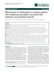
Figure 1<br />
New members of the TNF receptor lig<strong>an</strong>d family: role of RANKL (ODF,<br />
TRANCE, OPGL) <strong>an</strong>d its receptor RANK in <strong>osteoclast</strong> <strong>differentiation</strong>.<br />
RANKL, a membr<strong>an</strong>e-bound TNF-related factor, is expressed by<br />
osteoblast/stromal cells <strong>an</strong>d is up<strong>regulate</strong>d by osteotropic factors such<br />
as 1,25(OH) 2<br />
D 3<br />
, PTH, IL-6 or IL-11. Osteoclast (OC) precursors<br />
express RANK, a membr<strong>an</strong>e-bound TNF receptor, <strong>that</strong> recognizes<br />
RANKL through cell–cell interaction with osteoblast/stromal cells. This<br />
interaction enables <strong>osteoclast</strong> precursors to differentiate in the<br />
presence of M-CSF. Osteoprotegerin (OPG) is a member of the TNF<br />
receptor family <strong>that</strong> lacks a tr<strong>an</strong>smembr<strong>an</strong>e domain <strong>an</strong>d represents a<br />
secreted TNF receptor. OPG recognizes RANKL, <strong>an</strong>d this decoy<br />
receptor blocks the interaction between RANK <strong>an</strong>d RANKL, leading to<br />
inhibition of <strong>osteoclast</strong> <strong>differentiation</strong> <strong>an</strong>d activation.<br />
Osteoprotegerin<br />
Osteoprotegerin is a member of the TNF receptor family<br />
<strong>that</strong> lacks a tr<strong>an</strong>smembr<strong>an</strong>e domain <strong>an</strong>d represents a<br />
secreted receptor. Osteoprotegerin recognizes RANKL,<br />
<strong>an</strong>d this decoy receptor blocks the interaction between<br />
RANK <strong>an</strong>d RANKL, leading to <strong>an</strong> inhibition of <strong>osteoclast</strong><br />
<strong>differentiation</strong> <strong>an</strong>d activation [32 •• ,33 •• ]. Overexpression of<br />
osteoprotegerin in tr<strong>an</strong>sgenic mice results in a form of<br />
osteopetrosis <strong>that</strong> is characterized by a defect in <strong>osteoclast</strong><br />
<strong>differentiation</strong> [32 •• ]. By contrast, osteoprotegerindeficient<br />
mice develop severe osteoporosis because of<br />
increased <strong>osteoclast</strong> <strong>differentiation</strong> <strong>an</strong>d function [34 •• ]. In<br />
vitro studies have demonstrated the strong inhibitory action<br />
of osteoprotegerin on <strong>osteoclast</strong> <strong>differentiation</strong>, as well as<br />
on the bone-resorbing activity of <strong>osteoclast</strong>s [32 •• ,33 •• ].<br />
Role of RANK/RANKL <strong>an</strong>d osteoprotegerin in<br />
<strong>osteoclast</strong> <strong>differentiation</strong><br />
RANKL/osteoprotegerin bal<strong>an</strong>ce, signal tr<strong>an</strong>sduction <strong>an</strong>d<br />
<strong>osteoclast</strong> <strong>differentiation</strong><br />
Recent data suggest <strong>that</strong> M-CSF <strong>an</strong>d RANKL are two<br />
major factors involved in <strong>osteoclast</strong> <strong>differentiation</strong>. M-CSF<br />
is required for both proliferation <strong>an</strong>d <strong>differentiation</strong>, <strong>an</strong>d<br />
RANKL (which is not a growth factor) is required for <strong>differentiation</strong><br />
into mature <strong>osteoclast</strong>s <strong>an</strong>d for <strong>osteoclast</strong> activity<br />
[26]. In bone tissue, osteoprotegerin <strong>an</strong>d RANKL are<br />
expressed by osteoblast/stromal cells, <strong>an</strong>d the ratio of<br />
these products may modulate the ability of these cells to<br />
stimulate <strong>osteoclast</strong> <strong>differentiation</strong>/activity, as well as the<br />
rate of bone resorption [21 • ].<br />
In addition, it has been shown [26] <strong>that</strong> the interaction<br />
between RANKL <strong>an</strong>d RANK results in a tr<strong>an</strong>sduction<br />
signal in pre<strong>osteoclast</strong>s <strong>an</strong>d in mature <strong>osteoclast</strong>s <strong>that</strong><br />
may activate nuclear factor-κ (NF-κB). The role of NF-κB<br />
in the <strong>osteoclast</strong> <strong>differentiation</strong> has been previously<br />
demonstrated in mice with a double knockout for the p50<br />
<strong>an</strong>d p52 NF-κB subunits, in which a defect of <strong>osteoclast</strong><br />
<strong>differentiation</strong> leads to <strong>an</strong> osteopetrosis [35,36]. Other<br />
intracellular events are activated by tr<strong>an</strong>sduction signals,<br />
such as c-jun terminal kinase, <strong>an</strong>d TNF receptor-associated<br />
factors, which <strong>regulate</strong> activation of NF-κB <strong>an</strong>d/or<br />
c-jun terminal kinase [21 • ].<br />
RANK/RANKL, immune cells <strong>an</strong>d <strong>osteoclast</strong> <strong>differentiation</strong><br />
RANK <strong>an</strong>d RANKL have been shown to be expressed in<br />
dendritic cells <strong>an</strong>d T lymphocytes, respectively, in which<br />
they appear to be import<strong>an</strong>t regulators of the interactions<br />
between these cells [37,38]. These data suggest <strong>that</strong>,<br />
apart from <strong>osteoclast</strong> <strong>differentiation</strong> <strong>an</strong>d activation, RANK<br />
<strong>an</strong>d RANKL are involved in the immune system as suggested<br />
by the mice knockout models. RANKL-deficient<br />
mice lack lymph nodes <strong>an</strong>d exhibit defects in <strong>differentiation</strong><br />
of T <strong>an</strong>d B lymphocytes [27 •• ], <strong>an</strong>d RANK-deficient<br />
mice exhibit a marked deficiency in B cells in the spleen<br />
<strong>an</strong>d lack lymph nodes [31].<br />
Regulation of RANKL <strong>an</strong>d osteoprotegerin expression<br />
Recent studies have demonstrated <strong>that</strong> osteotropic<br />
factors <strong>an</strong>d hormones such as PTH, 1,25(OH) 2<br />
D 3<br />
, IL-11,<br />
IL-1β, TNF-α or prostagl<strong>an</strong>din E 2<br />
up<strong>regulate</strong> RANKL<br />
expression in osteoblast/stromal cells (Fig. 1). In addition,<br />
osteoprotegerin expression is down<strong>regulate</strong>d by<br />
prostagl<strong>an</strong>din E 2<br />
, <strong>an</strong>d is up<strong>regulate</strong>d by oestrogens [21 • ].<br />
RANK expression has not yet been extensively studied.<br />
An integrated view <strong>an</strong>d clinical implications<br />
An emerging concept is <strong>that</strong> cytokines <strong>an</strong>d hormonal<br />
factors <strong>that</strong> are involved in bone resorption may act by a<br />
common final pathway involving RANKL <strong>an</strong>d RANK [21 • ].<br />
In accord<strong>an</strong>ce with this concept, a recent in vivo study<br />
[39 •• ] has shown <strong>that</strong> a recombin<strong>an</strong>t chimaeric Fc fusion<br />
form of osteoprotegerin inhibited hypercalcaemia <strong>an</strong>d bone<br />
resorption induced by IL-1β, TNF-α, PTH <strong>an</strong>d 1,25(OH) 2<br />
D 3<br />
in mice. This convergence theory is probably not exclusive<br />
because recent studies [40,41] have suggested <strong>that</strong> the<br />
effects of TNF-α or IL-6 may involve different effectors.<br />
Therapeutic perspectives<br />
The concept presented above will probably lead to new<br />
therapeutic approaches in several diseases <strong>that</strong> are characterized<br />
by excessive bone resorption. Thus, osteoprote-
Arthritis Research Vol 2 No 6 Roux <strong>an</strong>d Orcel<br />
gerin (a specific inhibitor of RANKL) or <strong>an</strong> <strong>an</strong>alogue may<br />
be used to block the excess of bone resorption in pathological<br />
conditions such as hyper-resorption of malign<strong>an</strong>cy,<br />
in which this pathway seems to be primarily involved<br />
[42 •• ], or in osteoporosis, in which oestrogen deficiency<br />
could lead to decreased production of osteoprotegerin<br />
<strong>an</strong>d subsequent increased bone resorption.<br />
<strong>Bone</strong> erosions in rheumatoid arthritis<br />
Rheumatoid arthritis is <strong>an</strong>other interesting clinical model<br />
for the study of the role of RANK/RANKL in bone erosions,<br />
<strong>an</strong>d as a therapeutic target for osteoprotegerin.<br />
Rheumatoid arthritis is characterized by progressive bone<br />
<strong>an</strong>d cartilage destruction as a result of chronic synovitis.<br />
Numerous studies have pointed out the role of cytokines<br />
such as TNF-α or IL-1 in the joint destruction [43]. Recent<br />
studies suggest <strong>that</strong> RANKL mRNA is highly expressed in<br />
synovial tissues from patients with RA, but not in normal<br />
synovial tissues. This expression is detected in synovial<br />
fibroblasts, as well as in activated T cells derived from RA<br />
synovial tissues, suggesting <strong>that</strong> these cells may contribute<br />
to <strong>osteoclast</strong> formation at the specific sites of bone<br />
destruction in RA [44 • ,45 • ]. In addition, in rat adjuv<strong>an</strong>tinduced<br />
arthritis, RANKL is expressed on the surface of<br />
activated T cells isolated from affected rats, <strong>an</strong>d may be<br />
secreted in T cell cultures. Activated T cells could therefore<br />
directly induce <strong>osteoclast</strong>ogenesis through membr<strong>an</strong>e-bound<br />
<strong>an</strong>d soluble RANKL [28]. These data<br />
suggest <strong>that</strong> RANKL may have a major pathophysiological<br />
import<strong>an</strong>ce in the bone <strong>an</strong>d joint destruction observed in<br />
inflammatory arthritides such as RA. Activated T cells,<br />
which play a central role in the pathogenesis of RA, may<br />
(in addition to stromal cells) contribute to the <strong>osteoclast</strong>mediated<br />
bone resorption via RANKL expression [46 •• ].<br />
Conclusion<br />
Osteoclasts are multinucleated cells <strong>that</strong> are formed by<br />
fusion of <strong>osteoclast</strong> precursors from haematopoietic<br />
origin. These cells are responsible for bone resorption<br />
<strong>an</strong>d <strong>osteoclast</strong> <strong>differentiation</strong> <strong>an</strong>d represent <strong>an</strong> evident<br />
point of control of bone resorption. <strong>Bone</strong> resorption is<br />
closely <strong>regulate</strong>d in vivo by m<strong>an</strong>y cellular <strong>an</strong>d hormonal<br />
factors, which affect not only <strong>osteoclast</strong> activity, but also<br />
<strong>osteoclast</strong> formation. The recent discovery of new<br />
members of the TNF receptor lig<strong>an</strong>d family (ODF, TNFrelated<br />
induced cytokine, osteoprotegerin lig<strong>an</strong>d) have<br />
emphasized the crucial role of RANKL, which is<br />
expressed by osteoblast/stromal cells, <strong>an</strong>d its receptor<br />
RANK, which is expressed by <strong>osteoclast</strong> cells, in <strong>osteoclast</strong><br />
<strong>differentiation</strong> <strong>an</strong>d activation. This system is completed<br />
by osteoprotegerin, which is a secreted TNF<br />
receptor. Osteoprotegerin recognizes RANKL, <strong>an</strong>d this<br />
decoy receptor blocks the interaction between RANK<br />
<strong>an</strong>d RANKL. A number of osteotropic factors <strong>an</strong>d hormones<br />
may modulate bone resorption via this common<br />
final pathway, which may represent a potential therapeutic<br />
target in pathologic processes <strong>that</strong> are characterized<br />
by excessive bone resorption.<br />
References<br />
Articles of particular interest have been highlighted as:<br />
• of special interest<br />
•• of outst<strong>an</strong>ding interest<br />
1. Takahashi N, Udagawa N, T<strong>an</strong>aka S, Murakami H, Ow<strong>an</strong> I, Tamura T,<br />
Suda T: Postmitotic <strong>osteoclast</strong> precursors are mononuclear cells<br />
which express macrophage-associated phenotypes. Dev Biol<br />
1994, 163:212–221.<br />
2. Suda T, Takahashi N, Martin TJ: Modulation of <strong>osteoclast</strong> <strong>differentiation</strong>.<br />
Endocr Rev 1992, 13:66–80.<br />
3. Takahashi N, Akatsu T, Udagawa N, Sasaki T, Yamaguchi A, Moseley<br />
JM, Martin TJ, Suda T: Osteoblastic cells are involved in <strong>osteoclast</strong><br />
formation. Endocrinology 1988, 123:2600–2602.<br />
4. Quinn JM, McGee JO, Ath<strong>an</strong>asou NA: Cellular <strong>an</strong>d hormonal factors<br />
influencing monocyte <strong>differentiation</strong> to <strong>osteoclast</strong>ic bone-resorbing<br />
cells. Endocrinology 1994, 134:2416–2423.<br />
5. Kurihara N, Civin C, Roodm<strong>an</strong> GD: Osteotropic factor responsiveness<br />
of highly purified populations of early <strong>an</strong>d late precursors for<br />
hum<strong>an</strong> multinucleated cells expressing the <strong>osteoclast</strong> phenotype.<br />
J <strong>Bone</strong> Miner Res 1991, 6:257–261.<br />
6. Matayoshi A, Brown C, DiPersio JF, Haug J, Abu-Amer Y, Liapis H,<br />
Kuestner R, Pacifici R: Hum<strong>an</strong> blood-mobilized hematopoietic precursors<br />
differentiate into <strong>osteoclast</strong>s in the absence of stromal<br />
cells. Proc Natl Acad Sci USA 1996, 93:10785–10790.<br />
7. Roux S, Quinn J, Pichaud F, Orcel P, Chastre E, Jullienne A, De Vernejoul<br />
MC: Hum<strong>an</strong> cord blood monocytes undergo terminal <strong>osteoclast</strong><br />
<strong>differentiation</strong> in vitro in the presence of culture medium<br />
conditioned by gi<strong>an</strong>t cell tumor of bone. J Cell Physiol 1996, 168:<br />
489–498.<br />
8. de Vernejoul MC, Cohen-Solal M, Orcel P: <strong>Bone</strong> cytokines. Curr<br />
Opin Rheumatol 1993, 5:332–338.<br />
9. Roodm<strong>an</strong> GD: Interleukin-6: <strong>an</strong> osteotropic factor? J <strong>Bone</strong> Miner<br />
Res 1992, 7:475–478.<br />
10. Pfeilschifter J, Chenu C, Bird A, Mundy GR, Roodm<strong>an</strong> GD: Interleukin-1<br />
<strong>an</strong>d tumor necrosis factor stimulate the formation of<br />
hum<strong>an</strong> <strong>osteoclast</strong>like cells in vitro. J <strong>Bone</strong> Miner Res 1989,<br />
4:113–118.<br />
11. Yoshida H, Hayashi S, Kunisada T, Ogawa M, Nishikawa S, Okamura<br />
H, Sudo T, Shultz LD, Nishikawa S: The murine mutation osteopetrosis<br />
is in the coding region of macrophage colony stimulating<br />
factor gene. Nature 1990, 345:442–444.<br />
12. T<strong>an</strong>aka S, Takahashi N, Udagawa N, Tamura T, Akatsu T, St<strong>an</strong>ley ER,<br />
Kurokawa T, Suda T: Macrophage colony-stimulating factor is<br />
indispensable for both proliferation <strong>an</strong>d <strong>differentiation</strong> of <strong>osteoclast</strong><br />
progenitors. J Clin Invest 1993, 91:257–263.<br />
13. Orcel P, Feuga M, Bielakoff J, de Vernejoul MC: Local bone injections<br />
of LPS <strong>an</strong>d M-CSF increase bone resorption by different<br />
pathways in vivo in rats. Am J Physiol 1993, 264:E391–E397.<br />
14. Ishimi Y, Abe E, Jin CH, Miyaura C, Hong MH, Oshida M, Kurosawa H,<br />
Yamaguchi Y, Tomida M, Hozumi M: Leukemia inhibitory factor/ <strong>differentiation</strong>-stimulating<br />
factor (LIF/D-Factor): regulation of its<br />
production <strong>an</strong>d possible roles in bone metabolism. J Cell Physiol<br />
1992, 152:71–78.<br />
15. Girasole G, Passeri G, Jilka RL, M<strong>an</strong>olagas SC: Interleukin-11: a new<br />
cytokine critical for <strong>osteoclast</strong> development. J Clin Invest 1994,<br />
93:1516–1524.
http://arthritis-research.com/content/2/6/451<br />
16. Lacey DL, Erdm<strong>an</strong>n JM, Teitelbaum SL, T<strong>an</strong> H, Ohara J, Shioi A: Interleukin<br />
4, Interferon-γ <strong>an</strong>d prostagl<strong>an</strong>din E impact the <strong>osteoclast</strong>ic<br />
cell-forming potential of murine bone marrow macrophages.<br />
Endocrinology 1995, 136:2367–2376.<br />
17. Oreffo RO, <strong>Bone</strong>wald L, Kukita A, Garrett IR, Seyedin SM, Rosen D,<br />
Mundy GR: Inhibitory effects of bone-derived growth factors,<br />
osteoinductive factor <strong>an</strong>d tr<strong>an</strong>sforming growth factor-β on isolated<br />
<strong>osteoclast</strong>s. Endocrinology 1990, 126:3069–3075.<br />
18. Chenu C, Pfeilschifter J, Mundy GR, Roodm<strong>an</strong> GD: T<strong>an</strong>sforming<br />
growth factor β inhibits formation of <strong>osteoclast</strong>-like cells in longterm<br />
hum<strong>an</strong> marrow cultures. Proc Natl Acad Sci USA 1988, 85:<br />
5683–5687.<br />
19. Mbalaviele G, Orcel P, Bouizar Z, Julienne A, de Vernejoul MC: Tr<strong>an</strong>sforming<br />
growth factor β enh<strong>an</strong>ces calcitonin-induced cyclic AMP<br />
production <strong>an</strong>d the number of calcitonin receptors in long term<br />
cultures of hum<strong>an</strong> umbilical cord blood monocytes in the presence<br />
of 1,25-dihydroxycholecalciferol. J Cell Physiol 1992, 152:<br />
486–494.<br />
20. Orcel P, Bielakoff J, de Vernejoul MC: Effect of tr<strong>an</strong>sforming growth<br />
factor β on long-term hum<strong>an</strong> cord blood monocytes cultures. J<br />
Cell Physiol 1990, 142:293–298.<br />
21. Hofbauer LC, Khosla S, Dunst<strong>an</strong> CR, Lacey DL, Boyle WJ, Riggs BL:<br />
• The roles of osteoprotegerin <strong>an</strong>d osteoprotegerin lig<strong>an</strong>d in the<br />
paracrine regulation of bone resorption. J <strong>Bone</strong> Miner Res 2000,<br />
15:2–12.<br />
This review concerns RANKL, RANK <strong>an</strong>d osteoprotegerin. Osteoclast <strong>differentiation</strong><br />
may be determined by the relative ratio of RANKL to osteoprotegerin<br />
in the bone marrow microenvironment. These factors mediate the<br />
effects of large numbers of upstream hormones <strong>an</strong>d cytokines, suggesting a<br />
final common pathway in the regulation of <strong>osteoclast</strong>ogenesis.<br />
22. Suda T, Takahashi N, Udagawa N, Jimi E, Gillespie MT, Martin TJ:<br />
• Modulation of <strong>osteoclast</strong> <strong>differentiation</strong> <strong>an</strong>d function by the new<br />
members of the tumor necrosis factor receptor <strong>an</strong>d lig<strong>an</strong>d families.<br />
Endocr Rev 1999, 20:345–357.<br />
This review concerns RANKL, RANK <strong>an</strong>d osteoprotegerin – three key molecules<br />
<strong>that</strong> <strong>regulate</strong> <strong>osteoclast</strong> recruitment <strong>an</strong>d function.<br />
23. Lacey DL, Timms E, T<strong>an</strong> HL, Kelley MJ, Dunst<strong>an</strong> CR, Burgess T, Elliott<br />
•• R, Colombero A, Elliott G, Scully S, Hsu H, Sulliv<strong>an</strong> J, Hawkins N,<br />
Davy E, Capparelli C, Eli A, Qi<strong>an</strong> YX, Kaufm<strong>an</strong> S, Sarosi I, Shalhoub V,<br />
Senaldi G, Guo J, Del<strong>an</strong>ey J, Boyle WJ: Osteoprotegerin lig<strong>an</strong>d is a<br />
cytokine <strong>that</strong> <strong>regulate</strong>s <strong>osteoclast</strong> <strong>differentiation</strong> <strong>an</strong>d activation.<br />
Cell 1998, 93:165–176.<br />
The identification <strong>an</strong>d cloning of the lig<strong>an</strong>d of osteoprotegerin from a murine<br />
myelomonocytic cell line is discussed. Data are presented <strong>that</strong> suggest <strong>that</strong><br />
OPGL is <strong>an</strong> <strong>osteoclast</strong> <strong>differentiation</strong> <strong>an</strong>d activation factor.<br />
24. Yasuda H, Shima N, Nakagawa N, Yamaguchi K, Kinosaki M,<br />
•• Mochizuki S, Tomoyasu A, Y<strong>an</strong>o K, Goto M, Murakami A, Tsuda E,<br />
MorinagaT, Higashio K, Udagawa N, Takahashi N, Suda T: Osteoclast<br />
<strong>differentiation</strong> factor is a lig<strong>an</strong>d for osteoprotegerin/<strong>osteoclast</strong>ogenesis-inhibitory<br />
factor <strong>an</strong>d is identical to TRANCE/RANKL. Proc<br />
Natl Acad Sci USA 1998, 95:3597–3602.<br />
This paper describes the identification <strong>an</strong>d cloning of the lig<strong>an</strong>d of osteoprotegerin,<br />
ODF, from mouse stromal cell ST2. ODF mediates <strong>an</strong> essential<br />
signal to <strong>osteoclast</strong> progenitors for their <strong>differentiation</strong> into <strong>osteoclast</strong>s.<br />
25. Quinn JM, Elliott J, Gillespie MT, Martin TJ: A combination of osteo-<br />
•• clast <strong>differentiation</strong> factor <strong>an</strong>d macrophage-colony stimulating<br />
factor is sufficient for both hum<strong>an</strong> <strong>an</strong>d mouse <strong>osteoclast</strong> formation<br />
in vitro. Endocrinology 1998, 139:4424–4427.<br />
This study demonstrates <strong>that</strong> murine <strong>an</strong>d hum<strong>an</strong> <strong>osteoclast</strong> precursors,<br />
when cultured in the presence of M-CSF <strong>an</strong>d a soluble form of murine ODF,<br />
form bone-resorbing <strong>osteoclast</strong>s in the absence of osteoblast/stromal cells.<br />
26. Jimi E, Akiyama S, Tsurukai T, Okahashi N, Kobayashi K, Udagawa N,<br />
Nishihara T, Takahashi N, Suda T: Osteoclast <strong>differentiation</strong> factor<br />
acts as a multifunctional regulator in murine <strong>osteoclast</strong> <strong>differentiation</strong><br />
<strong>an</strong>d function. J Immunol 1999, 163:434–442.<br />
27. Kong YY, Yoshida H, Sarosi I, T<strong>an</strong> HL, Timms E, Capparelli C, Morony<br />
•• S, Oliveira-dos-S<strong>an</strong>tos AJ, V<strong>an</strong> G, Itie A, Khoo W, Wakeham A,<br />
Dunst<strong>an</strong> CR, Lacey DL, Mak TW, Boyle WJ, Penninger JM: OPGL is a<br />
key regulator of <strong>osteoclast</strong>ogenesis, lymphocyte development<br />
<strong>an</strong>d lymph-node org<strong>an</strong>ogenesis. Nature 1999, 397:315–323.<br />
Osteoprotegerin lig<strong>an</strong>d-deficient mice develop severe osteopetrosis <strong>an</strong>d a<br />
defect in tooth eruption, <strong>an</strong>d completely lack <strong>osteoclast</strong>s as a result of <strong>an</strong><br />
inability of osteoblasts to support <strong>osteoclast</strong>ogenesis.<br />
28. Kong YY, Feige U, Sarosi I, Bolon B, Tafuri A, Morony S, Capparelli C,<br />
Li J, Elliott R, McCabe S, Wong T, Campagnuolo G, Mor<strong>an</strong> E, Bogoch<br />
ER, V<strong>an</strong> G, Nguyen LT, Ohashi PS, Lacey DL, Fish E, Boyle WJ, Penninger<br />
JM: Activated T cells <strong>regulate</strong> bone <strong>loss</strong> <strong>an</strong>d joint destruction<br />
in adjuv<strong>an</strong>t arthritis through osteoprotegerin lig<strong>an</strong>d. Nature<br />
1999, 402:304–309.<br />
29. Nakagawa N, Kinosaki M, Yamaguchi K, Shima N, Yasuda H, Y<strong>an</strong>o K,<br />
•• Morinaga T, Higashio K: RANK is the essential signaling receptor<br />
for <strong>osteoclast</strong> <strong>differentiation</strong> factor in <strong>osteoclast</strong>ogenesis.<br />
Biochem Biophys Res Commun 1998, 253:395–400.<br />
This paper describes the cloning of ODF receptor, RANK, from a mouse<br />
macrophage-like <strong>osteoclast</strong> progenitor cell line.<br />
30. Hsu H, Lacey DL, Dunst<strong>an</strong> CR, Solovyev I, Colombero A, Timms E,<br />
T<strong>an</strong> HL, Elliott G, Kelley MJ, Sarosi I, W<strong>an</strong>g L, Xia XZ, Elliott R, Chiu L,<br />
Black T, Scully S, Capparelli C, Morony S, Shimamoto G, Bass MB,<br />
Boyle WJ: Tumor necrosis factor receptor family member RANK<br />
mediates <strong>osteoclast</strong> <strong>differentiation</strong> <strong>an</strong>d activation induced by<br />
osteoprotegerin lig<strong>an</strong>d. Proc Natl Acad Sci USA 1999, 96:3540–<br />
3545.<br />
31. Dougall WC, Glaccum M, Charrier K, Rohrbach K, Brasel K, De<br />
Smedt T, Daro E, Smith J, Tometsko ME, Maliszewski CR, Armstrong<br />
A, Shen V, Bain S, Cosm<strong>an</strong> D, Anderson D, Morrissey PJ, Peschon JJ,<br />
Schuh J: RANK is essential for <strong>osteoclast</strong> <strong>an</strong>d lymph node development.<br />
Genes Dev 1999, 13:2412–2424.<br />
32. Simonet WS, Lacey DL, Dunst<strong>an</strong> CR, Kelley M, Ch<strong>an</strong>g MS, Luthy R,<br />
•• NguyenHQ, Wooden S, Bennett L, Boone T, Shimamoto G, DeRose<br />
M, Elliott R, Colombero A, T<strong>an</strong> HL, Trail G, Sulliv<strong>an</strong> J, Davy E, Bucay N,<br />
Renshaw-Gegg L, Hughes TM, Hill D, Pattison W, Campbell P, Boyle<br />
WJ: Osteoprotegerin: a novel secreted protein involved in the regulation<br />
of bone density. Cell 1997, 89:309–319.<br />
This paper describes the identification of osteoprotegerin, a potent inhibitor<br />
of bone resorption, from a foetal rat intestine cDNA library. In vivo, overexpression<br />
of osteoprotegerin in tr<strong>an</strong>sgenic mice or administration of recombin<strong>an</strong>t<br />
osteoprotegerin into normal mice results in a severe osteopetrosis,<br />
secondary to a decrease in later stages of <strong>osteoclast</strong> <strong>differentiation</strong>.<br />
33. Tsuda E, Goto M, Mochizuki S, Y<strong>an</strong>o K, Kobayashi F, Morinaga T,<br />
•• Higashio K: Isolation of a novel cytokine from hum<strong>an</strong> fibroblasts<br />
<strong>that</strong> specifically inhibits <strong>osteoclast</strong>ogenesis. Biochem Biophys Res<br />
Commun 1997, 234:137–142.<br />
Identification of <strong>osteoclast</strong>ogenesis inhibitory factor from hum<strong>an</strong> embryonic<br />
lung fibroblasts is described. This factor, which is identical to osteoprotegerin,<br />
inhibits <strong>osteoclast</strong>-like cell formation stimulated through three distinct<br />
signalling pathways involving 1α,25-dihydroxyvitamin D 3<br />
, PTH or IL-11.<br />
34. Bucay N, Sarosi I, Dunst<strong>an</strong> CR, Morony S, Tarpley J, Capparelli C,<br />
•• Scully S, T<strong>an</strong> HL, Xu W, Lacey DL, Boyle WJ, Simonet WS: Osteoprotegerin-deficient<br />
mice develop early onset osteoporosis <strong>an</strong>d<br />
arterial calcification. Genes Dev 1998, 12:1260–1268.<br />
Osteoprotegerin-deficient mice develop <strong>an</strong> osteoporosis <strong>that</strong> is characterized<br />
by severe trabecular <strong>an</strong>d cortical bone porosity, marked thinning of the<br />
parietal bones of the skull, <strong>an</strong>d a high incidence of fractures. Osteoprotegerin-deficient<br />
mice also exhibit medial calcification of the aorta <strong>an</strong>d renal<br />
arteries.<br />
35. Fr<strong>an</strong>zoso G, Carlson L, Xing L, Poljak L, Shores EW, Brown KD,<br />
Leonardi A, Tr<strong>an</strong> T, Boyce BF, Siebenlist U: Requirement for NFkappaB<br />
in <strong>osteoclast</strong> <strong>an</strong>d B-cell development. Genes Dev 1997,<br />
11:3482–3496.<br />
36. Iotsova V, Caam<strong>an</strong>o J, Loy J, Y<strong>an</strong>g Y, Lewin A, Bravo R: Osteopetrosis<br />
in mice lacking NF-kappaB1 <strong>an</strong>d NF-kappaB2. Nature Med<br />
1997, 3:1285–1289.
Arthritis Research Vol 2 No 6 Roux <strong>an</strong>d Orcel<br />
37. Anderson DM, Maraskovsky E, Billingsley WL, Dougall WC, Tometsko<br />
ME, Roux ER, Teepe MC, DuBose RF, Cosm<strong>an</strong> D, Galibert L: A<br />
homologue of the TNF receptor <strong>an</strong>d its lig<strong>an</strong>d enh<strong>an</strong>ce T-cell<br />
growth <strong>an</strong>d dendritic-cell function. Nature 1997, 390:175–179.<br />
38. Wong BR, Rho J, Arron J, Robinson E, Orlinick J, Chao M, Kalachikov<br />
S, Cay<strong>an</strong>i E, Bartlett FS 3rd, Fr<strong>an</strong>kel WN, Lee SY, Choi Y: TRANCE is<br />
a novel lig<strong>an</strong>d of the tumor necrosis factor receptor family <strong>that</strong><br />
activates c-Jun N-terminal kinase in T cells. J Biol Chem 1997,<br />
272:25190–25194.<br />
39. Morony S, Capparelli C, Lee R, Shimamoto G, Boone T, Lacey DL,<br />
•• Dunst<strong>an</strong> CR: A chimeric form of osteoprotegerin inhibits hypercalcemia<br />
<strong>an</strong>d bone resorption induced by IL-1beta, TNF-alpha, PTH,<br />
PTHrP, <strong>an</strong>d 1, 25(OH)2D3. J <strong>Bone</strong> Miner Res 1999, 14:1478–1485.<br />
In vivo administration of osteoprotegerin prevented bone resorption <strong>an</strong>d<br />
hypercalcaemia induced by bone-resorbing factors (IL-1β, TNF-α, PTH,<br />
PTH related-protein, <strong>an</strong>d 1,25(OH) 2<br />
D 3<br />
).<br />
Authors’ affiliations: Sophie Roux <strong>an</strong>d Philippe Orcel (INSERM U349,<br />
Lariboisière Hospital, Paris, Fr<strong>an</strong>ce), Sophie Roux (Department of<br />
Rheumatology, Bicêtre Hospital, Bicêtre, Fr<strong>an</strong>ce), <strong>an</strong>d Philippe Orcel<br />
(Department of Rheumatology, Lariboisière Hospital, Paris, Fr<strong>an</strong>ce)<br />
Correspondence: Philippe Orcel, Professor of Rheumatology, MD,<br />
PhD, Department of Rheumatology, Lariboisière Hospital, Centre<br />
Viggo Petersen, 2, rue Ambroise Paré, 75475 Paris, Fr<strong>an</strong>ce.<br />
Tel: +33 1 4995 8825; fax: +33 1 4995 8631;<br />
e-mail: philippe.orcel@lrb.ap-hop-paris.fr<br />
40. Hofbauer LC, Lacey DL, Dunst<strong>an</strong> CR, Spelsberg TC, Riggs BL,<br />
Khosla S: Interleukin-1beta <strong>an</strong>d tumor necrosis factor-alpha, but<br />
not interleukin- 6, stimulate osteoprotegerin lig<strong>an</strong>d gene expression<br />
in hum<strong>an</strong> osteoblastic cells. <strong>Bone</strong> 1999, 25:255–259.<br />
41. Kobayashi K, Takahashi N, Jimi E, Udagawa N, Takami M, Kotake S,<br />
Nakagawa N, Kinosaki M, Yamaguchi K, Shima N, Yasuda H, Morinaga<br />
T, Higashio K, Martin TJ, Suda T: Tumor necrosis factor alpha stimulates<br />
<strong>osteoclast</strong> <strong>differentiation</strong> by a mech<strong>an</strong>ism independent of<br />
the ODF/RANKL-RANK interaction. J Exp Med 2000, 191:275–286.<br />
42. Chikatsu N, Takeuchi Y, Tamura Y, Fukumoto S, Y<strong>an</strong>o K, Tsuda E,<br />
•• Ogata E, Fujita T: Interactions between c<strong>an</strong>cer <strong>an</strong>d bone marrow<br />
cells induce <strong>osteoclast</strong> <strong>differentiation</strong> factor expression <strong>an</strong>d<br />
<strong>osteoclast</strong>-like cell formation in vitro. Biochem Biophys Res<br />
Commun 2000, 267:632–637.<br />
Enh<strong>an</strong>ced <strong>osteoclast</strong>ogenesis in the presence of c<strong>an</strong>cer cells might be due<br />
to <strong>an</strong> increase in ODF (RANKL) activity. The interactions between c<strong>an</strong>cer<br />
cells <strong>an</strong>d mouse bone marrow cells induce ODF expression <strong>an</strong>d suppress<br />
osteoprotegerin level in bone metastases, resulting in increased local bone<br />
destruction.<br />
43. Duff GW: Cytokines <strong>an</strong>d <strong>an</strong>ti-cytokines. Br J Rheumatol 1993,<br />
32(Suppl 1):15–20.<br />
44. Gravallese EM, M<strong>an</strong>ning C, Tsay A, Naito A, P<strong>an</strong> C, Amento E,<br />
• Goldring SR: Synovial tissue in rheumatoid arthritis is a source of<br />
<strong>osteoclast</strong> <strong>differentiation</strong> factor. Arthritis Rheum 2000, 43:250–258.<br />
ODF (RANKL) is expressed in synovial tissues from RA but not in normal<br />
synovium. This expression is detected in cultured synovial fibroblasts <strong>an</strong>d in<br />
activated T cells derived from RA synovial tissue.<br />
45. Takay<strong>an</strong>agi H, Iizuka H, Juji T, Nakagawa T, Yamamoto A, Miyazaki T,<br />
• Koshihara Y, Oda H, Nakamura K, T<strong>an</strong>aka S: Involvement of receptor<br />
activator of nuclear factor kappaB lig<strong>an</strong>d/<strong>osteoclast</strong> <strong>differentiation</strong><br />
factor in <strong>osteoclast</strong>ogenesis from synoviocytes in rheumatoid<br />
arthritis. Arthritis Rheum 2000, 43:259–269.<br />
RANKL is highly expressed in synovial tissues from RA, but not in normal<br />
synovium or in osteoarthritic synovium. Cultured rheumatoid synovial fibroblasts<br />
expressed RANKL <strong>an</strong>d are able to induce <strong>osteoclast</strong> <strong>differentiation</strong>,<br />
which requires cell–cell contact between synovial cells <strong>an</strong>d <strong>osteoclast</strong> precursors.<br />
46. Horwood NJ, Kartsogi<strong>an</strong>nis V, Quinn JM, Romas E, Martin TJ, Gillespie<br />
•• MT: Activated T lymphocytes support <strong>osteoclast</strong> formation in vitro.<br />
Biochem Biophys Res Commun 1999, 265:144–150.<br />
Hum<strong>an</strong> activated T lymphocytes produce RANKL. In addition, they support<br />
<strong>osteoclast</strong> <strong>differentiation</strong> in cocultures with murine spleen cells used as a<br />
source of <strong>osteoclast</strong> precursors.