Rezumatul tezei de doctorat - USAMV Cluj-Napoca
Rezumatul tezei de doctorat - USAMV Cluj-Napoca
Rezumatul tezei de doctorat - USAMV Cluj-Napoca

Create successful ePaper yourself
Turn your PDF publications into a flip-book with our unique Google optimized e-Paper software.
UNIVERSITATEA DE ŞTIINłE AGRICOLE<br />
ŞI MEDICINĂ VETERINARĂ CLUJ-NAPOCA<br />
ŞCOALA DOCTORALĂ<br />
FACULTATEA DE ZOOTEHNIE ŞI BIOTEHNOLOGII<br />
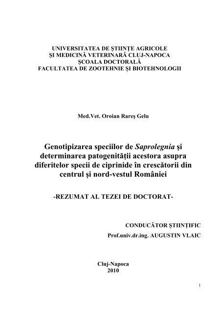
Med.Vet. Oroian Rareş Gelu<br />
Genotipizarea speciilor <strong>de</strong> Saprolegnia şi<br />
<strong>de</strong>terminarea patogenităŃii acestora asupra<br />
diferitelor specii <strong>de</strong> ciprini<strong>de</strong> în crescătorii din<br />
centrul şi nord-vestul României<br />
-REZUMAT AL TEZEI DE DOCTORAT-<br />
CONDUCĂTOR ŞTIINłIFIC<br />
Prof.univ.dr.ing. AUGUSTIN VLAIC<br />
<strong>Cluj</strong>-<strong>Napoca</strong><br />
2010<br />
1
CUPRINS<br />
INTRODUCERE.................................................................................................................................. 4<br />
CAPITOLUL I ..................................................................................................................................... 4<br />
IPOTEZA EXPERIMENTALĂ, OBIECTIVELE CERCETĂRII, DISPOZITIVUL<br />
EXPERIMENTAL, MATERIAL ŞI METODĂ DE LUCRU............................................................. 4<br />
I.1. IPOTEZA EXPERIMENTALĂ ŞI OBIECTIVELE CERCETĂRII ........................................ 4<br />
I.2. DISPOZITIVUL EXPERIMENTAL......................................................................................... 6<br />
I.3. MATERIAL ŞI METODĂ DE LUCRU ................................................................................... 7<br />
I.3.1. Prelevarea probelor ............................................................................................................. 7<br />
I.3.2. IniŃierea culturii <strong>de</strong> Saprolegnia în condiŃii <strong>de</strong> laborator.................................................... 8<br />
I.3.3. Mediile <strong>de</strong> cultură utilizate ................................................................................................. 9<br />
I.3.3.1. Mediile <strong>de</strong> cultură soli<strong>de</strong> utilizate în experiment....................................................... 10<br />
I.3.3.2. Mediile <strong>de</strong> cultură lichi<strong>de</strong> utilizate în experiment ..................................................... 10<br />
I.3.3.3. Antibioticele utilizate în experiment .......................................................................... 11<br />
I.3.4. Caracterizarea morfologică a tulpinilor <strong>de</strong> Saprolegnia................................................... 11<br />
I.3.5. ExtracŃia şi <strong>de</strong>tectarea ADN-ului din fungi ...................................................................... 12<br />
I.3.5.1. ExtracŃia ADN-ului din fungi utilizând kituri <strong>de</strong> extracŃie (QIAGEN) ...................... 12<br />
I.3.5.2. ExtracŃia ADN-ului din fungi utilizând soluŃii........................................................... 13<br />
I.3.6. Cuantificarea ADN prin metoda directă <strong>de</strong> <strong>de</strong>terminare a purităŃii şi concentraŃiei<br />
ADN cu spectofotometrul Nanodrop ND-1000 ......................................................................... 13<br />
I.3.7. Regiunea ITS <strong>de</strong> la fungi şi primerii utilizaŃi.................................................................... 13<br />
I.3.8. Tehnici moleculare utilizate în experiment....................................................................... 15<br />
I.3.8.1. Amplificarea PCR a probelor <strong>de</strong> Saprolegnia........................................................... 15<br />
I.3.8.2. Tehnica PCR-RFLP - Restriction Fragment Length Polymorphism ......................... 16<br />
I.3.9. Meto<strong>de</strong> utilizate în stabilirea diversităŃii şi....................................................................... 16<br />
înrudirii filogenetice a speciilor <strong>de</strong> fungi din familia Saprolegniaceae..................................... 16<br />
I.3.9.1. SecvenŃierea automată............................................................................................... 16<br />
CAPITOLUL II.................................................................................................................................. 17<br />
REZULTATELE CERCETĂRILOR PROPRII ................................................................................ 17<br />
II.1. REZULTATELE CULTURII DE SAPROLEGNIA ÎN<br />
CONDIłII DE LABORATOR ...................................................................................................... 17<br />
II.2. REZULTATE PRIVIND CREŞTERA ŞI DEVZOLTAREA<br />
SAPROLEGNIEI PE MEDIILE DE CULTURĂ.......................................................................... 18<br />
II.2.1. Rezultate comparative privind creşterea coloniilor<br />
<strong>de</strong> Saprolegnia pe mediile <strong>de</strong> cultură soli<strong>de</strong> .............................................................................. 18<br />
II.2.2. Rezultate comparative privind <strong>de</strong>zvoltarea coloniilor<br />
<strong>de</strong> Saprolegnia pe mediile <strong>de</strong> cultură lichi<strong>de</strong>............................................................................. 20<br />
II.3. CARACTERIZAREA MORFOLOGICĂ A TULPINILOR DE SAPROLEGNIA................ 20<br />
II.4. REZULTATE PRIVIND EXTRACłIA ŞI CUANTIFICAREA<br />
ADN-ULUI LA SAPROLEGNIA................................................................................................... 21<br />
II.4.1. ExtracŃia ADN ................................................................................................................. 21<br />
II.5. REZULTATELE AMPLIFICĂRII ADN-ULUI LA SPECII<br />
DE FUNGI DIN FAMILIA SAPROLEGNIACEAE ...................................................................... 22<br />
II.5.1. Amplificarea ADN prin PCR .......................................................................................... 22<br />
2
II.6. REZULTATELE RESTRICłIEI ENZIMATICE A ADN-ULUI DE LA SPECII DE<br />
FUNGI DIN FAMILIA SAPROLEGNIACEAE PRIN METODA PCR-RFLP............................. 23<br />
II.7. REZULTATE PRIVIND STUDIUL ÎNRUDIRII GENETICE A<br />
FUNGILOR DIN FAMILIA SAPROLEGNIACEAE..................................................................... 27<br />
II.8. REZULTATELE SECVENłIERII SPECIILOR DE FUNGI DIN FAMILIA<br />
SAPROLEGNIACEAE ÎN LOCAłIILE STUDIATE.................................................................... 29<br />
II.9. INCIDENłA SAPROLEGNIOZEI ÎN AREALUL STUDIAT<br />
DIN CENTRUL ŞI NORD-VESTUL ROMÂNIEI....................................................................... 29<br />
CAPITOLUL III................................................................................................................................. 33<br />
CONCLUZII ŞI RECOMANDĂRI................................................................................................... 33<br />
BIBLIOGRAFIE SELECTIVĂ ......................................................................................................... 37<br />
3
INTRODUCERE<br />
Saprolegnioza constituie una din cele mai importante cauze ale pier<strong>de</strong>rilor economice<br />
din acvacultură, infecŃiile cu fungi secondând doar bolile bacteriene ca importanŃă<br />
economică. InfecŃiile fungice sunt în general cronice, provocând pier<strong>de</strong>ri constante.<br />
Saprolegnia afectează un număr mare <strong>de</strong> peşti teleostei, cum sunt: somnul, ştiuca, bibanul,<br />
babuşca, crapul, salmoni<strong>de</strong>le, sturionii, barramundi, tilapia; fiind implicată în infestaŃii şi la<br />
specii <strong>de</strong> peşti tropicali: gurami, guppy, platy.<br />
În Japonia rata anuală <strong>de</strong> mortalitate la somonul Coho (Oncorhynchus kisuth) cauzată<br />
<strong>de</strong> Saprolegnia parasitica Coker este <strong>de</strong> 50%. Acelaşi procent se înregistrează şi la anghilă<br />
(Anguilla anguilla), tot în Japonia. În ScoŃia, saprolegnioza provoacă pier<strong>de</strong>ri economice<br />
importante în<strong>de</strong>osebi în crescătoriile <strong>de</strong> somoni. În sudul Statelor Unite ale Americii<br />
pier<strong>de</strong>rile înregistrate la somn au fost <strong>de</strong> 50%, iar pier<strong>de</strong>rea economică <strong>de</strong> 40 milioane <strong>de</strong><br />
dolari.<br />
În România nu există până la ora actuală o evaluare ştiinŃifică a pier<strong>de</strong>rilor din<br />
crescătoriile piscicole generate <strong>de</strong> saprolegnioză.<br />
CAPITOLUL I<br />
IPOTEZA EXPERIMENTALĂ, OBIECTIVELE CERCETĂRII,<br />
DISPOZITIVUL EXPERIMENTAL, MATERIAL ŞI METODĂ DE LUCRU<br />
I.1. IPOTEZA EXPERIMENTALĂ ŞI OBIECTIVELE CERCETĂRII<br />
Studiul bibliografic asupra saproleniozei la peşti în general, şi ciprini<strong>de</strong>lor în special,<br />
indică faptul că în lume datorită diferenŃelor generate <strong>de</strong> diversele tipuri <strong>de</strong> apă, ca şi <strong>de</strong><br />
solul pe care îl străbat sau în care sunt localizate, <strong>de</strong> temperaturile medii anuale, <strong>de</strong> gradul <strong>de</strong><br />
populare şi speciile piscicole existente, boala este generată <strong>de</strong> numeroase specii <strong>de</strong><br />
Saprolegnia, care au specificitate şi patogenitate diferite, în funcŃie <strong>de</strong> factorii mai sus<br />
enumeraŃi.<br />
Literatura <strong>de</strong> specialitate prezintă preocupări actuale privind i<strong>de</strong>ntificarea şi<br />
clasificarea diverselor specii şi subspecii <strong>de</strong> Saprolegnia, saprofite şi condiŃionat patogene la<br />
4
diverse specii piscicole. De aici se <strong>de</strong>sprin<strong>de</strong> faptul că nu este suficientă i<strong>de</strong>ntificarea strict<br />
morfologică a tulpinilor izolate, ci este necesară şi o caracterizare complementară<br />
moleculară privind structura ADN-ului, care să permită i<strong>de</strong>ntificarea corectă şi stabilirea<br />
distanŃelor genetice dintre populaŃiile şi subpopulaŃiile <strong>de</strong> Saprolegnia. Acest lucru este<br />
necesar întrucât pier<strong>de</strong>rile tehnologice în piscicultură raportate atât la nivel mondial, cât şi<br />
naŃional, datorită saprolegniozei, care afectează <strong>de</strong>zvoltarea peştilor <strong>de</strong> la faza <strong>de</strong> icră, larvă,<br />
alevin, tineret, adult ating până la 50% din producŃie, ceea ce se repercutează negativ asupra<br />
situaŃiei economico-financiare a crescătoriilor.<br />
Pornind <strong>de</strong> la aceste consi<strong>de</strong>rente, prin ipoteza cercetării ne-am propus i<strong>de</strong>ntificarea şi<br />
caracterizarea morfologică şi moleculară, prin analize <strong>de</strong> ADN, a speciilor şi subspeciilor <strong>de</strong><br />
Saprolegnia, care afectează populaŃiile <strong>de</strong> ciprini<strong>de</strong> din crescătoriile din centrul şi nordvestul<br />
României. În România nu s-au mai efectuat studii genetice asupra speciilor şi<br />
subspeciilor <strong>de</strong> Saprolegnia, care afectează speciile <strong>de</strong> ciprini<strong>de</strong>, şi care într-un viitor mai<br />
mult sau mai puŃin în<strong>de</strong>părtat ar putea constitui baza <strong>de</strong> plecare pentru crearea <strong>de</strong> vaccinuri<br />
<strong>de</strong> ADN, care să fie utilizate la efectivele <strong>de</strong> reproducŃie. Studiul izolatelor locale <strong>de</strong><br />
Saprolegnia vor contribui în mod evi<strong>de</strong>nt la <strong>de</strong>zvoltarea strategiilor <strong>de</strong> control a bolii.<br />
Obiective urmărite în cercetare:<br />
1. Stabilirea planului şi protocolului experimental;<br />
2. Optimizarea meto<strong>de</strong>lor <strong>de</strong> cultură a Saprolegniei;<br />
3. Optimizarea meto<strong>de</strong>lor <strong>de</strong> extracŃie şi amplificare a ADN-ului la Saprolegnia;<br />
4. Testarea diferitelor enzime <strong>de</strong> restricŃie prin tehnica PCR-RFLP;<br />
5. Stabilirea distanŃelor genetice dintre speciile familiei Saprolegniaceae i<strong>de</strong>ntificate;<br />
6. Monitorizarea inci<strong>de</strong>nŃei saprolegniozei la ciprini<strong>de</strong> din centrul si nord-vestul<br />
României.<br />
5
I.2. DISPOZITIVUL EXPERIMENTAL<br />
Dispozitivul experimental a fost localizat în zona <strong>de</strong> centru şi nord-vest a României,<br />
fiind cuprinse în control bazine piscicole localizate după cum urmează:<br />
Complexul Piscicol Ariniş, ju<strong>de</strong>Ńul Maramureş, cu 9 bazine;<br />
C.P.Motiş, ju<strong>de</strong>Ńul Sălaj, cu 7 bazine;<br />
C.P.Adrian, ju<strong>de</strong>Ńul Satu Mare, cu 10 bazine;<br />
Ferma Piscicolă łaga, ju<strong>de</strong>Ńul <strong>Cluj</strong>, cu 5 bazine;<br />
Ferma Piscicolă Ciurila, ju<strong>de</strong>Ńul <strong>Cluj</strong>, cu 6 bazine;<br />
Ferma Piscicolă Chiochiş, ju<strong>de</strong>Ńul BistriŃa-Năsăud, cu 5 bazine;<br />
Ferma Piscicolă Daia, ju<strong>de</strong>Ńul Alba, cu 5 bazine;<br />
Ferma Piscicolă Iernut, ju<strong>de</strong>Ńul Mureş, cu 5 bazine;<br />
Complexul Piscicol Cefa, ju<strong>de</strong>Ńul Bihor, cu 10 bazine;<br />
Ferma Piscicolă Ineu, ju<strong>de</strong>Ńul Arad, cu 7 bazine;<br />
În acest dispozitiv experimental, reprezentativ ca distribuŃie zonală, cuprinzând toată<br />
diversitatea <strong>de</strong> tipuri <strong>de</strong> apă şi sol existente, s-au organizat mai multe experimente.<br />
Prezentăm în continuare spre vizualizare repartizarea locaŃiilor marcate prin puncte<br />
albe pe harta Romaniei (fig.1).<br />
6
Fig.1. LocaŃiile în care s-au efectuat cercetările<br />
I.3. MATERIAL ŞI METODĂ DE LUCRU<br />
I.3.1. Prelevarea probelor<br />
Prelevarea probelor <strong>de</strong> apă s-a făcut prin organizarea unui plan experimental complet<br />
randomizat, pentru ca probele să fie reprezentative pentru crescătoriile piscicole din zona<br />
analizată.<br />
Au fost nominalizate bazinele din fiecare fermă, fiind monitorizate: suprafaŃa <strong>de</strong> luciu<br />
<strong>de</strong> apă, cu adâncimea şi caracteristicile biologice ale apei, precum şi structura fito- şi<br />
zooplanctonului şi speciile <strong>de</strong> peşti care le populează. Pentru acurateŃea rezultatelor, în<br />
i<strong>de</strong>ntificarea speciilor din familia Saprolegniaceae, pe lîngă observaŃiile curente efectuate pe<br />
7
perioada experimentală, s-a procedat la prelevarea <strong>de</strong> probe <strong>de</strong> apă din fiecare bazin luat în<br />
studiu, în trei luni diferite ale anului (<strong>de</strong>cembrie, martie, iunie), indiferent <strong>de</strong> suprafaŃa<br />
bazinului, care a fost cuprinsă între 0,5 şi 30 Ha. Mo<strong>de</strong>lul experimental utilizat a prevăzut<br />
prelevarea a 5 probe <strong>de</strong> apă din fiecare bazin, din cele patru laturi şi centru, <strong>de</strong> la adâncimi<br />
medii <strong>de</strong> 50 cm. Din cele 5 probe s-a făcut o singură probă <strong>de</strong> apă, care a fost ulterior<br />
analizată în condiŃii <strong>de</strong> laborator.<br />
Pentru i<strong>de</strong>ntificarea diferitelor specii din familia Saprolegniaceae s-au prelevat şi<br />
analizat probe <strong>de</strong> apă, specii <strong>de</strong> ciprini<strong>de</strong> şi icre afectate <strong>de</strong> boală din bazinele piscicole din<br />
Transilvania, luate în studiu. Probele <strong>de</strong> apă s-au prelevat în peturi <strong>de</strong> 2 litri, din cele 5<br />
puncte ale fiecărui bazin, după care cele 5 probe s-au amestecat într-un vas <strong>de</strong> 15 litri,<br />
prelevându-se pentru analiză o singură probă <strong>de</strong> amestec din fiecare bazin, într-o sticlă <strong>de</strong> 2<br />
litri. Transportul s-a efectuat în primele 12 până la 24 ore <strong>de</strong> la recoltare, procesele <strong>de</strong><br />
analiză şi experimentele fiind <strong>de</strong>rulate în ziua următoare.<br />
I.3.2. IniŃierea culturii <strong>de</strong> Saprolegnia în condiŃii <strong>de</strong> laborator<br />
Pentru ca eventualele diferenŃe, care se pot constata între speciile <strong>de</strong> fungi din familia<br />
Saprolegniaceae existente în ape, să poată fi atribuite strict diferenŃelor date <strong>de</strong> natura apei şi<br />
<strong>de</strong> speciile care o populează, s-a procedat în felul următor: din fiecare probă <strong>de</strong> apă, în<br />
cantitate <strong>de</strong> 2 litri, s-au constituit câte 3 probe a 100 ml <strong>de</strong> apă în recipiente <strong>de</strong> plastic sterile.<br />
În fiecare recipient au fost introduse 10-15 icre provenite <strong>de</strong> la aceeaşi femelă <strong>de</strong> crap.<br />
Pentru că literatura <strong>de</strong> specialitate oferă date contradictorii privind temperatura <strong>de</strong> <strong>de</strong>zvoltare<br />
a Saprolegniei în diferite zone ale lumii, noi ne-am propus testarea modului <strong>de</strong> creştere şi<br />
<strong>de</strong>zvoltare a fungului la 3 nivele <strong>de</strong> temperatură: 10°C, 15°C şi 22°C.<br />
Probele <strong>de</strong> apă în care au fost introduse spre infestare artificială cele 10-15 bucăŃi <strong>de</strong><br />
icre au fost introduse în incubator la temperaturile mai sus menŃionate, între 3-7 zile. În<br />
această perioadă s-au făcut observaŃii zilnice asupra fiecărei probe pentru a se constata<br />
momentul <strong>de</strong> <strong>de</strong>but al atacului fungului asupra icrelor şi a intensităŃii <strong>de</strong>zvoltării la<br />
temperatura respectivă.<br />
8
A fost constituit un număr <strong>de</strong> 207 probe pentru fiecare lună <strong>de</strong> recoltare, câte 3 probe<br />
pe fiecare bazin, care au fost urmărite la cele 3 temperaturi vizate, facându-se o apreciere a<br />
gradului <strong>de</strong> infestare a icrelor cu puncte <strong>de</strong> la 0 la 4, la 48, 96 şi 144 ore. Interpretarea<br />
datelor s-a făcut prin estimarea punctajului mediu a tuturor probelor <strong>de</strong> la acelaşi gradient<br />
termic, diferenŃele s-au exprimat procentual. Punctajele utilizate au următoarele semnificaŃii:<br />
0 - neevi<strong>de</strong>nŃiat<br />
1 - foarte slab infestat<br />
2 - slab infestat<br />
3 - mediu infestat<br />
4 - bine infestat<br />
I.3.3. Mediile <strong>de</strong> cultură utilizate<br />
Literatura <strong>de</strong> specialitate evi<strong>de</strong>nŃiază faptul că în general în <strong>de</strong>zvoltarea culturilor <strong>de</strong><br />
fungi pot fi utilizate atât medii soli<strong>de</strong>, cât şi lichi<strong>de</strong>. Mediile lichi<strong>de</strong> permit <strong>de</strong>zvoltarea<br />
miceliului fungic în cantitate mare, încât să poată fi utilizat prin tehnici ulterioare la extracŃia<br />
<strong>de</strong> ADN (Dieguez-Uribeondo şi col., 2007; Fernan<strong>de</strong>z-Benitez şi col., 2008).<br />
În această cercetare am utilizat comparativ două medii soli<strong>de</strong>: Potato Dextrose Agar<br />
(PDA-agar cu cartof-<strong>de</strong>xtroză) şi Sabouraud 2% Glucose Agar (SGA-agar Sabouraud cu<br />
glucoză) şi două medii lichi<strong>de</strong>: Potato Dextrose Broth (PDB-bulion cu cartof-<strong>de</strong>xtroză) şi<br />
Sabouraud Dextrose Broth (SDB-bulion Sabouraud cu <strong>de</strong>xtroză).<br />
Icrele infestate au fost prelevate steril în plăci Petri şi spălate cu apă distilată<br />
conŃinând 100 mg L -1 Penicilină G cu sare <strong>de</strong> potasiu. Apoi au fost însămânŃate pe două<br />
medii soli<strong>de</strong>: Potato Dextrose Agar (PDA-agar cu cartof-<strong>de</strong>xtroză) şi Sabouraud 2% Glucose<br />
Agar (SGA-agar Sabouraud cu glucoză), iar pentru a preîntâmpina creşterea bacteriană s-a<br />
adăugat acelaşi antibiotic, în concentraŃia recomandată. Coloniile au fost menŃinute apoi pe<br />
mediile PDA şi SGA timp <strong>de</strong> 3 zile la incubator, la temperaturile menŃionate (10, 15 şi<br />
22°C). În această perioadă s-a urmărit viteza <strong>de</strong> creştere şi diametrul coloniilor <strong>de</strong><br />
Saprolegnia (adaptat după Fernan<strong>de</strong>z-Benitez şi col., 2008). ContribuŃia originală a constat<br />
9
în faptul că s-au utilizat două medii <strong>de</strong> cultură soli<strong>de</strong> comparative şi trei gradiente <strong>de</strong><br />
temperatură pentru fiecare mediu.<br />
I.3.3.1. Mediile <strong>de</strong> cultură soli<strong>de</strong> utilizate în experiment<br />
Cele două medii <strong>de</strong> cultură soli<strong>de</strong> utilizate în experiment au fost următoarele<br />
(conform http://www.sigmaaldrich.com):<br />
Potato Dextrose Agar (PDA-agar cu cartof-<strong>de</strong>xtroză) (Fluka)<br />
Sabouraud 2% Glucose Agar (SGA-agar Sabouraud cu glucoză) (Fluka)<br />
Din fiecare bazin s-a făcut însămânŃare în cele două medii <strong>de</strong> cultură soli<strong>de</strong> utilizate<br />
(PDA, SGA), din cea mai bine <strong>de</strong>zvoltată probă, pentru a putea fi făcute observaŃii<br />
comparative privind modul <strong>de</strong> <strong>de</strong>zvoltare al speciilor <strong>de</strong> fungi din familia Saprolegniaceae.<br />
Pentru testarea ratei <strong>de</strong> creştere pe mediul solid cu agar, în plăcile Petri conŃinând<br />
cele două medii <strong>de</strong> cultură soli<strong>de</strong> (PDA, SGA) şi incubate la 22°C ± 2°C timp <strong>de</strong> 72 ore s-a<br />
urmărit rata <strong>de</strong> creştere a hifelor <strong>de</strong> Saprolegnia. Creşterea radială în diametru a fost<br />
măsurată tot la 24 ore. S-a consi<strong>de</strong>rat că hifele şi-au epuizat creşterea radială atunci când<br />
acestea au atins marginea plăcilor Petri (>40 mm) (prelucrare după Stueland şi col., 2005).<br />
La 72 ore <strong>de</strong> <strong>de</strong>zvoltare pe mediile soli<strong>de</strong>, coloniile fungice au fost pasate pe cele<br />
lichi<strong>de</strong>, în următorul mod: coloniile <strong>de</strong> pe mediul solid Potato Dextrose Agar (PDA-agar cu<br />
cartof-<strong>de</strong>xtroză) au fost transferate pe mediul lichid Potato Dextrose Broth (PDB-bulion cu<br />
cartof-<strong>de</strong>xtroză), iar coloniile <strong>de</strong> pe mediul solid Sabouraud 2% Glucose Agar (SGA-agar<br />
Sabouraud cu glucoză) pe mediul lichid Sabouraud Dextrose Broth (SDB-bulion Sabouraud<br />
cu <strong>de</strong>xtroză). Acest experiment s-a efectuat pentru a putea observa şi recomanda cea mai<br />
bună combinaŃie <strong>de</strong> transfer <strong>de</strong> la mediul solid la mediul lichid.<br />
I.3.3.2. Mediile <strong>de</strong> cultură lichi<strong>de</strong> utilizate în experiment<br />
Mediile lichi<strong>de</strong> utilizate în experiment au fost următoarele (conform<br />
http://www.sigmaaldrich.com):<br />
Potato Dextrose Broth (PDB-bulion cu cartof-<strong>de</strong>xtroză) (Fluka):<br />
10
Sabouraud Dextrose Broth (SDB-bulion Sabouraud cu <strong>de</strong>xtroză) (Fluka)<br />
Toate coloniile <strong>de</strong> fungi saprolegnieni <strong>de</strong> pe mediile soli<strong>de</strong> au fost pasate în pahare<br />
Erlenmeyer cu 100 ml mediu lichid cu adaos <strong>de</strong> antibiotic şi menŃinute într-un incubator cu<br />
agitator, la temperatura <strong>de</strong> 22°C ± 2°C, timp <strong>de</strong> 5-7 zile, în funcŃie <strong>de</strong> gradul <strong>de</strong> <strong>de</strong>zvoltare.<br />
Peletele miceliale au fost apoi filtrate, spălate cu apă distilată şi menŃinute la temperatura <strong>de</strong><br />
-20°C până la extracŃia ADN (metodă adaptată după Leclerc şi col., 2000).<br />
MenŃionez faptul că la această procedură au fost supuse probele <strong>de</strong> fungi obŃinuŃi din<br />
apa recoltată în cursul lunii iunie 2009, un număr total <strong>de</strong> 138 probe, din care 69 probe pe<br />
combinaŃia <strong>de</strong> mediu solid-lichid Potato Dextrose Agar (PDA-agar cu cartof-<strong>de</strong>xtroză)-<br />
Potato Dextrose Broth (PDB-bulion cu cartof-<strong>de</strong>xtroză) şi 69 probe combinaŃia <strong>de</strong> mediu<br />
solid-lichid Sabouraud 2% Glucose Agar (SGA-agar Sabouraud cu glucoză) - Sabouraud<br />
Dextrose Broth (SDB-bulion Sabouraud cu <strong>de</strong>xtroză).<br />
I.3.3.3. Antibioticele utilizate în experiment<br />
Pentru a preîntâmpina <strong>de</strong>zvoltarea şi contaminarea bacteriană, în mediile <strong>de</strong> cultură<br />
pentru fungi se utilizează mai multe tipuri <strong>de</strong> antibiotice (Lategan şi col., 2003, 2004). În<br />
experienŃa noastră, am optat pentru: Penicillin G potassium salt (sare <strong>de</strong> potasiu cu<br />
penicilină G) (Sigma). ConcentraŃia <strong>de</strong> lucru utilizată <strong>de</strong> noi pe mediile soli<strong>de</strong>, cât şi lichi<strong>de</strong><br />
a fost <strong>de</strong> 100 mg L -1 Penicilină G cu sare <strong>de</strong> potasiu.<br />
I.3.4. Caracterizarea morfologică a tulpinilor <strong>de</strong> Saprolegnia<br />
Pentru caracterizarea mofologică a tulpinilor <strong>de</strong> Saprolegnia <strong>de</strong>zvoltate pe mediile <strong>de</strong><br />
cultură, s-a urmărit pe cele 69 probe următoarele aspecte: tipurile <strong>de</strong> hife caracteristice<br />
genului Saprolegnia, reproducerea asexuată (prezenŃa flagelilor pe zoosporii primari) şi<br />
formarea structurilor sexuale (anteridiile şi oogoanele).<br />
Tulpinile fungice din fiecare bazin, la 72 ore <strong>de</strong> creştere pe mediul solid PDA, au fost<br />
reînsămânŃate în plăci Petri <strong>de</strong> plastic cu diamentrul <strong>de</strong> 9 cm, pe acelaşi mediu PDA, la<br />
temperatura <strong>de</strong> 22ºC, pH 5,5. Creşterea Saprolegniei a fost examinată periodic la microscop<br />
11
timp <strong>de</strong> 2-3 săptămâni, pentru a verifica <strong>de</strong>zvoltarea structurilor sexuale. Toate tulpinile au<br />
fost caracterizate şi i<strong>de</strong>ntificate conform cheilor <strong>de</strong> i<strong>de</strong>ntificare comunicate <strong>de</strong> Johnson Jr. şi<br />
col., 2002 (Dieguez-Uribeondo şi col., 2007).<br />
ObservaŃiile microscopice s-au efectuat pentru fiecare probă din fiecare bazin,<br />
procedându-se în felul următor: s-a prelevat cu o ansă microbiologică o porŃiune din colonia<br />
<strong>de</strong>zvoltată pe mediul PDA, s-a pus pe o lamă histologică, s-a mărunŃit cu o lamă <strong>de</strong> bisturiu,<br />
s-a adăugat o picătură <strong>de</strong> soluŃie Lugol pentru colorare, s-a omogenizat, iar la final<br />
preparatul a fost acoperit cu o lamelă histologică. ObservaŃiile s-au efectuat cu microscopul<br />
cu contrast <strong>de</strong> fază, imaginile preluându-se cu o camera digitală adaptată la microscop.<br />
I.3.5. ExtracŃia şi <strong>de</strong>tectarea ADN-ului din fungi<br />
ExtracŃia ADN s-a făcut după protocolul liu Colao Maria Chiara (1999). ExtracŃia<br />
ADN a fost efectuată din miceliumul fungic <strong>de</strong>zvoltat în cultură pură pe mediul lichid PDB<br />
(Potato <strong>de</strong>xtrose broth). S-au utilizat kitul <strong>de</strong> extracŃie <strong>de</strong> la Qiagen, Dneasy Plant Minikit<br />
(conform http://www.qiagen.com) şi extracŃia rapidă a ADN prin soluŃie PBS (tampon fosfat<br />
salin). ExtracŃia <strong>de</strong> ADN s-a efectuat pe 69 <strong>de</strong> probe, obŃinute pe combinaŃia <strong>de</strong> mediu solidlichid<br />
Potato Dextrose Agar (PDA-agar cu cartof-<strong>de</strong>xtroză)- Potato Dextrose Broth (PDBbulion<br />
cu cartof-<strong>de</strong>xtroză). Fiecare probă realizată pe fiecare bazin a fost divizată în două<br />
părŃi egale, fiind realizate în total 138 probe, pentru a se putea efectua extracŃia comparativă<br />
prin cele două meto<strong>de</strong> utilizate.<br />
I.3.5.1. ExtracŃia ADN-ului din fungi utilizând kituri <strong>de</strong> extracŃie (QIAGEN)<br />
DNeasy Plant minikit este o procedură bazată pe coloane “spin”, care încorporează:<br />
liza probelor, în<strong>de</strong>părtarea ARN-ului, în<strong>de</strong>părtarea proteinelor şi a polizahari<strong>de</strong>lor,<br />
precipitarea ADN-ului, şi legarea acestuia <strong>de</strong> membranele coloanelor “spin”. Sunt efectuate<br />
multiple spălări pentru a în<strong>de</strong>părta contaminanŃii, apoi ADN-ul este în<strong>de</strong>părtat <strong>de</strong> pe<br />
membrană.<br />
12
Echipament, materiale şi reactivi utilizate în experiment:<br />
- Colonii fungice în fază staŃionară (aproximativ 1 x 10 9 ), în număr <strong>de</strong> 69 pe probe, pe<br />
bazine <strong>de</strong> provenienŃă; Kitul <strong>de</strong> la Qiagen.<br />
I.3.5.2. ExtracŃia ADN-ului din fungi utilizând soluŃii<br />
La extracŃia rapidă a ADN utilizând soluŃie <strong>de</strong> PBS (fosfat tampon salin),<br />
efectuată pe un număr <strong>de</strong> 69 probe, s-a procedat în felul următor: într-un tub Eppendorf se<br />
cântăresc 120 mg Ńesut fungic, peste care se adaugă 200 µl soluŃie PBS. Probele se<br />
centrifughează la 3000 rpm timp <strong>de</strong> 20 minute, apoi se elimină supernatantul. OperaŃiunea a<br />
fost repetată <strong>de</strong> 5 ori, până la curăŃarea peletei <strong>de</strong> ADN fungic. Apoi probele s-au pus în baie<br />
<strong>de</strong> apă, la 95°C pentru spargerea peretelui celular, timp <strong>de</strong> 15 minute. Se uscă probele la<br />
etuvă 30 minute, după care se adaugă soluŃie TE (Tris-EDTA) <strong>de</strong> rehidratare a ADN-ului.<br />
Pentru metoda rapidă <strong>de</strong> extracŃie a ADN cu soluŃie NE se folosesc anumite<br />
materiale chimice, soluŃiile trebuind preparate în laborator. Materialele şi consumabilele au<br />
fost următoarele: pipete Eppendorf, cu volume reglabile; tuburi (Eppendorf); vârfuri <strong>de</strong><br />
pipete <strong>de</strong> dimensiuni diferite (dimensiuni mari, medii şi mici); apă sterilă.<br />
I.3.6. Cuantificarea ADN prin metoda directă <strong>de</strong> <strong>de</strong>terminare a purităŃii şi<br />
concentraŃiei ADN cu spectofotometrul Nanodrop ND-1000<br />
Pentru <strong>de</strong>terminarea purităŃii şi concentraŃiei ADN s-a utilizat spectofotometrul<br />
Nanodrop ND – 1000. Pentru a se putea afla concentraŃia ADN extras din probele <strong>de</strong> fungi s-<br />
a utilizat metoda directă <strong>de</strong> <strong>de</strong>terminare, care presupune măsurarea <strong>de</strong>nsităŃii optice a probei<br />
<strong>de</strong> ADN, la lungimea <strong>de</strong> undă 260/280 nm, pe un spectrofotometru, în domeniul UV/VIS.<br />
I.3.7. Regiunea ITS <strong>de</strong> la fungi şi primerii utilizaŃi<br />
La fungi şi alte eucariote, există două locaŃii pentru ADNr: genomul nuclear şi<br />
genomul mitocondrial. Cel din urmă conŃine două gene care codifică genele mitocondriale<br />
mici şi mari ale ARNr. ADNr nuclear la fungi este organizat, în general, într-o unitate<br />
13
nucleară care se repetă în tan<strong>de</strong>m. O unitate ADNr, ilustrată în fig.2, inclu<strong>de</strong> 3 gene ARNr:<br />
gena nucleară mică-small nuclear (18S) ARNr, 5,8S ARNr şi gena nucleară mare-large<br />
nuclear (28S) ARNr. Într-o unitate, genele sunt separate <strong>de</strong> două spaŃii transcrise interninternal<br />
transcribed spacers (ITS1 şi ITS2), şi două unităŃi ADNr separate <strong>de</strong> spaŃiul<br />
intergenic-intergenic spacer (IGS). Ultima genă ARNr (5S) poate fi sau nu în interiorul<br />
unităŃii repetate (Kamoun, 2003).<br />
Fig.2. Diagrama unităŃilor repetitive ale ADN ribozomal nuclear<br />
(Frisvad şi col., 1998)<br />
Cu excepŃia unor domenii variabile ale genelor ARNr, regiunile codificatoare sunt<br />
înalt conservate în cazul organismelor, acest lucru permiŃând comparaŃii între fungi înrudiŃi<br />
în<strong>de</strong>părtat. În contrast, <strong>de</strong>oarece aceştia evoluează repe<strong>de</strong>, regiunile necodificatoare au o<br />
variabilitate mai mare faŃă <strong>de</strong> regiunile codificatoare. SpaŃierile transcrise intern (ITS1 şi<br />
ITS2) necodificatoare pot fi utilizate în diferenŃierea speciilor înrudite strâns din interiorul<br />
unui gen fungic. Regiunea ITS, incluzând aici ITS1, gena 5,8S ARNr şi ITS2, este <strong>de</strong><br />
aproximativ 600 până la 1000 perechi <strong>de</strong> baze şi poate fi amplificată atât total, cât şi parŃial<br />
folosind primerii “universali” <strong>de</strong>scrişi <strong>de</strong> White şi col., în 1990.<br />
Regiunea ITS este acum poate cea mai secvenŃiată regiune a ADN la fungi. Aceasta<br />
este folosită în elaborarea studiilor <strong>de</strong> sistematică moleculară între specii şi chiar în interiorul<br />
speciilor (<strong>de</strong> exemplu, pentru a i<strong>de</strong>ntifica geografic rasele). Din cauza gradului mare <strong>de</strong><br />
variaŃie faŃă <strong>de</strong> alte regiuni genice ale ADNr (SSU şi LSU), variaŃia în interiorul unităŃilor<br />
repetitive individuale ale ADNr uneori poate fi observată atât în interiorul regiunii ITS, cât şi<br />
IGS. Pe lângă primerii standard utilizaŃi <strong>de</strong> majoritatea laboratoarelor (ITS1 şi ITS4), mai<br />
pot fi folosiŃi alŃi câŃiva primeri în amplificarea selectivă a secvenŃelor ITS a fungilor.<br />
14
Primerii folosiŃi <strong>de</strong> către noi în reacŃia PCR au fost următorii (standardizaŃi <strong>de</strong> White<br />
şi col., 1990):<br />
- ITS1, cu secvenŃa 5’-3’: TCCGTAGGTGAACCTGCGG;<br />
- ITS4, cu secvenŃa 5’-3’: TCCTCCGCTTATTGATATGC;<br />
- ITS5, cu secvenŃa 5’-3’: GGAAGTAAAAGTCGTAACAAGG;<br />
Am folosit comparativ un amestec <strong>de</strong> perechi <strong>de</strong> primeri, în următoarea combinaŃie:<br />
ITS1 cu ITS4 şi ITS4 cu ITS5. Primerul ITS1 se ataşează la capătul 3’al genei 18S ADNr şi<br />
primerul ITS4 la capătul 5’ al genei 28S ADNr (după Paul B. şi col., 2004). Ultima<br />
combinaŃie <strong>de</strong> primeri se utilizează în amplificarea regiunii unităŃii repetitive a ADNr, care<br />
inclu<strong>de</strong> două regiuni necodificatoare, <strong>de</strong>numite ITS1 şi ITS2, şi gena 5,8S ARNr (Llanos<br />
Frutos şi col., 2004).<br />
I.3.8. Tehnici moleculare utilizate în experiment<br />
I.3.8.1. Amplificarea PCR a probelor <strong>de</strong> Saprolegnia<br />
În experienŃa efectuată <strong>de</strong> noi am utilizat combinaŃiile <strong>de</strong> perechi <strong>de</strong> primeri ITS1 şi<br />
ITS4, ITS4 şi ITS5 (White şi col., 1990), care amplifică regiunea ITS1, gena 5,8S rRNA şi<br />
ITS2. ReacŃiile <strong>de</strong> amplificare au fost efectuate individual, într-un volum final <strong>de</strong> 25 µl, cu<br />
un termocycler Eppendorf.<br />
Parametrii ciclului <strong>de</strong> amplificare utilizat au fost următorii: <strong>de</strong>naturarea iniŃială la<br />
95°C timp <strong>de</strong> 3 minute, urmată <strong>de</strong> 35 cicluri <strong>de</strong> <strong>de</strong>naturare la 94°C timp <strong>de</strong> 1 minut,<br />
annealing la 55°C timp <strong>de</strong> 1 minut şi extensia la 72°C timp <strong>de</strong> 1 minut, şi extensia finală la<br />
72°C timp 7 minute. Produşii <strong>de</strong> amplificare au fost separaŃi prin electoforeză pe gel <strong>de</strong><br />
agaroză 1,5%, cu buffer TBE 1x, timp <strong>de</strong> 60 minute la 70V. ADN a fost apoi colorat cu Sybr<br />
Safe (Invitrogen) şi vizualizat la lumină UV. Se fotografiază gelul şi se salvează imaginea pe<br />
aparat (Biorad). Lungimile produşilor <strong>de</strong> amplificare au fost estimate în comparaŃie cu un<br />
Lad<strong>de</strong>r ADN Low Range (700 pb) sau <strong>de</strong> 50 pb (Fermentas) (adaptată după Ristaino şi col.,<br />
1998).<br />
15
Am mai testat o altă metodă <strong>de</strong> amplificare, care s-a efectuat într-un termocycler<br />
Eppendorf, după următorul protocol <strong>de</strong> lucru, cu etapele:<br />
- pre<strong>de</strong>naturare: 5 minute la 95°C<br />
- 35 cicluri: - <strong>de</strong>naturare 30 secun<strong>de</strong> la 95°C<br />
- annealing 30 secun<strong>de</strong> la 48°C<br />
- extensie 90 secun<strong>de</strong> la 68°C<br />
- extensie finală: 7 minute la 68°C (după Coalo Maria Chiara, 1999).<br />
Amplificarea s-a efectuat pe un număr <strong>de</strong> 69 probe, <strong>de</strong>zvoltate pe mediul Potato<br />
Dextrose Broth (PDB-bulion cu cartof-<strong>de</strong>xtroză), utilizând tehnica adaptată a lui Ristaino şi<br />
col., 1998.<br />
I.3.8.2. Tehnica PCR-RFLP - Restriction Fragment Length Polymorphism<br />
Fragmentele amlificate au fost digerate <strong>de</strong> noi cu 3 enzime <strong>de</strong> restricŃie <strong>de</strong> la<br />
Fermentas: RsaI, HindIII şi AluI. Protocolul <strong>de</strong> digestie a fost conform cu a<br />
manufacturierului (http://www.fermentas.com).<br />
Produşii PCR au fost incubaŃi peste noapte, la 37ºC, iar la final la 65ºC timp <strong>de</strong> 10<br />
minute. Produşii <strong>de</strong> digestie au fost apoi supuşi electroforezei, într-un gel <strong>de</strong> agaroză <strong>de</strong> 3%<br />
cu buffer TBE, la 60V, timp <strong>de</strong> 2 ore. ADN-ul a fost colorat cu Sybr Safe (Invitrogen),<br />
pentru a vizualiza polimorfismele fragmentelor amplificate, la lumină ultravioletă (Biorad).<br />
S-a utilizat un lad<strong>de</strong>r ADN <strong>de</strong> 700 pb pentru compararea fragmentelor (Fermentas).<br />
I.3.9. Meto<strong>de</strong> utilizate în stabilirea diversităŃii şi<br />
înrudirii filogenetice a speciilor <strong>de</strong> fungi din familia Saprolegniaceae<br />
I.3.9.1. SecvenŃierea automată<br />
Catena <strong>de</strong> ADN a fost secvenŃiată cu primerii universali ITS1 (sens) şi ITS4<br />
(antisens) la Mycrosinth (ElveŃia). Rezultatele secvenŃierilor au fost interpretate utilizânduse<br />
un software specific <strong>de</strong>numit Chromas Lite.<br />
Pentru stabilirea diversităŃii genetice au mai fost utilizate softuri genetice specifice.<br />
16
CAPITOLUL II<br />
REZULTATELE CERCETĂRILOR PROPRII<br />
II.1. REZULTATELE CULTURII DE SAPROLEGNIA ÎN<br />
CONDIłII DE LABORATOR<br />
S-a procedat la exprimarea procentuală din total probe analizate, a fiecărui scor cu<br />
care s-a apreciat gradul <strong>de</strong> infestare, pe fiecare nivel <strong>de</strong> temperatură, neŃinându-se cont <strong>de</strong><br />
bazine şi ferme (tabelul 1).<br />
Gradul <strong>de</strong> infestare a icrelor cu Saprolegnia în funcŃie <strong>de</strong> temperatură<br />
exprimat procentual din numărul total <strong>de</strong> probe (207)<br />
Temperatura<br />
Scor <strong>de</strong> infestare<br />
Tabelul 1<br />
0 1 2 3 4<br />
10°C 4,34 38,64 54,58 2,41 0<br />
15°C 0,96 11,11 32,36 51,20 4,34<br />
22°C 0,96 0,96 12,56 45,41 40,09<br />
Legendă: 0 – infestaŃie neevi<strong>de</strong>nŃiată; 1 - foarte slab infestat; 2 - slab infestat; 3 – mediu<br />
infestat; 4 – bine infestat.<br />
Din datele tabelare se evi<strong>de</strong>nŃiază faptul că <strong>de</strong>zvoltarea saprolegniozei este diferită <strong>de</strong><br />
la o temperatură <strong>de</strong> incubaŃie la alta. La 10°C, 38,64% dintre probe au fost punctate cu<br />
scorul <strong>de</strong> 1, ceea ce <strong>de</strong>notă o infestare foarte slabă la 144 ore <strong>de</strong> la incubaŃie. 54,58%, <strong>de</strong>ci<br />
mai mult din jumătatea probelor analizate <strong>de</strong>zvoltă slab fungul, doar 2,41% fiind mediu<br />
infestate.<br />
La temperatura <strong>de</strong> 15°C doar 0,96% din probe nu manifestă semne <strong>de</strong> infestare,<br />
comparativ cu 4,34% procent <strong>de</strong> neinfestare la 10°C. Un prag <strong>de</strong> infestare slab <strong>de</strong> 32,36%<br />
din probe este atins la temperatura <strong>de</strong> 15°C, iar nivelul mediu <strong>de</strong> infestare la 51,20% din ele.<br />
Spre <strong>de</strong>osebire <strong>de</strong> gradientul <strong>de</strong> temperatură <strong>de</strong> 10ºC, la cel <strong>de</strong> 15°C apare un procent <strong>de</strong><br />
4,34% din probe bine infestate.<br />
17
La temperatura <strong>de</strong> incubaŃie <strong>de</strong> 22ºC, 40,09% din probe prezintă un nivel bun <strong>de</strong><br />
infestare, 45,41% nivel mediu, şi doar 0,96% sunt neinfestate sau cu nivel foarte slab <strong>de</strong><br />
infestare. Aceste date evi<strong>de</strong>nŃiază faptul că indiferent <strong>de</strong> bazinul <strong>de</strong> recoltare, <strong>de</strong> zonă şi <strong>de</strong><br />
momentul anului, saprolegnioza ca boală se manifestă cu intensitate ridicată şi cu o durată<br />
mai scurtă <strong>de</strong> manifestare în timp la temperaturi <strong>de</strong> 20°C. Acest lucru vine să confirme<br />
datele <strong>de</strong> pier<strong>de</strong>ri la icrele şi larvele <strong>de</strong> ciprini<strong>de</strong> comunicate <strong>de</strong> crescători, ca şi <strong>de</strong><br />
cercetătorii din domeniu, care au loc în lunile mai-iunie, perioadă <strong>de</strong> reproducŃie, când se<br />
constată un puseu <strong>de</strong> saprolegnioză. La temperaturi mai scăzute, fungul este prezent chiar<br />
dacă pentru a afecta populaŃiile <strong>de</strong> peşti este necesară o perioadă mai lungă <strong>de</strong> incubaŃie a<br />
acestuia. La populaŃii <strong>de</strong> ciprini<strong>de</strong>, care iernează sub gheaŃă şi care realizează mişcări mai<br />
reduse, inci<strong>de</strong>nŃa este mai redusă <strong>de</strong>cât la alte specii, afectaŃi fiind doar indivizi care au intrat<br />
slăbiŃi la iernare sau prezintă răniri generate <strong>de</strong> manipulare.<br />
II.2. REZULTATE PRIVIND CREŞTERA ŞI DEVZOLTAREA<br />
SAPROLEGNIEI PE MEDIILE DE CULTURĂ<br />
II.2.1. Rezultate comparative privind creşterea coloniilor<br />
<strong>de</strong> Saprolegnia pe mediile <strong>de</strong> cultură soli<strong>de</strong><br />
S-au utilizat cele două medii soli<strong>de</strong> pentru a putea fi urmărit comparativ modul <strong>de</strong><br />
creştere al coloniilor la 24, 48 şi 72 ore. Din fiecare bazin al fiecărei locaŃii s-au constituit 2<br />
probe, câte una pentru fiecare mediu. Întrucât cele două probe <strong>de</strong> pe cele două medii, din<br />
fiecare bazin provin din aceeaşi cultură, însămânŃată la 6 zile pe aceste medii, consi<strong>de</strong>răm că<br />
diferenŃele <strong>de</strong> <strong>de</strong>zvoltare la acelaşi gradient <strong>de</strong> temperatură <strong>de</strong> 22°C, se pot atribui<br />
diferenŃelor <strong>de</strong> specii şi cerinŃelor <strong>de</strong> mediu.<br />
Pentru a se putea interpreta corect statistic diferenŃele <strong>de</strong> creştere a coloniilor pe cele<br />
2 medii şi la cele 3 gradiente <strong>de</strong> temperatură, în tabelul 2, prezentăm valorile medii <strong>de</strong><br />
<strong>de</strong>zvoltare a coloniilor în mm.<br />
18
Mediile diametrului coloniilor <strong>de</strong> Saprolegnia la 24, 48 şi 72 ore<br />
<strong>de</strong> <strong>de</strong>zvoltare pe cele două medii <strong>de</strong> cultură soli<strong>de</strong> (în mm)<br />
Timp citire 24 48 72<br />
Tabelul 2<br />
n<br />
Medii <strong>de</strong> cultură<br />
PDA* SGA** PDA SGA PDA SGA<br />
LocaŃia<br />
Ariniş 14,33 9,44 29,00 19,44 56,22 38,11 9+9<br />
Motiş 12,57 7,57 26,57 16,00 51,85 31,57 7+7<br />
Adrian 13,50 10,10 28,10 20,70 53,90 39,50 10+10<br />
łaga 12,00 7,60 24,20 16,20 48,80 32,20 5+5<br />
Ciurila 14,66 9,16 30,33 19,16 57,00 36,50 6+6<br />
Chiochiş 13,60 9,60 28,20 19,20 53,80 36,60 5+5<br />
Daia 11,80 8,00 24,80 16,60 49,40 33,60 5+5<br />
Iernut 11,80 8,20 25,00 17,40 50,80 34,80 5+5<br />
Cefa 12,20 8,00 22,50 16,60 51,30 33,20 10+10<br />
Ineu 13,14 8,28 26,85 17,14 54,00 35,71 7+7<br />
*PDA - Potato Dextrose Agar (agar cu cartof-<strong>de</strong>xtroză)<br />
**SGA - Sabouraud 2% Glucose Agar (agar Sabouraud cu glucoză)<br />
Se constată analizând mediile diametrelor, că la 24 ore (tabelul 2), pe mediul PDA,<br />
coloniile prezintă valori între 11,80 mm (în locaŃiile Daia şi Iernut) şi 14,66 mm (în locaŃia<br />
Ciurila). După aceleaşi 24 ore, coloniile cultivate pe SGA au o medie cuprinsă între 7,57<br />
mm (Motiş) şi 10,10 mm (Adrian).<br />
La 48 ore, cititrea aceloraşi plăci <strong>de</strong> cultură indică aproape o dublare <strong>de</strong> la cititrea<br />
anterioară, pe mediul PDA. Diametrul minim, în medie <strong>de</strong> 22,50 mm, se obŃine pe coloniile<br />
provenite <strong>de</strong> la Cefa, şi maximul <strong>de</strong> 30,33 mm pe probele provenite <strong>de</strong> la Ciurila (tabelul 2).<br />
La acelaşi interval <strong>de</strong> timp, comparativ cu probele <strong>de</strong> pe mediul PDA, cele <strong>de</strong> pe mediul<br />
SGA prezintă valori mult mai reduse, cu un minim <strong>de</strong> 16,00 mm în diametru (Motiş) şi un<br />
maxim <strong>de</strong> 20,70 mm (Adrian). După Stueland şi col., 2005, valori a diametrului coloniilor<br />
mai mari <strong>de</strong> 40 mm indică ajungerea la limita <strong>de</strong> creştere.<br />
La 72 ore, diametrele medii indică valori mai mari <strong>de</strong> 40 mm pentru mediul <strong>de</strong> cultură<br />
cu PDA şi sub această valoare pentru cele cu SGA. Diametrul maxim al coloniilor, <strong>de</strong> 57,00<br />
mm se realizează pe cele 6 probe însămânŃate <strong>de</strong> la Ciurila şi valori medii <strong>de</strong> 48,80 mm pe<br />
19
cele recoltate <strong>de</strong> la łaga (tabelul 2). Pe mediul SGA, diametrul minim <strong>de</strong> creştere, <strong>de</strong> 31,57<br />
mm realizează coloniile însămânŃate din apele <strong>de</strong> la Motiş, iar diametrul maxim, <strong>de</strong> 38,11<br />
mm, probele provenite <strong>de</strong> la Ariniş.<br />
II.2.2. Rezultate comparative privind <strong>de</strong>zvoltarea coloniilor<br />
<strong>de</strong> Saprolegnia pe mediile <strong>de</strong> cultură lichi<strong>de</strong><br />
Utilizarea mediilor <strong>de</strong> cultură lichi<strong>de</strong> este necesară pentru a se obŃine cantităŃi mai<br />
mari <strong>de</strong> masă fungică utilizată pentru extracŃia <strong>de</strong> ADN.<br />
Acest experiment s-a efectuat pentru a putea observa şi recomanda cea mai bună<br />
combinaŃie <strong>de</strong> transfer <strong>de</strong> la mediul solid la mediul lichid, prin aprecierea cantităŃii <strong>de</strong> masă<br />
fungică exprimată în cm 3 .<br />
Au fost urmărite şi analizate 138 <strong>de</strong> probe, câte 69 probe pe fiecare combinaŃie <strong>de</strong><br />
mediu. ObservaŃiile zilnice efectuate <strong>de</strong> la ziua <strong>de</strong> transfer la ziua a şaptea au evi<strong>de</strong>nŃiat un<br />
ritm <strong>de</strong> <strong>de</strong>zvoltare aproape dublu în cazul mediului lichid Potato Dextrose Broth (PDBbulion<br />
cu cartof-<strong>de</strong>xtroză), comparativ cu probele din mediul lichid Sabouraud Dextrose<br />
Broth (SDB-bulion Sabouraud cu <strong>de</strong>xtroză). În ziua a şaptea, în toate paharele Erlenmeyer<br />
cu 100 ml, peletele <strong>de</strong> fungi din mediul lichid Potato Dextrose Broth (PDB-bulion cu cartof<strong>de</strong>xtroză),<br />
aveau între 2,5 şi 4 cm 3 , iar în cele cu mediul lichid Potato Dextrose Broth (PDBbulion<br />
cu cartof-<strong>de</strong>xtroză) valori <strong>de</strong> 1-2 cm 3 .<br />
II.3. CARACTERIZAREA MORFOLOGICĂ A TULPINILOR DE SAPROLEGNIA<br />
Analiza microscopică evi<strong>de</strong>nŃiază faptul că hifele au un aspect neseptat caracteristic<br />
fungilor inferiori. Nu au fost analizate lungimea şi grosimea acestor hife, doar aspectul<br />
general <strong>de</strong> morfologie, care a confirmat prezenŃa fungilor saprolegnieni în cultură, fără a fi<br />
efectuate diferenŃieri morfologice între genuri şi specii. ObservaŃiile microscopice pe o<br />
perioadă <strong>de</strong> 21 <strong>de</strong> zile au evi<strong>de</strong>nŃiat la nivelul fiecărei probe prezenŃa sau absenŃa organelor<br />
<strong>de</strong> reproducŃie sexuată, şi a celei asexuate. Putem afirma cu certitudine că în condiŃii <strong>de</strong><br />
20
laborator specifice metodologiei <strong>de</strong>scrise <strong>de</strong> noi, la toate izolatele fungice a fost i<strong>de</strong>ntificată<br />
prezenŃa flagelilor pe zoosporii primari.<br />
În ceea ce priveşte <strong>de</strong>zvoltarea organelor <strong>de</strong> reproducŃie sexuată (anteridiile şi<br />
oogoanele), se constată că există diferenŃe <strong>de</strong> la un bazin la altul şi <strong>de</strong> la o locaŃie la alta.<br />
Această prezenŃă sau absenŃă a reproducŃiei sexuate o putem atribui în acest moment al<br />
cercetării faptului că speciile existente în bazine sunt diferite, iar unele dintre ele nu<br />
realizează reproducŃia sexuată in vitro, fapt confirmat şi <strong>de</strong> alŃi autori (Fregeneda Gran<strong>de</strong>s,<br />
2000; Dieguez Uribeondo, 2007).<br />
O analiză sintetică privind <strong>de</strong>zvoltarea organelor <strong>de</strong> reproducŃie sexuată la speciile <strong>de</strong><br />
fungi din familia Saprolegniaceae analizate (69 probe), ne permite să afirmăm că la un<br />
număr <strong>de</strong> 41 <strong>de</strong> izolate ceea ce reprezintă procentual 59,42%, ele sunt prezente, putând fi<br />
vizualizate microscopic. La 28 <strong>de</strong> probe, <strong>de</strong>ci la 40,58%, prezenŃa lor nu a putut fi<br />
vizualizată (tabelul 3).<br />
Aspectul<br />
hifelor<br />
Sinteză privind <strong>de</strong>zvoltarea organelor <strong>de</strong> reproducŃie<br />
la speciile <strong>de</strong> fungi din familia Saprolegniaceae analizate (69 probe)<br />
ReproducŃia sexuată<br />
Prezentă<br />
Absentă<br />
ReproducŃia<br />
asexuată<br />
(prezenŃa flagelilor)<br />
Tabelul 3<br />
Nr. probe<br />
Procent<br />
(%)<br />
Nr. probe<br />
Procent<br />
(%)<br />
Nr. probe<br />
Procent<br />
(%)<br />
Neseptat 41 59,42 28 40,58% 69 100%<br />
II.4. REZULTATE PRIVIND EXTRACłIA ŞI CUANTIFICAREA<br />
ADN-ULUI LA SAPROLEGNIA<br />
II.4.1. ExtracŃia ADN<br />
Fiecare probă <strong>de</strong> material biologic a fost divizată, cele două probe fiind supuse la câte<br />
o metodă <strong>de</strong> extracŃie, pentru a se putea aprecia pertinent care din cele două este mai<br />
eficientă pentru acest tip <strong>de</strong> fungi sub aspectul cantităŃii <strong>de</strong> ADN şi a purităŃii lui.<br />
21
Rezultatele prelucrării celor 69 probe extrase prin fiecare metodă arată că purităŃile şi<br />
cantităŃile <strong>de</strong> ADN redate <strong>de</strong> spectrofotomentrul Nanodrop ND 1000, variază <strong>de</strong> la un bazin<br />
la altul ca şi <strong>de</strong> la o metodă la alta în cadrul aceluiaşi bazin.<br />
Astfel, purităŃile ADN redate <strong>de</strong> spectrofotomentru, în urma extracŃiei cu kit pe<br />
microcoloane (Qiagen), oscilează între 1,02 - 1,51, cu o medie a purităŃii probelor <strong>de</strong> 1,250,<br />
la lungimea <strong>de</strong> undă 260/280, iar cantităŃile <strong>de</strong> ADN obŃinute prin aceeaşi metodă între 2,54<br />
– 14,00 ng/µl, cu o medie <strong>de</strong> 5,543 ng/µl. Analizând purităŃile ADN redate <strong>de</strong><br />
spectrofotomentru, în urma extracŃiei cu soluŃie ce conŃine PBS, constatăm că probele au<br />
valori cuprinse între 1,10 – 1,51, cu o medie <strong>de</strong> 1,394, la lungimea <strong>de</strong> undă 260/280, iar<br />
cantităŃile <strong>de</strong> ADN obŃinute prin aceeaşi metodă, între 5,23 ng/µl şi 86,56 ng/µl, cu o medie<br />
<strong>de</strong> 30,653 ng/µl.<br />
II.5. REZULTATELE AMPLIFICĂRII ADN-ULUI LA SPECII<br />
DE FUNGI DIN FAMILIA SAPROLEGNIACEAE<br />
II.5.1. Amplificarea ADN prin PCR<br />
ReacŃiile <strong>de</strong> amplificare au fost efectuate individual, într-un volum final <strong>de</strong> 25 µl, cu<br />
un termocycler Eppendorf. MenŃionăm faptul că procesul <strong>de</strong> amplificare în fază preliminară<br />
a fost efectuat pe câte 20 probe, din care 10 provenite din extracŃia ADN-ului din fungi<br />
utilizând kituri <strong>de</strong> extracŃie (QIAGEN) şi 10 probe provenite din extracŃia ADN-ului din<br />
fungi utilizând soluŃia PBS (tampon fosfat salin). Acest test a fost efectuat pentru a se urmări<br />
prin care din cele două meto<strong>de</strong> utilizate se realizează cea mai precisă amplificare a ADNului,<br />
fără obŃinerea <strong>de</strong> produşi nespecifici.<br />
MenŃionez faptul că al doilea protocol utilizat, <strong>de</strong>scris în material şi metodă, nu a dat<br />
rezultatele scontate la fungi, motiv pentru care s-a renunŃat la el. Prezentăm în continuare, în<br />
fig.3, rezultate comparative privind profilele electroforetice ale fragmentelor amplificate cu<br />
cele două seturi <strong>de</strong> primeri.<br />
22
Deoarece am utilizat în experimente extracŃiile ADN cu soluŃie PBS şi kit QIAGEN<br />
şi combinaŃia <strong>de</strong> primeri universali ITS1 şi ITS4, ITS4 şi ITS5, rezultatele electroforezei m-<br />
au <strong>de</strong>terminat să optez pentru extracŃia ADN cu kit (QIAGEN) şi perecherea <strong>de</strong> primeri<br />
ITS1 şi ITS4.<br />
1 2 3 4 5 6 7 8 9<br />
700pb-<br />
Fig.3. Profilul electroforetic ale unei probe <strong>de</strong> ADN extras din Saprolegnia şi amplificat cu<br />
perechile <strong>de</strong> primeri ITS1 şi ITS4, ITS4 şi ITS5 (original)<br />
1-Lad<strong>de</strong>r Low Range <strong>de</strong> 700 pb (Fermentas); 2-probă ADN extrasă cu kit (primeri ITS1-ITS<br />
4); 3-probă ADN extrasă cu soluŃie PBS (primeri ITS1-ITS4); 4-probă ADN extrasă cu<br />
soluŃie NE (primeri ITS1-ITS4); 5-probă ADN purificată cu kit PCR (primeri ITS1-ITS4);<br />
6-probă ADN purificată cu kit PCR (primeri ITS4-ITS5); 7-probă ADN extrasă cu soluŃie<br />
NE (primeri ITS4-ITS5); 8-probă ADN extrasă cu soluŃie PBS (primeri ITS4-ITS5);<br />
9-probă ADN extrasă cu kit (primeri ITS4-ITS5)<br />
II.6. REZULTATELE RESTRICłIEI ENZIMATICE A ADN-ULUI DE LA SPECII DE<br />
FUNGI DIN FAMILIA SAPROLEGNIACEAE PRIN METODA PCR-RFLP<br />
În cazul restricŃiei cu enzimele AluI şi HindIII, aceasta nu s-a produs, enzimele<br />
nedigerând fragmentul <strong>de</strong> aproximativ 700 pb amplificat, nerecunoscând situsul <strong>de</strong> restricŃie.<br />
Pentru acest consi<strong>de</strong>rent cea <strong>de</strong>-a treia enzimă testată (RsaI), care a restricŃionat toate<br />
23
probele testate a fost utilizată în continuare în analiza profilului electroforetic al celor 69 <strong>de</strong><br />
probe, aparŃinând bazinelor din cele 10 locaŃii.<br />
În continuare s-au efectuat profilele electroforetice ale analizelor <strong>de</strong> restricŃie cu<br />
enzima RsaI (Fermentas) a ADN amplificat cu perechile <strong>de</strong> primeri ITS1 şi ITS4 la specii <strong>de</strong><br />
fungi din familia Saprolegniaceae, pe fiecare bazin al fiecărei locaŃii.<br />
În cazul probelor <strong>de</strong> fungi din familia Saprolegniaceae, provenite din Complexului<br />
Piscicol Ariniş, ju<strong>de</strong>Ńul Maramureş, cu 9 bazine, în probele provenite din bazinele 1,2,3,4,5,8<br />
şi 9, se remarcă prezenŃa a 3 fragmente <strong>de</strong> ADN bine evi<strong>de</strong>nŃiate, primul fragment fiind<br />
cuprins între 350 şi 400 pb, al doilea fragment în jur <strong>de</strong> 200 pb, iar cel <strong>de</strong>-al treilea în jur <strong>de</strong><br />
150 pb. În probele din bazinele 6 şi 7 se evi<strong>de</strong>nŃiază 4 fragmente: primul este cuprins între<br />
450 şi 500 pb, aldoilea în jur <strong>de</strong> 300 pb, al treilea în jur <strong>de</strong> 200 pb, iar al patrulea la 150 pb.<br />
Aceste probe <strong>de</strong> ADN amplificate prezintă un polimorfism <strong>de</strong> lungime al fragmentelor, fapt<br />
confirmat <strong>de</strong> analiza secvenŃierii. Astfel, în urma rezultatelor secvenŃierii s-au i<strong>de</strong>ntificat<br />
două specii <strong>de</strong> fungi acvatici, fiecare aparŃinând la câte un alt gen din familia<br />
Saprolegniaceae. Profilele <strong>de</strong> ADN ilustrate în go<strong>de</strong>urile 2,3,4,5,6,9 şi 10, aparŃin speciei<br />
Saprolegnia ferax, iar profilele polimorfice <strong>de</strong> ADN, prezente în go<strong>de</strong>urile 7 şi 8, aparŃin<br />
speciei Achlya bisexualis.<br />
La probele <strong>de</strong> fungi din familia Saprolegniaceae, provenite din Complexul Piscicol<br />
Motiş, ju<strong>de</strong>Ńul Sălaj, în toate cele 7 bazine analizate se constată acelaşi profil electroforetic al<br />
produşilor PCR, constituit din câte 3 fragmente vizibile. Primul fragment este <strong>de</strong><br />
aproximativ 400 pb, al doilea fragment <strong>de</strong> 200 pb şi al treilea <strong>de</strong> 150 pb. În probele analizate<br />
din această locaŃie nu se semnalează existenŃa unor polimorfisme, toate fragmentele indicând<br />
prezenŃa unei singure specii <strong>de</strong> fungi, confirmată în urma secvenŃierii ca Saprolegnia ferax.<br />
La probele <strong>de</strong> fungi din familia Saprolegniaceae, din Complexul Piscicol Adrian,<br />
ju<strong>de</strong>Ńul Satu Mare, cu 10 bazine, în bazinele 1,2,3,4,6,7,8 şi 10, semnalăm prezenŃa a 3 benzi<br />
<strong>de</strong> migrare, prima la aproximativ 380-400 pb, a doua bandă la 200 pb şi a treia între 100 şi<br />
150 pb. În urma analizelor <strong>de</strong> secvenŃiere, în bazinele respective s-a i<strong>de</strong>ntificat specia<br />
Saprolegnia ferax. În bazinul 5 semnalăm prezenŃa a 2 benzi, din care banda 1 la<br />
aproximativ 450 pb, iar a doua bandă la 200 pb. În bazinul 9 pot fi observate clar 4<br />
24
fragmente: primul la aproximativ 450 pb, al doilea la 250 pb, al treilea la 200 pb şi al<br />
patrulea între 100 şi 150 pb. În probele din go<strong>de</strong>urile 6 şi 10 pot fi observate polimorfisme<br />
<strong>de</strong> lungime diferite faŃă <strong>de</strong> restul probelor <strong>de</strong> ADN fungic, confirmate prin secvenŃiere ca<br />
aparŃinând speciei Achlya bisexualis.<br />
La probele <strong>de</strong> fungi din familia Saprolegniaceae, provenite din Ferma Piscicolă łaga,<br />
din ju<strong>de</strong>Ńul <strong>Cluj</strong>, sunt evi<strong>de</strong>nŃiate la fiecare din cele 5 bazine analizate câte 4 fragmente <strong>de</strong><br />
restricŃie. Primul fragment este între 380-400 pb, al doilea fragment la 200 pb, al treilea<br />
fragment la 150 pb, iar al patrulea la aproximativ 100 pb. Profilele electroforetice ale<br />
probelor <strong>de</strong> ADN sunt asemănătoare ca şi lungime <strong>de</strong> perechi <strong>de</strong> baze. Datele secvenŃierii au<br />
relevat faptul că toate profilele electroforetice aparŃin speciei Saprolegnia ferax.<br />
La probele <strong>de</strong> fungi din familia Saprolegniaceae, provenite din Ferma Piscicolă Ciurila,<br />
ju<strong>de</strong>Ńul <strong>Cluj</strong>, cu 6 bazine, în bazinele 1,2,3,5 şi 6 se constată 3 fragmente <strong>de</strong> ADN restrictate,<br />
din care primul fragment este <strong>de</strong> aproximativ 400 pb, al doilea la 200 pb, iar al treilea la<br />
130-150 pb. În bazinul numărul 4, respectiv în go<strong>de</strong>ul 5, sunt evi<strong>de</strong>nŃiate 4 fragmente <strong>de</strong><br />
ADN, din care primul la 500 pb, al doilea la 250 pb, al treilea la 200 pb şi al patrulea între<br />
130-150 pb, ceea ce semnalează existenŃa unui polimorfism, confirmat prin secvenŃiere ca<br />
aparŃinând speciei Achlya bisexualis.<br />
La probele <strong>de</strong> fungi din familia Saprolegniaceae, provenite din Ferma Piscicolă<br />
Chiochiş, ju<strong>de</strong>Ńul BistriŃa-Năsăud, cu 5 bazine, în bazinele 1,2,3 şi 5, apar 3 fragmente cu<br />
următoarele mărimi: primul fragment între 400 şi 500 pb, al doilea fragment între 200 şi 250<br />
pb, iar al treilea fragment între 150-200 pb. Aceste profile electroforetice, corelate cu datele<br />
secvenŃierii şi confruntarea lor în GeneBank, au indicat prezenŃa speciei Saprolegnia ferax.<br />
În bazinul 4, cele 3 fragmente au lungimi diferite, astfel: primul fragment <strong>de</strong> 500 pb, al<br />
doilea <strong>de</strong> aproximativ 250 pb, iar al treilea <strong>de</strong> 150-200 pb. În urma analizei lungimii<br />
fragmentelor <strong>de</strong> amplificare prin electroforeză, în bazinul 4, se evi<strong>de</strong>nŃiază un polimorfism<br />
diferit faŃă <strong>de</strong> al celorlalte probe <strong>de</strong> ADN <strong>de</strong> la fungi, care pune în evi<strong>de</strong>nŃă prezenŃa unei<br />
alte specii, confirmată ca Achlya bisexualis.<br />
La probele <strong>de</strong> fungi din familia Saprolegniaceae, provenite din Ferma Piscicolă Daia,<br />
ju<strong>de</strong>Ńul Alba, cu 5 bazine, în bazinele 1,2,3,4 se remarcă prezenŃa a 3 benzi <strong>de</strong> restricŃie, din<br />
25
care prima bandă este la 400 pb, a doua bandă la 200 pb, iar a treia la 130-150 pb, în urma<br />
secvenŃierii i<strong>de</strong>ntificându-se specia <strong>de</strong> fungi acvatici Saprolegnia ferax. Se observă un<br />
polimorfism al lungimii fragmentelor la probele <strong>de</strong> fungi provenite din bazinul 5, confirmat<br />
ca aparŃinând speciei Achlya bisexualis. Primul fragment este <strong>de</strong> 500 pb, al doilea <strong>de</strong><br />
aproximativ 200 pb. Al treilea fragment este slab vizibil.<br />
La probele <strong>de</strong> fungi din familia Saprolegniaceae, din Ferma Piscicolă Iernut, ju<strong>de</strong>Ńul<br />
Mureş, cu 5 bazine, se observă aceleaşi lungimi ale fragmentelor în bazinele 1,2,3 şi 5.<br />
Primul fragment este <strong>de</strong> 400 pb, al doilea fragment <strong>de</strong> 200 pb, iar al treilea <strong>de</strong> 130-150 pb.<br />
Profilele electroforetice aparŃin speciei Saprolegnia ferax, secvenŃiată ulterior. În bazinul 4,<br />
lungimea fragmentelor diferă astfel: primul fragment este <strong>de</strong> 500 pb, al doilea <strong>de</strong><br />
aproximativ 210-220 pb, iar al treilea <strong>de</strong> 150 pb, fiind vorba <strong>de</strong> un polimorfism, respectiv <strong>de</strong><br />
o altă specie <strong>de</strong> fungi (Achlya bisexualis).<br />
În cazul probelor <strong>de</strong> fungi din familia Saprolegniaceae, provenite din Complexul<br />
Piscicol Cefa, ju<strong>de</strong>Ńul Bihor, cu 10 bazine, în bazinele 1,2,4,5,6,7 şi 9, cele 3 fragmente au<br />
următoarele lungimi: primul fragment este <strong>de</strong> 400 pb, al doilea <strong>de</strong> 200 pb, iar al treilea între<br />
130 şi 150 pb. Aceste polimorfisme aparŃin speciei Saprolegnia ferax. În bazinele 3, 8 şi 10,<br />
fragmentele sunt <strong>de</strong> mărimi diferite, confirmând în urma secvenŃierii specia Achlya<br />
bisexualis. Primul fragment se vizualizează mai clar, fiind la aproximativ 450 pb. La probele<br />
provenite din bazinele 8 şi 10, se observă prezenŃa unui fragment în plus, al doilea, <strong>de</strong> 250<br />
pb, al treilea <strong>de</strong> 200 pb şi al patrulea <strong>de</strong> aproximativ 130 pb sunt la aceeaşi lungime cu al<br />
doilea şi al treilea fragment <strong>de</strong> la probele provenite din celelalte bazine.<br />
La Ferma Piscicolă Ineu, ju<strong>de</strong>Ńul Arad, fragmentele tuturor probelor <strong>de</strong> fungi din<br />
familia Saprolegniaceae, din cele 7 bazine analizate au prezentat aceleaşi mărimi: primul <strong>de</strong><br />
400 pb, al doilea <strong>de</strong> 200 pb şi al treilea între 130 şi 150 pb. Analizele ulterioare <strong>de</strong><br />
secvenŃiere au confirmat prezenŃa speciei Saprolegnia ferax, în bazinele din locaŃia studiată.<br />
26
II.7. REZULTATE PRIVIND STUDIUL ÎNRUDIRII GENETICE A<br />
FUNGILOR DIN FAMILIA SAPROLEGNIACEAE<br />
Tree Diagram for 12 Variables<br />
Single Linkage<br />
Eucli<strong>de</strong>an distances<br />
Var1<br />
Var7<br />
Var3<br />
Var6<br />
Var2<br />
Var8<br />
Var10<br />
Var11<br />
Var4<br />
Var9<br />
Var5<br />
Var12<br />
0 10 20 30 40 50 60<br />
Linkage Distance<br />
Fig.4. Analiza <strong>de</strong> filogenie a celor 69 <strong>de</strong> probe <strong>de</strong> ADN<br />
<strong>de</strong> la fungi din familia Saprolegniaceae, provenite din 10 locaŃii<br />
Varianta 1: Ariniş, cu probele din bazinele 1-5, 8,9; Motiş, cu probele din cele 7 bazine;<br />
Varianta 2: Ariniş, cu probele din bazinele 6 şi 7;<br />
Varianta 3: Adrian, cu probele din bazinele 1–4, 6-8, 10;<br />
Varianta 4: Adrian, cu probele din bazinul 5;<br />
Varianta 5:Adrian, cu probele din bazinul 9;<br />
Varianta 6: łaga, cu probele din toate cele 5 bazine;<br />
Varianta 7: Ciurila, cu probele din bazinele 1-3, 5-6; Daia, cu probele din bazinele 1-4;<br />
Iernut, cu probele din bazinele 1-3, 5; Cefa, cu probele din bazinele 1-2, 4-7, 9;<br />
Ineu, cu probele din toate cele 7 bazine;<br />
Varianta 8: Ciurila, cu probele din bazinul 4;<br />
Varianta 9: Chiochiş, cu probele din bazinele 1-3 şi 5;<br />
Varianta 10: Chiochiş, cu probele din bazinul 4;<br />
Varianta 11: Iernut, cu probele din bazinul 4; Daia, cu probele din bazinul 5;<br />
Varianta 12: Cefa, cu probele din bazinele 8 şi 10.<br />
27
Interpretarea <strong>de</strong>ndrogramei efectuate în urma citirii lungimilor fragmentelor <strong>de</strong> ADN<br />
din toate cele 69 <strong>de</strong> bazine analizate ne permite să afirmăm următoartele aspecte privind<br />
filiaŃia fungilor din familia Saprolegniaceae i<strong>de</strong>ntificaŃi (fig.4).<br />
Toate tipurile <strong>de</strong> fungi i<strong>de</strong>ntificate au origine ancestrală comună, prima ruptură <strong>de</strong><br />
speciaŃie realizându-se la varietatea ancestrală, la o distanŃă <strong>de</strong> linkage <strong>de</strong> 58 cM (centi-<br />
Morgani). Una din speciaŃii a evoluat o lungă perioadă <strong>de</strong> timp, într-o parte a bazinelor<br />
analizate, când din cauza unei mutaŃii punctiforme, localizată la o distanŃă <strong>de</strong> 20 cM au<br />
apărut alte două speciaŃii. Una din acestea evoluează si în prezent, fiind localizată în bazinele<br />
1-5, 8,9 din locaŃia Ariniş, cele 7 bazine <strong>de</strong> la locaŃia Motiş (var.1), bazinele 1-3, 5-6 din<br />
Ciurila, bazinele 1-4 din Daia, bazinele 1-3 şi 5 din Iernut, bazinele 1-2, 4-7 şi 9 <strong>de</strong> la Cefa şi<br />
toate cele 7 bazine din locaŃie Ineu (var.7).<br />
Cealaltă speciaŃie care a evoluat în urma mutaŃiei punctiforme, <strong>de</strong> la nivelul <strong>de</strong> 20 cM<br />
a evoluat o perioadă <strong>de</strong> timp, după care în urma altei mutaŃii punctiforme, la distanŃa <strong>de</strong><br />
linkage <strong>de</strong> 10 cM a generat alte două speciaŃii. Una dintre ele (var.3) evoluează şi a fost<br />
i<strong>de</strong>ntificată în prezent în bazinele 1-4, 6-8 şi 10 în locaŃia Adrian şi în toate cele 5 bazine din<br />
locaŃia łaga (var.6). Cea <strong>de</strong>-a doua ramură care s-a <strong>de</strong>sprins din forma ancestrală, la distanŃa<br />
<strong>de</strong> 58 cM a evoluat în timp până când o mutaŃie punctiformă a <strong>de</strong>terminat crearea a două noi<br />
speciaŃii, la distanŃa <strong>de</strong> linkage <strong>de</strong> 42 cM. Una din ramuri a evoluat o perioadă scurtă <strong>de</strong><br />
timp, când la o distanŃă <strong>de</strong> linkage <strong>de</strong> 35 cM, a realizat o mutaŃie punctiformă care a<br />
<strong>de</strong>terminat crearea a două noi speciaŃii. Una a fost i<strong>de</strong>ntificată în bazinele 6 şi 7 din locaŃia<br />
Ariniş (var.2). Cealaltă speciaŃie, în urma unei alte mutaŃii punctiforme, la o distanŃă <strong>de</strong> 30<br />
cM a <strong>de</strong>terminat evoluŃia a două unităŃi taxonomice i<strong>de</strong>ntificate azi. Una din ele evoluează şi<br />
este foarte activă în bazinul 4 din localitatea Ciurila (var.8) şi bazinul 4 din localitatea<br />
Chiochiş (var.10). Cealaltă ramură <strong>de</strong>sprinsă este i<strong>de</strong>ntificată şi activează în bazinul 4 din<br />
localitatea Iernut şi bazinul 5 din Daia (var.11).<br />
Cea <strong>de</strong>-a doua ramură <strong>de</strong>sprinsă la distanŃa <strong>de</strong> linkage <strong>de</strong> 42 cM a evoluat o lungă<br />
perioadă <strong>de</strong> timp într-o parte a bazinelor analizate, după care, la distanŃa <strong>de</strong> 10 cM, în urma<br />
unei alte mutaŃii, au rezultat 3 speciaŃii, din care două apropiate genetic (var.4 şi 9),<br />
i<strong>de</strong>ntificate în bazinul 4 din locaŃia Adrian şi bazinele 1-3 şi 5 din Chiochiş. La o distanŃă<br />
28
mai mare genetică, indivizii <strong>de</strong>sprinşi din aceeaşi ramură evoluează în bazinul 9 din ferma<br />
Adrian (var.5) şi cei din bazinele 8 şi 10, localitatea Cefa (var.12).<br />
II.8. REZULTATELE SECVENłIERII SPECIILOR DE FUNGI DIN FAMILIA<br />
SAPROLEGNIACEAE ÎN LOCAłIILE STUDIATE<br />
În urma secvenŃierii automate cu secvenŃiatorul ABI Prism, efectuate la Mycrosinth<br />
(ElveŃia), au fost i<strong>de</strong>ntificate două specii <strong>de</strong> fungi acvatici în bazinele studiate, reprezentate<br />
<strong>de</strong> Saprolegnia ferax şi Achlya bisexualis.<br />
În cazul secvenŃierii speciei Saprolegnia ferax, fragmentul secvenŃiat cu primerii<br />
ITS1 (forward) şi ITS4 (reverse) are o lungime <strong>de</strong> 743 <strong>de</strong> perechi <strong>de</strong> baze, o i<strong>de</strong>ntitate <strong>de</strong><br />
99% şi prezintă două mutaŃii punctiforme faŃă <strong>de</strong> specia matriŃă din GeneBank. La poziŃia<br />
31, se observă o <strong>de</strong>leŃie a unei nucleoti<strong>de</strong> C, între nucleoti<strong>de</strong>le A–C şi la poziŃia 368, se<br />
remarcă o substituŃie A cu C.<br />
În cazul secvenŃierii speciei Achlya bisexualis, fragmentul secvenŃiat cu primerii ITS1<br />
(forward) şi ITS4 (reverse) are o lungime <strong>de</strong> 761 <strong>de</strong> perechi <strong>de</strong> baze, o i<strong>de</strong>ntitate <strong>de</strong> 99% şi<br />
prezintă 7 mutaŃii punctiforme faŃă <strong>de</strong> specia matriŃă din GeneBank. La poziŃia 112, se<br />
observă o substituŃie între nucleoti<strong>de</strong>le C cu T; la poziŃia 119, se remarcă o substituŃie G cu<br />
A; la poziŃia 194, o substituŃie T cu G; la poziŃiile 370 şi 371, două substituŃii C cu A; la<br />
poziŃia 416, o substituŃie A cu T şi la poziŃia 633 o substituŃie A cu G.<br />
II.9. INCIDENłA SAPROLEGNIOZEI ÎN AREALUL STUDIAT<br />
DIN CENTRUL ŞI NORD-VESTUL ROMÂNIEI<br />
Aşa cum s-a arătat la începutul acestei teze, saprolegnioza este consi<strong>de</strong>rat “ucigaşul<br />
tăcut” al speciilor piscicole, boala producând pier<strong>de</strong>ri extrem <strong>de</strong> importante în toate<br />
crescătoriile piscicole, în<strong>de</strong>osebi la cele ciprinicole.<br />
Saprolegnioza constituie una din cele mai importante cauze ale pier<strong>de</strong>rilor economice<br />
din acvacultură, infecŃiile cu fungi secondând doar bolile bacteriene ca importanŃă<br />
economică. InfecŃiile bacteriene sunt în general cronice, provocând pier<strong>de</strong>ri constante. În<br />
Japonia rata anuală <strong>de</strong> mortalitate la somonul Coho (Oncorhynchus kisuth) cauzată <strong>de</strong><br />
29
Saprolegnia parasitica Coker este <strong>de</strong> 50%. Acelaşi procent se înregistrează şi la anghilă<br />
(Anguilla anguilla), tot în Japonia. În ScoŃia, saprolegnioza provoacă pier<strong>de</strong>ri economice<br />
importante în<strong>de</strong>osebi în crescătoriile <strong>de</strong> somoni.<br />
În sudul Statelor Unite ale Americii pier<strong>de</strong>rile înregistrate la somn au fost <strong>de</strong> 50%, iar<br />
pier<strong>de</strong>rea economică <strong>de</strong> 40 milioane <strong>de</strong> dolari. În fiecare an crescătorii <strong>de</strong> somn din S.U.A.<br />
înregistreză pier<strong>de</strong>ri mai mari <strong>de</strong> 25 milioane <strong>de</strong> dolari din cauza bolilor acvatice, conform<br />
statisticilor.<br />
În Europa procentul <strong>de</strong> pier<strong>de</strong>ri la populaŃiile <strong>de</strong> ciprini<strong>de</strong>, în zonele în care pe lângă<br />
factorii predispozanŃi reprezentaŃi <strong>de</strong> suprapopulare, manipularea <strong>de</strong>fectuasă a peştilor,<br />
stresul <strong>de</strong> reproducŃie <strong>de</strong>terminat <strong>de</strong> excesul hormonilor corticosteroizi, infecŃiile asociate<br />
(Jeney şi col., 1995), se suprapun şi factorii <strong>de</strong> poluare a apei şi aerului, procentul <strong>de</strong><br />
mortalitate mediu la adulŃi <strong>de</strong>păşeşte 25%. Pier<strong>de</strong>ri între 50 şi 100% la icre şi <strong>de</strong> 14 până la<br />
30% la larve şi alevini, între 10 şi 15% la tineret semnalează mai mulŃi autori (Horvath şi<br />
col., 2005). Raportându-ne la situaŃia pe plan mondial şi european, constatăm că în arealul<br />
cuprins în cercetare evi<strong>de</strong>nŃiază şi în cazul nostru pier<strong>de</strong>ri însemnate <strong>de</strong>terminate <strong>de</strong><br />
saprolegnioză (tabelul 4).<br />
LocaŃia<br />
Icre în<br />
perioada <strong>de</strong><br />
incubaŃie<br />
Procentul <strong>de</strong> pier<strong>de</strong>ri <strong>de</strong>terminate <strong>de</strong> saprolegnioză<br />
în locaŃiile studiate la ciprini<strong>de</strong> (%)<br />
Larve Alevini Tineret<br />
AdulŃi<br />
Tabelul 4<br />
Iarna Primăvara Vara Toamna<br />
Ariniş 70 30 5 3 4 5 5 3<br />
Motiş 40 20 7 5 3 2 3 3<br />
Adrian 58 25 10 5 8 6 6 5<br />
łaga 60 28 10 8 9 5 4 4<br />
Ciurila 30 10 5 5 7 5 4 3<br />
Chiochiş 30 12 10 5 10 6 5 4<br />
Daia 25 15 5 3 5 3 3 3<br />
Iernut 50 25 13 10 10 7 5 5<br />
Cefa 20 5 5 2 3 4 4 4<br />
Ineu 28 7 6 3 4 3 4 3<br />
Media pe<br />
total locaŃii<br />
41,1 16,7 7,6 4,9 6,3 4,6 4,3 4,1<br />
30
MenŃionăm faptul că în toate bazinele piscicole analizate se proce<strong>de</strong>ază la <strong>de</strong>zinfecŃia<br />
anuală a bazinelor în care se practică reproducŃia naturală dirijată, prin golirea şi menŃinerea<br />
lor uscată pe perioada <strong>de</strong> iarnă. Înainte <strong>de</strong> umplere bazinele sunt <strong>de</strong>zinfectate cu var nestins<br />
pe toată suprafaŃa lor. Cu toate acestea, prin modul <strong>de</strong> manipulare al reproducătorilor şi prin<br />
explozia <strong>de</strong> tipuri <strong>de</strong> vegetaŃie, în perioada <strong>de</strong> reproducŃie se semnalează atacuri masive <strong>de</strong><br />
fungi patogeni asupra icrelor, cu pier<strong>de</strong>ri procentuale <strong>de</strong> până la 70% în locaŃia Ariniş, iar<br />
acolo un<strong>de</strong> măsurile <strong>de</strong> igienă sunt extrem <strong>de</strong> severe, pier<strong>de</strong>rile se ridică la 20% din icre<br />
(locaŃia Cefa). În medie, pe cele 69 bazine din cele 10 locaŃii luate în studiu, pier<strong>de</strong>rile <strong>de</strong><br />
icre <strong>de</strong> ciprini<strong>de</strong> se ridică la 41,10%. Perioada larvară, mai ales cea cuprinsă în primele 3 zile<br />
<strong>de</strong> viaŃă, când <strong>de</strong>plasarea lor este limitată <strong>de</strong> prezenŃa sacului vitelin, pier<strong>de</strong>rile medii<br />
semnalate pe arealul studiat se ridică la 16,70%, cu un maxim <strong>de</strong> pier<strong>de</strong>ri <strong>de</strong> 30% în<br />
Complexul Piscicol Ariniş, şi un minim <strong>de</strong> 5% în locaŃia Cefa. Perioada <strong>de</strong> alevini, extrem<br />
<strong>de</strong> pretenŃioasă ca nivel a oxigenului din apă şi a calităŃii hranei, este puternic afectată <strong>de</strong><br />
saprolegnioză. Pier<strong>de</strong>rile medii sunt ceva mai reduse faŃă <strong>de</strong> perioada larvară, fiind <strong>de</strong><br />
7,60%. Cel mai mare procent <strong>de</strong> pier<strong>de</strong>ri la această categorie, <strong>de</strong> 13% se semnalează în<br />
locaŃia Iernut, şi un minim <strong>de</strong> 5% în locaŃiile Ariniş, Ciurila, Daia, Cefa.<br />
Pe perioada <strong>de</strong> tineret, căreia i se acordă o <strong>de</strong>osebită atenŃie, în ceea ce priveşte<br />
calitatea apei şi a furajului, când se asigură o <strong>de</strong>nsitate optimă <strong>de</strong> indivizi şi când<br />
manipulările sunt interzise, pier<strong>de</strong>rile <strong>de</strong>terminate <strong>de</strong> saprolegnioză sunt mult mai reduse, în<br />
medie în arealul studiat fiind <strong>de</strong> 4,90%.<br />
Perioada <strong>de</strong> adult nu este ferită <strong>de</strong> atacul cu Saprolegnia. În majoritatea pescăriilor<br />
luate în studiu se practică pescuitul <strong>de</strong> toamnă şi mutarea peştilor reŃinuŃi pentru anul<br />
următor în bazinele <strong>de</strong> iernare. OperaŃiunea <strong>de</strong> prin<strong>de</strong>re, transport şi repopulare în alt bazin<br />
produc inerente răniri la nivelul cutanat şi al înotătoarelor. Pe perioada <strong>de</strong> iernare peştii nu<br />
consumă furaj, organismul fiind uşor slăbit şi pe acest fond, sub influenŃa schimbărilor<br />
bruşte <strong>de</strong> temperatură şi a stratului <strong>de</strong> gheaŃă, pier<strong>de</strong>rile sunt în medie <strong>de</strong> 6,30%. În perioada<br />
<strong>de</strong> primăvară, atât reproducătorii, cât şi peştii reŃinuŃi pentru creştere sunt verificaŃi privind<br />
starea lor <strong>de</strong> întreŃinere şi sănătate, prin pescuit <strong>de</strong> control cu plase. Manipularea produce<br />
răniri, care duc la creşterea inci<strong>de</strong>nŃei saprolegniozei, în medie pier<strong>de</strong>rile semnalate în areal<br />
31
fiind <strong>de</strong> 4,60%. Sfârşitul primăverii şi începutul verii coinci<strong>de</strong> la ciprini<strong>de</strong> cu perioada <strong>de</strong><br />
reproducŃie, când manipularea reproducătorilor, ca şi stresul dat <strong>de</strong> hormonii corticosteroizi<br />
<strong>de</strong>termină la exemplarele slăbite şi în asociere cu alte boli apariŃia saprolegniozei. Procentul<br />
mediu <strong>de</strong> pier<strong>de</strong>ri pe toate cele 69 <strong>de</strong> bazine studiate este <strong>de</strong> 4,30%.<br />
Un procent mai redus, <strong>de</strong> 4,10% se înregistrează pe perioada <strong>de</strong> toamnă, când toate<br />
efectivele <strong>de</strong> peşti prezintă în general o foarte bună stare <strong>de</strong> întreŃinere, fiind pregătiŃi <strong>de</strong><br />
iernare.<br />
32
CAPITOLUL III<br />
CONCLUZII ŞI RECOMANDĂRI<br />
1. PrezenŃa Saprolegniei în apă, dovedită prin infestarea icrelor, este semnalată indiferent<br />
<strong>de</strong> luna <strong>de</strong> recoltare a apei.<br />
2. Cel mai mare grad <strong>de</strong> infestare al icrelor <strong>de</strong> ciprini<strong>de</strong> cu Saprolegnia s-a semnalat la<br />
temperatura apei <strong>de</strong> 22ºC, când procentul <strong>de</strong> infestare mediu şi puternic atinge<br />
85,41%, indiferent <strong>de</strong> lunile calendaristice în care se prelevează probele.<br />
3. Indiferent <strong>de</strong> mediul <strong>de</strong> cultură solid utilizat (PDA şi SGA) şi timpul <strong>de</strong> citire (24, 48<br />
şi 72 ore) se semnalează diferenŃe <strong>de</strong> la un bazin la altul şi <strong>de</strong> la o locaŃie la alta a<br />
coloniilor <strong>de</strong> Saprolegnia analizate.<br />
4. Dezvoltarea coloniilor <strong>de</strong> Saprolegnia în mediul lichid Potato Dextrose Broth (PDBbulion<br />
cu cartof-<strong>de</strong>xtroză) este superioară celei realizate în mediul lichid Sabouraud<br />
Dextrose Broth (SDB-bulion Sabouraud cu <strong>de</strong>xtroză).<br />
5. Caracterizarea morfologică a tulpinilor <strong>de</strong> Saprolegnia din coloniile obŃinute,<br />
evi<strong>de</strong>nŃiază hife cu aspect neseptat specifice fungilor inferiori, fiind semnalate<br />
diferenŃe privind prezenŃa reproducŃiei sexuate (la 59,42%) şi absenŃa acesteia (la<br />
40,58%).<br />
6. La toate izolatele <strong>de</strong> Saprolegnia a fost i<strong>de</strong>ntificată prezenŃa flagelilor la zoosporii<br />
primari.<br />
7. PurităŃile ADN redate <strong>de</strong> spectrofotomentrul Nanodrop ND 1000 în urma extracŃiei cu<br />
kit pe microcoloane (Qiagen), oscilează între 1,02 - 1,51, cu o medie a purităŃii<br />
probelor <strong>de</strong> 1,250, la lungimea <strong>de</strong> undă 260/280 nm, iar cantităŃile <strong>de</strong> ADN obŃinute<br />
prin aceeaşi metodă între 2,54 – 14,00 ng/µl, cu o medie <strong>de</strong> 5,543 ng/µl, iar purităŃile<br />
ADN redate <strong>de</strong> spectrofotomentru, în urma extracŃiei cu soluŃie ce conŃine PBS, au<br />
valori cuprinse între 1,10 – 1,51, cu o medie <strong>de</strong> 1,394 la lungimea <strong>de</strong> undă 260/280<br />
nm, iar cantităŃile <strong>de</strong> ADN obŃinute prin aceeaşi metodă între 5,23 – 86,56 ng/µl, cu o<br />
medie <strong>de</strong> 30,653 ng/µl.<br />
33
8. În cazul probelor ADN extrase cu kit QIAGEN, în toate cazurile s-a produs<br />
amplificarea cu ambele perechi <strong>de</strong> primeri utilizate, cu evi<strong>de</strong>nŃierea fragmentelor <strong>de</strong><br />
743 pb la Saprolegnia ferax, respectiv 761 pb la Achlya bisexualis.<br />
9. În cazul probelor ADN extrase cu soluŃii PBS, cu toate că purităŃile şi cantităŃile <strong>de</strong><br />
ADN au fost în limite normale, amplificarea probelor <strong>de</strong> ADN provenite <strong>de</strong> la<br />
Saprolegnia cu cele două perechi <strong>de</strong> primeri s-a produs diferit, din cauza prezenŃei în<br />
probe a unor posibili inhibitori fungici, care nu permit ataşarea perechii <strong>de</strong> primeri<br />
ITS1 şi ITS4, iar în cazul utilizării unui kit <strong>de</strong> purificare PCR, amplificarea s-a<br />
produs.<br />
10. În cazul probelor extrase cu soluŃii NE, cu toate că purităŃile şi cantităŃile <strong>de</strong> ADN au<br />
fost în limite normale, amplificarea probelor <strong>de</strong> ADN provenite <strong>de</strong> la Saprolegnia cu<br />
cele două perechi <strong>de</strong> primeri nu s-a produs.<br />
11. Enzima RsaI are specificitate foarte bună pentru situsurile <strong>de</strong> restricŃie <strong>de</strong> la<br />
Saprolegnia şi Achlya, motiv pentru care s-a optat pentru utilizarea acesteia în toate<br />
restricŃiile <strong>de</strong> fragmente efectuate.<br />
12. Enzimele AluI şi HindIII testate <strong>de</strong> noi în experiment nu au specificitate pentru<br />
situsul <strong>de</strong> restricŃie <strong>de</strong> la probele provenite <strong>de</strong> la Saprolegnia şi Achlya, acestea<br />
nerealizând restricŃia fragmentelor <strong>de</strong> ADN.<br />
13. În cazul celor 10 locaŃii piscicole analizate şi în cele 69 <strong>de</strong> probe recoltate, se<br />
semnalează prezenŃa a două polimorfisme <strong>de</strong> lungime a ADN-ului, în gelurile<br />
analizate.<br />
14. Studiul arborelui filogenetic, pe baza distanŃelor <strong>de</strong> linkage şi a celor Euclidiene ne<br />
permite să afirmăm faptul că în locaŃiile şi bazinele studiate există diferite genuri din<br />
familia Saprolegniaceae, care manifestă patogenitate diferită faŃă <strong>de</strong> speciile <strong>de</strong><br />
ciprini<strong>de</strong>.<br />
15. În urma secvenŃierii ADN au fost i<strong>de</strong>ntificate două specii <strong>de</strong> fungi acvatici în<br />
bazinele studiate, reprezentate <strong>de</strong> Saprolegnia ferax (743 pb, o i<strong>de</strong>ntitate <strong>de</strong> 99% şi<br />
prezintă două mutaŃii punctiforme faŃă <strong>de</strong> specia matriŃă din GeneBank) şi Achlya<br />
34
isexualis (761 pb, o i<strong>de</strong>ntitate <strong>de</strong> 99% şi prezintă 7 mutaŃii punctiforme faŃă <strong>de</strong><br />
specia matriŃă din GeneBank).<br />
16. Cele mai mari pier<strong>de</strong>ri <strong>de</strong>terminate <strong>de</strong> speciile i<strong>de</strong>ntificate <strong>de</strong> noi (Saprolegnia ferax<br />
şi Achlya bisexualis), la speciile <strong>de</strong> ciprini<strong>de</strong> din România, sunt semnalate în perioada<br />
<strong>de</strong> reproducŃie, când procentul <strong>de</strong> pier<strong>de</strong>ri mediu al icrelor este <strong>de</strong> 41,10%, iar în<br />
perioada larvară, cu pier<strong>de</strong>ri medii <strong>de</strong> 16,70%.<br />
17. Speciile i<strong>de</strong>ntificate produc pier<strong>de</strong>ri în bazinele <strong>de</strong> creştere ale ciprini<strong>de</strong>lor din<br />
România, şi la categoriile <strong>de</strong> tineret (4,90%) şi adult (cu o medie <strong>de</strong> 6,30% pe<br />
perioada <strong>de</strong> iernare, 4,60% primăvara, 4,30% vara şi 4,10% toamna).<br />
18. Pier<strong>de</strong>rile semnalate din cauza saprolegniozei variază <strong>de</strong> la un sezon la altul, <strong>de</strong> la o<br />
crescătorie la alta în funcŃie <strong>de</strong> condiŃiile fizico-chimice şi biologice ale apei, <strong>de</strong><br />
acurateŃea tehnologiei <strong>de</strong> creştere aplicate şi <strong>de</strong> condiŃiile meteorologice generale.<br />
Pe baza rezultatelor obŃinute recomandăm:<br />
1. Recomandăm ca pentru analiza prezenŃei Saprolegniei în bazinele piscicole, probele<br />
<strong>de</strong> apă să fie prelevate din 4 laturi şi centrul bazinului, la adâncimea <strong>de</strong> aproximativ<br />
50 cm.<br />
2. Pentru creşterea diferitelor tulpini <strong>de</strong> Saprolegnia se pretează mediile soli<strong>de</strong> (PDA şi<br />
SGA), dar prin timpul <strong>de</strong> creştere mai rapid al coloniilor recomandăm mediul PDA.<br />
3. Întrucât în analiza <strong>de</strong> ADN sunt necesare cantităŃi mai mari <strong>de</strong> fungi şi Ńinând cont <strong>de</strong><br />
modul <strong>de</strong> <strong>de</strong>zvoltare a Saprolegniei în cele două medii lichi<strong>de</strong>, recomandăm utilizarea<br />
mediului lichid Potato Dextrose Broth (PDB-bulion cu cartof-<strong>de</strong>xtroză) în<br />
experimentele pe speciile genului.<br />
4. Recomandăm extracŃia ADN-ului fungic cu kituri (QIAGEN) şi neutilizarea extracŃiei<br />
cu soluŃii (PBS şi NE), care cu toate că realizează purităŃi şi cantităŃi <strong>de</strong> ADN în<br />
limite normale, realizează parŃial sau nu realizează amplificarea probelor <strong>de</strong> ADN<br />
provenite <strong>de</strong> la Saprolegnia.<br />
35
5. În cercetările pe fungi acvatici recomandăm utilizarea enzimei RsaI, care are o<br />
specificitate foarte bună pentru situsurile <strong>de</strong> restricŃie <strong>de</strong> la Saprolegnia şi Achlya, şi<br />
neutilizarea enzimelor AluI şi HindIII, care nu au specificitate pentru situsul <strong>de</strong><br />
restricŃie.<br />
6. Pentru caracterizarea genetică a fungilor acvatici din familia Saprolegniaceae,<br />
recomandăm utilizarea meto<strong>de</strong>i PCR-RFLP <strong>de</strong>scrisă <strong>de</strong> Ristaino şi col., 1998,<br />
adaptată <strong>de</strong> noi.<br />
7. Pentru i<strong>de</strong>ntificarea precisă a genurilor şi speciilor din familia Saprolegniaceae,<br />
recomandăm utilizarea secvenŃierii ADN şi compararea rezultatelor cu cele din<br />
GeneBank.<br />
8. Pentru evitarea pier<strong>de</strong>rilor datorate saprolegniozei, în bazinele <strong>de</strong> ciprini<strong>de</strong> din centrul<br />
şi nord-vestul României, recomandăm o <strong>de</strong>osebită vigilenŃă a crescătorilor<br />
concretizată în: controlul permanent al calităŃii apei, controlul tipurilor <strong>de</strong> vegetaŃie<br />
existentă în bazine, evitarea traumatizării peştilor prin manipulări, evitarea stresului<br />
prin oscilaŃiile nivelului apei, ca şi a tipului <strong>de</strong> furaj utilizat.<br />
36
BIBLIOGRAFIE SELECTIVĂ<br />
1. Colao Maria Chiara, 1999, Proprieta cinetiche e molecolari di laccasi fungine,<br />
Universita <strong>de</strong>gli Studi <strong>de</strong>lla Tuscia, Viterbo, 99 pg.;<br />
2. Coşier Viorica, 2007, Inginerie genetică, Ed. Risoprint, <strong>Cluj</strong>-<strong>Napoca</strong>;<br />
3. Dick, M.W., 1969, Morphology and taxonomy of the Oomycetes, with special<br />
reference to Saprolegniaceae, Leptomitaceae and Pithyaceae. I. Sexual reproduction,<br />
New Phytol., 68, 751–775;<br />
4. Dick, M.W., 1972, Morphology and taxonomy of the Oomycetes, with special<br />
reference to Saprolegniaceae, Leptomitaceae and Pithyaceae. II. Cytogenetic<br />
systems, New Phytol., 71, 1151–1159;<br />
5. Dieguez-Uribeondo, J., Fregeneda-Gran<strong>de</strong>s, J.M., Cerenius, L., Elena Perez-<br />
Iniesta, Aller-Gancedo, J.M., Teresa M. Telleri, So<strong>de</strong>rhall, K., Maria P. Martin,<br />
2007, Re-evaluation of the enigmatic species complex Saprolegnia diclina–<br />
Saprolegnia parasitica based on morphological, physiological and molecular data,<br />
Fungal Genetics and Biology, 44, 585–601;<br />
6. Fernan<strong>de</strong>z-Benitez Maria Jose, Ortiz-Santaliestra, M.E., Lizana, M., Dieguez-<br />
Uribeondo, J., 2008, Saprolegnia diclina: another species responsible for the<br />
emergent disease‘Saprolegnia infections’ in amphibians, FEMS Microbiol. Lett., 279,<br />
23–29;<br />
7. Francesconi, A., Kasai, M., Susan M. Harrington, Mara G. Beveridge, Ruta<br />
Petraitiene, Petraitis, V., Schaufele, R. L., Walsh, T J., 2008, Automated and<br />
manual methods of DNA extraction for Aspergillus fumigatus and Rhizopus oryzae<br />
analyzed by Quantitative Real-Time PCR, Journal of Clinical Microbiology, 46:6,<br />
1978–1984;<br />
8. Fregeneda Gran<strong>de</strong>s, J.M., Fernan<strong>de</strong>z Diez, M., Aller Gancedo, J.M., 2000,<br />
Ultrastructural analysis of Saprolegnia secondary zoospore cyst ornamentation from<br />
infected wild brown trout, Salmo trutta L., and river water indicates two distinct<br />
morphotypes amongst long-spined isolates, Journal of Fish Diseases, 23, 147–160;<br />
37
9. Frisvad, J.C., Bridge, P.D., Arora, D.K., 1998, Chemical fungal taxonomy, Marcel<br />
Dekker Inc., New York, 398 pg.;<br />
10. Heath, I.B., Karen Rethoret, 1981, Nuclear cycle of Saprolegnia ferax, J. Cell Set.,<br />
49, 353-367;<br />
11. Johnson Jr., T.W., Seymour, R.L., Padgett, D.E., 2002, Biology and systematics of<br />
the Saprolegniaceae, on-line publication: http://www.ilumina-dlib.org., 1028 pg.;<br />
12. Kamoun Sophien, 2003, Molecular genetics of pathogenic oomycetes, Eukaryotic<br />
Cell, 2:2, 191–199;<br />
13. Lategan, M.J., Gibson, L.F., 2003, Antagonistic activity of Aeromonas media strain<br />
A199 against Saprolegnia sp., an opportunistic pathogen of the eel, Anguilla australis<br />
Richardson, Journal of Fish Diseases, 26, 147-153;<br />
14. Lategan, M.J., Torpy, F.R., Gibson, L.F., 2004, Biocontrol of saprolegniosis in<br />
silver perch Bidyanus bidyanus (Mitchell) by Aeromonas media strain A199,<br />
Aquaculture, 235, 77-88;<br />
15. Leclerc, M.C., Guillot, J., Devilla, M., 2000, Taxonomic and phylogenetic analysis<br />
of Saprolegniaceae (Oomycetes) inferred from LSU rDNA and ITS sequence<br />
comparisons, Antonie van Leeuwenhoek, 77, 369–377;<br />
16. Llanos Frutos, R., M. Teresa Fernán<strong>de</strong>z-Espinar, Querol, A., 2004, I<strong>de</strong>ntification<br />
of species of the genus Candida by analysis of the 5.8S rRNA gene and the two<br />
ribosomal internal transcribed spacers, Antonie van Leeuwenhoek, 85, 175–185;<br />
17. Oroian, R.G., Oroian, T.E., Crina Teodora Carşai, Viorica Coşier, L. Sasca,<br />
2009, RAPD technique used in analyzing the genetic structure of Cyprinus carpio<br />
species – Galitian and Lausitz varieties, International symposium “Mo<strong>de</strong>rn animal<br />
husbandry-science, creativity and innovation”, Lucrări ştiinŃifice seria Zootehnie,<br />
<strong>USAMV</strong> Iaşi, cotaŃie CNCSIS B+, 52:14, 444-449;<br />
18. Oroian, R.G., Vlaic, A., Oroian, T.E., 2008, PCR technique used in Saprolegnia sp.<br />
genetical characterization, Lucrări ŞtiinŃifice - Universitatea <strong>de</strong> Ştiinte Agricole şi<br />
Medicină Veterinară Iaşi, Seria Zootehnie, vol. 51, 631-634;<br />
38
19. Oroian, T.E., 2006, SelecŃia asistată <strong>de</strong> markeri la crap, Ed. Risoprint, <strong>Cluj</strong>-<strong>Napoca</strong>,<br />
180 pg.;<br />
20. Oroian, T.E., Oroian, R.G., Cristina Hegeduş, Cighi, V., Dronca, D., 2009, The<br />
monitoring of phytoplankton evolution by biological year within Arinis-Maramures<br />
fishery complex, International symposium “Mo<strong>de</strong>rn animal husbandry-science,<br />
creativity and innovation”, Lucrări ştiinŃifice seria Zootehnie, <strong>USAMV</strong> Iaşi, cotaŃie<br />
CNCSIS B+, 52:14, 456-461;<br />
21. Paul, B., Monica M. Steciow, 2004, Saprolegnia multispora, a new oomycete<br />
isolated from water samples taken in a river in the Burgundian region of France,<br />
FEMS Microbiology Letters, 237, 393–398;<br />
22. Ristaino, J.B., Madritch, M., Trout, C.L., Parra, G., 1998, PCR amplification of<br />
ribosomal DNA for species i<strong>de</strong>ntification in the plant pathogen genus Phytophthora,<br />
Applied and Environmental Microbiology, 64:3, 948-954;<br />
23. Stueland, S., Hatai, K., Skaar, I., 2005, Morphological and physiological<br />
characteristics of Saprolegnia spp. strains pathogenic to Atlantic salmon, Salmo salar<br />
L., Journal of Fish Diseases, 28, 445-453;<br />
24. Vlaic, A., 2007, Genetica peştilor, Ed. Risoprint, <strong>Cluj</strong>-<strong>Napoca</strong>, 150 pg.;<br />
25. White, P.L., Barton, R., Guiver, M., Linton, C.J., Wilson, S., Smith, M., Beatriz<br />
L. Gomez, Carr, M.J., Kimmitt, P.T., Shila Seaton, Rajakumar, K., Tessa<br />
Holyoake, Chris C. Kibbler, Elizabeth Johnson, Hobson, R.P., Jones, B.,<br />
Rosemary A. Barnes, 2006, A consensus on fungal Polymerase Chain Reaction<br />
diagnosis, Journal of Molecular Diagnostics, 8:3, 376-384;<br />
26. White, T.J., Bruns, T., Lee, S., Taylor, J., 1990, Amplification and direct<br />
sequencing of fungal ribosomal RNA genes for phylogenetics. In: Innis, M., Gelfand,<br />
D.H., Sninsky, J., White, T.J. (Eds) PCR protocols (pg 315–322), Aca<strong>de</strong>mic Press,<br />
San Diego;<br />
27. *** http://www.fermentas.com<br />
28. *** http://www.ncbi.nlm.nih.gov<br />
29. *** http://www.qiagen.com<br />
39
30. *** http://www.sigmaaldrich.com<br />
31. *** http://www.tnfish.org/FishDiseasesParasites_TWRA/files/Saprolegnia.pdf<br />
40
UNIVERSITY OF AGRICULTURAL SCIENCES<br />
AND VETERINARY MEDICINE CLUJ-NAPOCA<br />
DOCTORAL SCHOOL<br />
FACULTY OF ANIMAL PRODUCTION<br />
AND BIOTECHNOLOGIES<br />
Vet. Oroian Rareş Gelu<br />
The genotypization of Saprolegnia species and the<br />
establishing of their pathogenicity upon different<br />
carp species from centern and north-western<br />
Romanian fisheries<br />
-SUMMARY OF THE PhD. THESIS-<br />
SCIENTIFIC COORDINATOR<br />
Prof.univ.dr.eng. AUGUSTIN VLAIC<br />
<strong>Cluj</strong>-<strong>Napoca</strong><br />
41
2010<br />
CONTENTS<br />
CHAPTER I ........................................................................................................................................... 4<br />
EXPERIMENTAL HYPOTHESIS, RESEARCH OBJECTIVES,<br />
EXPERIMENTAL DISPOSAL, MATERIAL AND METHOD........................................................... 4<br />
I.1. EXPERIMENTAL HYPOTHESIS AND RESEARCH OBJECTIVES ..................................... 4<br />
I.2. EXPERIMENTAL DISPOSAL................................................................................................... 5<br />
I.3. MATERIAL AND METHOD ..................................................................................................... 7<br />
I.3.1. Sample collecting.................................................................................................................. 7<br />
I.3.2. The initiation of Saprolegnia culture in laboratory conditions............................................. 7<br />
I.3.3. Culture media used ............................................................................................................... 8<br />
I.3.3.1. Solid culture media used in the experiment ................................................................... 9<br />
I.3.3.2. Liquid culture media used in the experiment............................................................... 50<br />
I.3.3.3. Antibiotics used in the experiment ............................................................................... 51<br />
I.3.4. Morphological characterization of Saprolegnia strains...................................................... 51<br />
I.3.5. Fungal DNA extraction and <strong>de</strong>tection ................................................................................ 52<br />
I.3.5.1. Fungal DNA extraction using extraction kits (QIAGEN) ............................................ 52<br />
I.3.5.2. Fungal DNA extraction using solutions....................................................................... 52<br />
I.3.6. DNA quantification using direct method of DNA purity and concentration with<br />
Nanodrop ND-1000 spectrophotometer ....................................................................................... 53<br />
I.3.7. ITS region from fungi and primers used............................................................................. 53<br />
I.3.8. Molecular techniques used in the experiment..................................................................... 55<br />
I.3.8.1. PCR amplification of Saprolegnia samples ................................................................. 55<br />
I.3.8.2. PCR-RFLP techique - Restriction Fragment Length Polymorphism .......................... 56<br />
I.3.9. Methods used in establishing the phylogenetic diversity<br />
and relationship of Saprolegniaceae family fungi species........................................................... 56<br />
I.3.9.1. Automatic sequencing .................................................................................................. 56<br />
CHAPTER II........................................................................................................................................ 56<br />
PERSONAL RESEARCH RESULTS................................................................................................. 56<br />
II.1. THE RESULTS OF SAPROLEGNIA CULTURE IN<br />
LABORATORY CONDITIONS ..................................................................................................... 57<br />
II.2. RESULTS REGARDING SAPROLEGNIA’S GROWTH AND<br />
DEVELOPMENT ON THE CULTURE MEDIA ........................................................................... 58<br />
II.2.1. Comparative results regarding the growth<br />
of Saprolegnia colonies on solid culture media ........................................................................... 58<br />
II.2.2. Comparative results regarding the <strong>de</strong>velopment<br />
of Saprolegnia colonies on the liquid culture media.................................................................... 19<br />
II.3. THE MORPHOLOGICAL DESCRIPTION OF SAPROLEGNIA STRAINS......................... 19<br />
II.4. RESULTS REGARDING SAPROLEGNIA’S DNA<br />
EXTRACTION AND QUANTIFICATION.................................................................................... 61<br />
II.4.1. DNA extraction.................................................................................................................. 61<br />
II.5. THE DNA AMPLIFICATION RESULTS OF FUNGAL SPECIES<br />
FROM SAPROLEGNIACEAE FAMILY ......................................................................................... 62<br />
II.5.1. DNA amplification using PCR .......................................................................................... 62<br />
II.6. THE DNA ENZYMATIC RESTRICTION RESULTS OF FUNGAL<br />
42
SPECIES FROM SAPROLEGNIACEAE FAMILY USING PCR-RFLP METHOD...................... 63<br />
II.7. RESULTS REGARDING THE GENETIC PHYLOGENY<br />
STUDY OF SAPROLEGNIACEAE FAMILY FUNGI.................................................................... 67<br />
II.8. SEQUENCING RESULTS OF THE SAPROLEGNIACEAE FAMILY<br />
FUNGAL SPECIES FROM THE STUDIED LOCATIONS .......................................................... 28<br />
II.9. THE INCIDENCE OF SAPROLEGNIASIS IN THE CENTRAL<br />
AND NORTH-WESTERN ROMANIAN AREAL......................................................................... 28<br />
CHAPTER III....................................................................................................................................... 72<br />
CONCLUSIONS AND RECOMMENDATIONS............................................................................... 72<br />
SELECTIVE BIBLIOGRAPHY.......................................................................................................... 76<br />
43
INTRODUCTION<br />
Saprolegniasis is one of the most important causes of economic losses in aquaculture,<br />
fungal infections are second only to bacterial diseases in economic importance. Fungal<br />
infections are generally restricted to chronic, steady losses. Saprolegnia infests a large<br />
number of teleosts, as: channel catfish, pike, bass, roach, carp, salmonids, sturgeon,<br />
barramundi, tilapia; being associated with tropical fish: gourami, guppy, platyfish.<br />
In Japan, there is an annual mortality rate of 50% in coho salmon (Oncorhynchus<br />
kisutch) due to Saprolegnia parasitica Coker. Fifty percent per year losses have also been<br />
reported in elver (Anguilla anguilla) culture in Japan. In Scotland, saprolegniasis causes<br />
important economic losses especially in salmon fisheries. In the southeastern of United<br />
States, major losses of 50% occur in channel catfish farming, and the economic loss of 40<br />
million dollars.<br />
In Romania, until now, there is not a scientific estimation of the fisheries losses due<br />
to saprolegniasis.<br />
CHAPTER I<br />
EXPERIMENTAL HYPOTHESIS, RESEARCH OBJECTIVES,<br />
EXPERIMENTAL DISPOSAL, MATERIAL AND METHOD<br />
I.1. EXPERIMENTAL HYPOTHESIS AND RESEARCH OBJECTIVES<br />
The bibliographic study on fish saprolegniasis in general, and particularly on carp<br />
species, indicates the fact that in the world, because of the differences generated by the<br />
diversity of water and soil types, by the medium annual temperatures, by the habitation<br />
<strong>de</strong>gree and by the existent fish species, the disease is generated by many species of<br />
Saprolegnia, which have different specificity and pathogenicity, <strong>de</strong>pending on the factors<br />
listed above.<br />
44
The specific literature presents updated preoccupations regarding the i<strong>de</strong>ntificationd<br />
and classification of different Saprolegnia species and subspecies, saprophytic and<br />
conditioned pathogenic at different fish species. This fact indicates that there is not sufficient<br />
the strain morphologic strict i<strong>de</strong>ntification, but there is nee<strong>de</strong>d a molecular additional<br />
characterization regarding DNA structure, which could permit a correct i<strong>de</strong>ntification and<br />
the establishing of genetic distances between Saprolegnia populations and subpopulations.<br />
This fact is necessary because the technological losses in fish industry, raported at<br />
international and national level, caused by saprolegniasis, which are affecting the fish<br />
<strong>de</strong>velopment from spawn, larva, alevin, young fish and adults, are reaching up to 50% from<br />
production, which has a negative impact on the fisheries economical and financial situation.<br />
Starting from those consi<strong>de</strong>rations, by the research hypothesis we proposed the<br />
morphologic and molecular i<strong>de</strong>ntification and <strong>de</strong>scription, using DNA samples, of<br />
Saprolegnia species and subspecies, which are affecting carp species populations from<br />
centern and north-western romanian fisheries. In Romania there haven’t been ma<strong>de</strong> any<br />
genetic studies on Saprolegnia species and subspecies, which affect carp species, and which<br />
could be the starting point in the future for DNA vaccination technology, at reproduction<br />
individuals. The study of Saprolegnia local isolates will have an obvious contribution at the<br />
<strong>de</strong>velopment of the disease control strategies.<br />
The research objectives were the following:<br />
1. The establishing of experimental plan and protocole;<br />
2. The optimization of Saprolegnia culture methods;<br />
3. The optimization of Saprolegnia DNA extraction and amplification methods;<br />
4. The testing of different restriction enzymes using PCR-RFLP technique;<br />
5. The establishing of genetic distances between i<strong>de</strong>ntified species from Saprolegniaceae<br />
family;<br />
6. The monitor of saprolegniasis inci<strong>de</strong>nce at carp species from centern and northwestern<br />
part of Romania.<br />
45
I.2. EXPERIMENTAL DISPOSAL<br />
The experimental disposal was placed in the centern and north-western part of<br />
Romania, including in the control, fishponds localized as it follows:<br />
Ariniş Fishery Complex, Maramureş county, with 9 fishponds;<br />
Motiş Fishery Complex, Sălaj county, with 7 fishponds;<br />
Adrian Fishery Complex, Satu Mare county, with 10 fishponds;<br />
łaga Fishery, <strong>Cluj</strong> county, with 5 fishponds;<br />
Ciurila Fishery, <strong>Cluj</strong> county, with 6 fishponds;<br />
Chiochiş Fishery, BistriŃa-Năsăud county, with 5 fishpons;<br />
Daia Fishery, Alba county, with 5 fishponds;<br />
Iernut Fishery, Mureş county, with 5 fishponds;<br />
Cefa Fishery Complex, Bihor county, with 10 fishponds;<br />
Ineu Fishery, Arad county, with 7 fishponds;<br />
In this experimental disposal, representative as areal distribution, including all types<br />
of water and soil existing, we have organized more experiments.<br />
We are presenting the repartition of the locations, marked with white points on the<br />
map of Romania (fig.1).<br />
46
Fig.1. The locations where the researches took place<br />
I.3. MATERIAL AND METHOD<br />
I.3.1. Sample collecting<br />
For the sample representativeness of the analyzed fisheries, the collecting of water<br />
samples inclu<strong>de</strong>d a completely randomized experimental plan. There were established the<br />
fishponds from each fishery, being monitored: the water surface area, with the <strong>de</strong>pth and<br />
biological characteristics, as well as the structure of phytoplankton and zooplankton, and the<br />
fish species which populate them. For the results accuracy in Saprolegniaceae family species<br />
i<strong>de</strong>ntification, besi<strong>de</strong> the current observations ma<strong>de</strong> in the experimental period, we took<br />
water samples from each fishpond studied, in 3 different months of the year (December,<br />
March, June), irrespective of the fishpond surface area, comprised between 0,5 and 30 Ha.<br />
47
The experimental mo<strong>de</strong>l used, inclu<strong>de</strong>d the collecting of 5 water samples from each<br />
fishpond, from the 4 lake si<strong>de</strong>s and from centre, and 50 cm medium <strong>de</strong>pth. From the 5<br />
samples we ma<strong>de</strong> a single water sample, which was subsequent analyzed in laboratory<br />
conditions.<br />
For the i<strong>de</strong>ntification of different species from Saprolegniaceae family, we collected<br />
and analyzed water samples, carp species and spawns affected by disease, from Transylvania<br />
monitored fishponds. The water samples were collected in 2 litres recipients, from the 5<br />
areas of each fishpond, then the 5 samples were mixed in a 15 litres tank, collecting for the<br />
analyze a single blen<strong>de</strong>d sample from each fishpond, in a 2 litres recipient. The transport<br />
was ma<strong>de</strong> in the first 12-24 hours from the collection and the analysis were performed in the<br />
next day.<br />
I.3.2. The initiation of Saprolegnia culture in laboratory conditions<br />
To ensure that the possible differences, which could appear between the fungal<br />
species from Saprolegniaceae family present in the water, could be strict attributed to the<br />
differences given by water nature and the species which populates it, we performed as it<br />
follows: from each water sample, in a quantity of 2 litres, we ma<strong>de</strong> up 3 samples of 100 ml,<br />
in plastic sterile recipients. In each recipient there were introduced 10-15 spawns from the<br />
same carp female. Because the literature offers contradictory data respecting Saprolegnia’s<br />
<strong>de</strong>velopment temperature in different world areals, we suggested the testing of the fungus<br />
growing and <strong>de</strong>velopment mo<strong>de</strong>, at 3 levels of temperature: 10°C, 15°C and 22°C.<br />
Water samples, containing 10-15 spawns for artificial infestation, were introduced in<br />
a incubator, following the temperatures mentioned, between 3-7 days. In this period we<br />
ma<strong>de</strong> daily observations on each sample, to find out the starting moment of fungus<br />
infestation upon spawns and the intensity of its <strong>de</strong>velopment at that temperature.<br />
There was constituted a number of 207 samples for each collecting month, 3 samples<br />
for each fishpond, which were monitored at the 3 temperature levels, and making an<br />
48
estimation of the spawns infestation <strong>de</strong>gree, with marks from 0 to 4, at 48, 96 and 144 hours.<br />
The data interpretation was ma<strong>de</strong> by estimating the medium score of all the samples from<br />
the same thermal gradient, the differences being percentual expressed.<br />
The marks used have the following significations:<br />
0 – uninfested<br />
1 – very weak infested<br />
2 – weak infested<br />
3 – medium infested<br />
4 –strong infested<br />
I.3.3. Culture media used<br />
The scientific literature un<strong>de</strong>rlines the fact that, generally, in the fungi cultures<br />
<strong>de</strong>velopment could be used either solid media, or liquid ones. Liquid media allow the<br />
<strong>de</strong>velopment of fungal mycelium in a large quantity, that could be used in techniques for<br />
DNA extraction (Dieguez-Uribeondo and al., 2007; Fernan<strong>de</strong>z-Benitez and al., 2008).<br />
In this research we utilized comparatively 2 solid media: Potato Dextrose Agar (PDA)<br />
and Sabouraud 2% Glucose Agar (SGA), and 2 liquid media: Potato Dextrose Broth (PDB)<br />
and Sabouraud Dextrose Broth (SDB).<br />
The spawns were sterile collected in Petri dishes and washed with distilled water<br />
containing 100 mg L -1 Penicillin G potassium salt. Then, were inoculated in two solid<br />
media: Potato Dextrose Agar (PDA) and Sabouraud 2% Glucose Agar (SGA), and for<br />
preventing the bacterial growing, we ad<strong>de</strong>d the same antibiotic, in the recommen<strong>de</strong>d<br />
concentration. The colonies were preserved then on PDA and SGA media, in the incubator<br />
for 3 days, at the mentioned temperatures (10, 15 and 22°C). In this period we observed<br />
Saprolegnia colonies growing speed and their diameter (adapted after Fernan<strong>de</strong>z-Benitez<br />
and al., 2008). The original contribution consisted in the fact that we used two comparative<br />
solid culture media and three temperature gradients for each medium.<br />
49
I.3.3.1. Solid culture media used in the experiment<br />
The two solid culture media used in the experiment were the following (according to<br />
http://www.sigmaaldrich.com):<br />
Potato Dextrose Agar (PDA) (Fluka)<br />
Sabouraud 2% Glucose Agar (SGA) (Fluka)<br />
From each fishpond, we ma<strong>de</strong> an inoculation in the two solid culture media used<br />
(PDA, SGA), from the most <strong>de</strong>veloped sample, for making comparative observations<br />
respecting the <strong>de</strong>veloping mo<strong>de</strong> of fungal species from Saprolegniaceae family.<br />
For testing the growing rate on agar solid medium, in Petri dishes containing the two<br />
solid culture media (PDA, SGA) and incubated at 22°C ± 2°C for 72 hours, we observed<br />
Saprolegnia’s hyphae growing rate. The radial diameter growth was measured at every 24<br />
hours. The hyphae reached the maximum capacity of radial growth, when those have get<br />
until the edge of Petri dises (>40 mm) (adapted after Stueland and al., 2005).<br />
At 72 hours of <strong>de</strong>velopment on the solid media, fungal colonies were moved on the<br />
liquid ones, in the following procedure: the colonies from Potato Dextrose Agar (PDA) solid<br />
medium were transfered on Potato Dextrose Broth (PDB) liquid medium, and the colonies<br />
from Sabouraud 2% Glucose Agar (SGA) solid medium, on Sabouraud Dextrose Broth<br />
(SDB) liquid medium. This experiment was performed to be able to observe and recommend<br />
the best transfer procedure from a solid medium to a liquid one.<br />
I.3.3.2. Liquid culture media used in the experiment<br />
The liquid media used in the experiment were the following (according to<br />
http://www.sigmaaldrich.com):<br />
Potato Dextrose Broth (PDB) (Fluka)<br />
Sabouraud Dextrose Broth (SDB) (Fluka)<br />
All the saprolegnian colonies from the solid media were moved in Erlenmeyer<br />
glasses, with 100 ml antibiotic liquid medium and kept in an rotary shaker incubator, at the<br />
temperature of 22°C ± 2°C, for 5-7 days, <strong>de</strong>pending on the <strong>de</strong>veloping <strong>de</strong>gree. The mycelial<br />
50
pellets were filtred, washed with distilled water and maintained at -20°C until DNA was<br />
extracted (adapted method after Leclerc and al., 2000).<br />
We mention the fact that this procedure was applied to fungi samples obtained from<br />
the water collected in June 2009, including 138 samples, whereby 69 samples on solid-liquid<br />
medium combination, Potato Dextrose Agar (PDA)- Potato Dextrose Broth (PDB), and the<br />
other 69 samples on solid-liquid Sabouraud 2% Glucose Agar (SGA) - Sabouraud Dextrose<br />
Broth (SDB).<br />
I.3.3.3. Antibiotics used in the experiment<br />
For preventing the bacterial <strong>de</strong>velopment and contamination, in the fungi culture<br />
media there are used more types of antibiotics (Lategan and al., 2003, 2004). In our<br />
experience, we opted for Penicillin G potassium salt (Sigma). We used the working<br />
concentration of <strong>de</strong> 100 mg L - Penicillin G potassium salt, on both solid and liquid media.<br />
I.3.4. Morphological characterization of Saprolegnia strains<br />
For the morphological characterization of Saprolegnia strains, <strong>de</strong>veloped on culture<br />
media, we analyzed the 69 samples with the following aspects: the hyphal type specific to<br />
Saprolegnia genus, the asexual reproduction (flagella presence on primary zoospores) and<br />
the formation of sexual structures (antheridia and oogonia).<br />
Fungal strains collected from each fishpond, at 72 hours of growing on PDA solid<br />
medium, were inoculated in 9 cm diameter plastic Petri dishes, on the same PDA medium, at<br />
22ºC, 5,5 pH. Saprolegnia’s growing was periodically examined at the microscope, for 2-3<br />
weeks, to check the <strong>de</strong>veloping of sexual structures. All the strains were characterized and<br />
i<strong>de</strong>ntified pursuant to the control keys communicated by Johnson Jr. and al., 2002 (Dieguez-<br />
Uribeondo and al., 2007).<br />
The microscope observations were performed for each sample from each fishpond,<br />
after the following procedure: we isolated with a microbiological dowser a part of the colony<br />
51
<strong>de</strong>veloped on PDA medium, then was placed on a histological sli<strong>de</strong>, minced with a scalpel,<br />
was ad<strong>de</strong>d a droplet of Lugol solution for coloring, homogenized, and in the end the sample<br />
was covered with a histological sli<strong>de</strong>. The observations were performed with the phase<br />
contrast microsope and the images were analyzed with a adapted digital camera.<br />
I.3.5. Fungal DNA extraction and <strong>de</strong>tection<br />
DNA extraction was ma<strong>de</strong> pursuant to the protocol of Colao Maria Chiara (1999).<br />
DNA was extracted from fungal mycellium <strong>de</strong>veloped in pure culture on PDB (Potato<br />
<strong>de</strong>xtrose broth) liquid medium. We used Qiagen extraction kit Dneasy Plant Minikit<br />
(pursuant to http://www.qiagen.com) and DNA rapid extraction using PBS (phosphate saline<br />
buffer) solution. The DNA extraction was applied to 69 samples, obtained on Potato<br />
Dextrose Agar (PDA) solid medium-Potato Dextrose Broth (PDB) liquid medium<br />
combination. Each sample from each fishpond was divi<strong>de</strong>d in two equal parts, being realized<br />
a total number of 138 samples, performing a comparative extraction for the two methods<br />
used.<br />
I.3.5.1. Fungal DNA extraction using extraction kits (QIAGEN)<br />
The DNeasy Plant minikit is a spin column procedure that incorporates sample lysis,<br />
removal of RNA, removal of proteins and polysacchari<strong>de</strong>s, DNA precipitation, and binding<br />
to the spin column membrane. Multiple washes are performed to remove contaminants, and<br />
DNA is then eluted from the membrane.<br />
Equipment, materials and reactives used:<br />
- Stationary phase fungal colonies (approximate 1 x 10 9 ), 69 samples, on fishponds<br />
provenance; Qiagen kit.<br />
I.3.5.2. Fungal DNA extraction using solutions<br />
52
The DNA rapid extraction using PBS solution, applied on 69 samples, performed<br />
in the following way: there was weighted 120 mg of fungal tissue in an Eppendorf tube and<br />
ad<strong>de</strong>d 200 µl PBS solution. The samples were centrifuged at 3000 rpm, for 20 minutes, then<br />
the supernatant was removed. This operation repeated five times, until the fungal DNA<br />
pellet was clean. Then, the samples were introduced in a marine bath, at 95°C for 15<br />
minutes, to disrupt the cell wall. The samples were dried for 30 minutes, and then was ad<strong>de</strong>s<br />
TE (Tris-EDTA) solution for DNA rehydrating.<br />
For the DNA rapid extraction method with NE solution are used some chemical<br />
substances, the solutions being ma<strong>de</strong> in a laboratory. The materials and expendables were<br />
the following: Eppendorf pipettes, with adjustable volumes; Eppendorf tubes; pipette tips of<br />
different dimensions (large, medium and small); sterile water.<br />
I.3.6. DNA quantification using direct method of DNA purity and concentration with<br />
Nanodrop ND-1000 spectrophotometer<br />
For the establishing of DNA purity and concentration, there was used Nanodrop ND-<br />
1000 spectrophotometer. To be able to find the DNA concentration extracted from fungal<br />
samples, we used the direct method, which involves the optical <strong>de</strong>nsity measurement of<br />
DNA sample, at 260/280 nm ripple length on a spectrophotometer, in UV/VIS domain.<br />
I.3.7. ITS region from fungi and primers used<br />
At the fungi and other eukaryotes, there are two locations for rDNA: the nuclear and<br />
the mitochondrial genome. The last one contains two genes which are coding the small and<br />
large mitochondrial genes of rDNA. Fungal nuclear rDNA is generally structured in a<br />
tan<strong>de</strong>m repeated nuclear unit. A rDNA unit, illustrated in fig.2, inclu<strong>de</strong>s 3 rRNA genes:<br />
small nuclear (18S) rRNA, 5,8S rRNA and large nuclear (28S) rRNA gene. In a unit, the<br />
53
genes are separated by two internal transcribed spacers (ITS1 and ITS2), and two rDNA<br />
units separated by intergenic spacer (IGS). The last rRNA gene (5S) can or can’t be in the<br />
interior of repeated unit (Kamoun, 2003).<br />
Fig.2. Diagram of nuclear ribosomal DNA repeat unit<br />
(Frisvad and al., 1998)<br />
With the exception of some variable domains of the rRNA genes, the coding regions<br />
are highly conserved among organisms, thus allowing comparisons between distantly related<br />
fungi. In contrast, because they evolve rapidly, noncoding regions have more variability than<br />
coding regions. The noncoding internal transcribed spacers (ITS1 and ITS2) can be used to<br />
discriminate between closely related species within a fungal genus. The ITS region,<br />
including the ITS1, the 5,8S rRNA gene, and the ITS2, is about 600 to 1000 base pairs and<br />
can be amplified either fully or partly, using “universal” primers <strong>de</strong>scribed by White and al.,<br />
in 1990.<br />
The ITS region is now perhaps the most wi<strong>de</strong>ly sequenced DNA region in fungi. It<br />
has typically been most useful for molecular systematics at the species level, and even<br />
within species (e.g., to i<strong>de</strong>ntify geographic races). Because of its higher <strong>de</strong>gree of variation<br />
than other genic regions of rDNA (SSU and LSU), variation among individual rDNA repeats<br />
can sometimes be observed within both the ITS and IGS regions. In addition to the standard<br />
primers used by most labs (ITS1 and ITS4), everal taxon-specific primers have been<br />
<strong>de</strong>scribed that allow selective amplification of fungal ITS sequences.<br />
The primers used in PCR reaction were the following (standardized by White and al.,<br />
1990):<br />
- ITS1, with 5’-3’ sequence: TCCGTAGGTGAACCTGCGG;<br />
- ITS4, with 5’-3’ sequence: TCCTCCGCTTATTGATATGC;<br />
- ITS5, with 5’-3’ sequence: GGAAGTAAAAGTCGTAACAAGG;<br />
54
We used a comparatively mixture of two primer pairs, in the following combination:<br />
ITS1 with ITS4 and ITS4 with ITS5. ITS1 primer is attaching at the 3’ end of the 18S rDNA<br />
gene and ITS4 primer is at the 5’end of the 28S rDNA gene (after Paul B. And al., 2004).<br />
The last combination of primers is used to amplify the region of the rDNA repeat unit that<br />
inclu<strong>de</strong>s the two non-coding regions <strong>de</strong>signated as the internal transcribed spacers ITS1 and<br />
ITS2, and the 5,8S rDNA gene (Llanos Frutos and al., 2004).<br />
I.3.8. Molecular techniques used in the experiment<br />
I.3.8.1. PCR amplification of Saprolegnia samples<br />
In our experience we used ITS1 and ITS4, ITS4 and ITS5 primer pair combinations<br />
(White and al., 1990), which amplify ITS1 region, 5,8S rRNA gene and ITS2. The<br />
amplification reactions were individually performed, in a final volume of 25 µl, with an<br />
Eppendorf thermocycler.<br />
The amplification cycling parameters were: initial <strong>de</strong>naturation at 95°C for 3 minutes,<br />
followed by 35 cycles of <strong>de</strong>naturation at 94°C for 1 minute, annealing at 55°C for 1 minute,<br />
and extension at 72°C for 1 minute, with a final extension at 72°C for 7 minutes. The<br />
amplification products were separated by electrophoresis in 1,5% agarose gels, in 1x TBE<br />
buffer, for 60 minutes at 70V. The DNA was stained with Sybr Safe (Invitrogen) and<br />
visualized un<strong>de</strong>r UV light. The gel was photographed and the image recor<strong>de</strong>d on the<br />
machine (Biorad).<br />
The lengths of amplification products were estimated by comparison to a DNA<br />
Lad<strong>de</strong>r Low Range (700 bp) or 50 bp (Fermentas) (adapted after Ristaino and al., 1998).<br />
We tested another amplification method, which was performed in an Eppendorf<br />
thermocycler, after the following protocol:<br />
- initial <strong>de</strong>naturation: 5 minutes at 95°C<br />
- 35 cycles: - <strong>de</strong>naturation 30 seconds at 95°C<br />
- annealing 30 seconds at 48°C<br />
55
- extension 90 seconds at 68°C<br />
- final extension: 7 minutes at 68°C (after Coalo Maria Chiara, 1999).<br />
The amplification performed on 69 samples, <strong>de</strong>veloped on Potato Dextrose Broth<br />
(PDB) medium, using an adapted method after Ristaino and al., 1998.<br />
I.3.8.2. PCR-RFLP techique - Restriction Fragment Length Polymorphism<br />
The amplified fragments were digested with 3 restriction enzymes from Fermentas:<br />
RsaI, HindIII and AluI. The digestion protocol was pursuant to the manufacturer<br />
(http://www.fermentas.com).<br />
The PCR products were overnight incubated, at 37ºC, and at the end at 65ºC for 10<br />
minutes. The digestion products were then separated by electrophoresis, in a 3% agarose gel,<br />
in 1x TBE buffer, for 2 hours at 60V. The DNA was stained with Sybr Safe (Invitrogen), for<br />
the observation of amplified fragments polymorphism, un<strong>de</strong>r UV light (Biorad). There was<br />
used a DNA Lad<strong>de</strong>r Low Range (700 bp), for the fragment comparison (Fermentas).<br />
I.3.9. Methods used in establishing the phylogenetic diversity<br />
and relationship of Saprolegniaceae family fungi species<br />
I.3.9.1. Automatic sequencing<br />
The DNA strand was sequenced with universal primers ITS1 (forward) and ITS4<br />
(reverse), by Mycrosinth (Switzerland). The sequencing results were interpreted using a<br />
specific software called Chromas Lite.<br />
For the establishing of genetic diversity there were used specific genetical software.<br />
CHAPTER II<br />
PERSONAL RESEARCH RESULTS<br />
56
II.1. THE RESULTS OF SAPROLEGNIA CULTURE IN<br />
LABORATORY CONDITIONS<br />
We expressed in percentage from the total number of analyzed samples, every<br />
infestational score from each temperature level, regardless the fishponds and fisheries (table<br />
1).<br />
Spawns infestation <strong>de</strong>gree with Saprolegnia based on temperature<br />
percentage estimated from the total number of samples (207)<br />
Temperature<br />
Infestational <strong>de</strong>gree<br />
Table 1<br />
0 1 2 3 4<br />
10°C 4,34 38,64 54,58 2,41 0<br />
15°C 0,96 11,11 32,36 51,20 4,34<br />
22°C 0,96 0,96 12,56 45,41 40,09<br />
Legend: 0 – uninfested; 1 - very weak infested; 2 - weak infested; 3 – medium infested; 4 –<br />
strong infested.<br />
From the table data results the fact that saprolegniasis <strong>de</strong>velopment is different from<br />
one incubation temperature to another. At 10°C, 38,64% from the samples had the score of<br />
1, which indicates a very weak infestation at 144 hours of incubation. A percentage of<br />
54,58%, more than half of the analyzed samples, are weak infested, and only 2,41% are<br />
medium infested.<br />
At 15°C, only 0,96% from the samples do not manifest any infestation sign,<br />
comparative to 4,34% uninfested samples at 10°C. A weak infestation <strong>de</strong>gree, of 32,36%<br />
from samples is reached at 15°C, and the infestation medium level at 51,20%. Compared to<br />
the temperature gradient of 10ºC, at 15°C there is a percentage of 4,34% with strong<br />
infestation.<br />
At 22ºC, 40,09% from the samples have a strong infestation <strong>de</strong>gree, 45,41% have a<br />
medium infestation, and only 0,96% are uninfested. Those data indicate the fact that<br />
regardless the fishpond, the area and the time of the year, saprolegniasis as disease spreads<br />
with a high intensity and a shorter period at 20°C. This result confirms the literature data<br />
regarding the carp spawns and larvae losses, communicated by fishery owners and<br />
57
esearchers, which take place in May-June months, the reproduction period, when there is<br />
observed a high inci<strong>de</strong>nce of saprolegniasis. At lower temperatures, the fungus is present<br />
even if to infest the fish populations it needs a longer incubation period. At carp populations,<br />
which are wintering un<strong>de</strong>r the ice and have reduced movements, the inci<strong>de</strong>nce is lower<br />
compared to the other species, being infested only the individuals which were weak in<br />
winter period or had wounds generated by manipulation procedures.<br />
II.2. RESULTS REGARDING SAPROLEGNIA’S GROWTH AND<br />
DEVELOPMENT ON THE CULTURE MEDIA<br />
II.2.1. Comparative results regarding the growth<br />
of Saprolegnia colonies on solid culture media<br />
We used two solid media to be able to test comparatively the growth type of the<br />
colonies, at 24, 48 and 72 hours. From each fishpond of each fishery, there were constituted<br />
2 samples, one for each medium. Because the two samples from the two media, are<br />
originating from the same culture, inoculated at 6 days, we consi<strong>de</strong>r that the growing<br />
differences at the same temperature gradient, of 22°C, are assigned to the differences<br />
between species and to the culture requests.<br />
To interpret statistical correctly the colonies growth differences on the two culture<br />
media and at the three temperature gradients, we present in table 2 the average values of<br />
colonies <strong>de</strong>velopment in mm.<br />
The diameter averages of Saprolegnia colonies at 24, 48 and 72 hours<br />
of <strong>de</strong>velopment on the two solid culture media (in mm)<br />
Table 2<br />
58
Reading time 24 48 72<br />
n<br />
Culture media<br />
PDA* SGA** PDA SGA PDA SGA<br />
Location<br />
Ariniş 14,33 9,44 29,00 19,44 56,22 38,11 9+9<br />
Motiş 12,57 7,57 26,57 16,00 51,85 31,57 7+7<br />
Adrian 13,50 10,10 28,10 20,70 53,90 39,50 10+10<br />
łaga 12,00 7,60 24,20 16,20 48,80 32,20 5+5<br />
Ciurila 14,66 9,16 30,33 19,16 57,00 36,50 6+6<br />
Chiochiş 13,60 9,60 28,20 19,20 53,80 36,60 5+5<br />
Daia 11,80 8,00 24,80 16,60 49,40 33,60 5+5<br />
Iernut 11,80 8,20 25,00 17,40 50,80 34,80 5+5<br />
Cefa 12,20 8,00 22,50 16,60 51,30 33,20 10+10<br />
Ineu 13,14 8,28 26,85 17,14 54,00 35,71 7+7<br />
*PDA - Potato Dextrose Agar<br />
**SGA - Sabouraud 2% Glucose Agar<br />
Analyzing the diameter averages at 24 hours (table 2), on PDA culture medium, the<br />
colonies had values between 11,80 mm (Daia and Iernut) and 14,66 mm (Ciurila). The<br />
colonies inoculated on SGA culture medium had an average between 7,57 mm (Motiş) and<br />
10,10 mm (Adrian).<br />
At 48 hours, the reading of the same culture Petri dishes indicated almost a doubling<br />
from the first reading, on PDA culture medium. The minimum diameter, in average of 22,50<br />
mm, was observed in the colonies from Cefa, and the maximum of 30,33 mm in the colonies<br />
from Ciurila (table 2). At the same time interval, compared to the samples from PDA culture<br />
medium, the samples from SGA medium had lower values, with a minimum of 16,00 mm in<br />
diameter (Motiş) and a maximum of 20,70 mm (Adrian). After Stueland and al., 2005,<br />
values of the colonies diameter bigger than 40 mm are indicating the reaching at the growing<br />
edge.<br />
At 72 hours, the diameter averages indicated values bigger than 40 mm on PDA<br />
culture medium, and un<strong>de</strong>r this value for SGA medium. The maximum diameter of the<br />
colonies, 57,00 mm, was observed on the 6 samples inoculated from Ciurila, and medium<br />
values of 48,80 mm on the samples collected from łaga (table 2). On SGA culture medium,<br />
59
the minimum growing diameter, of 31,57 mm, was realized by the colonies inoculated from<br />
Motiş water samples, and the maximum diameter, of 38,11 mm, the water samples from<br />
Ariniş.<br />
II.2.2. Comparative results regarding the <strong>de</strong>velopment<br />
of Saprolegnia colonies on the liquid culture media<br />
The usage of liquid culture media is necessary to obtain bigger quantities of fungal<br />
mass, for DNA extraction.<br />
This experiment was performed to observe and recommend the best transfer<br />
combination from solid culture medium to the liquid one, estimating the fungal mass<br />
quantity, in cm 3 .<br />
There were observed and analyzed 138 samples, 69 samples on each culture medium<br />
combination. Daily observations ma<strong>de</strong> from the transfer day up to the seventh day indicated<br />
almost a double rhythm of <strong>de</strong>velopment in Potato Dextrose Broth (PDB) liquid medium<br />
compared to the samples in Sabouraud Dextrose Broth (SDB) liquid medium. In day seven,<br />
in all 100 ml Erlenmeyer glasses, the fungal pellets from Potato Dextrose Broth (PDB)<br />
liquid medium had between 2,5 and 4 cm 3 , compared to the colonies <strong>de</strong>veloped in Potato<br />
Dextrose Broth (PDB) liquid medium, with values of 1-2 cm 3 .<br />
Further to the results, only the fungal samples obtained in bigger quantities on Potato<br />
Dextrose Broth (PDB) liquid medium were analyzed.<br />
II.3. THE MORPHOLOGICAL DESCRIPTION OF SAPROLEGNIA STRAINS<br />
The microscope observations indicated the fact that the hyphae have a characteristic<br />
unsegmented aspect of lower fungi. There were not analyzed the hyphal length and<br />
thickness, only the morphological general aspect, which confirmed the presence of<br />
saprolegnian fungi in the culture, without any morphological differentiation between genera<br />
and species. The microscope observations performed on a period of 21 days evaluated at<br />
every sample the presence or the absence of sexual reproduction organs, as well as the<br />
60
asexual one. We can precisely conclu<strong>de</strong> that in specific laboratory conditions, all fungal<br />
isolates had flagella on primary zoospores.<br />
Regarding the <strong>de</strong>velopment of sexual reproduction organs (antheridia and oogonia),<br />
there are differences from on fishpond and location to another. This presence or absence of<br />
the sexual reproduction can be attributed to the fact that the existent species are different,<br />
and some of them do not realyze the sexual reproduction in vitro (Fregeneda Gran<strong>de</strong>s, 2000;<br />
Dieguez Uribeondo, 2007).<br />
A statistical analysis regarding the <strong>de</strong>velopment of sexual reproduction organs at the<br />
analyzed fungal species from Saprolegniaceae family (69 samples), allows us to conclu<strong>de</strong><br />
that 41 isolates (59,42%) have sexual reproduction organs, observed at the microscope. At<br />
28 samples (40,58%), their presence was not visualized (table 3).<br />
Hyphal<br />
aspect<br />
Table 3<br />
Synthesis regarding the <strong>de</strong>velopment of reproduction organs<br />
at analyzed fungal species from Saprolegniaceae family<br />
Sexual reproduction<br />
Asexual<br />
Present<br />
Absent<br />
reproduction<br />
(Flagella presence)<br />
No.of<br />
samples<br />
Percent<br />
(%)<br />
No.of<br />
samples<br />
Percent<br />
(%)<br />
No.of<br />
samples<br />
Percent<br />
(%)<br />
Unsegmented 41 59,42 28 40,58% 69 100%<br />
II.4. RESULTS REGARDING SAPROLEGNIA’S DNA<br />
EXTRACTION AND QUANTIFICATION<br />
II.4.1. DNA extraction<br />
Each sample of biologic material was divi<strong>de</strong>d, the two samples being tested by an<br />
extraction method, to conclu<strong>de</strong> which one of the two methods is more efficient for this type<br />
of fungi, respecting the DNA quantity and purity.<br />
The results of the 69 samples extracted by each method, are indicating that DNA<br />
purities and quantities performed by Nanodrop ND 1000 spectrophotometer differ from one<br />
fishpond and method to another.<br />
61
So, DNA purities obtained after kit extraction (Qiagen) varied between 1,02 and 1,51,<br />
with an average of 1,250, at 260/280 nm, and the DNA quantity obtained was between 2,54<br />
and 14,00 ng/µl, with an average of 5,543 ng/µl. Analyzing the DNA purities after the<br />
extraction with PBS solution, we observate that the samples have values between 1,10 and<br />
1,51, with an average of 1,394, at 260/280 nm, and DNA quantities varied between 5,23<br />
ng/µl and 86,56 ng/µl, with an average of 30,653 ng/µl.<br />
II.5. THE DNA AMPLIFICATION RESULTS OF FUNGAL SPECIES<br />
FROM SAPROLEGNIACEAE FAMILY<br />
II.5.1. DNA amplification using PCR<br />
The amplification reactions were individually performed, in a final volume of 25 µl,<br />
with a thermocycler Eppendorf. In a preliminary phase, the amplification process was<br />
applied on 20 samples, 10 of the fungal DNA being extracted with kits (QIAGEN) and 10<br />
extracted with PBS solution. This test was done to observe which of the two methods used,<br />
realizes a more precisely DNA amplification, without any unspecific product.<br />
The second protocol used, <strong>de</strong>scribed in material and method, didn’t give the expected<br />
results at fungi, so we abandoned it. In the figure 3, we present the comparative results<br />
regarding the electrophoretic profiles of the fragments amplified with the two primer pairs.<br />
Because we used in the DNA extractions, PBS solution and QIAGEN kit, and the<br />
universal primers, the electrophoresis results convinced me to continue with the DNA kit<br />
extraction (QIAGEN), and ITS1 - ITS4 primer pair.<br />
62
1 2 3 4 5 6 7 8 9<br />
700pb-<br />
Fig.3. Electrophoretic profile of a DNA sample extracted from Saprolegnia and amplified<br />
with ITS1 and ITS4, ITS4 and ITS5 primer pairs<br />
1-Lad<strong>de</strong>r Low Range of 700 bp (Fermentas); 2-DNA sample extracted with kit (ITS1-ITS4<br />
primers); 3-DNA sample extracted with PBS solution (ITS1-ITS4 primers); 4-DNA sample<br />
extracted with NE solution (ITS1-ITS4 primers); 5-DNA sample purified with PCR kit<br />
(ITS1-ITS4 primers); 6-DNA sample purified with PCR kit (ITS4-ITS5 primers); 7-DNA<br />
sample extracted with NE solution (ITS4-ITS5 primers); 8-DNA sample extracted with PBS<br />
solution (ITS4-ITS5 primers); 9-DNA sample extracted with kit (ITS4-ITS5 primers)<br />
II.6. THE DNA ENZYMATIC RESTRICTION RESULTS OF FUNGAL<br />
SPECIES FROM SAPROLEGNIACEAE FAMILY USING PCR-RFLP METHOD<br />
When we used AluI and HindIII restriction enzymes, the restriction didn’t perform.<br />
The enzymes couldn’t digest the amplified fragment of 700 bp, because of the restriction<br />
site. Only the third enzyme tested (RsaI), which restricted all the samples, was used in the<br />
experiment to analyze the electrophoretic profile of the 69 samples, from the 10 locations.<br />
Then were performed the electrophoretic profiles of the restriction analyses, with<br />
RsaI (Fermentas) enzyme, of the DNA amplified with ITS1 and ITS4 primer pairs, at<br />
Saprolegniaceae family fungal species.<br />
The samples of fungal species from Saprolegniaceae family, from Ariniş fishery,<br />
Maramureş county, with 9 fishponds, collected from the fishponds 1,2,3,4,5,8 and 9, had 3<br />
63
DNA fragments, the first one between 350 and 400 bp, the second one around 200 bp, and<br />
the third one around 150 bp. In the samples collected from 6 and 7 fishponds, there are<br />
visualized 4 fragments: the first one between 450 bp and 500 bp, the second one around 300<br />
bp, the third one around 200 bp, and the fourth one at 150 pb. Those samples of amplified<br />
DNA are presenting a fragment length polymorphism, confirmed by the sequencing<br />
analyses. The results indicated the presence of two aquatic fungal species, each one<br />
belonging to another genera of Saprolegniaceae family. The DNA profiles, observed in<br />
2,3,4,5,6,9 and 10 lanes, are belonging to Saprolegnia ferax species, and the DNA<br />
polymorphic profiles, from 7 an 8 lanes, to Achlya bisexualis species.<br />
At the fungal samples from Saprolegniaceae family, collected from Motiş fishery,<br />
Sălaj county, in all the 7 analyzed fishponds, there is observed the same electrophoretic<br />
profile of the PCR products, with 3 visible fragments. The first fragment has about 400 bp,<br />
the second one 200 bp and the third one 150 bp. In all the analyzed samples, the<br />
polymorphisms were the same, all the fragments indicating the presence of a single fungal<br />
species, confirmed by sequencing as Saprolegnia ferax.<br />
At the fungal species from Saprolegniaceae family, collected from Adrian fishery,<br />
Satu Mare county, with 10 fishponds, in the 1,2,3,4,6,7,8 and 10 fishponds, there can be<br />
visualized 3 migration lanes, the first one at about 380-400 bp, the second one at 200 bp and<br />
the third one between 100 and 150 bp. After the sequence analyses performing, in the<br />
respective fishponds was i<strong>de</strong>ntified Saprolegnia ferax species. In the fishpond 5, there are<br />
present 2 lanes, the first one at about 450 bp, and the second one at 200 bp. In the fishpond 9<br />
can be observed precisely 4 fragments: the first one at about 450 bp, the second one at 250<br />
bp, the third one at 200 bp and the fourth one between 100 and 150 bp. At the samples from<br />
6 and 10 lanes, can be observed length polymorphisns, different from the rest of fungal DNA<br />
samples, confirmed by sequencing as belonging to Achlya bisexualis species.<br />
At the fungal species from Saprolegniaceae family, collected from łaga fishery, <strong>Cluj</strong><br />
county, there are present 4 restriction fragments, in all 5 analyzed fishponds. The first<br />
fragment has 380-400 bp, the second one has 200 bp, the third one has 150 bp, and the fouth<br />
one has around 100 bp. The electrophoretic profiles of DNA samples are similar as base<br />
64
pairs length. The sequencing results indicated the fact that all the electrophoretic profiles<br />
belonged to Saprolegnia ferax species.<br />
At the fungal species from Saprolegniaceae family, collected from Ciurila fishery,<br />
<strong>Cluj</strong> county, with 6 fishponds, in the fishponds 1,2,3,5 and 6 there can be observed 3<br />
fragments of restricted DNA, the first one is about 400 bp, the second one has 200 bp, and<br />
the third one has 130-150 bp. In the fishpond 4 there can be seen 4 DNA fragments, the first<br />
one at 500 bp, the second one at 250 bp, the third one at 200 bp and the fouth one between<br />
130-150 bp, which indicate the presence of a polymorphism, confirmed by sequencing as<br />
belonging to Achlya bisexualis species.<br />
At the fungal species from Saprolegniaceae family, collected from Chiochiş fishery,<br />
BistriŃa-Năsăud county, with 5 fishponds, in the 1,2,3 and 5 fishponds, can be observed 3<br />
fragments, with the following lengths: the first one between 400 bp and 500 bp, the second<br />
fragment between 200 bp and 250 bp, and the third one between 150-200 bp. Those<br />
electrophoretic profiles, correlated with the sequencing data and the GeneBank comparison,<br />
indicated the presence of Saprolegnia ferax species. In the fishpond 4, the 3 fragments have<br />
different lengths: the first fragment has 500 bp, the second one has about 250 bp and the<br />
third one has 150-200 bp. After the fragment length analyses by electrophoresis, in the<br />
fishpond 4 there is observed a different polymorphism compared to the other fungal DNA<br />
samples, which indicates the presence of another species, confirmed as Achlya bisexualis.<br />
At the fungal species from Saprolegniaceae family, collected from Daia fishery, Alba<br />
county, with 5 fishponds, in the 1,2,3,4 fishponds there is remarked the presence of 3<br />
restriction lanes, the first one having 400 bp, the second one 200 bp, and the third one 130-<br />
150 bp, after the sequencing analyses being i<strong>de</strong>ntified the aquatic fungal species named<br />
Saprolegnia ferax. It can be seen a fragment length polymorphism at the fungal species<br />
collected from fishpond 5, confirmed as belonging to Achlya bisexualis species. The first<br />
fragment has 500 bp, the second one has about 200 bp. The third fragment is low visible.<br />
At the fungal species from Saprolegniaceae family, collected from Iernut fishery,<br />
Mureş county, with 5 fishponds, can be observed the same fragment lengths, in 1,2,3 and 5<br />
fishponds. The first fragment has 400 bp, the second one had 200 bp, and the third one 130-<br />
65
150 bp. The electrophoretic profiles are belonging to Saprolegnia ferax species, ulterior<br />
sequenced. In the fishpond 4, the fragment length differs as it follows: the first fragment has<br />
500 bp, the second one has about 210-220 bp, and the third one has 150 bp, involving a<br />
polymorphism of another fungal species (Achlya bisexualis).<br />
In the case of the fungal species from Saprolegniaceae family, collected from Cefa<br />
fishery, Bihor county, with 10 fishponds, in the fishponds 1,2,4,5,6,7 and 9 , the 3 fragments<br />
have the following lengths: the first fragment 400 bp, the second fragment 200 bp and the<br />
third fragment between 130 and 150 bp. Those polymorphisms are belonging to Saprolegnia<br />
ferax species. In the fishponds 3,8 and 10, the fragments have different lengths, the<br />
sequencing confirming the presence of Achlya bisexualis species. The first fragment is<br />
visualized more clearly, having about 450 bp. At the samples originating from the fishponds<br />
8 and 10, it is observed an extra fragment the second one, of 250 bp, the third one having<br />
200 bp and the fourth one about 130 bp, have the same length with the second and third<br />
fragments from the other fishpond samples.<br />
At Ineu fishery, Arad county, the fragments of all the fungal samples from<br />
Saprolegniaceae family, collected from 7 fishponds analyzed, had the same lengths: the first<br />
one had 400 bp, the second one 200 bp, and the third one between 130 and 150 bp. The<br />
ulterior sequencing analyses confirmed the presence of Saprolegnia ferax species, in this<br />
location fishponds.<br />
66
II.7. RESULTS REGARDING THE GENETIC PHYLOGENY<br />
STUDY OF SAPROLEGNIACEAE FAMILY FUNGI<br />
Tree Diagram for 12 Variables<br />
Single Linkage<br />
Eucli<strong>de</strong>an distances<br />
Var1<br />
Var7<br />
Var3<br />
Var6<br />
Var2<br />
Var8<br />
Var10<br />
Var11<br />
Var4<br />
Var9<br />
Var5<br />
Var12<br />
0 10 20 30 40 50 60<br />
Linkage Distance<br />
Fig.4. Phylogenetic analysis of 69 DNA samples<br />
from Saprolegniaceae family fungi, isolated from 10 locations<br />
Variant 1: Ariniş, with the samples from 1-5, 8,9 fishponds; Motiş, with the samples from the 7 fishponds;<br />
Variant 2: Ariniş, with the samples from 6 şi 7 fishponds;<br />
Variant 3: Adrian, with the samples from 1–4, 6-8, 10 fishponds;<br />
Variant 4: Adrian, with the samples from fishpond 5;<br />
Variant 5:Adrian, with the samples from fishpond 9;<br />
Variant 6: łaga, with the samples from all the 5 fishponds;<br />
Variant 7: Ciurila, with the samples from 1-3, 5-6 fishponds; Daia, with the samples from 1-4 fishponds; Iernut, with<br />
the samples from 1-3, 5 fishponds; Cefa, with the samples from 1-2, 4-7, 9 fishponds; Ineu, with the<br />
samples from all the 7 fishponds;<br />
Variant 8: Ciurila, with the samples from fishpond 4;<br />
Variant 9: Chiochiş, with the samples from 1-3 and 5 fishponds;<br />
Variant 10: Chiochiş, with the samples from fishpond 4;<br />
Variant 11: Iernut, with the samples from fishpond 4; Daia, with the samples from fishpond 5;<br />
Variant 12: Cefa, with the samples from 8 and 10 fishponds.<br />
67
The <strong>de</strong>ndrogram interpretation, performed after the reading of DNA fragment lengths,<br />
from the 69 analyzed fishponds, allows us to conclu<strong>de</strong> the following aspects regarding the<br />
Saprolegniaceae family fungi phylogeny (fig.4).<br />
All the i<strong>de</strong>ntified fungal types have an common ancestral origin, the first speciation<br />
being realized at the ancestral variety, at a linkage distance of 58 cM (centiMorgans). One of<br />
the speciations evolved for a longer period of time, in a part of the analyzed fishponds, when<br />
caused by a point mutation, placed on a distance of 20 cM, appeared another 2 speciations.<br />
One of them evolves until today, being localized in the fishponds 1-5, 8,9 from Ariniş<br />
fishery, in the 7 fishponds from Motiş fishery (var.1), in the fishponds 1-3, 5-6 from Ciurila,<br />
Daia fishponds 1-4, Iernut fishponds 1-3 and 5, Cefa fishponds 1-2, 4-7 and 9, and all the 7<br />
fishponds from Ineu fishery (var.7).<br />
The other speciation, which evolved from a point mutation, at the level of 20 cM, for<br />
a period of time, suffered another point mutation, at the linkage distance of 10 cM,<br />
generating another two speciations. One of them (var.3) still evolves and was i<strong>de</strong>ntified in<br />
the fishponds 1-4, 6-8 and 10 from Adrian fishery and in all the 5 fishponds from łaga<br />
fishery (var.6). The second branch which broked from the ancestral form, at the distance of<br />
58 cM evolved until a point mutation caused the creation of two new speciations, at the<br />
linkage distance of 42cM. One of the branches evolved for a shorter period of time, when at<br />
the linkage distance of 35 cM realized a point mutation, which caused the appearance of<br />
another two new speciations. One of them was i<strong>de</strong>ntified in the fishponds 6 and 7 from<br />
Ariniş fishery (var.2). The other speciation, after a point mutation, at a linkage distance of 30<br />
cM, caused the evolution of two taxonomical units, i<strong>de</strong>ntified today. One of them evolves<br />
and it is very active in the fishpond 4 from Ciurila fishery (var.8), and in the fishpond 4 from<br />
Chiochiş fishery (var.10). The other broked branch is i<strong>de</strong>ntified and present today in the<br />
fishpond 4 from Iernut fishery and fishpond 5 from Daia (var.11).<br />
The second branch, broked at a linkage distance of 42 cM, evolved for a longer<br />
period of time, in a part of the analyzed fishponds, then, at a distance of 10 cM, after another<br />
mutation, resulted 3 speciations, two of them genetic related (var.4 and 9), i<strong>de</strong>ntified in the<br />
fishpond 4 from Adrian fishery and in fishponds 1-3 and 5 from Chiochiş fishery. At a<br />
68
higher genetic distance, the individuals broked from the same branch, are evolving today in<br />
the fishpond 9 from Adrian fishery (var.5) and in the fishponds 8 and 10, from Cefa fishery<br />
(var.12).<br />
II.8. SEQUENCING RESULTS OF THE SAPROLEGNIACEAE FAMILY<br />
FUNGAL SPECIES FROM THE STUDIED LOCATIONS<br />
After the automatic sequencing, performed with ABI Prism sequencer, by<br />
Mycrosinth (Switzerland), there were i<strong>de</strong>ntified two aquatic fungal species, in the studied<br />
fishponds, represented by Saprolegnia ferax and Achlya bisexualis.<br />
In the case of Saprolegnia ferax species sequencing, the fragment sequenced with<br />
the primers ITS1(forward) and ITS4 (reverse), has a length of 743 bp, a i<strong>de</strong>ntity of 99% and<br />
has two point mutations compared to the matrix species from GeneBank. At 31 position,<br />
there is observed a <strong>de</strong>letion between A-C nucleoti<strong>de</strong>s, of a C nucleoti<strong>de</strong>, and at 368 position,<br />
there is a substitution of A with C.<br />
After sequencing Achlya bisexualis species, the fragment sequenced with the<br />
primers ITS1(forward) and ITS4 (reverse), has a length of 761 bp, a i<strong>de</strong>ntity of 99% and has<br />
7 point mutations compared to the matrix species from GeneBank. At 112 position, there is<br />
rematked a substitution of C with T; at 119 position, a substitution of G with A; at 194<br />
position, a substitution of T with G; at 370 and 371 positions, two substitutions of C with A;<br />
at 416 position, a substitution of A with T, and at 633 position, a substitution of A with G.<br />
II.9. THE INCIDENCE OF SAPROLEGNIASIS IN THE CENTRAL<br />
AND NORTH-WESTERN ROMANIAN AREAL<br />
Saprolegniasis is the ”silent killer” of the aquatic species, the disease producing<br />
extremely important losses in the fisheries, particularly in the cyprinid ones.<br />
Saprolegniasis is one of the most important causes of economic losses in aquaculture,<br />
fungal infections are second only to bacterial diseases in economic importance. Fungal<br />
infections are generally restricted to chronic, steady losses. In Japan, there is an annual<br />
mortality rate of 50% in coho salmon (Oncorhynchus kisutch) due to Saprolegnia parasitica<br />
69
Coker. Fifty percent per year losses have also been reported in elver (Anguilla anguilla)<br />
culture in Japan. In Scotland, saprolegniasis causes important economic losses especially in<br />
salmon fisheries.<br />
In the southeastern of United States, major losses of 50% occur in channel catfish<br />
farming, and the economic loss of 40 million dollars. Every year, the channel catfish<br />
farmers, from United States, have losses of more than 25 million dollars caused by aquatic<br />
diseases.<br />
In Europe, the cyprinid species losses percentage is more than 25%, in the areals<br />
where over the predisposing factors, represented by overpopulation, bad manipulation of the<br />
fish, reproduction stress caused by corticosteroid hormone excess, associated infection<br />
(Jeney and al., 1995), there are cumulating the water and air pollution factors. Losses<br />
between 50 and 100% at spawns, and between 14% up to 30% at larvae and alevins,<br />
between 10 and 15% at young fish are signaled by different authors (Horvath and al., 2005).<br />
Taking in consi<strong>de</strong>ration the International and European situation, we observed in our<br />
research areal important losses caused by saprolegniasis (table 4).<br />
Location<br />
Spawns in<br />
hatching<br />
period<br />
The percentage of losses caused by saprolegniasis<br />
at carp species in the studied locations (%)<br />
Larvae Alevins Young<br />
fish<br />
Adults<br />
Table 4<br />
Winter Spring Summer Autumn<br />
Ariniş 70 30 5 3 4 5 5 3<br />
Motiş 40 20 7 5 3 2 3 3<br />
Adrian 58 25 10 5 8 6 6 5<br />
łaga 60 28 10 8 9 5 4 4<br />
Ciurila 30 10 5 5 7 5 4 3<br />
Chiochiş 30 12 10 5 10 6 5 4<br />
Daia 25 15 5 3 5 3 3 3<br />
Iernut 50 25 13 10 10 7 5 5<br />
Cefa 20 5 5 2 3 4 4 4<br />
Ineu 28 7 6 3 4 3 4 3<br />
Average on<br />
total<br />
locations<br />
41,1 16,7 7,6 4,9 6,3 4,6 4,3 4,1<br />
70
We mention the fact that in al the analyzed fisheries there is applied the annual<br />
disinfection, in the fishponds where the natural supervised reproduction takes place,<br />
emptying and keeping them dry on the winter period. Before the filling, the fishponds are<br />
disinfected with lime at all of their surface. With all this, because of the reproduction<br />
individuals manipulation and of the vegetation massive <strong>de</strong>velopment, in the reproduction<br />
period there are <strong>de</strong>scribed strong attacks of pathogenic fungi upon spawns, with losses up to<br />
70% in Ariniş fishery, and in the fisheries where the hygienic measures are very severe, the<br />
losses are up to 20% from affected spawns (Cefa location). In average, in the 69 fishponds<br />
from the 10 locations studied, the cyprinid spawn losses raise up to 41,10%. The larval<br />
period, specially the first 3 days of life one, when their movements are limited by the<br />
presence of vitelline membrane, the average losses raise up to 16,70%, with a 30%<br />
maximum of losses, in Ariniş fishery, and a 5% minimum of losses in Cefa fishery. The<br />
alevin period, extremely difficult as needs of water oxygen level and food quality, is strong<br />
affected by saprolegniasis. The average losses of 7,60%, are more reduced compared to the<br />
larval period. The biggest losses percentage at this category of age, is encountered in Iernut<br />
fishery (13%), and a minimum of 5% in Ariniş, Ciurila, Daia, Cefa fishries.<br />
In the young fish period, with its particular attention gived by the fishery owners<br />
regarding the water and food quality, when there is assured an optimal <strong>de</strong>nsity of the<br />
individuals and the manipulations restricted, the losses caused by saprolegniasis are more<br />
reduced, being in average of 4,90%.<br />
The adult period is not safe from Saprolegnia attack. In the majority of the studied<br />
fisheries, there is practiced the autumn fishing and the transfer of the kept fish for the next<br />
year, in the wintering fishponds. The manual labor of catching, transportation and<br />
repopulation in another fishpond causes body and fishfins inherent wounds. On the winter<br />
period, the fish do not eat any food, their organism being weakened, and un<strong>de</strong>r the<br />
influences of sud<strong>de</strong>n temperature changes and ice layer, the losses are in average of 6,30%.<br />
In springtime, the reproducers, as well as the fish kept for breeding, are verified regarding<br />
their state maintenance and health, using fishing nets. The manipulation produces wounds,<br />
which are causing the increase of saprolegniasis inci<strong>de</strong>nce, the average of the losses being of<br />
71
4,60%. The springtime end and the begining of the summer are overlapping with the<br />
cyprinid reproduction period, when the manipulations, as well as the stress caused by<br />
corticosteroid hormones are the factors of saprolegniasis inci<strong>de</strong>nce, at the weakened<br />
individuals. The average losses on the 69 samples studied is 4,30%.<br />
A lower percentage, of 4,10%, is remarked in the autumn period, when generally all<br />
the fish populations have a very good immune status, being prepared for wintering.<br />
CHAPTER III<br />
CONCLUSIONS AND RECOMMENDATIONS<br />
1. The water presence of Saprolegnia, prooved by the spawns infestation, is observed<br />
irrespective of the water collecting month.<br />
2. The higher infestation <strong>de</strong>gree of the cyprinid spawns with Saprolegnia was remarked<br />
at the water temperature of 22ºC, when the medium and strong infestation percentage<br />
reaches 85,41%, irrespective of sample collecting months.<br />
3. Irrespective of the solid culture media used (PDA and SGA), and of the reading<br />
interval (24, 48 and 72 hours), there are differences between the analyzed<br />
Saprolegnia colonies, from one fishpond and location to another.<br />
4. The growing of Saprolegnia colonies on Potato Dextrose Broth (PDB) liquid culture<br />
medium is superior to the one realized on Sabouraud Dextrose Broth (SDB) liquid<br />
medium.<br />
5. The morphologic characterization of Saprolegnia strains, from the obtained colonies,<br />
reveals unsegmented hyphae specific to the inferior fungi, and differences regarding<br />
the presence of sexual reproduction (at 59,42%) and its absence (at 40,58%).<br />
6. At all Saprolegnia isolates it was i<strong>de</strong>ntified the presence of flagella on the primary<br />
zoospores.<br />
7. DNA purities illustrated by Nanodrop ND 1000 spectrophotometer, after the kit<br />
extraction (Qiagen), varies between 1,02 - 1,51, with a sample purity average of<br />
1,250, at 260/280 nm ripple length, and the DNA quantities between 2,54 – 14,00<br />
ng/µl, with an average of 5,543 ng/µl, and the DNA purities after PBS solution<br />
72
extraction have values between 1,10 – 1,51, with an average of 1,394, at 260/280 nm<br />
ripple length, and the DNA quantities obtained by the same method, varied between<br />
5,23 – 86,56 ng/µl, with an average of 30,653 ng/µl.<br />
8. In the case of DNA samples extracted with kit QIAGEN, in all the situations the<br />
amplification produced with both primer pairs used, with the visualization of 743 bp<br />
fragments at Saprolegnia ferax, and of 761 bp at Achlya bisexualis.<br />
9. In the case of the DNA samples extracted with PBS solutions, in spite of the normal<br />
DNA purities and quantities, the amplification of Saprolegnia DNA samples, with the<br />
two primer pairs, produced differently, because of the presence of possible fungal<br />
inhibitors, which do not allow the attachment of ITS1 and ITS4 primer pair, and in<br />
the case of using a PCR purification kit, the amplification performed.<br />
10. In the case of the samples extracted with NE solutions, in spite of the normal purities<br />
and quantities, the amplification of Saprolegnia DNA samples, with the two primer<br />
pairs didn’t produced.<br />
11. RsaI enzyme has a very good specificity for Saprolegnia and Achlya restriction sites,<br />
so we opted for its usage in all the fragment restriction tests.<br />
12. AluI and HindIII enzymes, tested by us in the experiment, do not have any specificity<br />
for Saprolegnia and Achlya restriction sites, failing to produce any restricion of the<br />
DNA fragments.<br />
13. In the case of the 10 fisheries and 69 samples analyzed, there are visualized two DNA<br />
length polymorphisms, in the gels.<br />
14. The study of phylogenetic tree, using linkage and Eucli<strong>de</strong>an distances, allows us to<br />
conclu<strong>de</strong> the fact that in the studied fisheries and fishponds, there are different genera<br />
of fungi from Saprolegniaceae family, which have different pathogenicity upon<br />
cyprinid species.<br />
15. After the DNA sequencing, there were i<strong>de</strong>ntified two species of aquatic fungi, in the<br />
studied fishponds, represented by Saprolegnia ferax (743 bp, a i<strong>de</strong>ntity of 99% and<br />
has two point mutations compared to the matrix species from GeneBank) and Achlya<br />
73
isexualis (761 bp, a i<strong>de</strong>ntity of 99% and has 7 point mutations compared to the<br />
matrix species from GeneBank).<br />
16. The bigger losses caused by i<strong>de</strong>ntified species (Saprolegnia ferax and Achlya<br />
bisexualis), at the cyprinid species from Romania, are observed in the reproduction<br />
period, when the percentage of average losses at spawns reaches 41,10%, and 16,70%<br />
at larval period.<br />
17. The i<strong>de</strong>ntified species are causing losses in the cyprinid breeding fishponds from<br />
Romania, and at the young fish (4,90%) and adults (with an average of 6,30% in<br />
wintertime, 4,60% in springtime, 4,30% in summertime and 4,10% in autumn).<br />
18. The losses caused by saprolegniasis differ from one season and fishery to another,<br />
<strong>de</strong>pending on the water physical, chemical and biological conditions, on the breeding<br />
technology severity and on the general meteorological conditions.<br />
Based on the obtained results, we recommend:<br />
9. We recommend for the analyses of Saprolegnia presence in fishponds, that the water<br />
samples should be collected from the 4 lake si<strong>de</strong>s and centre, and from the <strong>de</strong>pth of<br />
about 50 cm.<br />
10. For the growth of different Saprolegnia strains, can be used solid culture media (PDA<br />
and SGA), but we recommend PDA culture medium for the colonies more rapid<br />
growing time.<br />
11. Because, in the DNA analyses there are nee<strong>de</strong>d big quantities of fungi, and making<br />
allowance for Saprolegnia <strong>de</strong>velopment mo<strong>de</strong> in the two culture liquid media, we<br />
recommend the use of Potato Dextrose Broth (PDB) liquid medium, in the<br />
experiments on the genus species.<br />
12. We recommend the fungal DNA extraction with kits (QIAGEN) and avoiding the<br />
solutions extraction (PBS and NE), which in spite of the fact that realizes normal<br />
DNA purities and quantities, the amplification of Saprolegnia DNA samples<br />
produces partialy or was absent.<br />
74
13. In researches on the aquatic fungi we recommend the use of RsaI enzyme, which has<br />
a very good specificity for Saprolegnia and Achlya restriction sites, and to avoid AluI<br />
and HindIII enzymes, which do not have any specificity for the restriction sites.<br />
14. For the genetic characterization of Saprolegniaceae family aquatic fungi, we<br />
recommend the use of PCR-RFLP method, <strong>de</strong>scribed by Ristaino and al., 1998,<br />
adapted by us.<br />
15. To be able to i<strong>de</strong>ntify precisely the genera and species from Saprolegniaceae family,<br />
we recommend the use of DNA sequencing and the results comparison with the ones<br />
from GeneBank.<br />
16. To avoid the losses caused by saprolegniasis, in the cyprinid fisheries from centern<br />
and north-western part of Romania, we recommend a special attention of the fishery<br />
owners in: the permanent water quality control, the vegetation type control, avoiding<br />
the fish traumatisms during manipulations, avoiding the stress caused by water level<br />
oscillations, and by the forage type used.<br />
75
SELECTIVE BIBLIOGRAPHY<br />
32. Colao Maria Chiara, 1999, Proprieta cinetiche e molecolari di laccasi fungine,<br />
Universita <strong>de</strong>gli Studi <strong>de</strong>lla Tuscia, Viterbo, 99 pg.;<br />
33. Coşier Viorica, 2007, Inginerie genetică, Ed. Risoprint, <strong>Cluj</strong>-<strong>Napoca</strong>;<br />
34. Dick, M.W., 1969, Morphology and taxonomy of the Oomycetes, with special<br />
reference to Saprolegniaceae, Leptomitaceae and Pithyaceae. I. Sexual reproduction,<br />
New Phytol., 68, 751–775;<br />
35. Dick, M.W., 1972, Morphology and taxonomy of the Oomycetes, with special<br />
reference to Saprolegniaceae, Leptomitaceae and Pithyaceae. II. Cytogenetic<br />
systems, New Phytol., 71, 1151–1159;<br />
36. Dieguez-Uribeondo, J., Fregeneda-Gran<strong>de</strong>s, J.M., Cerenius, L., Elena Perez-<br />
Iniesta, Aller-Gancedo, J.M., Teresa M. Telleri, So<strong>de</strong>rhall, K., Maria P. Martin,<br />
2007, Re-evaluation of the enigmatic species complex Saprolegnia diclina–<br />
Saprolegnia parasitica based on morphological, physiological and molecular data,<br />
Fungal Genetics and Biology, 44, 585–601;<br />
37. Fernan<strong>de</strong>z-Benitez Maria Jose, Ortiz-Santaliestra, M.E., Lizana, M., Dieguez-<br />
Uribeondo, J., 2008, Saprolegnia diclina: another species responsible for the<br />
emergent disease‘Saprolegnia infections’ in amphibians, FEMS Microbiol. Lett., 279,<br />
23–29;<br />
38. Francesconi, A., Kasai, M., Susan M. Harrington, Mara G. Beveridge, Ruta<br />
Petraitiene, Petraitis, V., Schaufele, R. L., Walsh, T J., 2008, Automated and<br />
manual methods of DNA extraction for Aspergillus fumigatus and Rhizopus oryzae<br />
analyzed by Quantitative Real-Time PCR, Journal of Clinical Microbiology, 46:6,<br />
1978–1984;<br />
39. Fregeneda Gran<strong>de</strong>s, J.M., Fernan<strong>de</strong>z Diez, M., Aller Gancedo, J.M., 2000,<br />
Ultrastructural analysis of Saprolegnia secondary zoospore cyst ornamentation from<br />
infected wild brown trout, Salmo trutta L., and river water indicates two distinct<br />
morphotypes amongst long-spined isolates, Journal of Fish Diseases, 23, 147–160;<br />
76
40. Frisvad, J.C., Bridge, P.D., Arora, D.K., 1998, Chemical fungal taxonomy, Marcel<br />
Dekker Inc., New York, 398 pg.;<br />
41. Heath, I.B., Karen Rethoret, 1981, Nuclear cycle of Saprolegnia ferax, J. Cell Set.,<br />
49, 353-367;<br />
42. Johnson Jr., T.W., Seymour, R.L., Padgett, D.E., 2002, Biology and systematics of<br />
the Saprolegniaceae, on-line publication: http://www.ilumina-dlib.org., 1028 pg.;<br />
43. Kamoun Sophien, 2003, Molecular genetics of pathogenic oomycetes, Eukaryotic<br />
Cell, 2:2, 191–199;<br />
44. Lategan, M.J., Gibson, L.F., 2003, Antagonistic activity of Aeromonas media strain<br />
A199 against Saprolegnia sp., an opportunistic pathogen of the eel, Anguilla australis<br />
Richardson, Journal of Fish Diseases, 26, 147-153;<br />
45. Lategan, M.J., Torpy, F.R., Gibson, L.F., 2004, Biocontrol of saprolegniosis in<br />
silver perch Bidyanus bidyanus (Mitchell) by Aeromonas media strain A199,<br />
Aquaculture, 235, 77-88;<br />
46. Leclerc, M.C., Guillot, J., Devilla, M., 2000, Taxonomic and phylogenetic analysis<br />
of Saprolegniaceae (Oomycetes) inferred from LSU rDNA and ITS sequence<br />
comparisons, Antonie van Leeuwenhoek, 77, 369–377;<br />
47. Llanos Frutos, R., M. Teresa Fernán<strong>de</strong>z-Espinar, Querol, A., 2004, I<strong>de</strong>ntification<br />
of species of the genus Candida by analysis of the 5.8S rRNA gene and the two<br />
ribosomal internal transcribed spacers, Antonie van Leeuwenhoek, 85, 175–185;<br />
48. Oroian, R.G., Oroian, T.E., Crina Teodora Carşai, Viorica Coşier, L. Sasca,<br />
2009, RAPD technique used in analyzing the genetic structure of Cyprinus carpio<br />
species – Galitian and Lausitz varieties, International symposium “Mo<strong>de</strong>rn animal<br />
husbandry-science, creativity and innovation”, Lucrări ştiinŃifice seria Zootehnie,<br />
<strong>USAMV</strong> Iaşi, cotaŃie CNCSIS B+, 52:14, 444-449;<br />
49. Oroian, R.G., Vlaic, A., Oroian, T.E., 2008, PCR technique used in Saprolegnia sp.<br />
genetical characterization, Lucrări ŞtiinŃifice - Universitatea <strong>de</strong> Ştiinte Agricole şi<br />
Medicină Veterinară Iaşi, Seria Zootehnie, vol. 51, 631-634;<br />
77
50. Oroian, T.E., 2006, SelecŃia asistată <strong>de</strong> markeri la crap, Ed. Risoprint, <strong>Cluj</strong>-<strong>Napoca</strong>,<br />
180 pg.;<br />
51. Oroian, T.E., Oroian, R.G., Cristina Hegeduş, Cighi, V., Dronca, D., 2009, The<br />
monitoring of phytoplankton evolution by biological year within Arinis-Maramures<br />
fishery complex, International symposium “Mo<strong>de</strong>rn animal husbandry-science,<br />
creativity and innovation”, Lucrări ştiinŃifice seria Zootehnie, <strong>USAMV</strong> Iaşi, cotaŃie<br />
CNCSIS B+, 52:14, 456-461;<br />
52. Paul, B., Monica M. Steciow, 2004, Saprolegnia multispora, a new oomycete<br />
isolated from water samples taken in a river in the Burgundian region of France,<br />
FEMS Microbiology Letters, 237, 393–398;<br />
53. Ristaino, J.B., Madritch, M., Trout, C.L., Parra, G., 1998, PCR amplification of<br />
ribosomal DNA for species i<strong>de</strong>ntification in the plant pathogen genus Phytophthora,<br />
Applied and Environmental Microbiology, 64:3, 948-954;<br />
54. Stueland, S., Hatai, K., Skaar, I., 2005, Morphological and physiological<br />
characteristics of Saprolegnia spp. strains pathogenic to Atlantic salmon, Salmo salar<br />
L., Journal of Fish Diseases, 28, 445-453;<br />
55. Vlaic, A., 2007, Genetica peştilor, Ed. Risoprint, <strong>Cluj</strong>-<strong>Napoca</strong>, 150 pg.;<br />
56. White, P.L., Barton, R., Guiver, M., Linton, C.J., Wilson, S., Smith, M., Beatriz<br />
L. Gomez, Carr, M.J., Kimmitt, P.T., Shila Seaton, Rajakumar, K., Tessa<br />
Holyoake, Chris C. Kibbler, Elizabeth Johnson, Hobson, R.P., Jones, B.,<br />
Rosemary A. Barnes, 2006, A consensus on fungal Polymerase Chain Reaction<br />
diagnosis, Journal of Molecular Diagnostics, 8:3, 376-384;<br />
57. White, T.J., Bruns, T., Lee, S., Taylor, J., 1990, Amplification and direct<br />
sequencing of fungal ribosomal RNA genes for phylogenetics. In: Innis, M., Gelfand,<br />
D.H., Sninsky, J., White, T.J. (Eds) PCR protocols (pg 315–322), Aca<strong>de</strong>mic Press,<br />
San Diego;<br />
58. *** http://www.fermentas.com<br />
59. *** http://www.ncbi.nlm.nih.gov<br />
60. *** http://www.qiagen.com<br />
78
61. *** http://www.sigmaaldrich.com<br />
62. ***http://www.tnfish.org/FishDiseasesParasites_TWRA/files/Saprolegnia.pdf<br />
79