
Create successful ePaper yourself
Turn your PDF publications into a flip-book with our unique Google optimized e-Paper software.
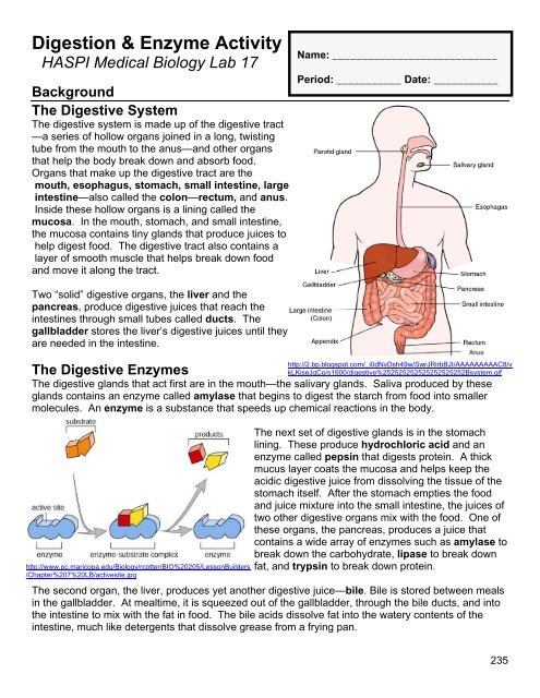
<strong>Digestion</strong> & Enzyme Activity<br />
HASPI Medical Biology Lab <strong>17</strong><br />
Background<br />
The Digestive System<br />
The digestive system is made up of the digestive tract<br />
—a series of hollow organs joined in a long, twisting<br />
tube from the mouth to the anus—and other organs<br />
that help the body break down and absorb food.<br />
Organs that make up the digestive tract are the<br />
mouth, esophagus, stomach, small intestine, large<br />
intestine—also called the colon—rectum, and anus.<br />
Inside these hollow organs is a lining called the<br />
mucosa. In the mouth, stomach, and small intestine,<br />
the mucosa contains tiny glands that produce juices to<br />
help digest food. The digestive tract also contains a<br />
layer of smooth muscle that helps break down food<br />
and move it along the tract.<br />
Name: ____________________________<br />
Period: ___________ Date: ___________<br />
Two “solid” digestive organs, the liver and the<br />
pancreas, produce digestive juices that reach the<br />
intestines through small tubes called ducts. The<br />
gallbladder stores the liver’s digestive juices until they<br />
are needed in the intestine.<br />
The Digestive Enzymes<br />
The digestive glands that act first are in the mouth—the salivary glands. Saliva produced by these<br />
glands contains an enzyme called amylase that begins to digest the starch from food into smaller<br />
molecules. An enzyme is a substance that speeds up chemical reactions in the body.<br />
http://www.pc.maricopa.edu/Biology/rcotter/BIO%20205/LessonBuilders<br />
/Chapter%207%20LB/activesite.jpg<br />
http://2.bp.blogspot.com/_i0dNvDsh49w/SwrJRirbBJI/AAAAAAAAAC8/v<br />
kLKiseJgCo/s1600/digestive%252525252525252525252Bsystem.gif<br />
The next set of digestive glands is in the stomach<br />
lining. These produce hydrochloric acid and an<br />
enzyme called pepsin that digests protein. A thick<br />
mucus layer coats the mucosa and helps keep the<br />
acidic digestive juice from dissolving the tissue of the<br />
stomach itself. After the stomach empties the food<br />
and juice mixture into the small intestine, the juices of<br />
two other digestive organs mix with the food. One of<br />
these organs, the pancreas, produces a juice that<br />
contains a wide array of enzymes such as amylase to<br />
break down the carbohydrate, lipase to break down<br />
fat, and trypsin to break down protein.<br />
The second organ, the liver, produces yet another digestive juice—bile. Bile is stored between meals<br />
in the gallbladder. At mealtime, it is squeezed out of the gallbladder, through the bile ducts, and into<br />
the intestine to mix with the fat in food. The bile acids dissolve fat into the watery contents of the<br />
intestine, much like detergents that dissolve grease from a frying pan.<br />
235
Name: __________________________________________ Date: ___________ Period: _________<br />
The 3 Major Digestible Macromolecules<br />
Protein Carbohydrate Lipids<br />
Polypeptide<br />
Polysaccharide<br />
Fat Molecule<br />
Polymer<br />
http://users.rcn.com/jkimball.ma.ultra<br />
net/BiologyPages/P/Peptide.gif<br />
Amino Acid<br />
http://bioweb.wku.edu/courses/biol115/<br />
wyatt/biochem/Carbos/Carb_poly.gif<br />
Monosaccharide<br />
http://www.foodmuseum.com/images<br />
/exfatMoleculeandCells.jpg<br />
Fatty Acid or Cholesterol<br />
Monomer<br />
Build and repair body<br />
tissues<br />
Glucose<br />
http://kentsimmons.uwinnipeg.ca/cm150<br />
4/Image69.gif<br />
Energy<br />
http://www.protocolsupplements.com/Sports-<br />
Performance-Supplements/wpcontent/uploads/2009/06/amino-acid-mcat1.png<br />
http://www.clker.com/clipart-<br />
10765.html<br />
Stored energy<br />
Use in the<br />
Body<br />
http://content.contentthatworks.com/image<br />
s_articles/2006/health/health_20060724_car<br />
egiver_flexedarm.jpg<br />
Meat, eggs, beans<br />
http://pullenl.wonecks.net/files/2010/09/<br />
Children-Running-Outside.jpg<br />
Breads, pasta, fruits,<br />
vegetables<br />
http://photos.demandstudios.com/ge<br />
tty/article/103/163/82370075_XS.jpg<br />
Oils, butter, meat, dairy,<br />
fast food<br />
Foods<br />
Sources<br />
http://www.gethenchnow.com/wpcontent/uploads/2011/01/high_protein<br />
_foods.jpg<br />
http://www.magazine.ayurvediccure.com/wpcontent/uploads/2009/02/carbohydrate.jpg<br />
http://cdn2-<br />
b.examiner.com/sites/default/files/styles/i<br />
mage_full_width/hash/af/13/af1309885ddd<br />
ea6427ea13af260f4ca4.jpg<br />
NIH. 2008. Your Digestive System and How it Works. National Digestive Diseases Information<br />
Clearinghouse, NIH Publication No. 08-2681. www.digestive.niddk.nih.gov<br />
Adapted from Neo/Sci Food <strong>Digestion</strong> Lab Activity 2011 236
Name: __________________________________________ Date: ___________ Period: _________<br />
Materials<br />
Spot plate 2% Albumin (protein) 1% Hydrochloric acid<br />
20 pH strips 3% Pepsin Biuret<br />
10 stirring sticks 1% Starch solution 2% Amylase<br />
Forceps Potassium iodine Corn oil<br />
Paper towels Water Liquid soap<br />
Procedure<br />
Purpose: The goal of this lab will be to observe how digestive enzymes are able to break down<br />
macromolecules. All data tables are located in the analysis portion of the lab.<br />
PART A: Protein <strong>Digestion</strong><br />
In part A the albumin represents a polypeptide protein. Pepsin is the enzyme needed to break down<br />
the albumin into amino acids, but can only work within a certain pH range. The Biuret will turn pink<br />
with smaller protein chains and will turn purple in the presence of large proteins.<br />
1. Using a pencil, label the wells on your spot plate 1-5.<br />
2. Place 5 drops of 2% Albumin in wells 1-4.<br />
3. Add 5 drops of 1% HCl to wellS 2, 4, and 5.<br />
4. Add 5 drops of 3% Pepsin to wells 3 and 4.<br />
5. Use separate stirring sticks to mix each well, and allow them to sit for 5-7 minutes.<br />
6. Use the forceps to dip a separate pH strip into each of the wells 1-5. Compare the color<br />
change of each strip to the pH strip chart to determine the pH of the mixture in each well.<br />
Record your results in Data Table 1.<br />
7. Add 2 drops of Biuret to wells 1-5. A positive test for protein breakdown will turn pink. If the<br />
well is positive, put a + in Data Table 1. If the well is negative put a -.<br />
8. Record your observations of each of the mixtures in Data Table 1.<br />
9. Rinse out your spot plate and dry it off with a paper towel.<br />
PART B: Carbohydrate <strong>Digestion</strong><br />
In part B starch is a polysaccharide, and amylase is the enzyme responsible for breaking starch down<br />
into monosaccharides. Since the potassium iodine tests for starch, you will be looking for a negative<br />
test to determine whether amylase actually broke down the starch.<br />
1. Using a pencil, label wells 1 and 2 on the spot plate.<br />
2. Add 15 drops of 1% Starch solution to wells 1 and 2.<br />
3. Add 5 drops of 2% Amylase solution to well 2.<br />
4. Use separate stirring sticks to mix each well, and allow them to sit for 5-7 minutes.<br />
5. Add 1 drop of Potassium iodine to wells 1 and 2. A positive test for starch will turn dark blueblack.<br />
If a well is positive for starch, put a + in the Data Table 2. If the test is negative for<br />
starch put a –.<br />
6. Record your observations of each of the mixtures in Data Table 2.<br />
7. Rinse out your spot plate and dry it off with a paper towel.<br />
Adapted from Neo/Sci Food <strong>Digestion</strong> Lab Activity 2011 237
Name: __________________________________________ Date: ___________ Period: _________<br />
PART C: Fat <strong>Digestion</strong><br />
In part C the corn oil represents the lipid polymer, and lipase is the enzyme responsible for breaking<br />
the lipid down. A change in pH will demonstrate that the enzyme is active. The soap works similarly<br />
to bile, and will break up the oil so lipase can work more easily.<br />
1. Using a pencil, label wells 1-3 on the spot plate.<br />
2. Add 10 drops of water to wells 1-3.<br />
3. Add 3 drops of Corn oil to wells 1-3.<br />
4. Use separate stirring sticks to mix each well thoroughly.<br />
5. Use the forceps to dip a separate pH strip into each of the wells 1-3. Compare the color<br />
change of each strip to the pH strip chart to determine the pH of the mixture in each well.<br />
Record your results in Data Table 3.<br />
6. Add 5 drops of lipase to well 2 and 3.<br />
7. Add 2 drops of liquid soap to well 3.<br />
8. Re-stir the mixture in each well, and allow them to sit for 20 minutes.<br />
9. Retest the pH and record your results in Data Table 3.<br />
10. Record your observations of each of the mixtures in Data Table 3.<br />
11. Rinse out your spot plate and dry it off with a paper towel.<br />
PART D: Testing for Macromolecules in Food<br />
In part D you will be performing portions of the tests from parts A-C to determine whether your food<br />
sample contains proteins, carbohydrates, and/or lipids.<br />
1. Choose a food item that is easily mixed with water.<br />
2. In a beaker, mix a small amount of your food sample in 10 ml of water. If the food sample is<br />
solid it will need to be thoroughly smashed.<br />
3. Using a pencil, label wells 1-5 on the spot plate.<br />
4. Add 5 drops of your food sample mixture to wells 1-5. Well 1 will act as the control.<br />
5. Use the forceps to dip a separate pH strip into each of the wells. Compare the color change of<br />
each strip to the pH strip chart to determine the pH of the mixture in each well. Record your<br />
results in Data Table 4.<br />
6. Test your food sample for protein by adding 5 drops of pepsin and 5 drops of HCl to well 2.<br />
7. Allow well 2 to sit for 5-7 minutes, then add 2 drops of Biuret. The mixture will turn pink if<br />
protein is present. Retest the pH of well 2. Record the results in Data Table 4.<br />
8. Test your food sample for carbohydrates by adding 5 drops of amylase to well 4.<br />
9. Allow well 3 and 4 to sit for 5-7 minutes then retest the pH of wells 3 and 4.<br />
10. Add 1 drop of potassium iodine to well 3 and 4. The mixture will turn blue-black in the<br />
presence of starch. Record the results in Data Table 4.<br />
11. Test your food sample for lipids by adding 5 drops of lipase and 2 drops of soap to well 5.<br />
12. Allow well 5 to sit for 20 minutes, then retest the pH of well 5, and record the results in Data<br />
Table 4.<br />
Adapted from Neo/Sci Food <strong>Digestion</strong> Lab Activity 2011 238
Name: __________________________________________ Date: ___________ Period: _________<br />
Analysis<br />
Data Table 1<br />
Well Contents pH Protein Test Observations<br />
1 Albumin<br />
2 Albumin + HCl<br />
3 Albumin +<br />
Pepsin<br />
4 Albumin +<br />
Pepsin + HCl<br />
5 HCl<br />
PART A Analysis Questions - on a separate sheet of paper complete the following<br />
1. Explain what may have happened to the protein in well #2 after the addition of hydrochloric<br />
acid? After the addition of pepsin in well #3? After the addition of pepsin and hydrochloric<br />
acid in well #4?<br />
2. What are the subunits that make up a protein?<br />
3. How could you tell that protein digestion took place?<br />
4. Which of the wells showed the greatest degree of protein digestion? Why?<br />
5. What is the purpose of well #1?<br />
6. What is the role of pepsin in human digestion? In what parts of the digestive system does<br />
protein digestion take place?<br />
7. Does the effectiveness of pepsin depend on pH? Explain.<br />
8. What can you conclude about the optimal pH for pepsin?<br />
Data Table 2<br />
Well Contents Starch Test Observations<br />
1 Starch<br />
2 Starch + Amylase<br />
PART B Analysis Questions - on a separate sheet of paper complete the following<br />
1. Explain what happened to the starch in well #2 after the addition of amylase?<br />
2. What are the subunits that make up carbohydrates?<br />
3. How could you tell that carbohydrate digestion had taken place?<br />
4. What is the purpose of well #1?<br />
5. What is the role of amylase in human digestion? Where is it produced?<br />
6. Where does carbohydrate digestion take place?<br />
7. What do you think may happen if HCl was added to the starch + amylase mixture? Explain<br />
your answer.<br />
Adapted from Neo/Sci Food <strong>Digestion</strong> Lab Activity 2011 239
Name: __________________________________________ Date: ___________ Period: _________<br />
Data Table 3<br />
Well Contents Initial pH Final pH Observations<br />
1 Oil + Water<br />
2 Oil + Water +<br />
Lipase<br />
3 Oil + Water +<br />
Lipase + Soap<br />
PART C Analysis Questions - on a separate sheet of paper complete the following<br />
1. How does the pH of each solution show that fat digestion has occurred?<br />
2. Which well showed the greatest degree of fat digestion? Explain.<br />
3. Where does fat digestion take place in the human digestive system?<br />
4. Describe the effects of bile on fat.<br />
5. Why is emulsification necessary for efficient digestion of fats, but not for proteins or<br />
carbohydrates?<br />
6. What effect would fat digestion have on the pH of the surrounding solution? Explain.<br />
7. Predict the effect of a blocked bile duct on the composition of a patient’s stool.<br />
Data Table 4<br />
Well Contents Initial pH Final pH Observations<br />
1 Food sample<br />
2 Food sample +<br />
Pepsin + HCl<br />
3 Food sample<br />
4 Food sample +<br />
Amylase<br />
5 Food sample +<br />
Lipase + Soap<br />
PART D Analysis Questions - on a separate sheet of paper complete the following<br />
1. Summarize the pH changes in each well.<br />
2. What macromolecules were present in your food sample? Was this what you expected?<br />
Explain your answer.<br />
3. What macromolecules do you think you might find in the following foods: bread, eggs,<br />
broccoli, apple, steak, potato chips.<br />
4. CONCLUSION: In 1-2 paragraphs summarize the procedure and results of this lab.<br />
Adapted from Neo/Sci Food <strong>Digestion</strong> Lab Activity 2011 240
Name: __________________________________________ Date: ___________ Period: _________<br />
Review Questions - on a separate sheet of paper complete the following<br />
1. What is the function of the digestive system?<br />
2. What is the role of the liver and pancreas?<br />
3. What is the responsibility of the gallbladder? How<br />
do you think having the gallbladder removed will<br />
affect digestion?<br />
4. What is an enzyme?<br />
5. Explain the function and location of the following<br />
enzymes: amylase, pepsin, trypsin, and lipase.<br />
6. Diagram A shows the rate of enzyme activity of<br />
digestive enzymes – amylase, pepsin, and trypsin –<br />
in a variety of pH. Using the knowledge of what you<br />
learned during this lab, identify which enzyme is most<br />
like A, B, and C in the graph. Explain your answer.<br />
7. What effect does cooking have on enzyme activity? Why?<br />
8. Why does freezing food preserve it?<br />
9. What is the role of bile in digestion?<br />
10. The polymer of protein is _______, and the monomer is ________.<br />
Diagram A<br />
http://mshuda.files.wordpress.com/2010/04/digestiv<br />
e-enzymes-ph.jpg<br />
11. Why does the body need proteins, and from what food sources can they be obtained?<br />
12. The polymer of carbohydrates is _______, and the monomer is _________.<br />
13. Why does the body need carbohydrates, and from what food sources can they be obtained?<br />
14. The polymer of lipids is _______, and the monomer is ________.<br />
15. Why does the body need lipids, and from what food sources can they be obtained?<br />
16. Based on what you learned about digestion, explain why it is important to eat a variety of foods<br />
to nourish your cells.<br />
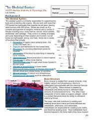
On the attached digestive system diagram, label the following:<br />
<strong>17</strong>. Mouth, pharynx, esophagus, stomach, small intestine, large intestine, rectum, liver,<br />
gallbladder, pancreas.<br />
18. Areas where the following occur; carbohydrate digestion, protein digestion, lipid digestion.<br />
19. Area where absorption of nutrients occurs.<br />
20. Area where water is reabsorbed.<br />
Adapted from Neo/Sci Food <strong>Digestion</strong> Lab Activity 2011 241
Name: __________________________________________ Date: ___________ Period: _________<br />
Adapted from Neo/Sci Food <strong>Digestion</strong> Lab Activity 2011 242