bilateral inguinal hernia with distinct hysterocele and ... - Jivaonline.net
bilateral inguinal hernia with distinct hysterocele and ... - Jivaonline.net
bilateral inguinal hernia with distinct hysterocele and ... - Jivaonline.net

You also want an ePaper? Increase the reach of your titles
YUMPU automatically turns print PDFs into web optimized ePapers that Google loves.
CLINICAL REPORT<br />
BILATERAL INGUINAL HERNIA WITH DISTINCT HYSTEROCELE<br />
AND OMENTOCELE IN A DACHSHUND BITCH<br />
1 2 3<br />
John Martin K. D. , Susannah Bijee Philip , Sherin B. Sarangom<br />
4<br />
<strong>and</strong> Ashay P. Kankonkar<br />
College of Veterinary <strong>and</strong> Animal Sciences, Mannuthy, Thrissur, Kerala 680651<br />
INTRODUCTION<br />
Inguinal <strong>hernia</strong>s may be congenital or acquired<br />
of which the former in dogs occur as a result of a<br />
defect in the <strong>inguinal</strong> ring through which the<br />
abdominal contents protrudes into the subcutaneous<br />
space (Pratschke, 2002). Inguinal <strong>hernia</strong>s may be<br />
congenital or acquired. The former are rare in dogs<br />
<strong>and</strong> may co-exist <strong>with</strong> the umbilical <strong>hernia</strong>s (Fossum,<br />
2007), while the latter are often seen in middle aged<br />
intact bitches (Waters et al., 1993). The potential<br />
factors involved in the development of <strong>inguinal</strong><br />
<strong>hernia</strong>s might be anatomical, hormonal <strong>and</strong><br />
metabolic in nature. However, the exact etiopathogenesis<br />
is still unknown (Smeak, 2003).<br />
Polygenic inheritance of <strong>inguinal</strong> <strong>hernia</strong> had been<br />
described in Cocker Spaniels <strong>and</strong> Dachshunds by<br />
Roberts (1986). The usual contents of <strong>inguinal</strong> <strong>hernia</strong><br />
may include omentum, fat, ovary, uterus, small<br />
intestine, colon, bladder <strong>and</strong> spleen, <strong>with</strong> omentum<br />
being the commonest. (Bellenger, 1996). Herniation<br />
of gravid uterus <strong>and</strong> pyometra uterus through the<br />
<strong>inguinal</strong> ring are also report (Munro <strong>and</strong> Stead, 1993;<br />
Byers, 2007). The present paper reports <strong>bilateral</strong><br />
<strong>inguinal</strong> <strong>hernia</strong> in a dachshund bitch <strong>and</strong> its surgical<br />
management.<br />
CASE HISTORY AND OBSERVATIONS<br />
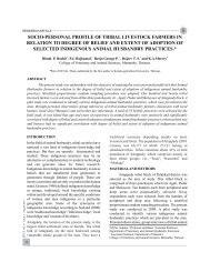
A five year old nulliparous dachshund bitch was<br />
presented to the Kerala Veterinary <strong>and</strong> Animal<br />
Sciences University Hospital, Kokkalai <strong>with</strong> a<br />
<strong>bilateral</strong> swelling in the <strong>inguinal</strong> region. The swelling<br />
was noticed since one month, which increased in size<br />
over the last two weeks. The animal was in estrus one<br />
month back <strong>and</strong> had no previous history of any<br />
1 2,3&4<br />
Associate Professor, M.V.Sc. Scholar<br />
Department of Veterinary Surgery <strong>and</strong> Radiology<br />
trauma. The animal was bright, active <strong>and</strong> alert. All<br />
the physiological parameters were <strong>with</strong>in the normal<br />
range. On palpation, the swellings were non-painful,<br />
soft <strong>and</strong> doughy in consistency. The left <strong>inguinal</strong><br />
swelling was bigger in size compared to the right<br />
one. The contents of both the swelling were nonreducible,<br />
even on application of moderate pressure.<br />
The bladder was catheterized, urine was relieved <strong>and</strong><br />
thus the possibility of vesicocele was ruled out.<br />
Ultrasonographic examination of the left <strong>inguinal</strong><br />
mass could revealed str<strong>and</strong>s of hypoechoic region<br />
<strong>with</strong> areas of normal echogenicity <strong>and</strong> the right<br />
<strong>inguinal</strong> mass <strong>with</strong> moderate echogenicity. Based on<br />
history, physical inspection <strong>and</strong> ultrasonographic<br />
examination, the condition was diagnosed as an<br />
acquired <strong>bilateral</strong> <strong>inguinal</strong> <strong>hernia</strong>. The reduction of<br />
the <strong>hernia</strong>l contents <strong>and</strong> herniorrhaphy under general<br />
anaesthesia were resorted to.<br />
SURGICAL INTERVENTION<br />
AND TREATMENT<br />
The dog was premedicated <strong>with</strong> atropine<br />
a<br />
sulphate at the rate of 0.045 mg/kg body weight<br />
b<br />
followed by xylazine hydrochloride at the rate of 1.5<br />
mg/kg body weight, both given intramuscularly. The<br />
the surgical site ware shaved, scrubbed <strong>and</strong> painted<br />
<strong>with</strong> Tr Iodine for aseptic surgery. General<br />
anaesthesia was induced <strong>with</strong> ketamine<br />
c<br />
hydrochloride at the rate of 5 mg/kg body weight<br />
intramuscularly <strong>and</strong> was maintained by incremental<br />
intravenous injection of a combination of xylazine<br />
hydrochloride <strong>and</strong> ketamine hydrochloride, equal<br />
d<br />
quantity by volume <strong>and</strong> diazepam given 'to effect'.<br />
The dog was positioned on dorsal recumbency. The<br />
site was painted <strong>with</strong> Tincture iodine <strong>and</strong> the patient<br />
JIVA Vol. 10 Issue 1 April 2012<br />
45
CLINICAL REPORT<br />
J. Ind. Vet. Assoc., Kerala. 10 (1)<br />
was draped. A four centimeter long cutaneous<br />
incision was made over the lateral aspect of the left<br />
<strong>inguinal</strong> swelling parallel to the flank fold. The<br />
incision was deepened through the subcutaneous<br />
tissue to expose <strong>hernia</strong>l sac by blunt dissection. The<br />
<strong>hernia</strong>l sac was opened <strong>and</strong> the contents were found<br />
to be non-gravid uterine horns <strong>with</strong> the broad<br />
ligaments (Fig.1). The kelotomy was performed by<br />
incising the <strong>inguinal</strong> ring in a cranio-medial direction<br />
to reduce whole non- gravid uterus <strong>with</strong> the broad<br />
ligaments into the abdominal cavity. The <strong>hernia</strong>l sac<br />
was ligated as close to the internal <strong>inguinal</strong> ring as<br />
possible <strong>and</strong> was sectioned distal to it. Both internal<br />
<strong>and</strong> external <strong>inguinal</strong> rings <strong>and</strong> the kelotomy wound<br />
were closed in separate layers by simple continuous<br />
sutures followed by a layer of subcutaneous sutures<br />
e<br />
using 3.5 metric polyglactin 910 . The skin wound<br />
was apposed <strong>with</strong> horizontal mattress sutures using<br />
3.0 metric nylon. A similar procedure was repeated<br />
on the right <strong>inguinal</strong> ring to reduce omentum. (Fig.<br />
2). A sterile gauze stent was sutured over both skin<br />
wounds <strong>and</strong> Tr. benzoin was applied.<br />
f<br />
Postoperatively, ceftriaxone was administered at the<br />
rate of 25 mg/kg body weight intravenously. Oral<br />
antibiotic therapy was continued for six more days.<br />
The animal had an uneventful recovery except for a<br />
seroma on the right side. The skin sutures were<br />
th<br />
removed on the 9 post-operative day (Fig. 3) <strong>and</strong> the<br />
seroma subsided subsequently. Ovariohysterectomy<br />
at another date was recommended to avoid<br />
complication is future.2.<br />
46<br />
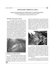
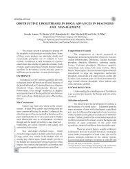
Fig. 1: The exposed contents of the left <strong>hernia</strong>l sac,<br />
the uterus <strong>and</strong> the broad ligament<br />
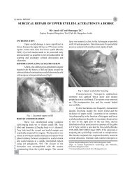
Fig. 2: The exposed content of the right<br />
<strong>hernia</strong>l sac, the omentum<br />
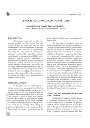
Fig. 3: Healed skin wound after <strong>bilateral</strong> <strong>hernia</strong>l<br />
repair <strong>with</strong> a seroma on right side<br />
RESULT AND DISCUSSION<br />
Acquired <strong>inguinal</strong> <strong>hernia</strong>s are relatively common<br />
in dogs <strong>and</strong> may often met <strong>with</strong> middle aged intact<br />
bitches. Although certain toy breeds are over<br />
represented, no breed predilection has been<br />
documented (Waters et al., 1993). According to<br />
Munro <strong>and</strong> Stead (1993), the potential for<br />
development of <strong>inguinal</strong> <strong>hernia</strong> is more in females<br />
because of the normal extension of the vaginal<br />
process through the <strong>inguinal</strong> canal. Furthermore,<br />
accumulation of fat around the round ligament may<br />
dilate the vaginal process <strong>and</strong> <strong>inguinal</strong> canal<br />
allowing <strong>hernia</strong>tion. The contents of the <strong>hernia</strong>l sac
CLINICAL REPORT<br />
in bitches most often consist of broad ligament <strong>and</strong><br />
uterine horns (Formston, 1990).<br />
In the present case, the history <strong>and</strong> the clinical<br />
signs were similar to the reportes of Jahromi et al.<br />
(2009). Byers et al. (2007) had reported a case of<br />
<strong>inguinal</strong> <strong>hernia</strong> in a dog <strong>with</strong> signs of oestrus<br />
approximately three to four weeks before<br />
presentation <strong>and</strong> substantial increase in size of<br />
<strong>inguinal</strong> swelling two to three weeks prior to<br />
presentation. The condition has to be differentially<br />
diagnosed from mammary tumours, cysts, lipomas,<br />
enlarged lymph nodes, abscesses <strong>and</strong> hematomas.<br />
The chances of traumatic <strong>inguinal</strong> <strong>hernia</strong> were also<br />
ruled out as the dog was in normal body condition<br />
<strong>and</strong> had no history of trauma. Though a diagnosis of<br />
<strong>bilateral</strong> <strong>inguinal</strong> <strong>hernia</strong>tion could be diagnosed<br />
before surgery, the <strong>hernia</strong>l contents could be<br />
confirmed only through surgical correction. Instead<br />
of a midline approach for <strong>bilateral</strong> <strong>inguinal</strong> <strong>hernia</strong><br />
repair in bitches, the conventional approach was<br />
selected as it could facilitate better visualisation of<br />
both <strong>inguinal</strong> rings (Waters et al., 1993). The<br />
contents of the left <strong>and</strong> right <strong>hernia</strong>l sacs were almost<br />
the same as in the report of Jahromi et al. (2009).<br />
Ovariohysterectomy was also recommended<br />
considering (Fossum, 2007) the probable heritable<br />
nature of this disease (Roberts, 1986). The most<br />
common post-operative complications observed in<br />
dogs undergone surgical repair of <strong>inguinal</strong> <strong>hernia</strong><br />
included hematoma or seroma formation, incisional<br />
infection, wound dehiscence, <strong>hernia</strong> recurrence,<br />
peritonitis, sepsis <strong>and</strong> death (Pratschke, 2002).<br />
However, the follow up study did not reveal any<br />
potential post-operative complications <strong>and</strong> the<br />
animal had an uneventful recovery.<br />
utrine horn <strong>and</strong> omentum in a beagle dog. J.<br />
Vet. Emerg. Crit. Care. 17:86-92<br />
Fossum, T. W. 2007. Surgery of the abdominal cavity.<br />
In: Small animal surgery (ed. Fossum, T. W.).<br />
rd<br />
3 Ed. Mosby Elsevier, Philadelphia, pp: 317-<br />
338<br />
Hayes, H. M. 1974. Congenital umbilical <strong>and</strong><br />
<strong>inguinal</strong> <strong>hernia</strong>s in cattle, horse, swine, dogs,<br />
<strong>and</strong> cats: Risk by breed <strong>and</strong> sex among<br />
hospital patients. Am. J. Vet. Res. 35: 839-842.<br />
Jahromi, A. R., Nazhvani, S. D., G<strong>and</strong>mani M. J. <strong>and</strong><br />
Mehrshad, S. 2009. Concurrent <strong>bilateral</strong><br />
<strong>inguinal</strong> <strong>and</strong> umbilical <strong>hernia</strong>s in a bitch-a case<br />
report. Vet. Arhiv. 79: 517-522.<br />
Munro, E. <strong>and</strong> Stead, C. 1993. Ultrasonographic<br />
diagnosis of uterine entrapment in an <strong>inguinal</strong><br />
<strong>hernia</strong>. J. Small Anim. Pract. 34: 139-141.<br />
Pratschke, K. 2002. Management of <strong>hernia</strong>s <strong>and</strong><br />
ruptures in small animals. In Pract. 24: 570-<br />
581<br />
Roberts, S. J. 1986. Veterinary Obstetrics <strong>and</strong> genital<br />
rd<br />
diseases (Theriogenology), 3 Ed. Woodstock,<br />
VT. 981p<br />
Smeak, D. D. 2003. Abdominal <strong>hernia</strong>s. In: Text book<br />
rd<br />
of small animal surgery (ed. Slatter, D.). 3 Ed.<br />
W. B. Saunders, Philadelphia, pp: 449-470<br />
Waters, D. J., Roy, R. G. <strong>and</strong> Stone, E. A. 1993. A<br />
retrospective study of <strong>inguinal</strong> <strong>hernia</strong> in 35<br />
dogs. Vet. Surg. 22: 44-49<br />
a<br />
Atropine Sulphate 1ml amp, Morvel<br />
b<br />
Xylaxin 10 ml vial, Indian Immunologicals<br />
REFERENCES<br />
Bellenger, C. R. 1996. Inguinal <strong>and</strong> scrotal<br />
<strong>hernia</strong>tion in 61 dogs. Aust. Vet. Practitioner.<br />
26: 58-59<br />
Byers, C. G., Williams, J. E. <strong>and</strong> Saylor, D. K. 2007.<br />
Pyometra <strong>with</strong> <strong>inguinal</strong> <strong>hernia</strong>tion of the left<br />
c<br />
Aneket 10 ml vial, Neon Laboratories Ltd.<br />
d<br />
Calmpose 2 ml ampoule, Ranbaxy<br />
e<br />
Vicryl 90 cm, Ethicon<br />
f<br />
Intacef 500 mg vial, Intas Pharmaceuticals<br />
JIVA Vol. 10 Issue 1 April 2012<br />
47