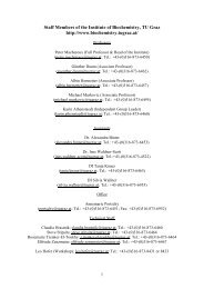
Staff Members of the Institute of Biochemistry, TU Graz http://www ...
Staff Members of the Institute of Biochemistry, TU Graz http://www ...
Staff Members of the Institute of Biochemistry, TU Graz http://www ...

Create successful ePaper yourself
Turn your PDF publications into a flip-book with our unique Google optimized e-Paper software.
<strong>Staff</strong> <strong>Members</strong> <strong>of</strong> <strong>the</strong> <strong>Institute</strong> <strong>of</strong> <strong>Biochemistry</strong>, <strong>TU</strong> <strong>Graz</strong><br />
<strong>http</strong>://<strong>www</strong>.biochemistry.tugraz.at/<br />
Pr<strong>of</strong>essors<br />
Peter Macheroux (Full Pr<strong>of</strong>essor & Head <strong>of</strong> <strong>the</strong> <strong>Institute</strong>)<br />
(peter.macheroux@tugraz.at; Tel.: +43-(0)316-873-6450)<br />
Gün<strong>the</strong>r Daum (Associate Pr<strong>of</strong>essor)<br />
(guen<strong>the</strong>r.daum@tugraz.at; Tel.: +43-(0)316-873-6462)<br />
Albin Hermetter (Associate Pr<strong>of</strong>essor)<br />
(albin.hermetter@tugraz.at; Tel.: +43-(0)316-873-6457)<br />
Assistants<br />
Dr. Karin A<strong>the</strong>nstaedt (until 9.12.2008)<br />
(karin.a<strong>the</strong>nstaedt@tugraz.at, Tel.:+43-(0)316-873-6460)<br />
DI Alexandra Binter (since 1.09.2008)<br />
(alexandra.binter@tugraz.at, Tel.: +43-(0)316-873-6453<br />
DI Silvia Wallner (since 1.10.2008)<br />
(silvia.wallner@tugraz.at, Tel.: +43-(0)316-873-6955)<br />
Office<br />
Annemarie Portschy<br />
portschy@tugraz.at; Tel.: +43-(0)316-873-6451; Fax: +43-(0)316-873-6952<br />
Technical <strong>Staff</strong><br />
Claudia Hrastnik; claudia.hrastnik@tugraz.at; Tel.: +43-(0)316-873-6460<br />
Martin Puhl; martin.puhl@tugraz.at; Tel.: 43-(0)316-873-6453<br />
Steve Stipsits; steve.stipsits@tugraz.at; Tel.: 43-(0)316-873-6464<br />
Rosemarie Trenker-El-Toukhy; r.trenker-el-toukhy@tugraz.at; Tel.: +43-(0)316-873-6464<br />
Elfriede Zenzmaier; elfriede.zenzmaier@tugraz.at; Tel.: +43-(0)316-873-6467<br />
Leo H<strong>of</strong>er (Workshop); leo.h<strong>of</strong>er@tugraz.at; Tel.: +43-(0)316-873-8431 or 8433<br />
1
Brief History <strong>of</strong> <strong>the</strong> <strong>Institute</strong> <strong>of</strong> <strong>Biochemistry</strong><br />
The <strong>Institute</strong> <strong>of</strong> <strong>Biochemistry</strong> and Food Chemistry was born out <strong>of</strong> <strong>the</strong> division <strong>of</strong> <strong>the</strong> <strong>Institute</strong><br />
<strong>of</strong> Biochemical Technology, Food Chemistry and Microchemistry <strong>of</strong> <strong>the</strong> former School <strong>of</strong><br />
Technology <strong>Graz</strong>. Toge<strong>the</strong>r with all <strong>the</strong> o<strong>the</strong>r Chemistry <strong>Institute</strong>s, it was located in <strong>the</strong> old<br />
Chemistry Building on Baron MANDELL's ground, corner Technikerstrasse-Mandellstrasse.<br />
1929 The <strong>Institute</strong> <strong>of</strong> Technical <strong>Biochemistry</strong> and Microbiology moved to <strong>the</strong> building <strong>of</strong><br />
<strong>the</strong> Fürstlich-Dietrichstein-Stiftung, Schlögelgasse 9, in which all <strong>the</strong> Bio-Sciences<br />
were <strong>the</strong>n concentrated.<br />
1945 Georg GORBACH - initially in <strong>the</strong> rank <strong>of</strong> a docent and soon <strong>the</strong>reafter as a.o.<br />
Pr<strong>of</strong>essor - took over to lead <strong>the</strong> <strong>Institute</strong>. The <strong>Institute</strong> was renamed <strong>Institute</strong> <strong>of</strong><br />
Biochemical Technology and Food Chemistry.<br />
1948 G. GORBACH was nominated Full Pr<strong>of</strong>essor and Head <strong>of</strong> <strong>the</strong> <strong>Institute</strong>. In succession<br />
<strong>of</strong> <strong>the</strong> famous <strong>Graz</strong> School <strong>of</strong> Microchemistry founded by PREGL and EMICH, Pr<strong>of</strong>.<br />
GORBACH was one <strong>of</strong> <strong>the</strong> most prominent and active leaders in <strong>the</strong> fields <strong>of</strong><br />
microchemistry, microbiology and nutritional sciences. After World War II, questions<br />
<strong>of</strong> water quality and waste water disposal became urgent; hence, <strong>the</strong> group <strong>of</strong> <strong>the</strong><br />
future Pr<strong>of</strong>. K. S<strong>TU</strong>NDL, which at that time was part <strong>of</strong> <strong>the</strong> <strong>Institute</strong>, was gaining<br />
importance. In addition, a division to fight dry-rot supervised by Dr. KUNZE and after<br />
his demise by H. SALOMON, was also affiliated with <strong>the</strong> <strong>Institute</strong>.<br />
1955 In honour <strong>of</strong> <strong>the</strong> founder <strong>of</strong> Microchemistry and former Pr<strong>of</strong>essor <strong>of</strong> <strong>the</strong> Technical<br />
University <strong>Graz</strong>, <strong>the</strong> extended laboratory was called EMICH-Laboratories. At <strong>the</strong> same<br />
time, <strong>the</strong> <strong>Institute</strong> was renamed <strong>Institute</strong> <strong>of</strong> Biochemical Technology, Food Chemistry<br />
and Microchemistry.<br />
Lectures and laboratory courses were held in <strong>Biochemistry</strong>, Biochemical Technology, Food<br />
Chemistry and Food Technology, Technical Microscopy and Microchemistry. In addition, <strong>the</strong><br />
<strong>Institute</strong> covered Technical Microbiology toge<strong>the</strong>r with Biological and Bacteriological<br />
Analysis - with <strong>the</strong> exception <strong>of</strong> Pathogenic Microorganisms - and a lecture in Organic Raw<br />
Materials Sciences.<br />
1970 After <strong>the</strong> decease <strong>of</strong> Pr<strong>of</strong>. GORBACH, Pr<strong>of</strong>. GRUBITSCH was appointed Head <strong>of</strong> <strong>the</strong><br />
<strong>Institute</strong>. Towards <strong>the</strong> end <strong>of</strong> <strong>the</strong> sixties, <strong>the</strong> division for water and waste water<br />
disposal headed by Pr<strong>of</strong>. S<strong>TU</strong>NDL was drawn out <strong>of</strong> <strong>the</strong> <strong>Institute</strong> and established as an<br />
<strong>Institute</strong> <strong>of</strong> its own. Pr<strong>of</strong>. SPITZY was nominated Pr<strong>of</strong>essor <strong>of</strong> General Chemistry,<br />
Micro- and Radiochemistry. This division was also drawn out <strong>of</strong> <strong>the</strong> mo<strong>the</strong>r <strong>Institute</strong><br />
and at <strong>the</strong> end <strong>of</strong> <strong>the</strong> sixties moved to a new building.<br />
1973 Division <strong>of</strong> <strong>the</strong> <strong>Institute</strong> for Biochemical Technology, Food Technology and<br />
Microchemistry took place. At first, Biochemical Technology toge<strong>the</strong>r with Food<br />
Technology formed a new <strong>Institute</strong> now called <strong>Institute</strong> <strong>of</strong> Biotechnology and Food<br />
Chemistry to which <strong>the</strong> newly nominated Pr<strong>of</strong>. LAFFERTY was appointed Head <strong>of</strong> <strong>the</strong><br />
<strong>Institute</strong>.<br />
2
1973 Dr. F. PALTAUF, docent <strong>of</strong> <strong>the</strong> Karl-Franzens-University <strong>Graz</strong>, was nominated o.<br />
Pr<strong>of</strong>essor and Head <strong>of</strong> <strong>the</strong> newly established <strong>Institute</strong> <strong>of</strong> <strong>Biochemistry</strong>. The interest <strong>of</strong><br />
Pr<strong>of</strong>. PALTAUF in studying biological membranes and lipids laid <strong>the</strong> foundation for<br />
<strong>the</strong> future direction <strong>of</strong> research at <strong>the</strong> <strong>Institute</strong>. G. DAUM, S. D. KOHLWEIN, and A.<br />
HERMETTER joined <strong>the</strong> <strong>Institute</strong>. All three young scientists were given <strong>the</strong> chance to<br />
work as post docs in renowned laboratories in Switzerland and <strong>the</strong> USA: G. DAUM<br />
with <strong>the</strong> groups <strong>of</strong> G. Schatz (Basel, Switzerland) and R. Schekman (Berkeley, USA),<br />
A. HERMETTER with J. R. Lakowicz (Baltimore, USA) and S. D. KOHLWEIN with<br />
S. A. Henry (New York, USA). Consequently, independent research groups<br />
specialized in cell biology (G. D.), biophysics (A. H.) and molecular biology (S. D. K.)<br />
evolved at <strong>the</strong> <strong>Institute</strong> in <strong>Graz</strong>, with <strong>the</strong> group <strong>of</strong> Pr<strong>of</strong>. F. PALTAUF still focusing on<br />
<strong>the</strong> chemistry and biochemistry <strong>of</strong> lipids.<br />
Teaching was always a major task <strong>of</strong> <strong>the</strong> <strong>Institute</strong>. Lectures, seminars and laboratory courses<br />
in basic biochemistry were complemented by special lectures, seminars, and courses held by<br />
<strong>the</strong> assistants who became docents in 1985 (G. D.), 1987 (A. H.), and 1992 (S. D. K.).<br />
Lectures in food chemistry and food technology were held by C. WEBER and H. SALOMON,<br />
who were staff members <strong>of</strong> <strong>the</strong> <strong>Institute</strong>. Hence <strong>the</strong> <strong>Institute</strong> was renamed <strong>Institute</strong> <strong>of</strong><br />
<strong>Biochemistry</strong> and Food Chemistry.<br />
1990 The <strong>Institute</strong> moved to a new building at Petersgasse 12. Expansion <strong>of</strong> <strong>the</strong> size <strong>of</strong><br />
individual research groups and acquisition <strong>of</strong> new equipment were essential for <strong>the</strong><br />
<strong>Institute</strong> to participate in novel collaborative efforts at <strong>the</strong> national and <strong>the</strong><br />
international level including joint projects (Forschungsschwerpunkte, Spezial-<br />
Forschungsbereiche) and EU-projects. Thus, <strong>the</strong> <strong>Institute</strong> <strong>of</strong> <strong>Biochemistry</strong>, toge<strong>the</strong>r<br />
with partner institutes from <strong>the</strong> Karl-Franzens-University was <strong>the</strong> driving force to<br />
establish <strong>Graz</strong> as a centre <strong>of</strong> competence in lipid research.<br />
1993 W. PFANNHAUSER was appointed as Pr<strong>of</strong>essor <strong>of</strong> Food Chemistry. Through his<br />
own enthusiasm and engagement and that <strong>of</strong> his collaborators, this new section <strong>of</strong> <strong>the</strong><br />
<strong>Institute</strong> rapidly developed and <strong>of</strong>fered students additional opportunities to receive a<br />
timely education.<br />
2000 The two sections, <strong>Biochemistry</strong> and Food Chemistry, being independent <strong>of</strong> each o<strong>the</strong>r<br />
with respect to personnel, teaching, and research, were separated. The new <strong>Institute</strong> <strong>of</strong><br />
<strong>Biochemistry</strong> (Head Pr<strong>of</strong>. PALTAUF) and <strong>the</strong> new <strong>Institute</strong> <strong>of</strong> Food Chemistry and<br />
Food Technology (Head Pr<strong>of</strong>. PFANNHAUSER) evolved.<br />
2001 F. PALTAUF, who had been Full Pr<strong>of</strong>essor <strong>of</strong> <strong>Biochemistry</strong> and Head <strong>of</strong> <strong>the</strong><br />
Department for 28 years, retired in September 2001. G. DAUM was elected Head <strong>of</strong><br />
<strong>the</strong> Department. S. D. KOHLWEIN was appointed Full Pr<strong>of</strong>essor <strong>of</strong> <strong>Biochemistry</strong> at<br />
<strong>the</strong> Karl-Franzens University <strong>Graz</strong> and left <strong>the</strong> <strong>Institute</strong> <strong>of</strong> <strong>Biochemistry</strong> <strong>of</strong> <strong>the</strong> <strong>TU</strong><br />
<strong>Graz</strong>.<br />
2003 P. MACHEROUX was appointed Full Pr<strong>of</strong>essor <strong>of</strong> <strong>Biochemistry</strong> in September 2003<br />
and Head <strong>of</strong> <strong>the</strong> <strong>Institute</strong> <strong>of</strong> <strong>Biochemistry</strong> in January 2004. His research interests<br />
revolve around topics in <strong>the</strong> area <strong>of</strong> protein biochemistry and enzymology which shall<br />
streng<strong>the</strong>n <strong>the</strong> already existing activities at <strong>the</strong> <strong>Graz</strong> University <strong>of</strong> Technology in this<br />
field (SFB Biocatalysis & Kplus).<br />
3
Highlights <strong>of</strong> 2008<br />
After <strong>the</strong> successful hearing in January, <strong>the</strong> FWF-funded PhD program “Molecular<br />
Enzymology” was approved for a second period (2008-2011) increasing <strong>the</strong> budget to 3.7<br />
Million Euro for <strong>the</strong> 20 laboratories at <strong>the</strong> participating universities (Karl-Franzens University<br />
and <strong>Graz</strong> University <strong>of</strong> Technology). In <strong>the</strong> meantime, <strong>the</strong> number <strong>of</strong> students in <strong>the</strong> program<br />
has risen to 48 with nine PhD students being trained at our institute.<br />
In June, <strong>the</strong> group <strong>of</strong> Peter Macheroux participated at <strong>the</strong> 16 th International Symposium on<br />
Flavins and Flavoproteins in Jaca, Spain. Six posters were presented at <strong>the</strong> conference with<br />
Sonja Sollner’s poster winning one <strong>of</strong> six Vincent Massey Poster Awards! The passed year<br />
has also seen some important publications: In collaboration with Karl Gruber’s group and our<br />
partner laboratory at <strong>the</strong> Ruder Boskovic <strong>Institute</strong> in Zagreb, we have published <strong>the</strong> first<br />
crystal structure <strong>of</strong> dipeptidylpeptidase III. Pravas Baral, a PhD student in Karl’s group,<br />
successfully crystallized <strong>the</strong> protein generated by Sigrid Deller and Nina Jajcanin-Jozic in our<br />
laboratory. The publication was selected best paper <strong>of</strong> <strong>the</strong> week by <strong>the</strong> editors <strong>of</strong> <strong>the</strong> Journal<br />
<strong>of</strong> Biological Chemistry. In December, Andreas Winkler’s study on berberine bridge enzyme<br />
was published in <strong>the</strong> journal Nature Chemical Biology. As before, this work was<br />
accomplished by a close collaboration in <strong>the</strong> PhD program “Molecular Enzymology” giving<br />
Andreas <strong>the</strong> opportunity to benefit from <strong>the</strong> expertise in structural biology in Karl’s group<br />
while carrying out biochemical and biophysical experiments in our institute.<br />
As in previous years, research in <strong>the</strong> laboratory <strong>of</strong> Gün<strong>the</strong>r Daum was supported by grants <strong>of</strong><br />
<strong>the</strong> Austrian Science Fund (FWF). In 2008, a new project devoted to “Phosphatidylserine<br />
Decarboxylases <strong>of</strong> <strong>the</strong> Yeast” was approved. During 2008, a number <strong>of</strong> new international<br />
collaborations were established which will be <strong>of</strong> mutual benefit in <strong>the</strong> future. Teaching and<br />
administrative activities <strong>of</strong> G. Daum included work as chairman <strong>of</strong> <strong>the</strong> Doctoral School<br />
„Molecular <strong>Biochemistry</strong> and Biotechnology“ at <strong>the</strong> <strong>TU</strong> <strong>Graz</strong>, Board Member <strong>of</strong> <strong>the</strong> FWF,<br />
Associate Editor <strong>of</strong> FEMS Yeast Research, and Member <strong>of</strong> <strong>the</strong> Editorial Board <strong>of</strong> <strong>the</strong> JBC.<br />
Planning <strong>of</strong> <strong>the</strong> upcoming Euro Fed Lipid (European Federation for <strong>the</strong> Science and<br />
Technology <strong>of</strong> Lipids) Conference to be held in October 2009 in <strong>Graz</strong> under <strong>the</strong> chairmanship<br />
<strong>of</strong> G. Daum was ano<strong>the</strong>r effort during <strong>the</strong> last year. This conference with 700 expected<br />
participants will be an important event for <strong>the</strong> lipid community in <strong>Graz</strong>.<br />
Basic research in Albin Hermetter’s group was performed in <strong>the</strong> framework <strong>of</strong> two joint<br />
projects (speaker R. Zechner, IMB, University <strong>of</strong> <strong>Graz</strong>) focusing on lipid (patho)physiology.<br />
His contribution to <strong>the</strong> GOLD project (GEN-AU program <strong>of</strong> <strong>the</strong> BM.W_F) is devoted to <strong>the</strong><br />
functional proteomic analysis <strong>of</strong> lipases relevant to lipid-associated disorders. Financial<br />
support <strong>of</strong> this project has just recently been approved for ano<strong>the</strong>r period <strong>of</strong> three years. In <strong>the</strong><br />
SFB Lipotox (FWF project), A. Hermetter studies <strong>the</strong> toxicity <strong>of</strong> oxidized phospholipids in<br />
<strong>the</strong> cells <strong>of</strong> <strong>the</strong> arterial wall. Applied research in lipid enzymology is performed in <strong>the</strong> Kplus<br />
center Applied Biocatalysis and will be continued in <strong>the</strong> planned Austrian Center for<br />
Industrial Biotechnology (ACIB, speaker A. Glieder, <strong>TU</strong> <strong>Graz</strong>) to be established in <strong>Graz</strong> and<br />
Vienna in 2010.<br />
Karin A<strong>the</strong>nstaedt, completed her “Habilitation” in 2007 and was appointed Docent,<br />
continued her scientific work on microbial lipid metabolism. She also continued her<br />
international collaborations with research groups in her field. In October 2008, her grant<br />
application was approved by <strong>the</strong> FWF, which will be <strong>the</strong> starting point for Karin to launch her<br />
own independent research group in our institute. As in <strong>the</strong> years before, Karin substantially<br />
contributed to our teaching activities in biochemistry and molecular biology.<br />
4
<strong>Biochemistry</strong> group<br />
Group leader: Peter Macheroux<br />
Secretary: Annemarie Portschy<br />
Senior Research Scientists: Sigrid Deller (until March 31 st ), Andrea Wagner (March-June)<br />
PhD students: Asma Asghar (until September 30), Alexandra Binter, Heidi Ehammer (until<br />
July 31), Karlheinz Flicker (until January 31), Martina Neuwirth (until May 31), Sonja<br />
Sollner, Gernot Rauch (until July 31), Silvia Wallner (since October 1), Andreas Winkler<br />
Diploma students: Katharina Fallmann, Tanja Knaus, Daniel Koller, Kerstin Motz, Martin<br />
Poms, Sabrina Riedl, Markus Schober, Silvia Wallner<br />
Guest student: Sebastian H<strong>of</strong>zumahaus (FH Jülich, Germany)<br />
Technicians: Eva Maria Pointner (karenziert), Martin Puhl (Karenzvertretung), Steve Stipsits<br />
(Karenzvertretung), Rosemarie Trenker-El-Toukhy<br />
General description<br />
The fundamental questions in <strong>the</strong> study <strong>of</strong> enzymes, <strong>the</strong> bio-catalysts <strong>of</strong> all living organisms,<br />
revolve around <strong>the</strong>ir ability to select a substrate (substrate specificity) and subject this<br />
substrate to a predetermined chemical reaction (reaction and regio-specificity). In general,<br />
only a few amino acid residues in <strong>the</strong> "active site" <strong>of</strong> an enzyme are involved in this process<br />
and hence provide <strong>the</strong> key to <strong>the</strong> processes taking place during enzyme catalysis. Therefore<br />
<strong>the</strong> focus <strong>of</strong> our research is to achieve a deeper understanding <strong>of</strong> <strong>the</strong> functional role <strong>of</strong> amino<br />
acids in <strong>the</strong> active site <strong>of</strong> enzymes with regard to substrate recognition and stereo- and<br />
regiospecificity <strong>of</strong> <strong>the</strong> chemical transformation. In addition, we are also interested in<br />
substrate-triggered conformational changes and how enzymes utilize c<strong>of</strong>actors (flavin,<br />
nicotinamide) to achieve catalysis. Towards <strong>the</strong>se aims we employ a multidisciplinary<br />
approach encompassing kinetic, <strong>the</strong>rmodynamic, spectroscopic and structural techniques. In<br />
addition, we use site-directed mutagenesis to generate mutant enzymes to probe <strong>the</strong>ir<br />
functional role in <strong>the</strong> before mentioned processes. Fur<strong>the</strong>rmore, we collaborate with our<br />
partners in academia and industry to develop inhibitors for enzymes, which can yield<br />
important new insights into enzyme mechanisms and can be useful as potential lead<br />
compounds in <strong>the</strong> design <strong>of</strong> new drugs.<br />
In our laboratory we have established kinetic (stopped-flow and rapid quench analysis <strong>of</strong><br />
enzymatic reactions), <strong>the</strong>rmodynamic (iso<strong>the</strong>rmal titration microcalorimetry) and<br />
spectroscopic (fluorescence, circular dichroism and UV/Vis absorbance) methods. We make<br />
also frequent use <strong>of</strong> MALDI-TOF and ESI mass spectrometry, protein purification techniques<br />
(chromatography and electrophoresis) and modern molecular biology methods to clone and<br />
express genes <strong>of</strong> interest to us. A brief description <strong>of</strong> our current research projects is given<br />
below.<br />
Berberine bridge enzyme<br />
Berberine bridge enzyme (BBE) is a central enzyme in <strong>the</strong> biosyn<strong>the</strong>sis <strong>of</strong> berberine, a<br />
pharmaceutically important alkaloid. The enzyme possesses a covalently attached FAD<br />
moiety, which is essential for catalysis. The reaction involves <strong>the</strong> oxidation <strong>of</strong> <strong>the</strong> N-methyl<br />
group <strong>of</strong> <strong>the</strong> substrate (S)-reticuline by <strong>the</strong> enzyme-bound flavin and concomitant formation <strong>of</strong><br />
a carbon-carbon bond (<strong>the</strong> “bridge”). The ultimate acceptor <strong>of</strong> <strong>the</strong> substrate-derived electrons<br />
is dioxygen, which reoxidizes <strong>the</strong> flavin to its resting state:<br />
5
The BBE-catalysed oxidative carbon-carbon bond formation is a new example <strong>of</strong> <strong>the</strong><br />
versatility <strong>of</strong> <strong>the</strong> flavin c<strong>of</strong>actor in biochemical reactions. Our goal is to understand <strong>the</strong><br />
oxidative cyclization reaction by a biochemical and structural approach.<br />
Recently, we have developed a new expression system for BBE (using cDNA from<br />
Eschscholzia california, gold poppy) in Pichia pastoris, which produces large amounts <strong>of</strong> <strong>the</strong><br />
protein (ca. 500 mg from a 10-L culture). This work was accomplished in collaboration with<br />
Pr<strong>of</strong>. Toni Kutchan (Donald Danforth Plant Science Center, U. S. A.) and Pr<strong>of</strong>. Toni Glieder<br />
(<strong>TU</strong> <strong>Graz</strong>). The availability <strong>of</strong> suitable quantities <strong>of</strong> BBE enabled us to crystallize <strong>the</strong> protein<br />
and to solve <strong>the</strong> structure in collaboration with Pr<strong>of</strong>. Karl Gruber at <strong>the</strong> Karl-Franzens<br />
University <strong>Graz</strong> (see below).<br />
Based on <strong>the</strong> three-dimensional structure <strong>of</strong> BBE, we have performed a site-directed<br />
mutagenesis program to investigate <strong>the</strong> role <strong>of</strong> amino acids present in <strong>the</strong> active site <strong>of</strong> <strong>the</strong><br />
enzyme. In conjunction with o<strong>the</strong>r experiments, this has led to <strong>the</strong> formulation <strong>of</strong> a new<br />
reaction mechanism for <strong>the</strong> enzyme (<strong>the</strong>sis project <strong>of</strong> Andreas Winkler; diploma project <strong>of</strong><br />
Sabrina Riedl and Kerstin Motz).<br />
6
Chorismate synthase<br />
Chorismate synthase is <strong>the</strong> seventh enzyme <strong>of</strong> <strong>the</strong> shikimate pathway, which leads to aromatic<br />
amino acids and o<strong>the</strong>r aromatic metabolites. Chorismate synthase catalyses <strong>the</strong> conversion <strong>of</strong><br />
5-enolpyruvylshikimate-3-phosphate to chorismate. The catalytic reaction requires reduced<br />
flavin for activity. This finding is very intriguing since flavins are essential c<strong>of</strong>actors in redox<br />
reactions and <strong>the</strong> elimination catalysed by chorismate synthase does not (formally) involve a<br />
reduction or oxidation <strong>of</strong> <strong>the</strong> substrate.<br />
Understanding <strong>the</strong> mechanism <strong>of</strong> chorismate synthase is <strong>of</strong> great importance because, as an<br />
enzyme <strong>of</strong> <strong>the</strong> shikimate pathway, it is only present in bacteria, fungi and plants which makes<br />
<strong>the</strong> enzyme an attractive target for <strong>the</strong> development <strong>of</strong> new antibiotics, fungicides and<br />
herbicides. More recently, <strong>the</strong> shikimate pathway was also discovered in apicomplexan<br />
parasites (Toxoplasma gondii, Plasmodium falciparum) emphasizing <strong>the</strong> important role <strong>of</strong> this<br />
pathway as a potential drug target. Therefore a mechanistic and structural investigation <strong>of</strong><br />
chorismate synthase may provide valuable information for <strong>the</strong> development <strong>of</strong> compounds<br />
with antimicrobial, fungicidal, herbicidal or antimalarial activity. The mechanistic studies are<br />
currently concentrating on a site-directed mutagenesis program with <strong>the</strong> aim to understand <strong>the</strong><br />
role <strong>of</strong> invariant amino acid residues in <strong>the</strong> active site <strong>of</strong> <strong>the</strong> enzyme. With this approach, we<br />
expect to gain fur<strong>the</strong>r insight into <strong>the</strong> functioning <strong>of</strong> <strong>the</strong> enzyme active site and <strong>the</strong> chemical<br />
reaction mechanism (<strong>the</strong>sis project <strong>of</strong> Gernot Rauch).<br />
Chorismate synthases can be classified according to <strong>the</strong> mode <strong>of</strong> reduction <strong>of</strong> <strong>the</strong> required<br />
c<strong>of</strong>actor: mon<strong>of</strong>unctional chorismate synthase requires an external source <strong>of</strong> reduced FMN<br />
whereas bifunctional enzymes possess an intrinsic NADPH:FMN oxidoreductase activity. The<br />
latter type <strong>of</strong> chorismate synthase has - so far - only been found in unicellular fungi (e.g.<br />
Saccharomyces cerevisiae and Neurospora crassa) and green algae (e.g. Euglena gracilis).<br />
Our current interest with regard to mono/bi-functionality <strong>of</strong> chorismate synthases focuses on<br />
<strong>the</strong> question whe<strong>the</strong>r mon<strong>of</strong>unctional chorismate synthases complement chorismate synthase<br />
deficient yeast (∆aro2). In order to tackle this issue, we have developed a system to evaluate<br />
complementation by mon<strong>of</strong>unctional bacterial and plant chorismate synthases in a ∆aro2 yeast<br />
strain. Our results indicate that bacterial and plant chorismate synthases are unable to confer<br />
prototrophy to <strong>the</strong> yeast knock-out despite expression <strong>of</strong> <strong>the</strong> proteins (see figure below, central<br />
panel; Ec, Escherichia coli; An, Anabaena; Le, Lycopersicon esculentum; Bs, Bacillus<br />
subtilis; Sc, Saccharomyces cerevisiae; Nc, Neurospora crassa; Tt, Tetrahymena<br />
<strong>the</strong>rmophila). This result suggests that <strong>the</strong> ability <strong>of</strong> chorismate synthase to utilize NADPH is<br />
essential for a fully functional shikimate pathway. This hypo<strong>the</strong>sis receives fur<strong>the</strong>r support by<br />
additional experiments where co-complementation with a bacterial NADPH:FMN<br />
oxidoreductase (YcnD) led to restoration <strong>of</strong> prototrophy even in <strong>the</strong> case <strong>of</strong> mon<strong>of</strong>unctional<br />
chorismate synthases (see figure below, right panel) (<strong>the</strong>sis project <strong>of</strong> Heidi Ehammer).<br />
7
Dipeptidylpeptidase III<br />
Dipeptidyl-peptidases III (DPPIII; EC 3.4.14.4) are zinc-dependent enzymes with molecular<br />
masses <strong>of</strong> ca. 80-85 kDa that specifically cleave <strong>the</strong> first two amino acids from <strong>the</strong> N-terminus<br />
<strong>of</strong> different length peptides. All known DPPIII sequences contain <strong>the</strong> unique motif HEXXGH,<br />
which enabled <strong>the</strong> recognition <strong>of</strong> <strong>the</strong> dipeptidyl-peptidase III family as a distinct evolutionary<br />
metallopeptidase family (M49). In mammals, DPPIII is associated with important<br />
physiological functions such as pain regulation and hence <strong>the</strong> enzyme is a potential drug<br />
target. Sigrid Deller and Nina Jajcanin-Jozic have successfully expressed, purified and<br />
characterized <strong>the</strong> recombinant yeast enzyme and Pravas Baral in Karl Gruber’s laboratory at<br />
<strong>the</strong> Karl-Franzens-University has elucidated <strong>the</strong> crystal structure <strong>of</strong> <strong>the</strong> protein. This work<br />
revealed that yeast DPPIII is <strong>the</strong> first representative <strong>of</strong> a novel protein topology:<br />
We are now focusing on structures with potential substrates or inhibitors bound in <strong>the</strong> active<br />
site. Towards that goal, we are generating inactive mutant proteins to enable cocrystallisation<br />
with peptide substrates. In addition, we have produced recombinant human DPPIII and are in<br />
<strong>the</strong> process to determine <strong>the</strong> crystal structure to provide <strong>the</strong> basis for <strong>the</strong> development <strong>of</strong><br />
potentially useful inhibitors <strong>of</strong> <strong>the</strong> enzyme (Gustavo Arruda’s <strong>the</strong>sis project in Karl Gruber’s<br />
laboratory assisted by Alexandra Binter)<br />
Nikkomycin biosyn<strong>the</strong>sis<br />
Nikkomycins are produced by several species <strong>of</strong> Streptomyces and exhibit fungicidal,<br />
insecticidal and acaricidal properties due to <strong>the</strong>ir strong inhibition <strong>of</strong> chitin synthase.<br />
Nikkomycins are promising compounds in <strong>the</strong> cure <strong>of</strong> <strong>the</strong> immunosuppressed, such as AIDS<br />
patients and organ transplant recipients as well as cancer patients undergoing chemo<strong>the</strong>rapy.<br />
Nikkomycin Z (R 1 = uracil & R 2 = OH, see below) is currently in clinical trial for its antifungal<br />
activity. Structurally, nikkomycins can be classified as peptidyl nucleosides containing two<br />
unusual amino acids, i.e. hydroxypyridylhomothreonine (HPHT) and aminohexuronic acid<br />
with an N-glycosidically linked base:<br />
8
Although <strong>the</strong> chemical structures <strong>of</strong> nikkomycins have been known since <strong>the</strong> 1970s, only a<br />
few biosyn<strong>the</strong>tic steps have been investigated in detail. The steps leading to <strong>the</strong><br />
aminohexuronic acid intermediate are unclear. Originally, it was hypo<strong>the</strong>sized that <strong>the</strong><br />
aminohexuronic acid moiety is generated by addition <strong>of</strong> an enolpyruvyl moiety from<br />
phosphoenolpyruvate (PEP) to ei<strong>the</strong>r <strong>the</strong> uridine or <strong>the</strong> 4-formyl-4-imidazolin-2-one analog to<br />
<strong>the</strong> 5’-position <strong>of</strong> ribose. This step is <strong>the</strong>n followed by ra<strong>the</strong>r speculative modifications to<br />
yield <strong>the</strong> aminohexuronic acid precursor. In contrast to this hypo<strong>the</strong>sis, we could recently<br />
demonstrate that UMP ra<strong>the</strong>r than uridine serves as <strong>the</strong> acceptor for <strong>the</strong> enolpyruvyl moiety, a<br />
reaction catalyzed by an enzyme encoded by a gene <strong>of</strong> <strong>the</strong> nikkomycin operon termed nikO.<br />
Fur<strong>the</strong>rmore, we could demonstrate that it is attached to <strong>the</strong> 3’- ra<strong>the</strong>r than <strong>the</strong> 5’-position <strong>of</strong><br />
UMP. These results are very intriguing since none <strong>of</strong> <strong>the</strong> nikkomycins syn<strong>the</strong>sized possess an<br />
enolpyruvyl group in this position <strong>of</strong> <strong>the</strong> sugar moiety. Hence, it must be concluded that <strong>the</strong><br />
resulting 3’-enolpyruvyl-UMP is subject to rearrangement reactions where <strong>the</strong> enolpyruvyl is<br />
detached from its 3’-position and transferred to <strong>the</strong> 5’-position <strong>of</strong> <strong>the</strong> ensuing aminohexuronic<br />
acid moiety. Currently, nothing is known about <strong>the</strong> enzymes involved in <strong>the</strong>se putative<br />
chemical steps. However, <strong>the</strong> genes that are co-transcribed with nikO have been reported and<br />
are designated nikI, nikJ, nikK, nikL, nikM and nikN. In order to investigate <strong>the</strong> role <strong>of</strong> <strong>the</strong><br />
encoded proteins in <strong>the</strong> biosyn<strong>the</strong>sis <strong>of</strong> <strong>the</strong> aminohexuronic acid moiety, <strong>the</strong>se genes will be<br />
cloned, expressed and <strong>the</strong> proteins purified for biochemical characterization (<strong>the</strong>sis project <strong>of</strong><br />
Alexandra Binter; diploma project <strong>of</strong> Sebastian H<strong>of</strong>zumahaus).<br />
Proteins in a new vitamin B 6 biosyn<strong>the</strong>tic pathway<br />
Vitamin B 6 (pyridoxine) is an essential nutritional compound required by animals and<br />
humans, which lack <strong>the</strong> biosyn<strong>the</strong>tic pathway for its production. Only bacteria, fungi and<br />
plants possess <strong>the</strong> capability to syn<strong>the</strong>sise vitamin B 6 from basic sugar components. The<br />
chemistry and biochemistry <strong>of</strong> <strong>the</strong> vitamin B 6 biosyn<strong>the</strong>sis has been studied in depth in <strong>the</strong><br />
model bacterium Escherichia coli and it has been thought that this pathway is shared by all<br />
o<strong>the</strong>r organisms in which vitamin B 6 biosyn<strong>the</strong>sis occurs. In recent years, however, it emerged<br />
that <strong>the</strong> biosyn<strong>the</strong>tic pathway found in E. coli is limited to a small group <strong>of</strong> eubacteria (chiefly<br />
<strong>the</strong> γ-subdivision) and <strong>the</strong> majority <strong>of</strong> organisms have developed an entirely different route to<br />
generate vitamin B 6 .<br />
In our studies, we focus on two new proteins that are associated with <strong>the</strong> biosyn<strong>the</strong>sis <strong>of</strong><br />
vitamin B 6 in <strong>the</strong> prokaryote Bacillus subtilis: Pdx1 and Pdx2. The major goal <strong>of</strong> our studies<br />
is to unravel <strong>the</strong>ir interaction which results in <strong>the</strong> generation <strong>of</strong> pyridoxal phosphate. In order<br />
9
to achieve this, both proteins have been recombinantly produced and purified in quantities that<br />
afford <strong>the</strong> characterisation <strong>of</strong> binding by iso<strong>the</strong>rmal titration calorimetry.<br />
Moreover, we have cloned and expressed <strong>the</strong> Pdx1 homolog Snz1 from Saccharomyces<br />
cerevisiae. The purified protein was crystallized and <strong>the</strong> three-dimensional structure solved by<br />
x-ray crystallography in collaboration with Dr. Ivo Tews, Biochemiezentrum Heidelberg.<br />
Interestingly, it was found that Snz1 exists as a hexamer ra<strong>the</strong>r than a dodecamer (see scheme<br />
below) and we are currently identifying <strong>the</strong> structural features that control oligomerization in<br />
<strong>the</strong> Pdx1 domain (<strong>the</strong>sis project <strong>of</strong> Martina Neuwirth and diploma project <strong>of</strong> Silvia Wallner).<br />
The absence <strong>of</strong> vitamin B 6 biosyn<strong>the</strong>sis in humans clearly suggests that <strong>the</strong> proteins involved<br />
are interesting new drug targets for <strong>the</strong> development <strong>of</strong> antibiotics, fungicides and herbicides.<br />
The recent discovery <strong>of</strong> vitamin B 6 biosyn<strong>the</strong>sis in <strong>the</strong> causative agent <strong>of</strong> malaria, Plasmodium<br />
falciparum, prompted us to engage in an international collaboration in order to characterise<br />
<strong>the</strong> chemistry and enzymology <strong>of</strong> vitamin B 6 biosyn<strong>the</strong>sis in this highly pathogenic organism.<br />
Our studies will provide a detailed understanding <strong>of</strong> <strong>the</strong> role(s) <strong>of</strong> Pdx1 and Pdx2 in P.<br />
falciparum and will enable us to initiate a drug design/screening programme which will<br />
receive additional support from <strong>the</strong> elucidation <strong>of</strong> <strong>the</strong> three-dimensional structures <strong>of</strong> <strong>the</strong><br />
proteins and <strong>the</strong>ir active complex (<strong>the</strong>sis project <strong>of</strong> Karlheinz Flicker).<br />
NAD(P)H:FMN quinone reductases<br />
Following our mechanistic and structural studies on YcnD, a nitroreductase from Bacillus<br />
subtilis, we have cloned ano<strong>the</strong>r NADPH-dependent flavin containing oxidoreductase from<br />
this organism (termed YhdA). We have achieved high level expression in E. coli BL 21 host<br />
cells and purified it to homogeneity. Steady-state kinetic studies have shown that YhdA<br />
reduces FMN and nitro-organic compounds at <strong>the</strong> expense <strong>of</strong> NADPH as <strong>the</strong> preferred<br />
electron donor. In parallel to <strong>the</strong> bacterial enzyme, we have also started to investigate <strong>the</strong><br />
properties <strong>of</strong> a homologous enzyme from Saccharomyces cerevisiae (termed LOT6p). Despite<br />
<strong>the</strong> availability <strong>of</strong> a three-dimensional x-ray structure for YhdA (1NNI) and LOT6p (1T0I),<br />
<strong>the</strong> physiological role <strong>of</strong> <strong>the</strong> enzyme was unclear. Our recent studies have now demonstrated<br />
that both enzymes rapidly reduce quinones at <strong>the</strong> expense <strong>of</strong> a reduced nicotinamide c<strong>of</strong>actor.<br />
In order to fur<strong>the</strong>r characterize <strong>the</strong> cellular role <strong>of</strong> LOT6p, we have carried out pull-down<br />
10
assays and identified <strong>the</strong> 20S core particle <strong>of</strong> <strong>the</strong> yeast proteasome as a protein interaction<br />
partner. Fur<strong>the</strong>r studies revealed that this complex recruits Yap4p, a member <strong>of</strong> <strong>the</strong> b-Zip<br />
transcription factor family, but only when <strong>the</strong> flavin-c<strong>of</strong>actor <strong>of</strong> Lot6p is in its reduced state.<br />
Oxidation <strong>of</strong> <strong>the</strong> flavin leads to dissociation <strong>of</strong> <strong>the</strong> transcription factor and relocalization to<br />
<strong>the</strong> nucleus. A similar system is known from mammalian cells, where a homologous quinone<br />
reductase (NQO1) binds to <strong>the</strong> 20S proteasome and recruits important tumor suppressor<br />
proteins such as p53 and p73α. Hence, <strong>the</strong> discovery <strong>of</strong> a homologous protein interaction in<br />
yeast provides an interesting model system to investigate <strong>the</strong> molecular basis for protein<br />
complex formation and regulation <strong>of</strong> proteasomal degradation <strong>of</strong> transcription factors (<strong>the</strong>sis<br />
project <strong>of</strong> Sonja Sollner and diploma project <strong>of</strong> Markus Schober).<br />
Diploma <strong>the</strong>ses completed<br />
Katharina Fallmann: Enzymes <strong>of</strong> Nikkomycin Biosyn<strong>the</strong>sis: NikJ, NikL, and NikO<br />
Nikkomycins are peptidyl nucleoside antibiotics that competitively inhibit chitin synthase and<br />
thus exhibit antifungal and insecticidal properties. The chemical structure <strong>of</strong> nikkomyins<br />
consists <strong>of</strong> <strong>the</strong> unusual amino acids hydroxypryridylhomothreonine (HPHT) and an<br />
aminohexuronic acid with, in <strong>the</strong> case <strong>of</strong> nikkomycin X, N-glycosidically linked 4-formyl-4-<br />
imidazolin-2-one (imidazolone base) or, in <strong>the</strong> case <strong>of</strong> nikkomycin Z, N-glycosidically linked<br />
uracil. Nikkomycin biosyn<strong>the</strong>sis in Streptomyces tendae requires a set <strong>of</strong> proteins which are<br />
grouped in few operons at <strong>the</strong> genetic level. Some Nik proteins have already been studied in<br />
detail, but <strong>the</strong> complete pathway <strong>of</strong> nikkomycin biosyn<strong>the</strong>sis is not yet known. In this work,<br />
<strong>the</strong> proteins NikJ, NikO, and <strong>the</strong> heterologous expression <strong>of</strong> <strong>the</strong> putative protein product <strong>of</strong><br />
nikL were studied. The nikJ gene was heterologously expressed with a C-terminal polyhistidine<br />
tag in E. coli. Suitable expression conditions and a purification protocol for <strong>the</strong><br />
protein NikJ were developed at laboratory scale. The expression requires iron<br />
supplementation <strong>of</strong> <strong>the</strong> growth medium in order to facilitate <strong>the</strong> formation <strong>of</strong> <strong>the</strong> iron-sulfur<br />
clusters during over-expression <strong>of</strong> NikJ. The optimised purification protocol employs affinity<br />
chromatography at a pH value <strong>of</strong> 8.5. The identity <strong>of</strong> <strong>the</strong> purified NikJ protein was confirmed<br />
by MALDI-TOF MS. Analytical gel filtration chromatography <strong>of</strong> NikJ showed a size between<br />
dimer and trimer in vitro. The results from UV-Vis spectroscopy indicated <strong>the</strong> presence <strong>of</strong><br />
coenzymes or pros<strong>the</strong>tic groups in NikJ. EPR spectroscopy revealed NikJ to be an iron-sulfur<br />
protein in accordance with <strong>the</strong> in silico sequence analysis results. For NikO, analytical gel<br />
filtration chromatography experiments confirmed earlier results on its quaternary structure<br />
showing a complex <strong>of</strong> <strong>the</strong> size between two and three monomers in vitro. Additionally, NikO<br />
was purified for protein crystallization. The attempts to express <strong>the</strong> respective protein from<br />
<strong>the</strong> nikL gene heterologously in E. coli did not result in any detectable protein amount.<br />
Possible reasons for <strong>the</strong> lack <strong>of</strong> expressed protein were analyzed, ruling out cloning errors by<br />
sequencing and restriction analysis, as well as expression failure induced by <strong>the</strong> different<br />
frequency <strong>of</strong> codons in <strong>the</strong> source organism S. tendae and <strong>the</strong> expression host E. coli, or an<br />
unfavourable mRNA secondary structure <strong>of</strong> <strong>the</strong> transcript, by means <strong>of</strong> in silico analysis.<br />
Tanja Knaus: Characterization <strong>of</strong> <strong>the</strong> NAD(P)H:FMN Oxidoreductase YcnD from Bacillus<br />
subtilis<br />
The results <strong>of</strong> <strong>the</strong> presented diploma <strong>the</strong>sis clearly indicate that <strong>the</strong> NAD(P)H:FMN<br />
oxidoreductase YcnD from <strong>the</strong> gram-positive bacterium Bacillus subtilis, which has<br />
previously been shown to exhibit azoreductase activity, also has a significant quinone<br />
reductase activity. Using steady-state kinetic and rapid reaction measurements, reduction <strong>of</strong><br />
11
several quinone substrates was investigated, showing K M values in <strong>the</strong> low micro molar range.<br />
For reactions with quinones <strong>the</strong> reductive half reaction <strong>of</strong> <strong>the</strong> redox process was found to be<br />
<strong>the</strong> rate-determining step. In comparison to o<strong>the</strong>r quinone reductases, YcnD does not have <strong>the</strong><br />
typical flavodoxin-fold, however reduction occurs by a two-electron reduction process, thus<br />
not stabilizing large amounts <strong>of</strong> radical semiquinone intermediates. Like many o<strong>the</strong>r FMN<br />
dependent quinone reductases, YcnD is inhibited by dicoumarol. In addition to this, <strong>the</strong><br />
enzyme under investigation shows a sulfite adduct formation with sodium sulfite, being<br />
typical ra<strong>the</strong>r for oxidases than quinone reductases. Apart from <strong>the</strong>se findings necessary for<br />
enzyme characterization, no clear evidence was found that YcnD is a part <strong>of</strong> <strong>the</strong> trimeric<br />
complex with two enzymes <strong>of</strong> <strong>the</strong> shikimate pathway, chorismate synthase and<br />
dehydroquinate synthase, respectively, by performing various pull-down assays and<br />
iso<strong>the</strong>rmal titration calorimetry measurements.<br />
Daniel Koller: Die Rolle von NikU und NikV in der Nikkomycinbiosyn<strong>the</strong>se von<br />
Streptomyces tendae<br />
Nikkomycins are nucleoside-peptide antibiotics, which have antifungal, insecticidal, and<br />
acaridal activity, by competitive inhibition <strong>of</strong> several chitin synthases. These enzymes are<br />
involved in cell wall syn<strong>the</strong>sis and are inhibited by nikkomycins because <strong>of</strong> <strong>the</strong>ir similarity to<br />
UDP-N-acetylglucosamine, <strong>the</strong> natural substrate <strong>of</strong> <strong>the</strong>se enzymes. Approximately 20<br />
biologically active nikkomycin structures were isolated from <strong>the</strong> culture filtrate <strong>of</strong><br />
Streptomyces tendae. The genes involved in biosyn<strong>the</strong>sis <strong>of</strong> nikkomycins in S. tendae were<br />
identified and based on <strong>the</strong> putative activity <strong>of</strong> <strong>the</strong> encoded enzymes a hypo<strong>the</strong>tical pathway<br />
was postulated. The purpose <strong>of</strong> this diploma <strong>the</strong>sis was <strong>the</strong> heterologous expression <strong>of</strong> two<br />
proteins <strong>of</strong> this pathway, NikU and NikV, in <strong>the</strong>ir native form in order to obtain sufficient<br />
material for protein crystallisation and subsequent x-ray analysis. NikU and NikV are subunits<br />
<strong>of</strong> a 22-heterotetramer, coenzyme B12-dependent glutamate mutase. Toward that goal,<br />
modified E. coli BL21 (DE3), E. coli Rosetta and strains containing rare t-RNAs were tested<br />
for protein expression. However, it appears that <strong>the</strong> difference in codon usage was<br />
unfavourable for high-level expression <strong>of</strong> Streptomyces genes in E. coli. Ano<strong>the</strong>r problem<br />
encountered during my study was <strong>the</strong> formation <strong>of</strong> inclusion bodies. To avoid this problem, I<br />
have studied <strong>the</strong> effect <strong>of</strong> temperature and coexpression on <strong>the</strong> solubility <strong>of</strong> heterologously<br />
expressed NikU and NikV. At 37 °C NikU is expressed partially native unlike NikV. At 20 °C<br />
both proteins are generated in a soluble form. In addition, it was found that coexpression did<br />
not result in a better yield <strong>of</strong> NikU or NikV at any <strong>of</strong> <strong>the</strong> temperatures investigated. In<br />
summary, <strong>the</strong> expression levels reached in bacterial host cells used in this diploma <strong>the</strong>sis were<br />
not satisfactory and sufficient protein material for crystallization could <strong>the</strong>refore not be<br />
obtained.<br />
Martin Poms: Backbone cyclisation <strong>of</strong> <strong>the</strong> conotoxin ρ-TA<br />
Conotoxins, small, disulfide-rich peptides isolated from <strong>the</strong> venom <strong>of</strong> cone snails, have<br />
created much interest as possible drug leads in <strong>the</strong> last decades due to <strong>the</strong>ir high potency and<br />
great specificity. Their ability to selectively inhibit ion channels could be valuable for a wide<br />
range <strong>of</strong> <strong>the</strong>rapeutical applications, while minimizing possible side effects. The main problem<br />
<strong>of</strong> peptides as <strong>the</strong>rapeutics presents itself in <strong>the</strong> susceptibility to enzymatic degradation. One<br />
solution to this problem could be <strong>the</strong> introduction <strong>of</strong> a cyclic backbone to <strong>the</strong> peptide to<br />
increase <strong>the</strong> stability while maintaining <strong>the</strong> activity. In this study, two cyclic analogues <strong>of</strong> <strong>the</strong><br />
conotoxin ρTIA were syn<strong>the</strong>sised and compared to <strong>the</strong> native peptide in accordance to <strong>the</strong>ir<br />
NMR structure, serum stability and activity towards <strong>the</strong> 1-adrenergic receptor. It shows that<br />
12
<strong>the</strong> cyclic analogues maintain <strong>the</strong>ir stability as well as a significant level <strong>of</strong> activity. Even<br />
though backbone cyclization has been applied to o<strong>the</strong>r conotoxins before, this means that <strong>the</strong><br />
technique <strong>of</strong> backbone cyclization can be applied to an even wider range <strong>of</strong> peptides and<br />
<strong>the</strong>refore serve as a useful tool in <strong>the</strong>rapeutics development to increase drug stability.<br />
Sabrina Riedl: Biochemical characterization <strong>of</strong> BBE mutants<br />
The berberine bridge enzyme (BBE) is an important enzyme <strong>of</strong> <strong>the</strong> alkaloid biosyn<strong>the</strong>sis in<br />
plants e.g. <strong>the</strong> California poppy (Eschscholzia californica), <strong>the</strong> Opium poppy (Papaver<br />
somniferum) and o<strong>the</strong>r members <strong>of</strong> <strong>the</strong> Papaveraceae and Berberidaceae. It catalyzes <strong>the</strong><br />
conversion <strong>of</strong> (S)-reticuline to (S)-scoulerine. Therefore, <strong>the</strong> transformation <strong>of</strong> <strong>the</strong> N-methyl<br />
group <strong>of</strong> (S)-reticuline into <strong>the</strong> berberine bridge carbon (C-8) <strong>of</strong> (S)-scoulerine is necessary.<br />
This is achieved by a 2-step mechanism. BBE is a flavoprotein containing a FAD c<strong>of</strong>actor<br />
which is bi-covalently attached to <strong>the</strong> protein via an 8-histidyl and 6-S-cysteinyl linkage. This<br />
type <strong>of</strong> bi-covalent linkage was just discovered about two years ago. For this reason only very<br />
little is known about <strong>the</strong> influence <strong>of</strong> <strong>the</strong> two covalent linkages. To investigate <strong>the</strong> importance<br />
<strong>of</strong> some active site amino acids mutations were introduced into <strong>the</strong> protein. Some mutations<br />
concerned <strong>the</strong> flavinylation <strong>of</strong> <strong>the</strong> protein e.g. at <strong>the</strong> site <strong>of</strong> covalent attachment or amino acids<br />
that are suggested to influence <strong>the</strong> formation <strong>of</strong> <strong>the</strong> covalent linkage to <strong>the</strong> protein. O<strong>the</strong>r<br />
mutations were made concerning <strong>the</strong> reaction mechanism e.g. amino acids which might be<br />
responsible for proton abstraction during <strong>the</strong> reaction. The mutations were expressed in Pichia<br />
pastoris host cells and purified using a 2-step purification protocol. They were <strong>the</strong>n analyzed<br />
regarding enzyme activity and <strong>the</strong> way <strong>of</strong> c<strong>of</strong>actor attachment. Three different mutants were<br />
analyzed: C166S, H174A and H104A. The H174A mutant is involved in <strong>the</strong> reaction<br />
mechanism. Introduction <strong>of</strong> this mutation led to a decreased enzyme activity <strong>of</strong> less than 1 %<br />
compared to <strong>the</strong> wild-type. The redoxpotential was reduced to 44.0 mV what is a difference <strong>of</strong><br />
88 mV (wild-type: 132 mV). The C166S and <strong>the</strong> H104A mutation concern <strong>the</strong> covalent<br />
attachment to <strong>the</strong> c<strong>of</strong>actor. It was shown that serine could not replace cysteine and form <strong>the</strong><br />
covalent linkage which was <strong>the</strong> point <strong>of</strong> interest. The H104A mutant also showed a substantial<br />
decrease <strong>of</strong> activity (only 6 %) although <strong>the</strong> redox potential was only slightly affected. This<br />
led to <strong>the</strong> conclusion that bi-covalent attachment is necessary for sufficient enzyme activity.<br />
Markus Schober: The transcription factor Yap4p and its interaction with <strong>the</strong> quinone<br />
reductase Lot6p<br />
In <strong>the</strong> course <strong>of</strong> time organisms have evolved complex strategies to counteract oxidative<br />
stress. These responses are rarely triggered by a single factor but involve a cascade <strong>of</strong> protein<br />
interactions, which are <strong>the</strong> subject <strong>of</strong> countless studies. Transcriptional regulation and<br />
proteolytic degradation both play major roles in stress response. The tumor suppressing<br />
transcription factor p53, whose cellular level is known to be regulated by <strong>the</strong> ubiquitin-26S<br />
proteasome-Mdm2 system, was found to be accumulated in times <strong>of</strong> oxidative stress. It is<br />
thought that this accumulation is caused by a different ubiquitin- and Mdm2-independent<br />
pathway, which is regulated by NAD(P)H quinone oxidoreductase 1 (NQO1) that protects p53<br />
from proteasomal degradation by <strong>the</strong> 20S proteasome. Recently, a potential interaction<br />
between <strong>the</strong> 20S proteasome, <strong>the</strong> quinone reductase Lot6p and <strong>the</strong> transcription factor Yap4p,<br />
a member <strong>of</strong> <strong>the</strong> AP1-like yeast activator protein family involved in <strong>the</strong> oxidative stress<br />
response was discovered in our research group. This interaction is only occurring when Lot6p<br />
is present in its reduced form but does not depend on <strong>the</strong> presence <strong>of</strong> NADH, which has yet<br />
not been shown for <strong>the</strong> p53-20S proteasome-NQO1 pathway. Our in vitro experiments have<br />
shown that Yap4p is also degraded by <strong>the</strong> 20S proteasome in an ubiquitin independent<br />
13
fashion. We hypo<strong>the</strong>size that Yap4p is protected from proteasomal degradation when it is<br />
bound to Lot6p or that interaction <strong>of</strong> Yap4p with Lot6p regulates Yap4p’s localization in <strong>the</strong><br />
cell.<br />
Silvia Wallner: Thermodynamic interaction studies on <strong>the</strong> Pdx2/Pdx2 interface <strong>of</strong> Bacillus<br />
subtilis.<br />
Vitamin B6 is a versatile c<strong>of</strong>actor, which is absolutely required for physical and mental health<br />
in humans. Today two different pathways for de novo biosyn<strong>the</strong>sis <strong>of</strong> pyridoxal 5-phosphate<br />
(PLP) are characterized, which are designated as DXP-dependent and DXP-independent<br />
pathway. In <strong>the</strong> predominant route, referred to as DXP-independent pathway, PLP is<br />
syn<strong>the</strong>sized directly from a pentasaccharide and a trisaccharide in presence <strong>of</strong> L-glutamine.<br />
This reaction is catalyzed by a heteromeric glutamine amidotransferase complex consisting <strong>of</strong><br />
12 Pdx1 synthase and 12 Pdx2 glutaminase subunits. In this diploma project various aspects<br />
<strong>of</strong> complex formation <strong>of</strong> <strong>the</strong> Bacillus subtilis PLP synthase complex have been analyzed.<br />
Today <strong>the</strong> crystal structure <strong>of</strong> <strong>the</strong> ternary Pdx1:Pdx2H170N:L-glutamine complex and <strong>of</strong> <strong>the</strong><br />
individual Pdx1 and Pdx2 moieties are available. Moreover, in previous studies iso<strong>the</strong>rmal<br />
titration calorimetry has been applied to determine <strong>the</strong> <strong>the</strong>rmodynamics <strong>of</strong> complex formation.<br />
In order to understand <strong>the</strong> complex mode <strong>of</strong> interaction between <strong>the</strong> B. subtilis Pdx1 and Pdx2<br />
moieties, <strong>the</strong> contribution <strong>of</strong> specific amino acid residues to complex assembly has been<br />
investigated. Candidates for site-directed mutagenesis have been selected according to<br />
structural predictions. For this study all chosen amino acid residues concern ei<strong>the</strong>r Pdx2<br />
oxyanion hole formation or <strong>the</strong> correct placing <strong>of</strong> <strong>the</strong> Pdx1 N-helix. Thermodynamics <strong>of</strong><br />
complex formation have been evaluated for all generated mutant proteins applying iso<strong>the</strong>rmal<br />
titration calorimetry. Microcalorimetry experiments show that Pdx2 Gln10 actually affects<br />
oxyanion hole formation and <strong>the</strong> tightness <strong>of</strong> <strong>the</strong> PLP synthase complex. Moreover,<br />
<strong>the</strong>rmodynamic data approve <strong>the</strong> importance <strong>of</strong> Pdx1 Lys18 for stabilization <strong>of</strong> Pdx1 N. Pdx1<br />
Lys18 forms polar contacts to Pdx2 Gln10 and Glu15 and hence holds <strong>the</strong> crucial N-helix in<br />
place. These new insights in interaction processes on <strong>the</strong> Pdx1/Pdx2 interface may help to get<br />
a better understanding <strong>of</strong> <strong>the</strong> mode <strong>of</strong> PLP synthase complex formation. Due to <strong>the</strong> absence <strong>of</strong><br />
a PLP biosyn<strong>the</strong>tic pathway in humans this pathway could eventually be a potential target for<br />
new drug development and hence a holistic understanding <strong>of</strong> <strong>the</strong> PLP synthase complex is<br />
required.<br />
Doctoral <strong>the</strong>ses completed<br />
Heidemarie Ehammer: Biochemical studies on <strong>the</strong> enzymology and evolution <strong>of</strong> <strong>the</strong><br />
shikimate pathway<br />
The shikimate pathway has been found in plants, bacteria, fungi and some protozoa and is<br />
essential for <strong>the</strong> biosyn<strong>the</strong>sis <strong>of</strong> aromatic compounds. As mammals do not utilise this<br />
pathway, <strong>the</strong> enzymes involved can be used as potential targets for <strong>the</strong> development <strong>of</strong> new<br />
antibiotics. Chorismate synthase catalyses <strong>the</strong> last step <strong>of</strong> <strong>the</strong> shikimate pathway that leads to<br />
<strong>the</strong> formation <strong>of</strong> chorismate, which is <strong>the</strong> branch point metabolite in <strong>the</strong> syn<strong>the</strong>sis <strong>of</strong> aromatic<br />
amino acids and a variety <strong>of</strong> aromatic compounds. This enzyme has an absolute requirement<br />
for reduced flavin. There are two classes <strong>of</strong> chorismate synthases, which are distinguished<br />
according to <strong>the</strong> origin <strong>of</strong> <strong>the</strong> reduced flavin c<strong>of</strong>actor. Mon<strong>of</strong>unctional chorismate synthases<br />
have been identified in plants and bacteria and depend on <strong>the</strong> level <strong>of</strong> reduced FMN from <strong>the</strong><br />
cellular environment. However, bifunctional enzymes from fungi and <strong>the</strong> ciliated protozoan<br />
Euglena gracilis can generate reduced FMN at <strong>the</strong> expense <strong>of</strong> NADPH. The main focus <strong>of</strong><br />
14
this work lies in <strong>the</strong> classification <strong>of</strong> hi<strong>the</strong>rto uncharacterised chorismate synthases by<br />
introducing an in vivo screen, which is based on a chorismate synthase deficient<br />
Saccharomyces cerevisiae strain. This analysis revealed that bifunctionality is present in<br />
enzymes <strong>of</strong> protozoan species such as <strong>the</strong> parasite Toxoplasma gondii. In contrast, all bacterial<br />
and plant chorismate synthases tested are mon<strong>of</strong>unctional. In addition, I have demonstrated<br />
that a mon<strong>of</strong>unctional chorismate synthase confers prototrophy in conjunction with an<br />
NADPH:FMN oxidoreductase indicating that bifunctionality is required due to <strong>the</strong> lack <strong>of</strong> free<br />
reduced FMN in fungal and possibly protozoan species. Interestingly, I have found that <strong>the</strong><br />
presence <strong>of</strong> a bifunctional enzyme correlates with <strong>the</strong> occurrence <strong>of</strong> a pentafunctional arom<br />
complex. The conclusion that fungal and protozoan species have a common ancestor is<br />
consistent with phylogenetic studies <strong>of</strong> <strong>the</strong> shikimate pathway. The arom complex comprises<br />
<strong>the</strong> enzymes catalysing steps 2 to 6 <strong>of</strong> <strong>the</strong> shikimate pathway. Along with <strong>the</strong> bifunctional<br />
chorismate synthase, <strong>the</strong> arom complex is present in fungal and protozoan species whereas in<br />
bacteria <strong>the</strong> same reactions are catalysed by five mon<strong>of</strong>unctional enzymes. We have been<br />
interested in <strong>the</strong> investigation <strong>of</strong> <strong>the</strong> crystal structure <strong>of</strong> <strong>the</strong> arom complex from<br />
Saccharomyces cerevisiae, Aro1p, to get insights into <strong>the</strong> enzyme mechanism <strong>of</strong><br />
multifunctional complexes. Although a good level <strong>of</strong> Aro1p expression has been observed in<br />
E. coli, fur<strong>the</strong>r investigations will be necessary to isolate higher amounts <strong>of</strong> pure protein.<br />
Karlheinz Flicker: Thermodynamic interaction studies on <strong>the</strong> assembly <strong>of</strong> heteromeric<br />
pyridoxal-5’-phosphate synthase from Plasmodium falciparum<br />
Pyridoxal-5'-phosphate (PLP) is an essential nutrient for animals and humans as it acts as a<br />
c<strong>of</strong>actor in many enzymes. The predominant de novo biosyn<strong>the</strong>tic route is catalysed by PLP<br />
synthase consisting <strong>of</strong> twelve individual Pdx1/Pdx2 glutamine amidotransferase heterodimers<br />
in which Pdx1 is <strong>the</strong> synthase and Pdx2 <strong>the</strong> glutaminase. In this study, complex formation <strong>of</strong><br />
PLP synthase from <strong>the</strong> human malaria pathogen Plasmodium falciparum has been<br />
characterised using iso<strong>the</strong>rmal titration calorimetry. The <strong>the</strong>rmodynamic data are integrated in<br />
a 3D-homology model <strong>of</strong> plasmodial PLP synthase and are compared in detail with data from<br />
<strong>the</strong> previously investigated PLP synthase from B. subtilis. Presence <strong>of</strong> L-Gln increases <strong>the</strong><br />
affinity <strong>of</strong> <strong>the</strong> complex 30-fold and alters <strong>the</strong> <strong>the</strong>rmodynamic signature <strong>of</strong> complex formation.<br />
According to experimental heat capacities <strong>the</strong> interface in <strong>the</strong> presence <strong>of</strong> L-Gln is less<br />
dominated by hydrophobic interactions than in its presence. Fluorescence studies also verify<br />
<strong>the</strong> hydrophobic character <strong>of</strong> <strong>the</strong> PfPdx1 protein interface. In addition, <strong>the</strong> role <strong>of</strong> <strong>the</strong> N-<br />
terminal region <strong>of</strong> PfPdx1 for complex formation has been verified. A swap mutant in which<br />
<strong>the</strong> complete αN-helix <strong>of</strong> PfPdx1 was exchanged for <strong>the</strong> corresponding segment from BsPdx1<br />
shows cross-binding to BsPdx2 and also elicits partial glutaminase activity in BsPdx2. This<br />
demonstrates that formation <strong>of</strong> <strong>the</strong> protein complex interface via αN and catalytic activation<br />
<strong>of</strong> <strong>the</strong> glutaminase are linked processes.<br />
Martina Neuwirth: Interaction studies on Pdx1 and Pdx2 – two proteins involved in vitamin<br />
B6 biosyn<strong>the</strong>sis<br />
Pyridoxal 5-phosphate (PLP), <strong>the</strong> biologically active form <strong>of</strong> vitamin B6, is one <strong>of</strong> <strong>the</strong> most<br />
versatile organic c<strong>of</strong>actors in nature. While animals and humans are not capable to syn<strong>the</strong>size<br />
this vitamin, most bacteria, fungi, protozoa, and plants utilize a de novo PLP biosyn<strong>the</strong>sis<br />
pathway catalyzed by a glutamine amidotransferase. This PLP synthase, composed <strong>of</strong> a<br />
synthase- (Pdx1) and a glutaminase-subunit (Pdx2), is not a single heterodimer, but a complex<br />
<strong>of</strong> approximately 650 kDa. In this complex twelve Pdx1 molecules form a double-ring shaped<br />
15
dodecameric core. Each <strong>of</strong> <strong>the</strong> twelve Pdx1 subunits independently interacts with one <strong>of</strong><br />
twelve Pdx2 molecules, forming <strong>the</strong> whole 24meric PLP synthase complex. In this study,<br />
complex formation <strong>of</strong> <strong>the</strong> bacterium Bacillus subtilis was <strong>the</strong>rmodynamically characterized. It<br />
could be shown that complex formation depends on <strong>the</strong> presence <strong>of</strong> <strong>the</strong> substrate L-glutamine,<br />
which has a tightening effect on <strong>the</strong> preformed Pdx1/Pdx2 complex. In o<strong>the</strong>r words, L-<br />
glutamine increases <strong>the</strong> affinity <strong>of</strong> <strong>the</strong> two molecules to each o<strong>the</strong>r and alters <strong>the</strong>rmodynamic<br />
properties <strong>of</strong> <strong>the</strong> complex. Fur<strong>the</strong>rmore, <strong>the</strong> contribution <strong>of</strong> single amino acids located in <strong>the</strong><br />
Pdx1/Pdx2 interface to complex formation, were investigated. Based on <strong>the</strong> crystal structure<br />
<strong>of</strong> <strong>the</strong> B. subtilis PLP synthase complex, single amino acid residues were exchanged and <strong>the</strong><br />
mutated proteins were analyzed in terms <strong>of</strong> <strong>the</strong>ir <strong>the</strong>rmodynamic and enzymatic properties.<br />
Correlation <strong>of</strong> <strong>the</strong> obtained data with structural data yielded a deeper insight into <strong>the</strong> reaction<br />
mechanism related to structural changes upon complex formation. Fur<strong>the</strong>rmore, <strong>the</strong> crystal<br />
structure <strong>of</strong> Snz1, <strong>the</strong> homologues protein to Pdx1 in <strong>the</strong> fungus Saccharomyces cerevisiae,<br />
could be solved. Surprisingly, Snz1 did not show <strong>the</strong> expected dodemcameric form, but<br />
appeared to be a hexamer assembled in <strong>the</strong> same manner as <strong>the</strong> B. subtilis Pdx1 core. It could<br />
be shown that an additional lysine residue in Snz1, lacking in B. subtilis Pdx1, leads to this<br />
effect. Moreover, <strong>the</strong> importance <strong>of</strong> <strong>the</strong> C-terminal end, entirely visible in <strong>the</strong> Snz1 3D<br />
structure and showing a close vicinity to <strong>the</strong> enzymes active site, could be demonstrated. Due<br />
to its absence in humans, PLP biosyn<strong>the</strong>sis, considered to be a potential new drug target, is <strong>of</strong><br />
great interest in <strong>the</strong> medical field. The new findings described in this study lead to a better<br />
understanding <strong>of</strong> <strong>the</strong> Pdx1/Pdx2 interaction, and thus may contribute to <strong>the</strong> development <strong>of</strong><br />
new <strong>the</strong>rapeutic strategies.<br />
Gernot Rauch: Mechanistic studies on chorismate synthase<br />
Chorismate synthase catalyzes <strong>the</strong> anti-1,4-elimination <strong>of</strong> <strong>the</strong> 3-phosphate and <strong>the</strong> C(6proR)<br />
hydrogen from 5-enolpyruvylshikimate 3-phosphate (EPSP) to generate chorismate, <strong>the</strong> final<br />
product <strong>of</strong> <strong>the</strong> common shikimate pathway and a precursor for <strong>the</strong> biosyn<strong>the</strong>sis <strong>of</strong> aromatic<br />
compounds. The enzyme has an absolute requirement for reduced FMN, which is thought to<br />
facilitate cleavage <strong>of</strong> <strong>the</strong> C-O bond by transiently donating an electron to <strong>the</strong> substrate. The<br />
crystal structure <strong>of</strong> <strong>the</strong> enzyme revealed that EPSP is bound near <strong>the</strong> flavin isoalloxazine ring<br />
with several invariant amino acid residues in contact with <strong>the</strong> substrate and/or c<strong>of</strong>actor. In this<br />
work I present <strong>the</strong> results <strong>of</strong> two mutagenesis studies. In <strong>the</strong> first study, <strong>the</strong> invariant aspartate<br />
367 was replaced through alanine and asparagine. Both single mutant proteins (Asp367Ala<br />
and Asp367Asn) were comparable to <strong>the</strong> wild-type enzyme with respect to substrate and<br />
c<strong>of</strong>actor binding, indicating that Asp367 is not required for binding <strong>of</strong> ei<strong>the</strong>r <strong>the</strong> flavin or <strong>the</strong><br />
substrate. In sharp contrast to <strong>the</strong>se results, <strong>the</strong> activity <strong>of</strong> both single mutant proteins was<br />
found to be 620 and 310 times lower for <strong>the</strong> Asp367Ala and Asp367Asn mutant proteins,<br />
respectively. The data provides <strong>the</strong> first experimental evidence that <strong>the</strong> carboxylate group <strong>of</strong><br />
aspartate 367 participates in <strong>the</strong> deprotonation <strong>of</strong> N(5) <strong>of</strong> <strong>the</strong> reduced flavin c<strong>of</strong>actor, which in<br />
turn abstracts <strong>the</strong> C(6proR) hydrogen yielding chorismate as <strong>the</strong> product. In <strong>the</strong> second study<br />
two invariant serine residues at positions 16 and 127 <strong>of</strong> <strong>the</strong> Neurospora crassa chorismate<br />
synthase were replaced with alanine, producing two single-mutant proteins (Ser16Ala and<br />
Ser127Ala) and a double-mutant protein (Ser16AlaSer127Ala). The residual activity <strong>of</strong> <strong>the</strong><br />
Ser127Ala and Ser16Ala single-mutant proteins was found to be six- and 70-fold lower,<br />
respectively, than that <strong>of</strong> <strong>the</strong> wild-type protein. No residual activity was detected for <strong>the</strong><br />
Ser16AlaSer127Ala double-mutant protein, and formation <strong>of</strong> <strong>the</strong> typical transient<br />
intermediate, characteristic for <strong>the</strong> chorismate synthase-catalysed reaction, was not observed,<br />
in contrast to <strong>the</strong> single-mutant proteins. Based on <strong>the</strong> structure <strong>of</strong> <strong>the</strong> enzyme, this study<br />
provides strong evidence that Ser16 and Ser127 form part <strong>of</strong> a proton relay system among <strong>the</strong><br />
16
isoalloxazine ring <strong>of</strong> FMN, histidine 106 and <strong>the</strong> phosphate group <strong>of</strong> EPSP that is essential for<br />
<strong>the</strong> formation <strong>of</strong> <strong>the</strong> transient intermediate and for substrate turnover.<br />
International cooperations<br />
M. Abramic, Ruder Boskovic <strong>Institute</strong> Zagreb, Croatia<br />
B. Kappes, Universitätsklinikum Heidelberg, Germany<br />
T. Kutchan, Donald Danforth Plant Science Center, U.S.A.<br />
M. Groll, Technical University Munich, Germany<br />
B. A. Palfey, University <strong>of</strong> Michigan, U.S.A.<br />
I. Tews, Universität Heidelberg, Germany<br />
Research projects<br />
FWF P19858: “Enzymes <strong>of</strong> nikkomycin biosyn<strong>the</strong>sis”<br />
FWF P17215: “Proteins involved in a novel vitamin B 6 pathway”<br />
FWF P17471: “Mechanism <strong>of</strong> chorismate synthase”<br />
FWF-Doktoratskolleg “Molecular Enzymology”<br />
Invited lectures<br />
1) Macheroux, P., Schober, M., Sollner, S.<br />
The role <strong>of</strong> quinone reductase as a redox sensor in eukaryotic cells<br />
16 th International Symposium on Flavins & Flavoproteins, Jaca, Spain, 12.06.2008<br />
2) Sollner, S., Schober, M., Wagner, A., Prem, A., Deller, S., Groll, M., Palfey, B.,<br />
Macheroux, P.<br />
Toward an understanding <strong>of</strong> <strong>the</strong> cellular role <strong>of</strong> yeast quinone reductase Lot6p<br />
Vincent Massey Award Lecture at <strong>the</strong> 16 th International Symposium on Flavins &<br />
Flavoproteins, Jaca, Spain, 12.06.2008<br />
3) Winkler, A., Gruber, K., Macheroux, P.<br />
Structure function studies on berberine bridge enzyme<br />
65 th Harden Conference on Enzymes: Nature’s Molecular Machines, University <strong>of</strong><br />
Cumbria, Ambleside, U. K., 17.08.2008<br />
4) Macheroux, P., Winkler, A., Gruber, K.<br />
Structure and mechanism <strong>of</strong> berberine bridge enzyme<br />
2 nd International Interdisciplinary Conference on Vitamins, Coenzymes & Bi<strong>of</strong>actors,<br />
A<strong>the</strong>ns, Georgia, U. S. A., 26.10.2008<br />
Publications<br />
1) Deller, S., Macheroux, P. and Sollner, S.:<br />
Flavin-dependent quinone reductases<br />
Cell. Mol. Life Sci. 2008, 65:141-160.<br />
17
2) Hall, M., Stueckler, C., Ehammer, H., Pointner, E., Oberdorfer, G., Gruber, K., Hauer,<br />
B., Stuermer, R., Kroutil, W., Macheroux, P., and Faber, K.:<br />
Asymmetric bioreduction <strong>of</strong> C=C bonds using enoate reductases OPR1, OPR3 and<br />
YqjM: Enzyme-based stereocontrol<br />
Advanced Syn<strong>the</strong>sis & Catalysis 2008, 350:411-418.<br />
3) Rauch, G., Ehammer, H., Bornemann, S., and Macheroux, P.:<br />
Replacement <strong>of</strong> two invariant serine residues in chorismate synthase provides evidence<br />
that a proton relay system is essential for catalytic activity<br />
FEBS Journal 2008, 275:1464-1473.<br />
4) Ande, S. R., Fussi, H., Knauer, H., Murkovic, M., Ghisla, S., Fröhlich, K.-U., and<br />
Macheroux, P.:<br />
Induction <strong>of</strong> apoptosis in yeast by ophidian L- amino acid oxidase<br />
Yeast 2008, 25:349-357.<br />
5) Baral, P. K., Jajcanin-Jozic, N., Deller, S., Macheroux, P., Abramic, M., and Gruber, K.:<br />
The first structure <strong>of</strong> dipeptidylpeptidase III (DPP-III) provides insight into <strong>the</strong> catalytic<br />
mechanism and <strong>the</strong> mode <strong>of</strong> substrate binding<br />
J. Biol. Chem. 2008, 283:22316-22324.<br />
6) Huang, C.-H., Winkler, A., Chen, C.-L., Lai, W.-L., Tsai, Y.-C., Macheroux, P., and<br />
Liaw, S.-H.:<br />
Functional roles <strong>of</strong> <strong>the</strong> 6-S-cysteinyl, 8α-N1-histidyl FAD in glucooligosaccharide<br />
oxidase from Acremonium strictum<br />
J. Biol. Chem. 2008, 283:30990-30996.<br />
7) Voss, C. V., Gruber, C. C., Faber, K., Knaus, T., Macheroux, P., and Kroutil, W.:<br />
Orchestration <strong>of</strong> a concurrent oxidation and reduction cycle<br />
J. Am. Chem. Soc. 2008, 130:13969-13972.<br />
8) Winkler, A., Lyskowski, A., Riedl, S., Puhl, M., Kutchan, T., Macheroux, P., and<br />
Gruber, K.:<br />
A concerted mechanism for berberine bridge enzyme<br />
Nat. Chem. Biol., 2008, 4:739-741.<br />
Award<br />
Sonja Sollner: Vincent Massey Award at <strong>the</strong> 16 th International Symposium on Flavins and<br />
Flavoproteins (June 8-13, 2008), Jaca, Spain.<br />
18
Cell Biology Group<br />
Group leader: Gün<strong>the</strong>r Daum<br />
Graduate students: Andrea Wagner, Tamara Wriessnegger, Irmgard Schuiki, Tibor Czabany,<br />
Melanie Connerth, Sona Rajakumari, Karlheinz Grillitsch, Miroslava Spanova, Susanne<br />
Horvath (since Nov. 2008)<br />
Diploma student: Susanne Horvath (till Oct. 2008), Sabine Zitzenbacher (since Oct. 2008)<br />
Technician: Claudia Hrastnik<br />
Visiting scientists: Meng Lin, Christian Albrecht University, Kiel, Germany<br />
General description<br />
The existence <strong>of</strong> functional organelles is <strong>the</strong> basis for regulated processes within a cell. To<br />
sequester organelles from <strong>the</strong>ir environment, membranes are required which not only protect<br />
<strong>the</strong> interior <strong>of</strong> <strong>the</strong> organelles but also govern communication with <strong>the</strong> rest <strong>of</strong> <strong>the</strong> cell.<br />
Therefore, it is very important to understand <strong>the</strong> biogenesis and maintenance <strong>of</strong> biological<br />
membranes. To study this question our laboratory makes use <strong>of</strong> <strong>the</strong> yeast as a well established<br />
experimental system. Our studies are focused on <strong>the</strong> syn<strong>the</strong>sis <strong>of</strong> lipids and <strong>the</strong>ir assembly into<br />
yeast organelle membranes. We make use <strong>of</strong> <strong>the</strong> advantages <strong>of</strong> this experimental system to<br />
combine biochemical, molecular and cell biological methods addressing problems <strong>of</strong> lipid<br />
metabolism, lipid depot formation and membrane biogenesis.<br />
The majority <strong>of</strong> yeast lipids are syn<strong>the</strong>sized in <strong>the</strong> endoplasmic reticulum (ER) with some<br />
significant contributions <strong>of</strong> mitochondria, <strong>the</strong> Golgi and <strong>the</strong> so-called lipid particles. O<strong>the</strong>r<br />
subcellular fractions are devoid <strong>of</strong> lipid-syn<strong>the</strong>sizing activities. Spatial separation <strong>of</strong> lipid<br />
biosyn<strong>the</strong>tic steps and lack <strong>of</strong> lipid syn<strong>the</strong>sis in several cellular membranes necessitate<br />
efficient transfer <strong>of</strong> lipids from <strong>the</strong>ir site <strong>of</strong> syn<strong>the</strong>sis to <strong>the</strong>ir proper destination(s). The<br />
maintenance <strong>of</strong> organelle lipid pr<strong>of</strong>iles requires strict coordination <strong>of</strong> biosyn<strong>the</strong>tic and<br />
translocation activities. Specific aspects studied currently in our laboratory are (i) assembly<br />
and homeostasis <strong>of</strong> phosphatidylethanolamine in yeast organelle membranes with emphasis on<br />
peroxisomes and <strong>the</strong> plasma membrane, (ii) neutral lipid storage in lipid particles/droplets and<br />
mobilization <strong>of</strong> <strong>the</strong>se depots with emphasis on <strong>the</strong> involvement <strong>of</strong> lipases and hydrolases, and<br />
(iii) characterization <strong>of</strong> organelle membranes from <strong>the</strong> industrial yeast Pichia pastoris.<br />
Phosphatidylethanolamine, a key component <strong>of</strong> yeast organelle membranes<br />
Work from our laboratory and from o<strong>the</strong>r groups had shown that phosphatidylethanolamine<br />
(PtdEtn), one <strong>of</strong> <strong>the</strong> major phospholipids <strong>of</strong> yeast membranes, is highly important for cellular<br />
function and cell proliferation. PtdEtn syn<strong>the</strong>sis in <strong>the</strong> yeast is accomplished by a network <strong>of</strong><br />
reactions including (i) syn<strong>the</strong>sis <strong>of</strong> phosphatidylserine (PtdSer) in <strong>the</strong> endoplasmic reticulum,<br />
(ii) decarboxylation <strong>of</strong> PtdSer by mitochondrial phosphatidylserine decarboxylase 1 (Psd1p)<br />
or (iii) Psd2p in a Golgi/vacuolar compartment, (iv) <strong>the</strong> CDP-ethanolamine pathway<br />
(Kennedy pathway) in <strong>the</strong> endoplasmic reticulum, and (v) <strong>the</strong> lysophospholipid acylation<br />
route catalyzed by Ale1p. To obtain more insight into biosyn<strong>the</strong>sis, assembly and homeostasis<br />
<strong>of</strong> PtdEtn single and multiple mutants bearing defects in <strong>the</strong> respective pathways can be used.<br />
Previous investigations in our laboratory were aimed at <strong>the</strong> molecular biological<br />
identification <strong>of</strong> novel components involved in PtdEtn homeostasis <strong>of</strong> <strong>the</strong> yeast Saccharomyces<br />
cerevisiae. For this purpose, a number <strong>of</strong> genetic screenings were performed. To<br />
19
obtain a global view <strong>of</strong> <strong>the</strong> role <strong>of</strong> PtdEtn in <strong>the</strong> cell and to study <strong>the</strong> effects <strong>of</strong> an unbalanced<br />
PtdEtn level we recently subjected a psd1∆ deletion mutant and <strong>the</strong> corresponding wild-type<br />
to DNA microarray analysis and examined genome-wide changes in gene expression caused<br />
by a PSD1 deletion. Comparison <strong>of</strong> <strong>the</strong> gene expression pattern <strong>of</strong> <strong>the</strong> psd1∆ mutant with <strong>the</strong><br />
wild-type led to <strong>the</strong> identification <strong>of</strong> 55 differentially expressed genes. Grouping <strong>of</strong> <strong>the</strong>se<br />
genes into functional categories revealed that PtdEtn formation by Psd1p influenced <strong>the</strong><br />
expression <strong>of</strong> genes involved in diverse cellular pathways including transport, carbohydrate<br />
metabolism and stress response. Most <strong>of</strong> <strong>the</strong> proteins identified are present in <strong>the</strong> cytosol<br />
followed by <strong>the</strong> nucleus, cellular membranes, <strong>the</strong> cell wall and mitochondria. Moreover, this<br />
genome-wide analysis identified a number <strong>of</strong> gene products with unknown function which<br />
will be subjected to detailed molecular analysis addressing PtdEtn homeostasis in yeast cells.<br />
Figure 1:<br />
Three-dimensional reconstruction <strong>of</strong><br />
yeast cells grown on oleic acid.<br />
Transmission electron micrographs <strong>of</strong><br />
ultrathin sections (A, C; bar = 1µm)<br />
and corresponding 3D reconstructions<br />
<strong>of</strong> serial sections (B, D) <strong>of</strong> a<br />
chemically fixed wild type cell are<br />
shown. Cells shown in A and B were<br />
grown on YPD, and cells shown in C<br />
and D were grown on oleic acid to<br />
induce formation <strong>of</strong> peroxisomes. CW<br />
cell wall; ER endoplasmic reticulum;<br />
LP lipid particle; M mitochondrion; N<br />
nucleus; V vacuole; PX peroxisomes.<br />
By courtesy <strong>of</strong> G. Zellnig, KFUG.<br />
To understand PtdEtn homeostasis in some more detail, we performed experiments defining<br />
traffic routes <strong>of</strong> PtdEtn within <strong>the</strong> yeast cell. For <strong>the</strong>se investigations we have chosen two<br />
subcellular fractions as destinations for PtdEtn translocation, namely peroxisomes and <strong>the</strong><br />
plasma membrane. These two membranes have in common <strong>the</strong>ir lack <strong>of</strong> capacity to<br />
syn<strong>the</strong>size PtdEtn. We employed yeast mutants bearing defects in <strong>the</strong> different pathways <strong>of</strong><br />
PtdEtn syn<strong>the</strong>sis, which are distributed among mitochondria, endoplasmic reticulum and<br />
Golgi. These experiments demonstrated that PtdEtn formed in all three organelles can be<br />
supplied to peroxisomes and to <strong>the</strong> plasma membrane. The fatty acid composition <strong>of</strong><br />
peroxisomal and plasma membrane phospholipids was mostly influenced by <strong>the</strong> available<br />
pool <strong>of</strong> total species syn<strong>the</strong>sized, although a certain balancing effect was observed regarding<br />
<strong>the</strong> assembly <strong>of</strong> PtdEtn species into <strong>the</strong> two target compartments. We assume that <strong>the</strong><br />
phospholipid composition <strong>of</strong> peroxisomes and <strong>the</strong> plasma membrane is mainly affected by<br />
<strong>the</strong> syn<strong>the</strong>sis <strong>of</strong> <strong>the</strong> respective components and subject to equilibrium, and to a lesser extent<br />
affected by specific transport and assembly processes. Organelle association and membrane<br />
contact between organelles may be a relevant mechanism for lipid transport between<br />
20
organelles, but components governing this process and related events have to be studied in<br />
future investigations.<br />
Growth <strong>of</strong> yeast on oleic acid not only induces formation <strong>of</strong> peroxisomes, but also leads to an<br />
intense syn<strong>the</strong>sis <strong>of</strong> triacylglycerols which are stored in lipid particles. The link between<br />
neutral lipid metabolism and peroxisome proliferation is subject to current investigations<br />
with emphasis on <strong>the</strong> role <strong>of</strong> enzymes involved. These studied are related to <strong>the</strong> possible<br />
lipotoxic effect <strong>of</strong> fatty acids in yeast.<br />
Neutral lipid storage in lipid particles and mobilization<br />
Yeast cells have <strong>the</strong> ability to store neutral lipids TAG (triacylglycerols) and SE (steryl esters)<br />
in subcellular structures named lipid particles/droplets. Upon requirement, TAG and SE can<br />
be mobilized and serve as building blocks for membrane biosyn<strong>the</strong>sis. In an ongoing project<br />
in our laboratory, we investigate and characterize enzymatic steps which lead to <strong>the</strong> storage <strong>of</strong><br />
TAG and SE and at <strong>the</strong> same time to <strong>the</strong> biogenesis <strong>of</strong> lipid particles. Moreover, we study<br />
processes involved in <strong>the</strong> mobilization <strong>of</strong> TAG and SE depots.<br />
Prerequisite for <strong>the</strong> understanding <strong>of</strong> LP function and structure is elucidation <strong>of</strong> <strong>the</strong>ir<br />
molecular equipment. For this purpose, we performed conventional analysis and mass<br />
spectrometric analysis <strong>of</strong> lipids (TAG, STE, phospholipids), but also <strong>of</strong> proteins which are<br />
present on <strong>the</strong> surface <strong>of</strong> LP. These analyses were carried out with LP from cells grown on<br />
glucose or oleic acid. Results obtained by <strong>the</strong>se methods revealed marked differences in <strong>the</strong><br />
lipidome, but also in <strong>the</strong> proteome <strong>of</strong> LP isolated from yeast grown under different conditions.<br />
Moreover, proteome analysis <strong>of</strong> LP led to identification <strong>of</strong> several new putative LP proteins.<br />
In <strong>the</strong> yeast, <strong>the</strong> SE synthases Are1p and Are2p, and <strong>the</strong> TAG synthases Dga1p and Lro1p<br />
contribute to <strong>the</strong> formation <strong>of</strong> lipid depots. Triple mutants with only one <strong>of</strong> <strong>the</strong>se four<br />
enzymes active were used to obtain detailed information about <strong>the</strong> coordinate process <strong>of</strong><br />
neutral lipid syn<strong>the</strong>sis and <strong>the</strong> specific contribution <strong>of</strong> each <strong>of</strong> <strong>the</strong> four acyltransferases to lipid<br />
particle biogenesis. All four triple mutants with only one <strong>of</strong> <strong>the</strong> gene products, Dga1p, Lro1p,<br />
Are1p or Are2p, active form lipid particles, although at different rate and lipid composition.<br />
Through <strong>the</strong>se experiments we showed that TAG or SE alone are sufficient for lipid particle<br />
biogenesis. Biophysical methods revealed that individual neutral lipids strongly affected <strong>the</strong><br />
internal structure <strong>of</strong> lipid particles. SE form several ordered shells below <strong>the</strong> surface<br />
phospholipid monolayer <strong>of</strong> lipid particles, whereas TAG are more or less randomly packed in<br />
<strong>the</strong> center <strong>of</strong> <strong>the</strong> droplets (Figure 2). We propose that this structural arrangement <strong>of</strong> neutral<br />
lipids in lipid particles may be important for <strong>the</strong>ir physiological role especially with respect to<br />
mobilization <strong>of</strong> TAG and SE reserves.<br />
~5 shells SE TAG-core<br />
TG<br />
d~3500 Å<br />
d~ 200-<br />
300nm<br />
Figure 2: Internal structure<br />
<strong>of</strong> yeast lipid particles.<br />
A core <strong>of</strong> TAG is surrounded<br />
by several shells<br />
<strong>of</strong> SE. The surface <strong>of</strong> lipid<br />
particles consists <strong>of</strong> a<br />
phospholipid monolayer<br />
with proteins embedded<br />
21
Similar to TAG and SE syn<strong>the</strong>sizing enzymes, proteins involved in degradation <strong>of</strong> neutral<br />
lipids occur in redundancy. Three TAG lipases named Tgl3p, Tgl4p and Tgl5p, and three SE<br />
hydrolases named Yeh1p, Yeh2p and Tgl1p were identified in <strong>the</strong> yeast. Recently, we studied<br />
enzymatic properties <strong>of</strong> <strong>the</strong> three SE hydrolytic enzymes in some detail. Our investigations<br />
demonstrated that each SE hydrolase exhibits certain substrate specificity. Using single,<br />
double and triple deletion mutants and strains overexpressing <strong>the</strong> three SE hydrolases, we<br />
showed <strong>the</strong> contribution <strong>of</strong> Tgl1p, Yeh1p and Yeh2p, respectively, to <strong>the</strong> mobilization <strong>of</strong> SE<br />
from <strong>the</strong>ir site <strong>of</strong> storage, <strong>the</strong> lipid particles. We also demonstrated that sterol intermediates<br />
stored as SE are readily mobilized through SE hydrolases, recycled to <strong>the</strong> sterol biosyn<strong>the</strong>tic<br />
pathway and converted to <strong>the</strong> final product, ergosterol. Finally, we propose that defects in SE<br />
cleavage affect sterol biosyn<strong>the</strong>sis in a type <strong>of</strong> feedback regulation. Conclusively, SE storage<br />
and mobilization although being dispensable for yeast viability under laboratory conditions<br />
contribute markedly to sterol homeostasis in this microorganism.<br />
Previous work from our laboratory had demonstrated that deletion <strong>of</strong> TGL3, <strong>the</strong> major yeast<br />
TAG lipase located to lipid particles, resulted in a decreased mobilization <strong>of</strong> triacylglycerols<br />
(TAG) from <strong>the</strong> lipid particles. TAG stored in a tgl3∆ mutant contained slightly increased<br />
amounts <strong>of</strong> 22:0 and 26:0 very long chain fatty acids (VLCFAs). These VLCFAs are<br />
indispensable for sphingolipid biosyn<strong>the</strong>sis and crucial for raft association in <strong>the</strong> yeast. To<br />
understand <strong>the</strong> possible role <strong>of</strong> TAG lipolysis in sphingolipid metabolism, we carried out<br />
radiolabeling experiments with sphingolipid precursors in wild type and tgl mutants. These<br />
results indicated that <strong>the</strong> mutants have a significantly reduced rate <strong>of</strong> sphingolipid<br />
biosyn<strong>the</strong>sis and an increased rate <strong>of</strong> sphingolipid turnover compared to wild type.<br />
Competition experiments with labeled palmitic acid (16:0) and non-radiolabeled cerotic acid<br />
(26:0) demonstrated that <strong>the</strong> 26:0 fatty acid competed with <strong>the</strong> 16:0 fatty acid during labeling<br />
<strong>of</strong> sphingolipids. In summary, we hypo<strong>the</strong>size that stored TAG can be a fatty acid donor for<br />
sphingolipid formation and TAG lipases contribute to this pathway in <strong>the</strong> yeast.<br />
Ano<strong>the</strong>r potential non-polar storage lipid is squalene. This component belongs to <strong>the</strong> group <strong>of</strong><br />
isoprenoids and is precursor for <strong>the</strong> syn<strong>the</strong>sis <strong>of</strong> sterols, steroids and ubiquinons. It has<br />
become popular for biotechnological purposes but its isolation from natural sources, e.g.,<br />
shark liver oil, is ra<strong>the</strong>r expensive and questionable from <strong>the</strong> ecological viewpoint. Therefore,<br />
alternative sources to isolate this compound were suggested among <strong>the</strong>m <strong>the</strong> yeast<br />
Saccharomyces cerevisiae. In <strong>the</strong> yeast, <strong>the</strong> amount <strong>of</strong> squalene can be increased by varying<br />
culture conditions or by genetic manipulation. As an example, in strains deleted <strong>of</strong> HEM1<br />
squalene accumulates and is stored mainly in lipid particles. Interestingly, a heme deficient<br />
dga1∆lro1∆are1∆are2∆ quadruple mutant (Qhem1∆) which is devoid <strong>of</strong> <strong>the</strong> classical storage<br />
lipids, TAG and SE, accumulates substantial amounts <strong>of</strong> squalene in cellular membranes,<br />
especially in microsomes and <strong>the</strong> plasma membrane. The fact that Qhem1∆ does not form<br />
lipid particles suggests that biogenesis <strong>of</strong> <strong>the</strong>se storage particles cannot be initiated by<br />
squalene which is syn<strong>the</strong>sized in <strong>the</strong> ER similar to TAG and SE. This result also indicates that<br />
squalene accumulation, at least under <strong>the</strong> conditions tested, is not lipotoxic. Finally, our<br />
experiments demonstrate that squalene incorporated into organelle membranes does not<br />
compromise cellular function. In summary, subcellular distribution <strong>of</strong> squalene can be<br />
regarded as an example for highly flexible lipid storage in yeast.<br />
Lipids <strong>of</strong> Pichia pastoris organelles<br />
The yeast Pichia pastoris is an important experimental system for heterologous expression <strong>of</strong><br />
proteins. Never<strong>the</strong>less, surprisingly little is known about organelles <strong>of</strong> this microorganism.<br />
For this reason, we started a systematic biochemical and cell biological study to establish<br />
22
standardized methods <strong>of</strong> Pichia pastoris organelle isolation and characterization. The<br />
expertise <strong>of</strong> our laboratory in cell fractionation and organelle isolation from <strong>the</strong> yeast<br />
Saccharomyces cerevisiae has been developed through a number <strong>of</strong> investigations in <strong>the</strong> past.<br />
Some <strong>of</strong> <strong>the</strong> methods applied for Saccharomyces cerevisiae can be used for Pichia pastoris<br />
cell fractionation, whereas o<strong>the</strong>rs cannot or need to be adapted. Recent work focused on <strong>the</strong><br />
biochemical characterization <strong>of</strong> mitochondrial membranes and <strong>the</strong> plasma membrane from<br />
Pichia pastoris grown on different carbon sources. The major aim <strong>of</strong> this project was <strong>the</strong><br />
qualitative and quantitative analysis <strong>of</strong> phospholipids, fatty acids and sterols from both types<br />
<strong>of</strong> membranes; when Pichia pastoris was grown on different carbon sources.<br />
Recently, we started to study selected lipid syn<strong>the</strong>sizing enzymes from Pichia pastoris with<br />
emphasis on <strong>the</strong> phosphatidylethanolamine (PE) biosyn<strong>the</strong>tic machinery. We identified two<br />
Pichia pastoris PS decarboxylases (PSD) encoded by genes homologous to PSD1 and PSD2<br />
from Saccharomyces cerevisiae (Figure 3). Using Pichia pastoris psd1 and psd2 mutants we<br />
investigated <strong>the</strong> role <strong>of</strong> <strong>the</strong>se two gene products in PE syn<strong>the</strong>sis and <strong>the</strong> effect <strong>of</strong> <strong>the</strong>ir<br />
deletions on cell growth. Mitochondrial Psd1p is <strong>the</strong> major enzyme for PE syn<strong>the</strong>sis in Pichia<br />
pastoris. Deletion <strong>of</strong> PSD1 caused a loss <strong>of</strong> PSD activity in mitochondria and a severe growth<br />
defect on minimal media which was partially cured by supplementation with ethanolamine.<br />
This result indicated that <strong>the</strong> CDP-ethanolamine but not <strong>the</strong> Psd2p pathway provided<br />
sufficient PE for cell viability in a psd1 background. Under <strong>the</strong>se conditions, however, cellular<br />
and mitochondrial PE levels were dramatically reduced. Fatty acid analysis from wild type,<br />
psd1 and psd2 demonstrated <strong>the</strong> selectivity <strong>of</strong> both PSDs in vivo for <strong>the</strong> syn<strong>the</strong>sis <strong>of</strong><br />
unsaturated PE species. Short chain saturated (16:0) PE species were preferentially imported<br />
into mitochondria irrespective <strong>of</strong> psd1 or psd2 deletions. Thus, biosyn<strong>the</strong>sis and subcellular<br />
distribution contribute to PE homeostasis in mitochondria <strong>of</strong> Pichia pastoris.<br />
.<br />
Figure 3<br />
Phylogenetic tree <strong>of</strong> PSD sequences from different organisms. The tree was constructed using<br />
<strong>the</strong> neighbor joining (NJ) method <strong>of</strong> <strong>the</strong> CLUSTALW program.<br />
23
Diploma Thesis completed<br />
Susanne Horvath: Phosphatidylserine Decarboxylase 1 from <strong>the</strong> Yeast Saccharomyces<br />
cerevisiae<br />
In <strong>the</strong> yeast Saccharomyces cerevisiae, phosphatidylethanolamine is syn<strong>the</strong>sized via 4<br />
different pathways including phosphatidylserine decarboxylation by Psd1p and Psd2p, <strong>the</strong><br />
CDP-ethanolamine pathway and <strong>the</strong> lysophospholipid acylation pathway (Ale1p). The PSD1<br />
proenzyme is composed <strong>of</strong> a mitochondrial targeting sequence, a ß-subunit, a cleavage site<br />
(LGST motif) and an α-subunit. To study <strong>the</strong> contribution <strong>of</strong> <strong>the</strong> α-subunit to posttranslational<br />
processing and enzymatic activation, mutants which lack <strong>the</strong> α-subunit, namely PSD1(-α)-<br />
GFP and PSD1(-α), were constructed. Both mutant strains were cloned into a psd2<br />
background and used for phenotype studies. Analysis <strong>of</strong> <strong>the</strong> growth phenotype <strong>of</strong> PSD1(-α)-<br />
GFP psd2 showed similarity to growth behaviour <strong>of</strong> psd1 psd2 which revealed an<br />
ethanolamine auxotrophy on minimal media containing lactate or glucose. In contrast, cells<br />
from PSD1(-α) psd2 were not able to grow on nonfermentable carbon sources such as glycerol<br />
and lactate even if ethanolamine was supplemented. Analysis <strong>of</strong> mitochondrial phospholipid<br />
patterns <strong>of</strong> <strong>the</strong> strains PSD1(-α)-GFP, PSD1(-α), PSD1(-α)-GFP psd2 and PSD1(-α) psd2<br />
exhibited a decreased level <strong>of</strong> phosphatidylethanolamine similar to psd1 and psd1 psd2<br />
mutants. This result suggests that <strong>the</strong> PSD1 proenzyme in yeast undergoes similar<br />
posttranslational processing steps as bacterial and mammalian PSD where a pyruvoyl<br />
pros<strong>the</strong>tic group at <strong>the</strong> amino terminus <strong>of</strong> <strong>the</strong> a-subunit is formed which is <strong>the</strong>n part <strong>of</strong> <strong>the</strong><br />
catalytic centre. Moreover, possible links between phosphatidylethanolamine and<br />
triacylglycerol metabolism were investigated. Therefore, triacylglycerol levels <strong>of</strong> mutant<br />
strains which bear defects in <strong>the</strong> four phosphatidylethanolamine biosyn<strong>the</strong>tic pathways,<br />
namely psd1, psd2, psd1 psd2, cki1 dpl1 eki1 and ale1 were analyzed and compared to <strong>the</strong><br />
wild type BY4741. On minimal lactate medium supplemented with ethanolamine, deletion <strong>of</strong><br />
PSD1 led to decreasing triacylglycerol levels whereas in ale1 and cki1 dpl1 eki1 increased<br />
levels <strong>of</strong> triacylglycerol were observed, and psd2 showed a similar triacylglycerol level to wild<br />
type. These data suggest that phosphatidylethanolamine syn<strong>the</strong>sis by Psd1p is directly linked<br />
to triacylglycerol metabolism.<br />
Doctoral Theses completed<br />
Andrea Wagner: Neutral lipid storage and mobilization in <strong>the</strong> yeast Saccharomyces<br />
cerevisiae<br />
In all types <strong>of</strong> eukaryotic cells, neutral lipids are stored as energy reserve and a source <strong>of</strong><br />
building blocks needed for membrane formation. This is also true for <strong>the</strong> yeast S. cerevisiae<br />
which syn<strong>the</strong>sizes triacylglycerols (TAG) and steryl esters (SE) as <strong>the</strong> most prominent storage<br />
lipids. These lipids are stored in so-called lipid particles (LP) which consist <strong>of</strong> a hydrophobic<br />
core <strong>of</strong> neutral lipids surrounded by a phospholipid monolayer with a small amount <strong>of</strong><br />
proteins embedded. This <strong>the</strong>sis dealt with <strong>the</strong> role <strong>of</strong> LP in neutral lipid metabolism <strong>of</strong> <strong>the</strong><br />
yeast. The first important point <strong>of</strong> this study was identification and characterization <strong>of</strong> SE<br />
hydrolases, and <strong>the</strong> second aspect was a structural investigation <strong>of</strong> LP. In <strong>the</strong> first part <strong>of</strong> this<br />
work <strong>the</strong> two SE hydrolases, Yeh1p and Tgl1p, both localized on LP were characterized. The<br />
role <strong>of</strong> two enzymes with overlapping activities and slightly different substrate specificities is<br />
discussed. In <strong>the</strong> second part <strong>of</strong> this <strong>the</strong>sis, yeast strains mutated in <strong>the</strong> neutral lipid<br />
syn<strong>the</strong>sizing enzymes Dga1p, Lro1p, Are1p and Are2p were used for biochemical und<br />
biophysical investigations. It was demonstrated that <strong>the</strong> four acyltransferases act<br />
independently <strong>of</strong> each o<strong>the</strong>r and are able to syn<strong>the</strong>size individually a sufficient amount <strong>of</strong><br />
24
neutral lipids for functional LP. A model <strong>of</strong> yeast LP suggesting SE to form several ordered<br />
shells around a central core <strong>of</strong> randomly packed TAG is proposed.<br />
Irmgard Schuiki: Assembly <strong>of</strong> phospholipids into mitochondrial membranes<br />
Phospholipids are essential components for function and integrity <strong>of</strong> membranes. In <strong>the</strong> yeast<br />
Saccharomyces cerevisiae, phosphatidylethanolamine (PtdEtn) plays a specific and important<br />
role. Yeast PtdEtn can be formed by four different pathways, with <strong>the</strong> major route mediated<br />
by phosphatidylserine decarboxylase 1 (Psd1p) localized to <strong>the</strong> inner mitochondrial membrane<br />
(IMM). Assembly <strong>of</strong> Psd1p to <strong>the</strong> IMM is affected by deletion <strong>of</strong> OXA1 encoding an IMM<br />
protein translocase, which leads to decreased PtdEtn syn<strong>the</strong>sis in mitochondria. The IMM<br />
protease Yme1p contributes substantially to <strong>the</strong> proteolytic turnover <strong>of</strong> Psd1p. To obtain a<br />
genome wide view <strong>of</strong> <strong>the</strong> role <strong>of</strong> Psd1p, DNA microarray analyses were performed. This<br />
analyses revealed involvement <strong>of</strong> PtdEtn formed by Psd1p in numerous cellular pathways.<br />
Subcellular distribution <strong>of</strong> PtdEtn was studied with emphasis on <strong>the</strong> supply <strong>of</strong> this<br />
phospholipid to <strong>the</strong> plasma membrane <strong>of</strong> yeast. It was assumed that specific lipid transport<br />
from <strong>the</strong> intracellular sites <strong>of</strong> syn<strong>the</strong>sis, <strong>the</strong> endoplasmic reticulum, mitochondria and Golgi<br />
compartment, has to occur towards <strong>the</strong> cell periphery. These investigations showed that<br />
supply <strong>of</strong> PtdEtn to <strong>the</strong> cell periphery can be accomplished by all pathways <strong>of</strong> PtdEtn<br />
syn<strong>the</strong>sis. However, <strong>the</strong>re is a preference for PtdEtn produced by phosphatidylserine<br />
decarboxylase 2 (Psd2p). In summary, this Thesis underlined <strong>the</strong> importance <strong>of</strong> PtdEtn, not<br />
only for <strong>the</strong> structural integrity <strong>of</strong> cellular membranes, but also for complex physiological<br />
processes.<br />
Tamara Wriessnegger: Characterization <strong>of</strong> organelles from <strong>the</strong> methylotrophic yeast Pichia<br />
pastoris<br />
The methylotrophic yeast Pichia pastoris is widely used as efficient expression system for <strong>the</strong><br />
production <strong>of</strong> heterologous proteins, but despite <strong>the</strong> extensive use in biotechnology hardly any<br />
information is available about cell biological and biochemical properties <strong>of</strong> Pichia pastoris<br />
organelles. This <strong>the</strong>sis provided first insight into <strong>the</strong> membrane composition <strong>of</strong> peroxisomes,<br />
mitochondria and <strong>the</strong> plasma membrane <strong>of</strong> Pichia pastoris. We described appropriate<br />
methods for <strong>the</strong> isolation <strong>of</strong> highly purified organelles, which are prerequisite for subsequent<br />
protein and lipid analyses. Peroxisome induction by methanol or oleic acid leads to <strong>the</strong><br />
accumulation <strong>of</strong> proteins <strong>of</strong> alcohol oxidation or fatty acid beta-oxidation, respectively, in<br />
peroxisomes. Morphological changes <strong>of</strong> Pichia pastoris in response to <strong>the</strong> growth medium<br />
were investigated by electron microscopy. The lipid composition <strong>of</strong> peroxisomal membranes<br />
shows only minor differences in response to <strong>the</strong> used carbon source. The exception was <strong>the</strong><br />
use <strong>of</strong> oleic acid, because this fatty acid became predominant in phospholipids from total cell<br />
and peroxisomal extracts. On ei<strong>the</strong>r C-source, phosphatidylcholine (PtdCho) and phosphatidylethanolamine<br />
(PtdEtn) were <strong>the</strong> major peroxisomal phospholipids. In mitochondria, a<br />
characteristic feature is <strong>the</strong> higher phospholipid to protein ratio and <strong>the</strong> lower ergosterol to<br />
phospholipid ratio <strong>of</strong> <strong>the</strong> outer membrane compared to <strong>the</strong> inner membrane. Variation <strong>of</strong> <strong>the</strong><br />
C-source, however, leads to marked changes <strong>of</strong> <strong>the</strong> fatty acid pattern both in total and<br />
mitochondrial membranes. The inner mitochondrial membrane is also enriched in cardiolipin,<br />
whereas <strong>the</strong> outer membrane contains a high amount <strong>of</strong> phosphatidylinositol. Our study shows<br />
that PtdEtn syn<strong>the</strong>sis by phosphatidylserine decarboxylase 1 (Psd1p) is mainly localized to<br />
mitochondria and Psd1p is responsible for <strong>the</strong> major PSD activity in Pichia pastoris. Defects<br />
in <strong>the</strong> enzyme lead to disability <strong>of</strong> <strong>the</strong> cells to grow on minimal glucose media without<br />
supplementation <strong>of</strong> ethanolamine. Psd2p, <strong>the</strong> second PSD enzyme, is not able to compensate<br />
25
for <strong>the</strong> loss <strong>of</strong> Psd1p, whereas <strong>the</strong> CDP-ethanolamine branch <strong>of</strong> <strong>the</strong> Kennedy pathway<br />
achieves major importance in PtdEtn syn<strong>the</strong>sis <strong>of</strong> Pichia pastoris. Finally, we show that <strong>the</strong><br />
lipid composition <strong>of</strong> <strong>the</strong> plasma membrane is very flexible and highly depends on <strong>the</strong> used C-<br />
source. Especially methanol and oleic acid lead to a change in plasma membrane lipid<br />
composition which may have a strong impact on membrane structure and fluidity.<br />
International Cooperations<br />
J. Cregg, Keck Graduate <strong>Institute</strong> <strong>of</strong> Applied Life Sciences, Claremont, CA, USA<br />
I. Hapala, Slovak Academy <strong>of</strong> Sciences, <strong>Institute</strong> <strong>of</strong> Animal <strong>Biochemistry</strong> and Genetics,<br />
Ivanka pri Dunaji, Slovak Republic<br />
I. Feussner, Plant <strong>Biochemistry</strong>, Albrecht-von-Haller-<strong>Institute</strong> <strong>of</strong> Plant Sciences, Georg-<br />
August University Göttingen, Germany<br />
R. Erdmann, <strong>Institute</strong> <strong>of</strong> Physiological Chemistry, Ruhr-University Bochum, Germany<br />
T. Höfken, <strong>Institute</strong> <strong>of</strong> <strong>Biochemistry</strong>, Christian Albrecht University, Kiel, Germany<br />
M. Karas, <strong>Institute</strong> <strong>of</strong> Pharmaceutical Chemistry, Johann Wolfgang Goe<strong>the</strong> University,<br />
Frankfurt, Germany<br />
R. Rajasekharan, Department <strong>of</strong> <strong>Biochemistry</strong>, Indian <strong>Institute</strong> <strong>of</strong> Science, Bangalore, India<br />
A. S. Sandelius, Department <strong>of</strong> Plant and Environmental Sciences, University <strong>of</strong> Go<strong>the</strong>nburg,<br />
Sweden<br />
Research Projects<br />
FWF 17321: Phosphatidylethanolamine <strong>of</strong> yeast mitochondria<br />
FWF 18857: Neutral lipid storage and mobilization in <strong>the</strong> yeast Saccharomyces cerevisiae<br />
FWF L-178 (Translational research): Characterization <strong>of</strong> organelles from <strong>the</strong> yeast Pichia<br />
pastoris<br />
FWF PhD Program: Molecular Enzymology<br />
Invited Lectures<br />
1) G. Daum<br />
Lipid storage and mobilization in <strong>the</strong> yeast<br />
Fluxome Company, Lyngby, Denmark, 16 October 2008<br />
2) G. Daum<br />
Lipid storage and mobilization in <strong>the</strong> yeast<br />
Croatian Biochemical Society, Osijek, Croatia, 17-20 September 2008<br />
Publications<br />
1) Czabany, T., Wagner, A., Zweytick, D., Lohner, K., Leitner, E., Ingolic, E. and Daum,<br />
G.:<br />
Structural and biochemical properties <strong>of</strong> lipid particles from <strong>the</strong> yeast Saccharomyces<br />
cerevisiae.<br />
J. Biol. Chem. 2008, 283:17065-17074.<br />
26
2) Rajakumari, S., Grillitsch, K. and Daum, G.:<br />
Syn<strong>the</strong>sis and turnover <strong>of</strong> non-polar lipids in yeast<br />
Prog. Lipid Res. 2008,47:157-171.<br />
3) Schuiki, I., and Daum, G.:<br />
Phosphatidylserine decarboxylases, key enzymes <strong>of</strong> lipid metabolism<br />
IUBMB Life 2009 in press.<br />
4) Wagner, A., Grillitsch, K., Leitner, E. and Daum, G.:<br />
Mobilization <strong>of</strong> steryl esters from lipid particles <strong>of</strong> <strong>the</strong> yeast Saccharomyces cerevisiae.<br />
Biochim. Biophys. Acta. 2009 [Epub ahead <strong>of</strong> print].<br />
5) Wriessnegger, T., Leitner, E., Belegratis, M. R., Ingolic, E. and Daum, G.:<br />
Lipid analysis <strong>of</strong> mitochondrial membranes from <strong>the</strong> yeast Pichia pastoris.<br />
Biochim. Biophys. Acta. 2009 [Epub ahead <strong>of</strong> print].<br />
6) Connerth. M., Grillitsch, K., Köfeler, H. and Daum, G.:<br />
Analysis <strong>of</strong> Lipid Particles from Yeast<br />
Lipidomics, Humana Press, D. Armstrong ed. (2009) in press.<br />
Award<br />
Tibor Czabany received a FEBS Short term Fellowship for a stay in <strong>the</strong> laboratory <strong>of</strong> Pr<strong>of</strong>. I.<br />
Feussner, Plant <strong>Biochemistry</strong>, Albrecht-von-Haller-<strong>Institute</strong> <strong>of</strong> Plant Sciences, Georg-August<br />
University Göttingen, Germany<br />
27
Molecular Biology Group<br />
Karin A<strong>the</strong>nstaedt<br />
Triacylglycerol syn<strong>the</strong>sis in <strong>the</strong> oleaginous yeast Yarrowia lipolytica<br />
In <strong>the</strong> oleaginous yeast Yarrowia lipolytica mainly triacylglycerols (TAG) and only minor<br />
quantities <strong>of</strong> steryl esters accumulate as storage lipids. Like in all eukaryotic cells <strong>the</strong>se<br />
neutral lipids are sequestered from <strong>the</strong> cytosolic environment in so-called lipid particles. The<br />
structure <strong>of</strong> <strong>the</strong>se storage compartments <strong>of</strong> neutral lipids is ra<strong>the</strong>r simple. TAG and steryl<br />
esters are forming a hydrophobic core which is surrounded by a phospholipid monolayer with<br />
few proteins embedded. Under specific growth conditions, e.g., when industrial fats or<br />
glycerol are used as a carbon source, <strong>the</strong> amount <strong>of</strong> TAG can be increased up to 40% <strong>of</strong> cell<br />
dry weight. Microscopic inspection <strong>of</strong> cells grown under such conditions reveals that nearly<br />
<strong>the</strong> entire cell is filled with lipid particles. This outstanding capacity <strong>of</strong> Yarrowia lipolytica to<br />
accumulate neutral lipids (mainly TAG) leads to its application in biotechnological processes<br />
such as single cell oil production or production <strong>of</strong> nutrients enriched in essential fatty acids<br />
which can serve as nutritional complements. However, Yarrowia lipolytica may also serve as<br />
a model organism to study lipid turnover in adipocytes, since not only <strong>the</strong> ability to store<br />
excessive amounts <strong>of</strong> TAG in lipid particles but also <strong>the</strong> composition <strong>of</strong> this cell compartment<br />
resembles adipocytes <strong>of</strong> higher eukaryotes. Despite <strong>the</strong>se potentials <strong>of</strong> Yarrowia lipolytica<br />
information about TAG (lipid) metabolism in this yeast is ra<strong>the</strong>r limited. Thus, recently we<br />
started to investigate <strong>the</strong> proteome <strong>of</strong> Yarrowia lipolytica for proteins involved in TAG<br />
syn<strong>the</strong>sis. Computational analyses revealed two candidate gene-products <strong>of</strong> <strong>the</strong> oleaginous<br />
yeast homologous to TAG synthases <strong>of</strong> o<strong>the</strong>r organisms. A decreased amount <strong>of</strong> TAG in<br />
mutant cells defective in <strong>the</strong>se candidate genes already pinpointed to a function <strong>of</strong> <strong>the</strong>se<br />
polypeptides in TAG formation. To investigate whe<strong>the</strong>r <strong>the</strong>se candidate genes encode true<br />
TAG synthases <strong>the</strong>se genes were heterologously expressed in cells <strong>of</strong> a Saccharomyces<br />
cerevisiae mutant defective in neutral lipid syn<strong>the</strong>sis and as a consequence lacking lipid<br />
particles.<br />
A<br />
C<br />
B<br />
D<br />
Figure 1: Restoration <strong>of</strong> lipid particle<br />
formation upon heterologous<br />
expression <strong>of</strong> Yarrowia lipolytica TAG<br />
synthase candidates in mutant cells <strong>of</strong><br />
<strong>the</strong> budding yeast Saccharomyces<br />
cerevisiae lacking lipid particles.<br />
Upper panels: Positive (A; wild-type)<br />
and negative (B; mutant) control <strong>of</strong><br />
Saccharomyces cerevisiae cells bearing<br />
empty plasmids.<br />
Lower panels: Saccharomyces<br />
cerevisiae mutant cells transformed<br />
with plasmids bearing ei<strong>the</strong>r <strong>of</strong> <strong>the</strong><br />
respective candidate genes <strong>of</strong> Yarrowia<br />
lipolytica (C, D).<br />
Arrows point to lipid particles.<br />
Size bar: 10 µm.<br />
28
Fluorescent microscopic inspection and lipid analyses <strong>of</strong> <strong>the</strong> respective mutants clearly<br />
demonstrated that <strong>the</strong>se Yarrowia lipolytica genes encode true TAG synthases. Currently, <strong>the</strong><br />
characteristics <strong>of</strong> <strong>the</strong>se two TAG synthases <strong>of</strong> <strong>the</strong> oleaginous yeast are under investigation.<br />
Is Nte1p <strong>of</strong> Saccharomyces cerevisiae a true member <strong>of</strong> <strong>the</strong> NTE1-family?<br />
Neuropathy target esterase (NTE) was originally identified as <strong>the</strong> target <strong>of</strong> organophosphorous<br />
esters (OP) present in many pesticides as well as warfare agents. Toxifications with OPs are<br />
causing delayed neuropathy in man and some o<strong>the</strong>r vertebrates. The syndrome <strong>of</strong> <strong>the</strong><br />
organophosphorous-induced delayed neuropathy (OPIDN) is characterised by paralysis <strong>of</strong> <strong>the</strong><br />
lower limbs and degeneration <strong>of</strong> long axons in <strong>the</strong> spinal cord. These effects are common<br />
responses <strong>of</strong> neurons in metabolic disorders (e.g., diabetes), toxic states and advanced age.<br />
Nei<strong>the</strong>r NTE nor its homologs in o<strong>the</strong>r organisms are closely related to any o<strong>the</strong>r esterase or<br />
protease. The importance <strong>of</strong> NTE can be seen by <strong>the</strong> fact that mice lacking this protein die as<br />
early embryos, and inactivation <strong>of</strong> SWS (swiss cheese), <strong>the</strong> homolog <strong>of</strong> NTE in <strong>the</strong> fruit fly<br />
Drosophila melanogaster, leads to a progressive degeneration <strong>of</strong> <strong>the</strong> nervous system, which<br />
can be observed as big “holes” in <strong>the</strong> brain <strong>of</strong> <strong>the</strong> fly.<br />
Figure 2: “Holes” in <strong>the</strong> brain <strong>of</strong> a sws-mutant <strong>of</strong> <strong>the</strong> fruit fly Drosophila melanogaster. First<br />
“holes” in <strong>the</strong> brain <strong>of</strong> a sws-mutant can be already observed after 5 days (B). The number <strong>of</strong><br />
“holes” increases progressively with age (20d; C). In contrast, no “holes” can be observed in<br />
wild-type brains after 20 days (A).<br />
Heterologous expression <strong>of</strong> <strong>the</strong> murine NTE can completely substitute SWS in Drosophila,<br />
demonstrating <strong>the</strong> functional conservation <strong>of</strong> this protein. A homolog <strong>of</strong> NTE is also present<br />
in <strong>the</strong> yeast Saccharomyces cerevisiae encoded by NTE1. In contrast to higher eukaryotes,<br />
deletion <strong>of</strong> this protein in <strong>the</strong> model organism yeast does not result in a phenotype under<br />
several standard conditions tested. However, due to <strong>the</strong> presence <strong>of</strong> this polypeptide in a<br />
single cell organism, NTE has to be involved in a more general context as in development,<br />
neural survival and brain integrity. All efforts to identify <strong>the</strong> biological function, substrate,<br />
and interaction partners <strong>of</strong> NTE in higher eukaryotes have failed so far. In contrast, Nte1p <strong>of</strong><br />
<strong>the</strong> yeast has been reported to exhibit phospholipase B activity, thus indicating a similar<br />
function <strong>of</strong> <strong>the</strong> respective counterparts <strong>of</strong> higher eukaryotes. In this project, we are<br />
investigating whe<strong>the</strong>r Nte1p <strong>of</strong> Saccharomyces cerevisiae is a true member <strong>of</strong> <strong>the</strong> NTE<br />
protein family and thus suitable as a model for unravelling <strong>the</strong> biological function <strong>of</strong> <strong>the</strong> NTE<br />
protein family. In vitro, <strong>the</strong> functional conservation between NTE <strong>of</strong> higher eukaryotes and<br />
Nte1p <strong>of</strong> <strong>the</strong> yeast has already been demonstrated. But is this also <strong>the</strong> case in vivo? For<br />
answering this question, I expressed NTE1 <strong>of</strong> <strong>the</strong> yeast heterologously in <strong>the</strong> fruit fly<br />
Drosophila melanogaster. A functional compensations would be indicated by reversion <strong>of</strong> <strong>the</strong><br />
phenotype <strong>of</strong> an sws mutant fly, i.e., observable as a decrease in <strong>the</strong> size and / or number <strong>of</strong><br />
“holes” in <strong>the</strong> brain <strong>of</strong> Drosophila. Several different mutant lines were constructed expressing<br />
<strong>the</strong> yeast NTE1 to different extent and in different tissues. Subsequently, <strong>the</strong> brains <strong>of</strong> <strong>the</strong>se<br />
29
mutants were analyzed by fluorescent microscopy for <strong>the</strong> presence <strong>of</strong> “holes”. Any result from<br />
full reversion <strong>of</strong> <strong>the</strong> phenotype to no effect has been considered, however, <strong>the</strong> phenotype was<br />
even worse in <strong>the</strong> transfectants, showing a higher number <strong>of</strong> “holes”. In addition, <strong>the</strong> life span<br />
<strong>of</strong> <strong>the</strong> sws-mutants expressing NTE1 <strong>of</strong> yeast was significantly reduced compared to control<br />
flies. This result indicates that <strong>the</strong>re is no functional conservation among <strong>the</strong> NTE protein<br />
family in vivo. However, my searches for an explanation what caused this ra<strong>the</strong>r striking effect<br />
provided some evidence that Nte1p interacts with enzymes <strong>of</strong> <strong>the</strong> lipid metabolism and forms<br />
a protein complex with at least one more polypeptide. Preliminary results indicated that this is<br />
also true for Sws <strong>of</strong> <strong>the</strong> fruit fly. Fur<strong>the</strong>rmore, mutations in certain lipid syn<strong>the</strong>sizing<br />
pathways led to smaller and a fewer number <strong>of</strong> holes in <strong>the</strong> brain <strong>of</strong> a sws-mutant fly, thus<br />
indicating that <strong>the</strong> phenotype <strong>of</strong> <strong>the</strong> sws mutant is caused by major changes in <strong>the</strong> lipid<br />
pattern. Analysis <strong>of</strong> <strong>the</strong> lipid pattern <strong>of</strong> <strong>the</strong> respective mutant flies will reveal which lipid<br />
parameters are required for normal development <strong>of</strong> <strong>the</strong> nervous system, and will provide<br />
valuable information for potential targets to compensate effects caused by neurons in<br />
metabolic disorders (e.g., diabetes), toxic states and advanced age.<br />
International Cooperations<br />
T. Chardot and J.-M. Nicaud, <strong>Institute</strong> National de la Recherche Agronomique, UMR Chimie<br />
Biologique, Thiverval Grignon, France<br />
D. Kretzschmar, Oregon Health and Sciences University, Portland, Oregon, USA<br />
P. Glynn, Leicester University, Leicester, UK<br />
Publications<br />
1) A<strong>the</strong>nstaedt, K.:<br />
Neutral lipids in yeast: syn<strong>the</strong>sis, storage and degradation.<br />
In Microbiology <strong>of</strong> Hydrocarbons, Oils, Lipids, and Derived Compounds. (Ed. by K. N.<br />
Timmis) Springer-Heidelberg, 2008 in press.<br />
2) A<strong>the</strong>nstaedt, K.:<br />
Players in <strong>the</strong> neutral lipid game – proteins involved in neutral lipid metabolism in yeast.<br />
In Microbiology <strong>of</strong> Hydrocarbons, Oils, Lipids, and Derived Compounds. (Ed. by K. N.<br />
Timmis) Springer-Heidelberg, 2008 in press.<br />
3) A<strong>the</strong>nstaedt, K.:<br />
Isolation and characterization <strong>of</strong> lipid particles <strong>of</strong> yeast.<br />
In Microbiology <strong>of</strong> Hydrocarbons, Oils, Lipids, and Derived Compounds. (Ed. by K. N.<br />
Timmis) Springer-Heidelberg, 2008 in press.<br />
4) A<strong>the</strong>nstaedt, K.:<br />
Neutral lipid metabolism in yeast as a template for biomedical research.<br />
In Microbiology <strong>of</strong> Hydrocarbons, Oils, Lipids, and Derived Compounds. (Ed. by K. N.<br />
Timmis) Springer-Heidelberg, 2008 in press.<br />
30
Biophysical chemistry group<br />
Group leader: Albin Hermetter<br />
Post-docs: Heidrun Susani-Etzerodt, Jo-Anne Rasmussen, Heidemarie Ehammer<br />
Graduate students: Jessica Schatte, Maria Morak, Maximilian Schicher, Ute Stemmer, Tomas<br />
Bobula, Zsuzsanna Dunai, Daniel Koller, Claudia Ramprecht, Lingaraju Marlingapla<br />
Halasiddappa<br />
Undergraduate students: Martina Geier<br />
Technicians: Elfriede Zenzmaier<br />
General description<br />
Our research deals with <strong>the</strong> role <strong>of</strong> glycero(phospho)lipids and lipid modifying enzymes as<br />
components <strong>of</strong> membranes and lipoproteins, as mediators in cellular (patho)biochemistry, and<br />
<strong>the</strong>ir application as analytical tools in enzyme technology. Fluorescence spectroscopy is used<br />
as a main technique to investigate <strong>the</strong> behaviour <strong>of</strong> <strong>the</strong>se biomolecules in <strong>the</strong> respective<br />
supramolecular systems.<br />
Section 1 <strong>of</strong> <strong>the</strong> following report summarizes our studies on lipid oxidation, <strong>the</strong> effects <strong>of</strong><br />
oxidized lipids on intracellular signalling, and <strong>the</strong> inhibition <strong>of</strong> <strong>the</strong>se processes by syn<strong>the</strong>tic<br />
and natural antioxidants.<br />
Section 2 describes <strong>the</strong> development <strong>of</strong> fluorescence and chip technology for functional<br />
proteomic analysis <strong>of</strong> lipolytic enzymes in microbial, animal and human cells.<br />
1. Lipid oxidation and a<strong>the</strong>rosclerosis – Lipotoxicity<br />
1.1. Interaction <strong>of</strong> oxidized phospholipids with vascular cells<br />
Sphingomyelin<br />
acid<br />
Sphingomyelinase<br />
Oxidized -<br />
Phospholipids<br />
O<br />
H<br />
O<br />
O<br />
O<br />
O<br />
O<br />
O<br />
O<br />
O<br />
O<br />
O P O<br />
OH<br />
O<br />
O<br />
O<br />
O P O<br />
OH<br />
+<br />
N<br />
N +<br />
Ceramide<br />
JNK<br />
p38 MAPK<br />
Caspase 3<br />
ERK<br />
AKT/PKB<br />
NFkB<br />
PROLIFERATION<br />
SURVIVAL<br />
Interactions <strong>of</strong> oxidized lipoproteins with<br />
<strong>the</strong> cells <strong>of</strong> <strong>the</strong> arterial wall induce and<br />
influence <strong>the</strong> progress <strong>of</strong> a<strong>the</strong>rosclerosis.<br />
Accumulation <strong>of</strong> foam cells originating<br />
from macrophages and excessive intimal<br />
growth <strong>of</strong> vascular smooth muscle cells<br />
(SMC) alternating with focal massive cell<br />
death are characteristics typical <strong>of</strong> <strong>the</strong> a<strong>the</strong>rosclerotic lesion. These phenomena are largely<br />
mediated by <strong>the</strong> oxidized phospholipid components <strong>of</strong> <strong>the</strong> modified particles that are<br />
generated under <strong>the</strong> conditions <strong>of</strong> oxidative stress. In <strong>the</strong> framework <strong>of</strong> <strong>the</strong> research<br />
consortium LIPOTOX, we investigate <strong>the</strong> “Toxicity <strong>of</strong> oxidized phospholipids in<br />
macrophages”. This study aims at identifying <strong>the</strong> molecular and cellular effects <strong>of</strong> an<br />
important subfamily <strong>of</strong> <strong>the</strong> oxidized phospholipids containing long hydrocarbon chains in<br />
position sn-1 and short polar acyl residues in position sn-2 <strong>of</strong> <strong>the</strong> glycerol backbone. The<br />
respective compounds trigger an intracellular signaling network including sphingomyelinases,<br />
(MAP) kinases and transcription factors <strong>the</strong>reby inducing proliferation or apoptosis <strong>of</strong><br />
vascular cells. Both phenomena largely depend on <strong>the</strong> specific action <strong>of</strong> sphingolipid<br />
31
mediators that are acutely formed upon cell stimulation by <strong>the</strong> oxidized lipids. Fluorescence<br />
microscopic studies on labeled lipid analogs revealed that <strong>the</strong> short-chain<br />
phosphatidylcholines are easily transferred from <strong>the</strong> aqueous phase into <strong>the</strong> cell plasma<br />
membrane and eventually spread throughout <strong>the</strong> cells. Under <strong>the</strong>se circumstances, <strong>the</strong> biologi-<br />
cally active compounds are very likely to interfere<br />
directly with various signaling components inside<br />
<strong>the</strong> cells, which finally decide about cell growth or<br />
death. Investigations are being performed on <strong>the</strong><br />
levels <strong>of</strong> <strong>the</strong> lipidome, <strong>the</strong> apparent enzyme<br />
activities, <strong>the</strong> proteome an <strong>the</strong> transcriptome to find<br />
<strong>the</strong> primary molecular targets and <strong>the</strong>ir downstream<br />
elements that are <strong>the</strong> key compnents <strong>of</strong> lipid-induced<br />
cell death.<br />
1.2. Oxidative stress and antioxidants<br />
Antioxidants protect biomolecules against<br />
oxidative stress and thus, may prevent its<br />
pathological consequences. Specific fluorescent<br />
markers have been established for high-throughput<br />
screening <strong>of</strong> lipid and protein oxidation and its<br />
inhibition by natural and syn<strong>the</strong>tic antioxidants.<br />
These methods can also be used for <strong>the</strong><br />
determination <strong>of</strong> antioxidant capacities <strong>of</strong><br />
biological fluids (e.g. serum) and edible oils. A<br />
prominent example is pumpkin seed oil which<br />
contains a variety <strong>of</strong> antioxidants (e.g. tocopherols<br />
and phenolic substances) and o<strong>the</strong>r secondary plant<br />
components that possess useful biological<br />
properties.<br />
2. Functional proteomic analysis <strong>of</strong> lipolytic enzymes<br />
The functional properties <strong>of</strong> lipases and esterases are <strong>the</strong> subject <strong>of</strong> our studies in <strong>the</strong><br />
framework <strong>of</strong> two joint research programs (GOLD-Genomics <strong>of</strong> Lipid-associated Disorders at<br />
KFU <strong>Graz</strong> and <strong>the</strong> Research center Applied Biocatalysis at <strong>TU</strong> <strong>Graz</strong>). Lipolytic enzymes<br />
catalyze intra- and extracellular lipid degradation. Dysfunctions in lipid metabolism may lead<br />
to various diseases including obesity, diabetes or a<strong>the</strong>rosclerosis. In chemistry, lipases and<br />
esterases are important biocatalysts for (stereo)selective reactions on syn<strong>the</strong>tic and natural<br />
substrates leading to defined products for pharmaceutical or agrochemical use.<br />
2.1. Fluorescent suicide inhibitors for in-gel analysis <strong>of</strong> lipases and esterases<br />
Fluorescent suicide inhibitors have been developed as activity recognition probes (ARPs) for<br />
qualitative and quantitative analysis <strong>of</strong> active lipolytic enzymes in complex biological samples<br />
(industrial enzyme preparations, serum, cells, tissues). Since inhibitor binding to <strong>the</strong> active<br />
32
sites <strong>of</strong> lipases and esterases is specific and stoichiometric, accurate information can be<br />
obtained about <strong>the</strong> type <strong>of</strong> enzyme and <strong>the</strong> moles <strong>of</strong> active protein (active sites) in<br />
electrophoretically pure or heterogeneous enzyme preparations.<br />
Labelling <strong>of</strong> enzymes with an ARP specific for serine hydrolases<br />
BG<br />
Inactive<br />
Inactive<br />
RG NBD<br />
RG: Reacti v e p h osphonate gr o up,<br />
irreversibly inhibits <strong>the</strong> catalytic serine<br />
Active<br />
BG<br />
BG: Binding group: is specifically recognized<br />
by <strong>the</strong> active enzyme<br />
Inactive<br />
Inactive<br />
Inhibited<br />
RG<br />
NBD<br />
NBD : fluorescent tag<br />
λ : 488 nm; λ : 540 nm<br />
ex<br />
em<br />
Fluorescent inhibitors are currently applied to proteomic analysis <strong>of</strong> <strong>the</strong> lipolytic enzymes in<br />
human and animal cells. These studies are performed in <strong>the</strong> framework <strong>of</strong> <strong>the</strong> joint project<br />
GOLD (Genomics <strong>of</strong> Lipid-associated Disorders/ coordinated by KFU <strong>Graz</strong>) which aims at<br />
discovering novel genes, processes and pathways that regulate lipid homeostasis in humans,<br />
mice and yeast. This is one <strong>of</strong> <strong>the</strong> projects in <strong>the</strong> field <strong>of</strong> functional genomics (GEN-AU,<br />
GENome research in AUstria) funded by <strong>the</strong> Austrian Federal Ministry for Education, Science<br />
and Culture (bm:bwk).<br />
Activity-based enzyme stain<br />
Total protein stain<br />
The lipolytic proteome <strong>of</strong> mouse<br />
adipose tissue<br />
Proteins <strong>of</strong> brown adipose tissue from fasted wild-type mice were labelled with a fluorescent<br />
lipase inhibitor followed by 2-D electrophoretic separation. The fluorescently tagged enzymes<br />
were detected with a laser scanner. A total protein stain (Sypro rubyTM) is shown as a<br />
control. Fluorescent spots were cut out and after tryptic in gel-digest, <strong>the</strong> isolated peptides<br />
were analyzed by nanoHPLC-MS/MS. The lipolytic proteomes <strong>of</strong> various mouse tissues,<br />
human liver and cultured human adipocytes have been determined using this method. The<br />
activities <strong>of</strong> new enzyme candidates on physiologically relevant lipid substrates are currently<br />
being identified.<br />
33
2.2. Protein microarrays – Enzyme chips<br />
In <strong>the</strong> framework <strong>of</strong> <strong>the</strong> Kplus Research Centre<br />
Applied Biocatalysis (<strong>TU</strong> <strong>Graz</strong>), we have<br />
developed "biochips" representing ordered<br />
microarrays <strong>of</strong> biospecific ligands for <strong>the</strong> detection<br />
and quantification <strong>of</strong> lipolytic enzymes in<br />
biological samples. In addition, microarrays <strong>of</strong><br />
enzymes are generated to determine protein<br />
affinities for substrate analogs. The development<br />
<strong>of</strong> <strong>the</strong>se tools basically requires three steps, namely<br />
specific loading <strong>of</strong> activated supports with<br />
bioaffinity ligands, measurement <strong>of</strong> lipase binding<br />
(e.g. by fluorescence techniques), and application<br />
Angewandte<br />
Biokatalyse<br />
Kompetenzzentrum GmbH<br />
<strong>of</strong> <strong>the</strong> bio chips to <strong>the</strong> investigation <strong>of</strong> real analytical problems. These systems will be used<br />
for <strong>the</strong> screening <strong>of</strong> lipases with specific substrate acceptance.<br />
Diploma <strong>the</strong>sis completed:<br />
Martina Geier: Functional screening <strong>of</strong> lipolytic enzymes in mouse tissues.<br />
In <strong>the</strong> postgenomic era, <strong>the</strong> focus <strong>of</strong> research is on <strong>the</strong> identification <strong>of</strong> <strong>the</strong> gene products and<br />
on <strong>the</strong> assignment <strong>of</strong> <strong>the</strong>ir cellular and physiological functions. In this <strong>the</strong>sis, various mouse<br />
tissues were pr<strong>of</strong>iled for esterolytic and lipolytic proteins using biotinylated<br />
p-nitrophenylphosphonates with distinct selectivities. The functional screens showed that <strong>the</strong><br />
complexity <strong>of</strong> <strong>the</strong> lipolytic proteome varies notably between <strong>the</strong> examined tissues. A<br />
comparison <strong>of</strong> <strong>the</strong> data sets obtained with <strong>the</strong> novel biotinylated probes on <strong>the</strong> one hand and<br />
established fluorescent inhibitors on <strong>the</strong> o<strong>the</strong>r hand revealed that information from both types<br />
<strong>of</strong> inhibitors is complementary. The functional screen <strong>of</strong> mouse tissues revealed new potential<br />
esterase and lipase candidates: a hydrolase domain containing protein 10 (Abhd10),<br />
lysophospholipase 3 (LPL) and an unknown protein product (UPP) were found in white<br />
adipose tissue. These proteins were cloned, transiently over-expressed in COS-7 cells and<br />
examined with regard to <strong>the</strong>ir substrate preferences. Finally, a set <strong>of</strong> matched fluorescence<br />
inhibitors for detection <strong>of</strong> lipases was syn<strong>the</strong>sized, characterized and applied to differential<br />
activity-based analysis (DABGE) <strong>of</strong> isolated enzymes.<br />
Doctoral <strong>the</strong>sis completed:<br />
Jessica Schatte: Molecular identification <strong>of</strong> lipases in liver and bile<br />
This work aimed at <strong>the</strong> molecular identification <strong>of</strong> lipolytic enzymes in liver and bile by a<br />
functional proteomic approach. Bile has been thought to be mainly involved in lipid<br />
metabolism by emulsifying lipids in <strong>the</strong> upper intestine. In addition, bile expresses lipolytic<br />
activity but no respective enzymes have been identified at <strong>the</strong> molecular level so far. It was<br />
<strong>the</strong> aim <strong>of</strong> <strong>the</strong> here presented study to identify <strong>the</strong>se proteins. We determined <strong>the</strong> lipolytic<br />
activity <strong>of</strong> murine bile with structurally defined glycerolipids. Additionally, we identified<br />
several lipolytically and esterolytically active enzymes using active site-directed fluorescent<br />
inhibitors followed by two-dimensional gel electrophoresis and mass spectroscopy. Two <strong>of</strong><br />
<strong>the</strong> identified lipases, Triacylglycerol Hydrolase (TGH) and Carboxylesterase ML1 (CES<br />
34
ML1) were chosen for detailed analysis. Both enzymes hydrolised triacylglycerols in vitro. In<br />
contrast to TGH, CES ML1 also hydrolised diacylglycerols. Thus we assume that in addition<br />
to o<strong>the</strong>r lipases, CES ML1 and TGH account for a considerable share <strong>of</strong> <strong>the</strong> lipolytic activity<br />
<strong>of</strong> bile. In addition, we identified <strong>the</strong> lipolytic and esterolytic proteome <strong>of</strong> human liver and<br />
cultured human cells (HepG2), which are responsible for protein secretion into bile, using <strong>the</strong><br />
same activity-based proteomics approach mentioned above. A quantitative comparison <strong>of</strong><br />
liver tissue from different donors using differential activity-based gel electrophoresis<br />
(DABGE) showed that every individual expressed a distinct enzyme pattern. This work<br />
represents <strong>the</strong> first detailed study describing <strong>the</strong> lipolytic enzymes in human liver tissue and<br />
liver cells. The functional proteomic approach used <strong>the</strong>rein can be expected to become a<br />
valuable tool for future investigations on liver lipid (patho) physiology<br />
International Cooperations:<br />
T. Hianik, Department <strong>of</strong> Biophysics and Chemical Physics, Comenius University, Bratislava,<br />
Slovakia<br />
E. Lacôte, Chargé de recherche CNRS, Université Pierre et Marie Curie, Paris, France<br />
C. Ferreri, I.S.O.F. - BioFreeRadicals , Consiglio Nazionale delle Ricerche, Bologna Italy<br />
C. Tringali, Dipartimento di Scienze Chimiche, Universita di Catania, Italy<br />
D. Russell, Department <strong>of</strong> Microbiology & Immunology, College <strong>of</strong> Veterinary Medicine;<br />
Cornell University, Ithaca, USA<br />
R. Yates, Department <strong>of</strong> <strong>Biochemistry</strong> and Molecular Biology, Faculty <strong>of</strong> Medicine,<br />
University <strong>of</strong> Calgary, Calgary, Canada,<br />
N. Faergeman, Department <strong>of</strong> Biochemisty and Molecular Biology, University <strong>of</strong> Sou<strong>the</strong>rn<br />
Denmark, Odense, Denmark<br />
Research Projects:<br />
BM.W_F (Genomforschung in Österreich, GEN-AU): Functional and differential analysis <strong>of</strong><br />
lipolytic enzymes by affinity tagging (GOLD II project-Genomics <strong>of</strong> Lipid-associated<br />
Disorders, coordinated by KFU <strong>Graz</strong>).<br />
FWF Special Research Program (SFB) LIPOTOX (coordinated by KFU <strong>Graz</strong>): Toxicity <strong>of</strong><br />
oxidized phospholipids in macrophages.<br />
FWF Doctoral Program Molecular Enzymology: Functional enzyme analysis (coordinated by<br />
KFU <strong>Graz</strong>).<br />
TIG Research center Applied Biocatalysis (coordinated by <strong>TU</strong> <strong>Graz</strong>): Novel lipases with<br />
specific substrate acceptance.<br />
Invited lectures:<br />
1) Mechanisms <strong>of</strong> signaling by oxidized phospholipids in vascular cells. J. Heyrovský<br />
<strong>Institute</strong> <strong>of</strong> Physical Chemistry, Academy <strong>of</strong> Sciences <strong>of</strong> <strong>the</strong> Czech R., Czech Republic,<br />
September 26, 2008.<br />
2) Functional proteomic analysis <strong>of</strong> lipolytic enzymes in mammalian cells. Medical<br />
University <strong>of</strong> Innsbruck, Austria, June 5, 2008<br />
3) Apoptotic signalling <strong>of</strong> oxidized phospholipids in vascular cells. COST meeting<br />
“Bioradicals”, Bologna, Italy, April 11, 2008<br />
35
Publications:<br />
1) Deigner, H. P., Hermetter, A.:<br />
Oxidized phospholipids: Emerging mediators <strong>of</strong> pathophysiology<br />
Curr. Opin. Lipidol. 2008, 19:289-294.<br />
2) Birner-Gruenberger, R., Susani-Etzerodt, H., Kollroser, M., Rechberger, G., Hermetter,<br />
A.:<br />
Activity-based proteome mapping <strong>of</strong> lipases and esterases in murine liver<br />
Proteomics 2008, 8:3645-3656.<br />
3) Rasmussen, A., Hermetter, A.:<br />
Chemical syn<strong>the</strong>sis <strong>of</strong> fluorescent glycero- and sphingolipids<br />
Progr. Lipid Res. 2008, 47:436-460.<br />
4) Steiner, A., Stütz ,A., Wrodnigg, T. M., Tarling, C. A., Wi<strong>the</strong>rs, S. G., Hermetter, A.,<br />
Schmidinger, H.:<br />
Glycosidase pr<strong>of</strong>iling with immobilized glycosidase inhibiting iminoalditols - A pro<strong>of</strong><strong>of</strong>-concept<br />
study<br />
Bioorg. Med. Chem. Lett. 2008, 18:1922-1925.<br />
5) Fruhwirth G. O., Hermetter A.:<br />
Production technology and characteristics <strong>of</strong> Styrian pumpkin seed oil<br />
Dossier “Virgin (cold-pressed) oils”, B. Matthäus, ed.<br />
Eur. J. Lipid Sci. Technol. 2008, 110:637-644.<br />
6) Chatilialoglu, C., Ferreri, C., Hermetter, A., Lacote, E., Mihaljevic, B., Nicolaides, A.,<br />
Siafaka-Kapadai, A.:<br />
Lipidomics and free radical modifications <strong>of</strong> lipids<br />
Chimia 2008, 62:713-720.<br />
7) Fruhwirth, G. O., Hermetter, A.:<br />
Mediation <strong>of</strong> apoptosis by oxidized phospholipids<br />
Subcellular <strong>Biochemistry</strong> 2008, 49:351-367.<br />
8) Grimard, V., Massier, J., Richter, D., Schwudke, D., Kalaidzidis, Y., Fava, E.,<br />
Hermetter, A., Thiele, C.:<br />
siRNA screening reveals JNK2 as an evolutionary conserved regulator <strong>of</strong> triglyceride<br />
homeostasis<br />
J. Lipid. Res. 2008, 49:2427-2440.<br />
9) Schicher, M., Polsinger, M., Hermetter, A., Prassl, R., Zimmer, A.:<br />
In vitro release <strong>of</strong> prop<strong>of</strong>ol and binding capacity with regard to<br />
plasma constituents<br />
Eur. J. Pharm. Biopharm. 2008, 70:882-888.<br />
10) Rhode, S., Grurl, R., Brameshuber, M., Hermetter, A., Schütz, G. J.:<br />
Plasma membrane fluidity affects transient immobilization <strong>of</strong> oxidized phospholipids in<br />
endocytotic sites for subsequent uptake.<br />
J. Biol. Chem., 2008, Epub ahead <strong>of</strong> print.<br />
36
11) Watschinger, K., Keller, M. A., Hermetter, A., Golderer, G., Werner-Felmayer, G.,<br />
Werner, E. R.<br />
Glyceryl e<strong>the</strong>r monooxygenase resembles aromatic amino acid hydroxylases in metal<br />
ion and tetrahydrobiopterin dependence.<br />
Biol. Chem., 2008, Epub ahead <strong>of</strong> print.<br />
Patents:<br />
Optisch detektierbare (Alkylacyl-2-Aminodesoxy-glycero)-p- nitrophenyl-phosphonate.<br />
Europäische Patentschrift EP 1 603 927 B1 (20. 8. 2008)<br />
Erfinder: A. Hermetter und O. Oskolkova<br />
37
Lectures and Laboratory Courses<br />
LU aus Biochemie I<br />
5,33 LU <strong>Staff</strong><br />
Biochemie II 1,5 VO Macheroux P<br />
LU aus Biochemie II 4 LU <strong>Staff</strong><br />
Bachelorarbeit 1 SE Daum G<br />
Seminar zur Masterarbeit aus Biochemie und<br />
Molekularer Biomedizin<br />
2 SE Daum G, Hermetter A,<br />
Macheroux P<br />
Anleitung zu wissenschaftlichen Arbeiten aus Biochemie 2 SE Daum G, Hermetter A,<br />
und Molekularer Biomedizin<br />
Macheroux P<br />
Molekulare Physiologie 2 VO Macheroux P<br />
Projektlabor Bioanalytik 6 PR Daum G, Hermetter A,<br />
Macheroux P<br />
Molekulare Enzymologie I 3 PV Macheroux P<br />
GL Chemie (BMT) 2 VO Hermetter A<br />
Membran Biophysik 3 PV Lohner K<br />
Membran-Mimetik 1 VO Lohner K<br />
Biophysikalische Chemie 3 PV Laggner P<br />
Biophysikalische Chemie der Lipide 3 PV Hermetter A<br />
Zellbiologie der Lipide 3 PV Daum G<br />
Zellbiologie 1 1 SE Daum G<br />
Physik.Meth.i.d.Biochemie 2 LU Laggner P<br />
Physikalische Methoden in der Biochemie 1 VO Laggner P<br />
Biochemie I<br />
3,75 VO Macheroux P<br />
LU aus Molekularbiologie 3 LU A<strong>the</strong>nstaedt K<br />
Seminar zu den LU aus Molekularbiologie 1 SE A<strong>the</strong>nstaedt K<br />
Immunologische Methoden 2 VO Daum G<br />
Immunologische Methoden 2 LU Daum G<br />
Bachelorarbeit 1 SE Daum G<br />
Seminar zur Masterarbeit aus Biochemie und<br />
Molekularer Biomedizin<br />
2 SE Daum G, Hermetter A,<br />
Macheroux P<br />
Anleitung zu wissenschaftlichen Arbeiten aus Biochemie 2 SE Daum G, Hermetter A,<br />
und Molekularer Biomedizin<br />
Macheroux P<br />
Spezielle Kapitel der Biochemie 1 VO Daum G, Hermetter A,<br />
Macheroux P<br />
Fluoreszenztechnologie 2 VO Hermetter A<br />
Fluoreszenztechnologie 1,5 LU Hermetter A<br />
Molekulare Enzymologie 2 VO Gruber K, Macheroux<br />
P, Nidetzky B<br />
Projektlabor Bioanalytik 6 PR Daum G, Hermetter A,<br />
Macheroux P<br />
Molekulare Enzymologie II 3 PV Macheroux P<br />
GL Biochemie (BMT) 2 VO Macheroux P<br />
Membran Biophysik 3 PV Lohner K<br />
Biophysikalische Chemie 3 PV Laggner P<br />
Biophysikalische Chemie der Lipide 3 PV Hermetter A<br />
Zellbiologie der Lipide 3 PV Daum G<br />
Strukturanalyse in Biophysik u. Materialforschung 2 VO Laggner P<br />
Zellbiologie 2 1 SE Daum G<br />
Immunologische Methoden 1 VO Daum G<br />
38