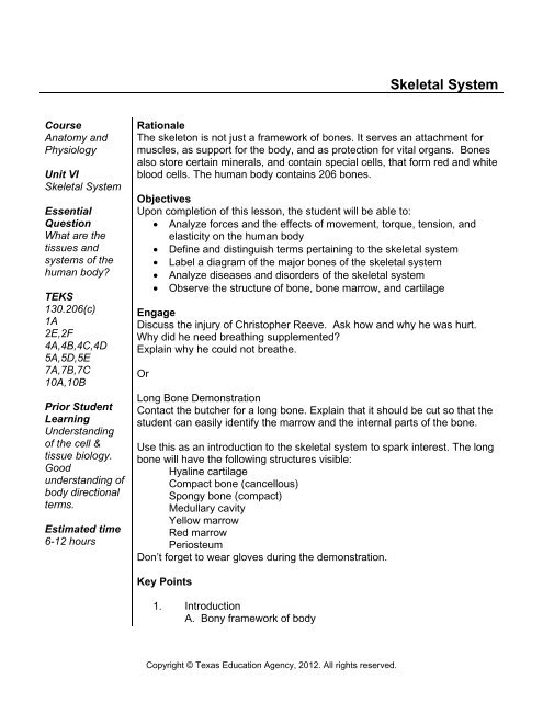
Skeletal System - Health Science Technology Education
Skeletal System - Health Science Technology Education
Skeletal System - Health Science Technology Education

You also want an ePaper? Increase the reach of your titles
YUMPU automatically turns print PDFs into web optimized ePapers that Google loves.
<strong>Skeletal</strong> <strong>System</strong><br />
Course<br />
Anatomy and<br />
Physiology<br />
Unit VI<br />
<strong>Skeletal</strong> <strong>System</strong><br />
Essential<br />
Question<br />
What are the<br />
tissues and<br />
systems of the<br />
human body?<br />
TEKS<br />
130.206(c)<br />
1A<br />
2E,2F<br />
4A,4B,4C,4D<br />
5A,5D,5E<br />
7A,7B,7C<br />
10A,10B<br />
Prior Student<br />
Learning<br />
Understanding<br />
of the cell &<br />
tissue biology.<br />
Good<br />
understanding of<br />
body directional<br />
terms.<br />
Estimated time<br />
6-12 hours<br />
Rationale<br />
The skeleton is not just a framework of bones. It serves an attachment for<br />
muscles, as support for the body, and as protection for vital organs. Bones<br />
also store certain minerals, and contain special cells, that form red and white<br />
blood cells. The human body contains 206 bones.<br />
Objectives<br />
Upon completion of this lesson, the student will be able to:<br />
• Analyze forces and the effects of movement, torque, tension, and<br />
elasticity on the human body<br />
• Define and distinguish terms pertaining to the skeletal system<br />
• Label a diagram of the major bones of the skeletal system<br />
• Analyze diseases and disorders of the skeletal system<br />
• Observe the structure of bone, bone marrow, and cartilage<br />
Engage<br />
Discuss the injury of Christopher Reeve. Ask how and why he was hurt.<br />
Why did he need breathing supplemented?<br />
Explain why he could not breathe.<br />
Or<br />
Long Bone Demonstration<br />
Contact the butcher for a long bone. Explain that it should be cut so that the<br />
student can easily identify the marrow and the internal parts of the bone.<br />
Use this as an introduction to the skeletal system to spark interest. The long<br />
bone will have the following structures visible:<br />
Hyaline cartilage<br />
Compact bone (cancellous)<br />
Spongy bone (compact)<br />
Medullary cavity<br />
Yellow marrow<br />
Red marrow<br />
Periosteum<br />
Don’t forget to wear gloves during the demonstration.<br />
Key Points<br />
1. Introduction<br />
A. Bony framework of body<br />
Copyright © Texas <strong>Education</strong> Agency, 2012. All rights reserved.
B. 206 bones in adult<br />
C. Functions<br />
1. Support: body structure and shape<br />
2. Protection: vital organs surrounded<br />
3. Movement and anchorage of muscles (levers for<br />
muscular action)<br />
a. Tendons: attach muscle to bone<br />
b. Ligaments: attach bone to bone<br />
4. Mineral storage: calcium and phosphorus<br />
5. Blood cell formation - hematopoiesis<br />
2. Bone Composition<br />
A. Collagen: chief organic constituent (protein)<br />
B. Inorganic calcium salts (Vitamin D essential for absorption of<br />
minerals i.e. calcium)<br />
1. Deposition favored by<br />
a. Estrogen, testosterone<br />
b. Alkaline phosphatase<br />
c. Thyrocalcitonin<br />
d. Mechanical stress, i.e. traction<br />
2. Withdrawal favored by<br />
a. Alkaline phosphatase<br />
b. Parathormone<br />
c. Inactivity<br />
C. Cells<br />
1. Osteoblasts: bone building, bone repairing cells in the<br />
periosteum<br />
2. Osteocytes: mature bone cells within the bone matrix<br />
3. Osteoclast: causes reabsorption of bone<br />
D. Periosteum<br />
1. Dense, fibrous membrane covering bone<br />
2. Contains blood vessels<br />
3. Essential for bone cell survival and bone formation<br />
3. Types of Bones Based on Composition<br />
A. Compact bone<br />
1. Very dense, stress bearing<br />
2. Haversian systems<br />
a. Lamellae: concentric cylinder shaped calcified<br />
structure<br />
b. Lacunae: small spaces containing tissue fluid<br />
c. Osteocytes: facilitate exchange of calcium<br />
between blood and bone<br />
d. Canaliculi: canals connecting the lacunae<br />
together and to the haversian canal which carries<br />
nutrients and wastes to and from the osteocytes<br />
B. Cancellous bone<br />
1. Light, spongy<br />
Copyright © Texas <strong>Education</strong> Agency, 2012. All rights reserved.
2. Low stress areas where weight of bone would be a<br />
problem<br />
3. Found at ends of long bones, ribs, sternum, hips,<br />
vertebrae, cranium<br />
4. No haversian systems<br />
5. Web-like arrangement<br />
4. Classification of Bones According to Shape<br />
A. Long bones (extremities): levers<br />
1. Epiphysis: at the ends, covered with hyaline cartilage<br />
for articulating bones; cancellous bone<br />
2. Diaphysis: shaft, covered with periosteum; medullary<br />
canal with yellow and red marrow (lined with<br />
endosteum); covered with periosteum for bone growth,<br />
repair, and nutrition; compact bone<br />
3. Femur, tibia, fibula, humerus, ulna, radius, clavicle<br />
B. Short: cube shaped, allows flexible movement<br />
1. Cancellous bone covered by compact bone<br />
2. Carpals, tarsals, metacarpals, metatarsals, phalanges<br />
C. Flat: flat plates; protect vital organs and provide broad surface<br />
area for muscle attachment<br />
1. Cranial bones, facial bones, scapula, sternum<br />
D. Irregular: peculiarly shaped to provide support and protection,<br />
yet allow flexibility<br />
1. Vertebrae, ribs, ear, hip, hyoid<br />
E. Sesamoid bones<br />
1. Extra bones found in certain tendons, i.e. patella<br />
5. Bone Formation<br />
A. Initially collagen fibers secreted by fibroblasts<br />
B. Cartilage deposited between fibers<br />
C. Skeleton fully formed by 2 nd month of fetal development (all<br />
cartilage)<br />
D. After 8 th week of fetal development ossification (mineral matter<br />
deposited and replaces cartilage) begins<br />
E. Childhood and adolescence: ossification exceeds bone loss<br />
F. Early adulthood thru middle age: ossification equals bone loss<br />
G. After age 35: bone loss exceed ossification<br />
H. Skull<br />
1. Begins as fibrous membrane<br />
2. Ossification center in the middle of the membrane:<br />
begins in the middle and radiates out<br />
3. Ossification not complete at birth: fontanels (soft<br />
spots) on infant’s head allow molding of skull during<br />
birth and with the open joints allows for growth of the<br />
brain<br />
I. Other bones<br />
1. Begin as hyaline cartilage<br />
Copyright © Texas <strong>Education</strong> Agency, 2012. All rights reserved.
2. Short bones: one ossification center in middle and<br />
proceeds toward the periphery<br />
3. Long bones: three ossification centers (one at each<br />
end and one in the center of the shaft) ossification<br />
from center toward each end and from each end<br />
toward the center<br />
6. Bone Growth<br />
A. Grow in length at the epiphyseal line<br />
B. Grow in width by the addition of bone to the surface<br />
C. Controlled by the anterior pituitary (growth hormone)<br />
1. Dwarfism: hypofunction<br />
2. Giantism: hyperfunction<br />
3. Acromegaly: hyperfunction after puberty; enlarges<br />
bones of hands, feet, face<br />
7. Bone Markings<br />
A. Purpose<br />
1. Join one bone to another<br />
2. Provide a surface for attachment of muscles<br />
3. Create an opening for passage of blood vessels and<br />
nerves<br />
4. Used as landmarks<br />
B. Examples<br />
1. Process: bony prominence or projection<br />
2. Condyle: a rounded knuckle-like prominence usually at<br />
a point of articulation<br />
3. Epicondyle: small projection<br />
4. Head: a rounded articulating process at the end of a<br />
bone<br />
5. Spine: a sharp, slender projection<br />
6. Tubercle: a small rounded process<br />
7. Tuberosity: a large rounded process<br />
8. Trochanter: a large process for muscle attachment<br />
9. Fossa: a depression or hollow<br />
10. Foramen: a hole<br />
11. Crest: sharp ridge<br />
12. Line: a less prominent ridge of a bone than a crest<br />
13. Meatus: a tube-like passage<br />
14. Sinus/antrum: a cavity within a bone<br />
15. Depression: hollow region or opening<br />
16. Fissure: narrow, slit-like opening<br />
17. Sulcus: a groove<br />
18. Facet: a small area on a bone<br />
8. Bone Marrow<br />
A. Yellow marrow<br />
1. Medullary cavity of long bones<br />
2. Fat storage<br />
Copyright © Texas <strong>Education</strong> Agency, 2012. All rights reserved.
B. Red marrow: hematopoietic tissue<br />
1. In all cancellous bone in children<br />
2. In adults: cancellous bone of vertebrae, hips, sternum,<br />
ribs, cranial bones, proximal ends of femur and<br />
humerus<br />
3. Forms RBCs, platelets, some WBCs, and destroys old<br />
RBCs and some foreign materials<br />
9. Axial Skeleton<br />
A. Skull: 22 bones<br />
B. Cranium: houses and protects the brain with 8 bones<br />
1. Frontal: forms forehead and orbits of eyes;<br />
supraorbital margins (ridge to protect eyes)<br />
2. Ethmoid: forms roof of nasal cavity, very light bone has<br />
horizontal plate, perpendicular plate, and two lateral<br />
masses<br />
3. Parietal -- Right and Left: form sides and roof of skull,<br />
internal surface rough to accommodate the brain<br />
4. Temporal -- Right and Left: forms temple, cheek, ear<br />
openings<br />
a. Squamous portion: forms temple<br />
b. Zygomatic process: forms cheek<br />
c. Petrous portion: forms auditory canal<br />
d. Mastoid portion: behind the ear<br />
e. Tympanic portion: walls of acoustic meatus<br />
5. Occipital: back of skull, inferior portion has foramen<br />
magnum where spinal cord passes through; sides of<br />
foramen have two projections (condyles) that articulate<br />
with the first cervical vertebra (atlas)<br />
6. Sphenoid: fills space between orbital plates, contains<br />
sphenoidal sinuses; upper surface has a depression<br />
called the sella turcica, where the pituitary gland rests<br />
7. Wormian Bones: extra bones formed by irregular<br />
connections of cranial sutures<br />
8. Cranial Sutures: unite the bones of the cranium; as<br />
child grows, irregular bands of connective tissue ossify<br />
and turns into hard bone<br />
a. Coronal suture: between frontal and parietal<br />
bones<br />
b. Sagittal suture: between right and left parietal<br />
bones<br />
c. Lambdoidal suture: between parietal and occipital<br />
bones<br />
d. Squamous suture: between temporal and parietal<br />
bones<br />
e. Abnormalities<br />
(1) Microcephalus: premature fusion<br />
Copyright © Texas <strong>Education</strong> Agency, 2012. All rights reserved.
(2) Hydrocephalus: delayed fusion (increases<br />
intracranial pressure)<br />
9. Fontanels: fusion of the cranial bones is not complete<br />
at birth, so a space between the bones remains<br />
a. Anterior (Bregmatic): “soft spot”, closes at 18<br />
months<br />
b. Posterior (Occipital): triangular, closes at 2-3<br />
months<br />
c. Anteriolateral (Sphenoidal): at 2 temples, closes<br />
at 2-3 months<br />
d. Posterolateral (Mastoidal): behind ears (2),<br />
closes at 1 year<br />
C. Facial Bones: guard and support the eyes, ears, nose, and<br />
mouth; 14 bones<br />
1. Nasal bones (2): form bridge of nose<br />
2. Vomer: forms central nasal septum<br />
3. Maxillary (2): upper jaw bones, fusion occurs before<br />
birth (if not, cleft palate occurs), forms roof of mouth,<br />
walls of nose, floor of orbitals; body has maxillary<br />
sinuses, alveolar process upper teeth, palatine<br />
process anterior palate; largest bone of the upper<br />
face<br />
4. Mandible: lower jawbone, largest bone of face, two<br />
perpendicular portions called rami (have 2 processes:<br />
condylar process posterior forms temporalmandibular<br />
joint; coronoid process anterior for<br />
muscle attachment)<br />
5. Zygoma (2): cheek bones<br />
6. Lacrimal (2): small bones form medial wall of eye<br />
socket, tear duct passes through, smallest, fragile<br />
7. Palatine (2): forms back roof of mouth and floor of<br />
nose, L-shaped<br />
8. Inferior turbinate (2): forms curved ledge inside side<br />
wall of nose<br />
D. Ear Bones: tiny bones in middle ear cavity in temporal bone<br />
1. Malleus (2): the hammer<br />
2. Incus (2): the anvil<br />
3. Stapes (2): the stirrups<br />
E. Hyoid Bone: u-shaped bone in the neck at the base of the<br />
tongue; the only bone that does not touch another bone.<br />
F. Cranial Sinuses: cavities within the cranium, function as<br />
resonance chambers in the production of the voice, decrease<br />
weight of skull, lined with mucous membrane<br />
1. Frontal sinuses (2): above eyebrows, open into nasal<br />
cavity<br />
2. Ethmoid sinuses (2): between eyes<br />
Copyright © Texas <strong>Education</strong> Agency, 2012. All rights reserved.
3. Sphenoidal sinus (1): posterior to ethmoidal sinuses,<br />
opens into nasopharynx<br />
4. Maxillary sinuses (2): on either side of nose, opens on<br />
lateral wall of nasal cavity<br />
G. Vertebral Column<br />
1. Functions<br />
a. Supports trunk and neck<br />
b. Protects spinal cord<br />
c. Multiple joint spaces allow for bending and<br />
twisting<br />
2. Curves: (lateral view) allow for resilience and spring for<br />
walking<br />
a. Thoracic: present at birth<br />
b. Sacral: low back<br />
c. Cervical: begins at 3 months when infant first<br />
begins to lift head<br />
d. Lumbar: begins when child first walks<br />
3. Vertebrae: 26 separated by intervertebral disk to<br />
cushion joints for movement<br />
a. Body: thick, disk-shaped anterior portion<br />
b. Arch: encloses space for spinal cord (neural<br />
canal); has 3 processes for muscle attachment<br />
(spinous process dorsally directed, 2<br />
transverse processes)<br />
c. Articular processes: provide for articulation with<br />
other vertebrae (2 superior and 2 inferior)<br />
d. Pedicles (2): originate from body of vertebrae<br />
notched to allow spinal cord nerves to pass<br />
e. Lamina: posterior wall of vertebrae, weakest point<br />
4. Sections<br />
a. Cervical (7): smallest, oblong bodies, wide<br />
transverse processes<br />
(1) Atlas: 1 st cervical vertebra; supports head<br />
by articulating with condyles of occipital<br />
bone; bony ring with no body; has short<br />
wing-like transverse processes; allows<br />
forward and backward motion<br />
(2) Axis: 2 nd vertebra; small body with<br />
projection called the odontoid process that<br />
acts as the axis of rotation of the skull<br />
(3) 3 rd , 4 th , 5 th , and 6 th vertebrae forked to<br />
cradle strong ligaments of head<br />
(4) 7 th vertebra has very prominent spinous<br />
process called the vertebral prominence<br />
that can be felt at the base of the neck<br />
b. Thoracic (12): progressively increase in size from<br />
Copyright © Texas <strong>Education</strong> Agency, 2012. All rights reserved.
neck; have long spinous process (pointed<br />
downward), 6 articular facets for rib attachment<br />
c. Lumbar (5): largest and strongest, have short<br />
projections for muscle attachment<br />
d. Sacral: 5 fused bones, triangular, forms dorsal<br />
part of pelvis, joins ileum bone at iliosacral joint<br />
e. Coccyx: 3-4 fused bones, articulates with tip of<br />
sacrum, slightly movable (to assist in childbirth)<br />
5. Injuries and Diseases<br />
a. Kyphosis: hunchback, posterior thoracic<br />
exaggerated<br />
b. Lordosis: swayback, exaggerated anterior curve<br />
of lumbar region<br />
c. Scoliosis: lateral curvature of the spine<br />
d. Fractures & Dislocations: most often if fracture of<br />
lamina, can cause spinal cord damage, paralysis<br />
e. Intervertebral disk herniation: causes pressure on<br />
spinal nerve, pain<br />
f. Tuberculosis of spine: by tubercle bacillus,<br />
destroys body of vertebrae<br />
H. Thorax: 25 bones and cartilage; walls covered by skin and<br />
muscles; floor is formed by the diaphragm<br />
1. Functions<br />
a. Protect and support heart and lungs<br />
b. Supports bones of pectoral girdle<br />
c. Plays a leading role in respiration<br />
d. Ribs and sternum aid in RBC formation<br />
2. Sternum: breast bone; sword and handle shape<br />
a. Manubrium: handle; notched for 1 st 7 costal<br />
cartilage; articulates with acromium end of<br />
clavicle and 1 st rib<br />
b. Body: blade; notched for 1 st 7 costal cartilage<br />
c. Xiphoid process: tip; attachment site for<br />
diaphragm<br />
3. Costal cartilages: hyaline cartilage connecting ribs to<br />
sternum in 1-7 and to anterior ribs in 8-10<br />
4. Ribs (12 pairs): attached posteriorly with vertebrae and<br />
anteriorly with costal cartilage<br />
a. True ribs: 1 st seven pairs<br />
b. False ribs: 8-12 (11 and 12 are floating ribs)<br />
10. Appendicular Skeleton (126 bones)<br />
A. Shoulder Girdle<br />
1. Clavicles (2): collar bones<br />
2. Scapulae (2): shoulder blade<br />
B. Upper Extremities<br />
1. Humerus: upper arm<br />
Copyright © Texas <strong>Education</strong> Agency, 2012. All rights reserved.
2. Radius: thumb side of forearm<br />
3. Ulna: little finger side of forearm<br />
4. Carpals (8): wrist bones<br />
5. Metacarpals (5): hand bones<br />
6. Phalanges (14): finger bones<br />
C. Pelvic Girdle<br />
1. Os coxae (2): contains the acetabulum (hip socket)<br />
a. Ilium<br />
b. Ischium<br />
c. Pubis<br />
2. Sacrum<br />
D. Lower Extremities<br />
1. Femur: thigh bone<br />
2. Patella: kneecap<br />
3. Tibia: shin bone<br />
4. Fibula: lateral bone of lower leg<br />
5. Tarsals (7): ankle bones<br />
a. Talus<br />
b. Calcaneus<br />
6. Metatarsals (5): foot bones<br />
7. Phalanges (14): toe bones<br />
11. Articulations<br />
A. Synarthrotic: immovable<br />
B. Amphiarthrotic: limited movement i.e. pubic symphysis,<br />
vertebral joints, sacroiliac joint<br />
C. Diarthrotic: freely movable<br />
1. Gliding: wrist<br />
2. Pivot: between radius and ulna<br />
3. Ball and socket: hip<br />
4. Hinge: elbow<br />
12. Diseases/Disorders<br />
A. Arthritis: inflammation of the bones at the joints, usually with<br />
pain and changes in bone structure<br />
B. Bunion: abnormal lateral displacement of big toe causing<br />
inflammation and thickening of the bursa<br />
C. Bursitis: inflammation of the bursa, which is a sac or cavity<br />
filled with synovial fluid<br />
D. Dislocation: the displacement of a bone from a joint, tearing<br />
ligaments, tendons, and capsules<br />
E. Fracture: a break in a bone<br />
1. Simple<br />
2. Compound<br />
3. Spiral<br />
4. Comminuted<br />
5. Greenstick<br />
F. Osteitis: inflammation or infection of the bone<br />
Copyright © Texas <strong>Education</strong> Agency, 2012. All rights reserved.
G. Osteomyelitis: bone infection that involves the bone marrow<br />
H. Osteoporosis: condition in which bones become softer and<br />
more brittle, and thus more liable to fracture due to loss of<br />
mineral content; associated with aging<br />
I. Rickets: condition in which bones fail to calcify and growth is<br />
hampered usually due to a deficiency of vitamin D and<br />
phosphorus in the diet<br />
J. Spina bifida: congenital defect in which the vertebrae fail to<br />
unite in the midline<br />
K. Sprain: wrenching of a joint with injury to ligaments<br />
Activity<br />
I. Color code a diagram of the skeleton (axial and appendicular) then<br />
label the structures.<br />
II. Participate in the Hokie-Pokie Osteokey.<br />
III. Participate in Paper Bones Game.<br />
IV. Participate in Bag o' Bones Game.<br />
V. Participate in Skeletonary Game.<br />
VI. Complete the <strong>Skeletal</strong> <strong>System</strong> and Long Bones Laboratory<br />
VII.<br />
Investigation.<br />
Identify anatomical structures of skeletal system on dissected cat.<br />
(This can be accomplished as a virtual tour on the internet, or if your<br />
budget allows, the students can dissect cats.)<br />
VIII. Research and report on the cause and effect of disease, trauma, and<br />
congenital defects on the structure and function of cells, tissues,<br />
organs, and systems as well as research technological advances and<br />
limitations in the treatment of the skeletal system disorders.<br />
Assessment<br />
Successful identification of skeletal muscles<br />
Successful completion of activities<br />
Laboratory Investigation Rubric<br />
Materials<br />
Activity I- Diagram of the skeleton and colored pencils<br />
Activity II- no outside materials needed<br />
Activity III- Each group of students will need the following:<br />
Cardboard Halloween skeleton about four feet long<br />
Post-it stickers (1/2” x 2”) – 25<br />
List of bones and labeled atlas (from text)<br />
Activity IV- disarticulated bones from a skeleton model<br />
Activity V- Modeling clay (2 sticks)<br />
Deck of cards (1 set) with the name of a bone on each<br />
Copyright © Texas <strong>Education</strong> Agency, 2012. All rights reserved.
Desk and chair in front of room<br />
List of bones and atlas<br />
Activity VI –<br />
Prepared slide of hyaline cartilage<br />
Prepared slide of bone<br />
Prepared slide of bone marrow<br />
Microscope for viewing microscope slides<br />
Stereomicroscope<br />
Knife<br />
Long bone from chicken leg - contact local butcher<br />
Dissecting tray<br />
Colored pencils<br />
Activity VII- Dissection Cat 1 for every 2-4 students, and dissection tools<br />
AND/OR computers with internet access<br />
Activity VIII- Computers with internet access<br />
Visit with a local butcher about making some special cuts on some cow joint<br />
and long bones in order to show layers and function.<br />
Utah State Office of <strong>Education</strong>, (2005). Medical Anatomy and Physiology<br />
Teacher Resource CD. Utah.<br />
Accommodations for Learning Differences<br />
For reinforcement, the student will make flashcards of key terms.<br />
For enrichment, the student will research and report on how forensic<br />
scientists use the skeletal system when investigating homicides.<br />
National and State <strong>Education</strong> Standards<br />
National <strong>Health</strong> <strong>Science</strong> Cluster Standards<br />
HLC01.01 Academic Foundations<br />
<strong>Health</strong> care workers will know the academic subject matter required (in<br />
addition to state high school graduation requirements) for proficiency within<br />
their area. They will use this knowledge as needed in their role.<br />
HLC1O.01 Technical Skills<br />
<strong>Health</strong> Care Workers will apply technical skills required for all career<br />
specialties. They will demonstrate skills and knowledge as appropriate.<br />
TEKS<br />
130.206(c)<br />
130.206(c)(1)(A) demonstrate safe practices during laboratory and field<br />
investigations;<br />
130.206(c)(2)(E) plan and implement descriptive, comparative, and<br />
experimental investigations, including asking questions, formulating testable<br />
hypotheses, and selecting equipment and technology;<br />
Copyright © Texas <strong>Education</strong> Agency, 2012. All rights reserved.
130.206(c)(2)(F) collect and organize qualitative and quantitative data and<br />
make measurements with accuracy and precision using tools such as<br />
calculators, spreadsheet software, data-collecting probes, computers,<br />
standard laboratory glassware, microscopes, various prepared slides,<br />
stereoscopes, metric rulers, electronic balances, hand lenses, Celsius<br />
thermometers, hot plates, lab notebooks or journals, timing devices, Petri<br />
dishes, lab incubators, dissection equipment, meter sticks, and models,<br />
diagrams, or samples of biological specimens or structures;<br />
130.206(c)(2)(H) communicate valid conclusions supported by the data<br />
through methods such as lab reports, labeled drawings, graphic organizers,<br />
journals, summaries, oral reports, and technology-based reports;<br />
130.206(c)(4)(A) analyze the chemical reactions that provide energy for the<br />
body;<br />
130.206(c)(4)(D) analyze the effects of energy excess in disorders such as<br />
obesity as it relates to cardiovascular and musculoskeletal systems;<br />
130.206(c)(5)(A) explain the coordination of muscles, bones, and joints that<br />
allows movement of the body;<br />
130.206(c)(5)(D) analyze and describe the effects of pressure, movement,<br />
torque, tension, and elasticity on the human body;<br />
130.206(c)(5)(E) perform an investigation to determine causes and effects<br />
of force variance and communicate findings;<br />
130.206(c)(7)(A) illustrate conduction systems such as nerve transmission<br />
or muscle stimulation;<br />
130.206(c)(7)(B) investigate the therapeutic uses and effects of external<br />
sources of electricity on the body system;<br />
130.206(c)(7)(C) evaluate the application of advanced technologies such as<br />
electroencephalogram, electrocardiogram, bionics, transcutaneous electrical<br />
nerve stimulation, and cardioversion;<br />
130.206(c)(10)(A) analyze the relationships between the anatomical<br />
structures and physiological functions of systems, including the<br />
integumentary, nervous, skeletal, musculoskeletal, cardiovascular,<br />
respiratory, gastrointestinal, endocrine, and reproductive;<br />
130.206(c)(10)(B) evaluate the cause and effect of disease, trauma, and<br />
congenital defects on the structure and function of cells, tissues, organs, and<br />
systems; and<br />
130.206(c)(10)(C) research technological advances and limitations in the<br />
treatment of system disorders.<br />
Texas College and Career Readiness Standards<br />
English Language Arts<br />
II. B. Understand new vocabulary and concepts and use them accurately in<br />
reading, writing, and speaking.<br />
III. B. Develop effective speaking styles for both group and one-on-one<br />
situations.<br />
IV. A. Apply listening skills as an individual, and as a member of a group in a<br />
variety of settings.<br />
Copyright © Texas <strong>Education</strong> Agency, 2012. All rights reserved.
IV. B. 2. Listen actively and effectively in one-on-one communication<br />
situations.<br />
<strong>Science</strong><br />
1.A.1. Utilize skepticism, logic, and professional ethics in science.<br />
1.A.2. Use creativity and insight to recognize and describe patterns in natural<br />
phenomena.<br />
1.A.3. Formulate appropriate questions to test understanding of a natural<br />
phenomenon.<br />
1.A.4. Relay on reproducible observations of empirical evidence when<br />
constructing analyzing, and evaluating explanations of natural events and<br />
processes.<br />
1.E.2. Use essential vocabulary of the discipline being studied.<br />
3.A.1. Use correct applications of writing practices in scientific<br />
communication<br />
Copyright © Texas <strong>Education</strong> Agency, 2012. All rights reserved.
Skeleton, Anterior View<br />
Copyright © Texas <strong>Education</strong> Agency, 2012. All rights reserved.
Skeleton, Posterior View<br />
Copyright © Texas <strong>Education</strong> Agency, 2012. All rights reserved.
Label the Skeleton, Anterior View<br />
Copyright © Texas <strong>Education</strong> Agency, 2012. All rights reserved.
Label the Skeleton, Posterior View<br />
Copyright © Texas <strong>Education</strong> Agency, 2012. All rights reserved.
Activity: Hokie-Pokie Osteokey<br />
Objective:<br />
Help students practice location of bones.<br />
Instructions:<br />
1. Write the following anatomical terms on the board:<br />
Phalanges (hands)<br />
Left patella<br />
Right humeral head<br />
Olecranons<br />
Coccyx<br />
Left ulna<br />
Right tarsals<br />
Left innominate<br />
Cranium<br />
Right tibia<br />
Left calcaneous<br />
Mandible<br />
Right radius<br />
Left great toe<br />
Right parietal<br />
Right carpels<br />
skeleton<br />
2. Ask students to stand. (It’s fun to get everyone in a circle.)<br />
3. Have everyone sing Hokey-pokey song and substitute bones on board for usual body parts.<br />
“Put you phalanges in. Take your phalanges out.<br />
Put your phalanges in and shake them all about.<br />
Do the hokey-pokey and turn yourself around. That’s what it’s all about!”<br />
Clap, Clap.<br />
Copyright © Texas <strong>Education</strong> Agency, 2012. All rights reserved.
Paper Bones<br />
Objective:<br />
Materials:<br />
Label bones on a cardboard “Halloween-type” skeleton<br />
Each group of students will need the following:<br />
1. Cardboard Halloween skeleton about four feet long<br />
2. Sticky memo paper (1/2” x 2”) – 25<br />
3. List of bones and labeled atlas (from text)<br />
Warm-up<br />
1. Break the class into groups of 3 or 4 (no more)<br />
2. Give each group the materials above and tell them to choose a place to work together (table<br />
top or floor)<br />
3. Give them ten minutes to correctly label all the bones they can find on the list<br />
4. Have each group present their labeled skeleton while the class critiques and gives feedback.<br />
The students are encouraged to defend their choices using the reference atlas. The<br />
instructor is the referee.<br />
5. Repetition of this process for each group reinforces the learning.<br />
Competition<br />
1. Have each group choose a team name (related to anatomy, e.g., Patellas)<br />
2. Each group prepares to start the exercise over but without the atlas.<br />
3. This time they must label the skeleton as quickly as possible while they are being timed.<br />
Give them a few minutes to organize task assignments before starting. Be sure they do not<br />
fill out the label beforehand.<br />
4. Give the starting signal and keep track of the time.<br />
5. Instruct them that when finished they should signal completion by standing up and holding<br />
the labeled skeleton up.<br />
6. Note times and finishing order on the board.<br />
7. Have the groups represent their labeling results in order with group critique.<br />
8. Score by adding 10-15 seconds per error to their elapsed time.<br />
9. The team with the fastest resulting time wins.<br />
10. Present an award.<br />
Comment<br />
Be certain that most of the bones are accurately portrayed on the cardboard skeleton. If there is<br />
an error, point it out before play starts and agree to call it something.<br />
Copyright © Texas <strong>Education</strong> Agency, 2012. All rights reserved.
Bag o’ Bones<br />
Objective:<br />
Identify a bone in a bag by touch only<br />
Materials: 1. A set of disarticulated bones<br />
2. Pillowcase<br />
1. Divide the students randomly into two teams and decide on team names (anatomy names).<br />
2. The instructor places a bone into the pillowcase in such a manner that the students cannot<br />
see it (students can be asked to put their heads down, or the instructor can select the bone<br />
behind a cabinet door).<br />
3. Flip a coin to determine which team starts. That team puts forth the first player to represent<br />
them.<br />
4. The student player is given the choice of feeling the bone inside the bag or feeling the bone<br />
from the outside (which is more difficult through the cloth). Award one point for inside and<br />
two points for outside examination.<br />
5. Give the student only 10 seconds (you can vary this according to the skill of your students) to<br />
feel the bone, counting during the examination.<br />
6. After withdrawing his hand, the student is given another 10 seconds to announce the name<br />
of the bone.<br />
7. If the student chooses to examine the bone from outside the bag, he is given only 5 seconds<br />
and then 5 seconds to announce the bone name.<br />
8. If correct, the instructor takes the bone out of the bag and awards the points accordingly (1<br />
point for in-bag exam or 2 points for external exam).<br />
9. Ask the student to point out what features caused them to decide on the bone name.<br />
10. If the bone named is incorrect, the instructor give an opportunity to a student on the other<br />
team and that team may now reap the points. The process continues to alternate from one<br />
team to the other until it is correctly identified. Obviously the chance for success becomes<br />
greater with each wrong answer put forth. Eventually someone will identify the bone.<br />
11. The instructor (or a designated student) keeps track of the team scores.<br />
12. After every student has had at least one turn, the game is finished and a winner declared.<br />
Comments<br />
This game should only be played when the class is fairly familiar with the bones and their<br />
surface features. It is a good opportunity to point out features like the hinge-like joint on the<br />
proximal end of the ulna that help differentiate this bone from the radius. The process also<br />
reinforces some of the anatomic features like the tibia is much thicker than fibula. It is best to<br />
use the long bones and the irregular bones. Small bones (carpals, tarsals, and phalanges) are<br />
difficult to exactly identify. Vertebrae can be a challenge, but it is possible to state from what<br />
region on the spine the bone comes from (cervical, thoracic).<br />
Copyright © Texas <strong>Education</strong> Agency, 2012. All rights reserved.
Skeletonary<br />
Objective:<br />
Make clay images of the bones<br />
Materials: 1. Modeling clay (2 sticks)<br />
2. Deck of cards (1 set) with the name of a bone on each<br />
3. Desk and chair in front of room<br />
4. List of bones and atlas<br />
Competition<br />
1. Divide the class into two equal groups and have them sit on opposing sides.<br />
2. Have each team choose a name.<br />
3. Flip a coin to decide which team will go first and then have the team choose one member to<br />
start. This person comes to the front of the class.<br />
4. The student randomly chooses a card from the deck of bones and shows it to the teacher.<br />
5. For one point, the student must identify the bone on the atlas (unseen by the class).<br />
6. Sitting at the desk, in front of the room, the student has 30 –40 seconds to mold the clay into<br />
a suitable representation of the selected bone.<br />
7. The team members then have 30 seconds to make three guesses. If correct, the team gets<br />
one more point.<br />
8. If not guessed correctly, the other team has an opportunity to guess once and get the point.<br />
9. Then the student-sculptor is given an additional question by the teacher related to the<br />
selected bone. If answered correctly, the team gets one more point. Maximum points are<br />
three per turn.<br />
10. The other team then takes a turn.<br />
11. The team with the highest score wins and each team member gets an award.<br />
Additional Rules<br />
1. The sculptor may not speak during their clay rendition. Speaking nullifies the point.<br />
2. The sculptor may hold the clay model to their body to show its location.<br />
3. Any sculpted form is allowable to give the hint. More than one bone may be made and the<br />
sculptor can then point to the unknown bone.<br />
Comments<br />
This game develops psychomotor skills and encourages creativity in remembering features of<br />
the bones. For instance, the sculptor with the task of communicating the metacarpal may take<br />
all five of them with the attached phalanges or carpal bones and then point to the metacarpals.<br />
Kids get creative.<br />
Copyright © Texas <strong>Education</strong> Agency, 2012. All rights reserved.
<strong>Skeletal</strong> <strong>System</strong> and Long Bones Laboratory Investigation<br />
Purpose:<br />
In this lab, students will observe closely the structure of bone and cartilage. Students will also<br />
observe closely the structure of long bones and become familiar with bone marrow.<br />
Background Information:<br />
Materials:<br />
Prepared slide of hyaline cartilage<br />
Prepared slide of bone<br />
Prepared slide of bone marrow<br />
Microscope for viewing microscope slides<br />
Stereomicroscope<br />
Knife<br />
Long bone from chicken leg<br />
Dissecting tray<br />
Colored pencils<br />
Procedure and Observations:<br />
I. Observing the Structure of a Long Bone<br />
1. Obtain a long bone of a chicken leg and place it in a dissecting tray. Observe the outside<br />
covering and features of the bone. Note the smooth covering at both ends of the bone.<br />
Also notice the presence of small holes along the surface of the bone. Describe the<br />
surface of the chicken leg bone.<br />
2. With a knife, cut one end of the bone to expose the fine structure of the spongy bone and<br />
some bone marrow. Be careful when handling the knife. Be sure to cut in a direction<br />
away from your hands and body. If you are having trouble cutting, see your instructor for<br />
help.<br />
3. Using your book for a diagram of the long bone as a guide, continue to observe the<br />
chicken bone. For better observation of structures, you will want to use a dissecting<br />
microscope at this point. Observe the periosteum, which covers and protects bone.<br />
Beneath the periosteum is a hard dense layer called compact bone. Notice the softer<br />
spongy bone is near the ends of the bone. Spongy bone contains many small spaces.<br />
Copyright © Texas <strong>Education</strong> Agency, 2012. All rights reserved.
In long bones the spongy bone also contains red marrow, which produces red and white<br />
blood cells. Note the internal cavity of the bone, which may contain some fat-storing<br />
yellow marrow. On the outer edge of the end of the bone, find the plate of articular<br />
cartilage. Remember that this cartilage helps in bone movement. Each of the wide ends<br />
of a long bone is called the epiphysis, whereas the long shaft of the bone is called the<br />
diaphysis.<br />
4. Sketch the internal structure of the chicken leg bone. Label the periosteum, compact<br />
bone, spongy bone, red marrow, yellow marrow, articular cartilage, epiphysis, diaphysis.<br />
5. Answer the following questions about the chicken leg bone.<br />
a. Why is it necessary that the ends of the bones be smooth?<br />
b. What is the function of the small holes in the bone surface?<br />
c. What is the importance of compact and spongy bone?<br />
Copyright © Texas <strong>Education</strong> Agency, 2012. All rights reserved.
II. Observing Cartilage, Bone and Bone Marrow<br />
1. Cartilage is more flexible than bone, thus it can take a great deal of stress. Live cartilage<br />
cells, called chondrocytes, are found in cavities called lacunae. Cartilage contains no<br />
blood vessels. Materials enter and exit the chondrocytes by diffusion to and from the<br />
blood vessels in adjacent layers of tissue. Hyaline cartilage is composed of close<br />
collagen fibers, making it tough and slightly flexible. Hyaline cartilage is found on the<br />
articular surfaces of bones. It also connects the ribs to the sternum and is the chief<br />
component of the embryonic skeleton.<br />
2. Obtain a slide of hyaline cartilage. Note the chondrocytes within the lacunae. The<br />
collagen fibers are too fine to be seen using ordinary staining procedures and ordinary<br />
microscopes. Look around the slide and assure yourself that the cartilage is indeed<br />
avascular.<br />
3. Draw the hyaline cartilage. Be sure and label the chondrocyte and the lacunae.<br />
4. Bone is the hard tissue chemically identified by crystals of calcium phosphate or<br />
calcium carbonate. The calcium salts are quite strong but are brittle. The main<br />
component of the rest of bone is collagen. Collagen fibers are long, straight, unbranched,<br />
three-ply protein chains that are highly flexible. Since the calcium salts are organized<br />
around the collagen fibers, the combined portions of the osseous tissue exhibit the best<br />
of both worlds. The result is a strong, flexible and relatively shatter resistant structure.<br />
Remember back to the histology unit when we studied about bones. We learned about<br />
the Haversian systems which are responsible for supplying the bone with nutrients and<br />
removing wastes. Making up the bone are many thin, concentric circles known as<br />
lamella. The black spaces within the lamella are the lacunae, wherein reside the<br />
osteocyte or bone cell. The canaliculi are the canals running between the lacunae. In<br />
addition to housing the cytoplasmic streamers of the osteocytes, the canaliculi serve as<br />
Copyright © Texas <strong>Education</strong> Agency, 2012. All rights reserved.
the passageway for the osteocytic nutrients and waste products. Within the central canal<br />
or the Haversian canal, are the blood vessels of the bone. The Haversian vessels and<br />
canals are longitudinal. The bone also contains transverse vessels found in the<br />
transverse perforating canals (or Volkmann’s canals or communicating canals).<br />
5. Obtain a prepared slide of ground bone tissue. (The term “ground” means the tissue<br />
proper. In other words, the bone is complete with a matrix and assorted landmarks.)<br />
Sketch what you see. Label the lamella, lacuna, canaliculi, and the Haversian canal.<br />
Bone<br />
Bone Marrow<br />
6. Bone marrow is the region in which blood cells are made. Even though we have not<br />
studied hemopoiesis in detail, try to identify some of the tissues and cells that you see.<br />
Refer to the lesson on blood and see how much you can actually identify. Draw, in the<br />
circle above, and label what you see.<br />
7. Answer the following questions about the microslides.<br />
a. Why is red bone marrow important?<br />
b. How is cartilage similar to bone?<br />
c. How do cartilage and bone differ?<br />
Copyright © Texas <strong>Education</strong> Agency, 2012. All rights reserved.
d. The skeleton of an unborn baby consists of a large amount of cartilage, which will<br />
later change to bone. Of what advantage to the unborn child is a skeleton made of<br />
cartilage?<br />
e. Based on your knowledge of cartilage and bone, are the fontanels of the fetal skull<br />
cartilage or bone and why?<br />
f. Describe how a chondrocyte is able to obtain nutrients.<br />
g. Describe how an osteocyte receives nutrients from a blood vessel that is<br />
penetrating through the periosteum. Please be specific with the bone structures<br />
through which nutrients must pass. You should end up at the osteocyte.<br />
("Medical anatomy and," 2005)<br />
Copyright © Texas <strong>Education</strong> Agency, 2012. All rights reserved.
Laboratory Investigation Rubric<br />
Student:____________________________<br />
Course:____________________________<br />
Date:______________________________<br />
Scoring Criteria<br />
4<br />
Excellent<br />
3<br />
Good<br />
2<br />
Needs Some<br />
Improvement<br />
1<br />
Needs Much<br />
Improvement<br />
N/A<br />
Problem is appropriately<br />
identified.<br />
Problem is precise, clear, and<br />
relevant.<br />
Association between the<br />
problem and the predicted<br />
results is direct and relevant.<br />
All variables are clearly<br />
operationalized.<br />
Demonstrates comprehension<br />
of the use of scientific<br />
concepts and vocabulary.<br />
All significant data is<br />
measured.<br />
Data is recorded effectively<br />
and efficiently.<br />
Conclusion relates directly to<br />
hypothesis.<br />
Conclusion has relevancy in<br />
resolution of the original<br />
problem.<br />
Conclusion relates the study<br />
to general interest.<br />
Copyright © Texas <strong>Education</strong> Agency, 2012. All rights reserved.