Chapter 12: Centrioles
Chapter 12: Centrioles
Chapter 12: Centrioles

You also want an ePaper? Increase the reach of your titles
YUMPU automatically turns print PDFs into web optimized ePapers that Google loves.
554 CENTRIOLES<br />
arguments based upon analysis of partially purified preparations have the inherent<br />
weakness of possible contamination with components of the neighboring cytoplasm.<br />
Although the evidence is highly suggestive of the presence of RNA, a resolution of this<br />
problem will require further study.<br />
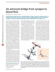
<strong>Centrioles</strong> serving as basal bodies for cilia or flagella usually retain their simple cylindrical form<br />
(A), but their shape may be modified by the development of lateral appendages and one or both ends<br />
may be closed (B, C, D). In many invertebrates and some vertebrates, cross-striated ciliary rootlets<br />
extend from the lower end of the basal body for variable distances into the apical cytoplasm (C, D).<br />
In rare instances in protozoa (viz, Euplotees), the central pair of microtubules of the axoneme may<br />
form a loop extending into the central cavity of the basal body (E). In cells with a single flagellum, a<br />
second centriole is often at right angles to the one serving as basal body (F). (From Fawcett, in The<br />
Cell, Vol 2. J. Brachet and A. Mirsky, eds., Academic Press 1961.)<br />
<strong>Centrioles</strong> are usually positioned so that their long axes form a right angle. Their<br />
perpendicular orientation is maintained even though they may be half a micrometer or<br />
more apart and have no visible structural elements connecting them. The nature of the<br />
long-range forces or organization of the centrosomal cytoplasm that are responsible for<br />
this relationship are unknown. Departures from the usual orthogonal arrangement are<br />
occasionally encountered in normal cells and are reported to be common in malignant<br />
tumors.<br />
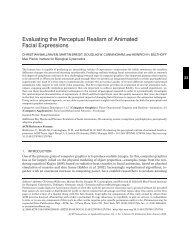
<strong>Centrioles</strong> vary somewhat in length from one cell type to another but are usually<br />
about 0.5 pm long and 0.2 pm in diameter. In the accompanying micrograph a pair of<br />
centrioles near the lumenal surface of an epithelial cell shows the usual perpendicular<br />
orientation. The discrepancy in length of the two members of this pair is unusual.<br />
The two ends of a centriole can often be distinguished in that one is slightly<br />
narrower and appears to be closed and the other appears open.<br />
Figure 301. Intestinal epithelium of a chicken embryo. (Micrograph courtesy of Sergei Sorokin.)<br />
Figure 301