Chapter 12: Centrioles
Chapter 12: Centrioles
Chapter 12: Centrioles

Create successful ePaper yourself
Turn your PDF publications into a flip-book with our unique Google optimized e-Paper software.
552 CENTRIOLES CENTRIOLES 553<br />
<strong>Centrioles</strong> do not undergo transverse division nor do they split longitudinally.<br />
Instead, the new centriole develops in end-to-side relationship to a specific region of a<br />
preexisting centriole but separated from it by a narrow electron lucent space. The<br />
anlage from which it develops, called the procentriole, is an annular condensation of<br />
dense material approximately the same diameter as a mature centriole but initially<br />
devoid of microtubules. The procentriole elongates by accretion of material to its free<br />
end, and the pinwheel arrangement of triplet microtubules gradually appears within the<br />
previously homogeneous dense ring. The forming centriole maintains its orientation<br />
perpendicular to the parent centriole. After reduplication the original members of the<br />
diplosome separate and each, with its newly formed centriole, moves to one pole of the<br />
division figure. Thus in the cell cycle, centrioles are duplicated but not, as previously<br />
believed, by division. A template mechanism cannot be invoked, since the new<br />
centriole arises perpendicular to its precursor and not in intimate contact with it. The<br />
preexisting centriole seems to act merely as a site of induction of nucleation for<br />
self-assembly of the new centriole from precursors synthesized elsewhere in the cell.<br />
Studies of ciliogenesis have shown that centrioles can arise apart from preexisting<br />
centrioles (Stockinger and Cirelli, 1965; Steinman, 1968; Sorokin, 1968). The great<br />
majority of the basal bodies develop around dense spherical bodies variously called<br />
deuterosomes, or procentriole organizers. These in turn arise by condensation of<br />
smaller aggregations of filamentous material (filosomes). Several annular procentrioles<br />
may arise radially around the same procentriole organizer. They elongate rapidly and<br />
acquire the microtubular internal structure typical of centrioles. When they have<br />
attained their definitive length they dissociate from the organizer and move to the cell<br />
surface, where each initiates polymerization of the nine doublet microtubules forming<br />
the axoneme of a cilium.<br />
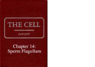
Cilia<br />
**.<br />
Cilia<br />
Fibrous granules<br />
Diagrammatic representation of the two modes of formation of basal bodies during ciliogenesis.<br />
The two original centrioles may become sites of nucleation of multiple procentrioles as shown at the<br />
left. Also, fibrogranular elements of unknown provenance may form deuterosomes or procentriole<br />
organizers around which new centrioles develop as shown at the right. (From Fawcett, in Genetics<br />
of the Spermatozoon, Bogtrykkeriet Forum, Copenhagen, 1971.)<br />
In its role as basal body of a cilium or flagellum, the centriole can be considered to<br />
serve a template function, since the polymerization of tubulin takes place directly on<br />
the distal end of the triplet microtubules in its wall and the ninefold symmetry of the<br />
centrioles is expressed in the nine doublets of the axoneme. For reasons that remain<br />
obscure only two members of each centriolar triplet nucleate the assembly of tubulin<br />
under these conditions, since doublet and not triplet microtubules are formed as<br />
peripheral elements of the axoneme.<br />
Assembly of single microtubules evidently does not require a preexisting site of<br />
nucleation, for the axial pair of single microtubules in the axoneme arises without<br />
a corresponding structure in the axis of the centriole. The nucleating function<br />
of centrioles for microtubule assembly has been demonstrated in vitro using centrioles<br />
isolated from human cells and tubulin extracted from bovine brain (McGill and<br />
Brinkley, 1975).<br />
The important role traditionally assigned to the centrioles in formation of the<br />
mitotic spindle seems not to have been valid. Mitosis occurs with formation of a<br />
typical spindle in both lower and higher plants in the absence of centrioles (Pickett-<br />
Heaps, 1969, 1971). In electron micrographs of animal cells, the spindle microtubules<br />
do converge upon the centriolar region but they rarely contact the centrioles themselves.<br />
They terminate in the pericentriolar cytoplasm in dense spherical granules<br />
called centriolar satellites. Selective damage to the pericentriolar material by laser<br />
microbeam irradiation in prophase results in failure of the chromosomes to separate at<br />
anaphase. This occurs with little or no morphological evidence of structural damage to<br />
the centrioles themselves (Berns et al., 1977). Investigative attention has therefore<br />
shifted to the surrounding specialized zone of cytoplasm which constitutes the other<br />
major component of the centrosome. It has been possible to isolate centrosomes from<br />
certain strains of cultured cells that have been treated with colchicine to block spindle<br />
formation. When these are incubated in vitro with tubulin extracted from brain, large<br />
numbers of microtubules polymerize around each centrosome. These emanate from the<br />
pericentriolar material and not from the centrioles (Gould and Borisy, 1977), It is<br />
concluded therefore that centriolar satellites and possibly other components of the<br />
pericentriolar cytoplasm can initiate assembly of microtubules both in vivo and in vitro.<br />
This does not necessarily mean that the centrioles are inactive during cell division.<br />
They may control aggregation of the pericentriolar material or may be involved in its<br />
activation at the appropriate time in the cell cycle. The interrelations of the several<br />
components of the centrosome have yet to be worked out.<br />
The ubiquitous occurrence of centrioles, their replication, and their continuity<br />
from generation to generation of cells prompted the speculation that they might possess<br />
their own DNA, as do mitochondria and chloroplasts. In several early cytochemical<br />
and biochemical analyses of basal bodies isolated in bulk from protozoa, the<br />
presence of DNA was reported (Seaman, 1960; Randall and Disbrey, 1965). This could<br />
not be confirmed in later investigations with purer fractions (Flavell and Jones, 1970).<br />
Further cytochemical studies using acridine orange staining suggested, however, that<br />
RNA was associated with basal bodies of cilia (Hartman et al., 1974). RNA in centrioles<br />
and in their satellites was also reported on the basis of the sensitivity of certain of their<br />
components to digestion with ribonuclease (Stubblefield and Brinkley, 1967; Brinkley<br />
and Stubblefield, 1970; Dippel, 1976).<br />
Evidence of a functional role for RNA in centrioles has been presented more<br />
recently. When basal bodies isolated from protozoa are injected into Xenopus eggs, they<br />
induce the formation of conspicuous radial arrays of microtubules (asters). The<br />
aster-inducing activity of basal bodies was eliminated by prior treatment with ribonuclease,<br />
but this treatment did not interfere with their capacity to serve as templates<br />
for polymerization of microtubules at their ends (Heidemann et al., 1977). It was<br />
concluded that centrioles contain RNA and that this component is necessary for<br />
initiation of aster formation. Other interpretations of the observations are possible and