

Reappraisal of the Regnauld Procedure for Hallux Valgus and
Reappraisal of the Regnauld Procedure for Hallux Valgus and
Reappraisal of the Regnauld Procedure for Hallux Valgus and

You also want an ePaper? Increase the reach of your titles
YUMPU automatically turns print PDFs into web optimized ePapers that Google loves.
CHAPTER I8<br />
RE,APPRAISAL OF THE, RE.GNAULD PROCEDURE FOR<br />
FIALLUXVALGUS AND FIALLUX RIGIDUS<br />
John V Vdnore, DPM<br />
The <strong>Regnauld</strong> procedure has been part <strong>of</strong> this investigatort<br />
surgical armamentarium <strong>for</strong> de<strong>for</strong>mities <strong>of</strong> <strong>the</strong> first ray <strong>for</strong><br />
more than a decade. The <strong>Regnauld</strong> has been called by<br />
various names in an attempt to describe <strong>the</strong> procedure as<br />
per<strong>for</strong>med by various authors. <strong>Regnauld</strong> himself call this<br />
procedure an "autograft" describing <strong>the</strong> complete removal<br />
<strong>of</strong> <strong>the</strong> phalangeal base from <strong>the</strong> foot, followed by<br />
remodeling <strong>and</strong> <strong>the</strong>n its re-insertion. He did not fixate <strong>the</strong><br />
graft <strong>and</strong> described avascular necrosis as a consequence <strong>of</strong><br />
<strong>the</strong> procedure.'<br />
Several American Podiatrists including Gudas, Weil,<br />
Shelton <strong>and</strong> Clarke, Kashuk, Jacobs have utilized this<br />
procedure in <strong>the</strong> 1980s <strong>and</strong> 1990s following interaction<br />
with <strong>Regnauld</strong> at European <strong>and</strong> American venues. It<br />
was at <strong>the</strong> Hershey Surgical Seminar <strong>of</strong> 1990 with a<br />
presentation by Bernard <strong>Regnauld</strong> <strong>and</strong> particularly<br />
following discussions with Valente Valenti also a<br />
proponent <strong>of</strong> this procedure that I chose to investigate its<br />
potential usefulness. Somewhere along <strong>the</strong> way, this<br />
procedure was labeled an "enclavement" <strong>and</strong> I also have<br />
utilized this term to describe <strong>the</strong> <strong>Regnauld</strong> type<br />
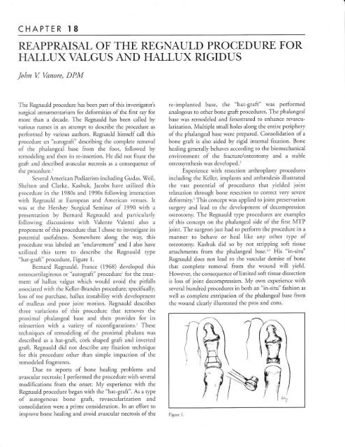
"hat-graft" procedure, Figure 1.<br />
Bernard <strong>Regnauld</strong>, France (1968) developed this<br />
osteocartilaginous or "autograft" procedure' <strong>for</strong> <strong>the</strong> treatment<br />
<strong>of</strong> hallux valgus which would avoid <strong>the</strong> pitfalls<br />
associated with <strong>the</strong> Keller-Br<strong>and</strong>es procedure; specifically,<br />
Ioss <strong>of</strong> toe purchase, hallux instability with development<br />
<strong>of</strong> malleus <strong>and</strong> poor joint motion. <strong>Regnauld</strong> describes<br />
three variations <strong>of</strong> this procedure that removes <strong>the</strong><br />
proximal phalangeal base <strong>and</strong> <strong>the</strong>n provides <strong>for</strong> its<br />
reinsertion with a variery <strong>of</strong> reconfigurations.l These<br />
techniques <strong>of</strong> remodeling <strong>of</strong> <strong>the</strong> proximal phalanx was<br />
described as a hat-graft, cork shaped graft <strong>and</strong> inverted<br />
graft. <strong>Regnauld</strong> did not describe any fixation technique<br />
<strong>for</strong> this procedure o<strong>the</strong>r than simple impaction <strong>of</strong> <strong>the</strong><br />
remodeled fragments.<br />
Due to reports <strong>of</strong> bone healing problems <strong>and</strong><br />
avascular necrosis; I per<strong>for</strong>med <strong>the</strong> procedure with several<br />
modifications from <strong>the</strong> onset. My experience with <strong>the</strong><br />
<strong>Regnauld</strong> procedure began with <strong>the</strong> "hat-graft". As a type<br />
<strong>of</strong> autogenous bone graft, revascularization <strong>and</strong><br />
consolidation were a prime consideration. In an ef<strong>for</strong>t to<br />
improve bone healing <strong>and</strong> avoid avascular necrosis <strong>of</strong> <strong>the</strong><br />
re-implanted base, <strong>the</strong> "hat-graft" was per<strong>for</strong>med<br />
analogous to o<strong>the</strong>r bone graft procedures. The phalangeal<br />
base was remodeled <strong>and</strong> fenestrated to enhance revascularization.<br />
Multiple small holes along <strong>the</strong> entire periphery<br />
<strong>of</strong> <strong>the</strong> phalangeal base were prepared. Consolidation <strong>of</strong> a<br />
bone graft is also aided by rigid internal fixation. Bone<br />
healing generally behaves according to <strong>the</strong> biomechanical<br />
environment <strong>of</strong> <strong>the</strong> fracture/osteotomy <strong>and</strong> a stable<br />
osteosyn<strong>the</strong>sis was developed.'<br />
Experience with resection arthroplasry procedures<br />
including <strong>the</strong> Keller, implants <strong>and</strong> arthrodesis illustrated<br />
<strong>the</strong> vast potential <strong>of</strong> procedures that yielded joint<br />
relaxation through bone resection to correct very severe<br />
de<strong>for</strong>mity.'This concept was applied to joint Preservation<br />
surgery <strong>and</strong> lead to <strong>the</strong> deveiopment <strong>of</strong> decompression<br />
osteotomy. The <strong>Regnauld</strong> type procedures are examples<br />
<strong>of</strong> this concept on <strong>the</strong> phalangeal side <strong>of</strong> <strong>the</strong> first MTP<br />
joint. The surgeon just had to per<strong>for</strong>m <strong>the</strong> procedure in a<br />
manner to behave or heal like any o<strong>the</strong>r type <strong>of</strong><br />
osteotomy. Kashuk did so by not stripping s<strong>of</strong>t tissue<br />
attachments from <strong>the</strong> phalangeal base.'', His "in-situ"<br />
<strong>Regnauld</strong> does not lead to <strong>the</strong> vascular demise <strong>of</strong> bone<br />
that complete removal from <strong>the</strong> wound will yield.<br />
Howeveq <strong>the</strong> consequence <strong>of</strong> limited s<strong>of</strong>t tissue dissection<br />
is loss <strong>of</strong> joint decompression. My own experience with<br />
several hundred procedures in both an "in-situ" fashion as<br />
well as complete extripation <strong>of</strong> <strong>the</strong> phalangeal base from<br />
<strong>the</strong> wound clearly illustrated <strong>the</strong> pros <strong>and</strong> cons.<br />
Figure 1.<br />
J-)<br />
it (tr<br />
)\<br />
m<br />
A<br />
fi/
CHAPTER 18<br />
The following discussion will describe presenr<br />
techniques <strong>of</strong> <strong>the</strong> <strong>Regnauld</strong> procedure. Clearly, both<br />
variants <strong>of</strong> <strong>the</strong> procedure with complete versus<br />
incomplete dissection have <strong>the</strong>ir advantages <strong>and</strong><br />
appropriate patient selection is <strong>the</strong> proper determinant.<br />
Phalangeal osreoromies <strong>of</strong> <strong>the</strong> haliux have traditionally<br />
been utilized <strong>for</strong> <strong>the</strong> treatment <strong>of</strong> hallux valgus <strong>and</strong><br />
hallux valgus interphalangeus with <strong>the</strong> primary goal <strong>of</strong><br />
reduction <strong>of</strong> rhe abduction <strong>of</strong> <strong>the</strong> great toe through a<br />
wedge osteoromy.6,7 Tiaditionally, surgeons have viewed<br />
<strong>the</strong> hallux osteoromy or Akin rFpe osreotomy as an<br />
adjunctive procedure to improve <strong>the</strong> cosmetic alignment<br />
<strong>of</strong> <strong>the</strong> great toe. The <strong>Regnauld</strong> enclavemenr procedure<br />
provides much grearer porenrial but preoperative<br />
assessment must identifi, <strong>the</strong> problems <strong>and</strong> <strong>the</strong> surgeon<br />
plan corrective maneuvers.<br />
Long-st<strong>and</strong>ing hallux valgus <strong>of</strong>ten presents with<br />
significant s<strong>of</strong>t tissue conrractures in <strong>and</strong> around <strong>the</strong> first<br />
MTP joint. Joint decompression procedures allow <strong>for</strong><br />
reduction <strong>of</strong> significant proportions <strong>of</strong> transverse or<br />
frontal plane de<strong>for</strong>mity. Use <strong>of</strong> <strong>the</strong> procedure in senile<br />
hallux valgus <strong>and</strong> hallux valgus rigidus illustrated its<br />
potential <strong>and</strong> usefulness. Radiographically, surgeons like<br />
to quantitate de<strong>for</strong>mity <strong>and</strong> devise a surgical plan on <strong>the</strong><br />
basis <strong>of</strong> osseous relationships, <strong>for</strong> example radiographic<br />
angles, such as <strong>the</strong> intermetatarsal (IM) or proximal<br />
articular set angle (PASA).s Determining <strong>the</strong> corrective<br />
potential <strong>of</strong> joint decompression procedures is more<br />
difficult. The bone resection <strong>of</strong> a decompression<br />
osteotomy provides s<strong>of</strong>t tissue <strong>and</strong> joint relaxation that<br />
allows <strong>for</strong> not only positional or s<strong>of</strong>t tissue components <strong>of</strong><br />
de<strong>for</strong>mity but also indirect reduction <strong>of</strong> structural<br />
components. Joint relaxation plays an important role in<br />
reduction <strong>of</strong> de<strong>for</strong>miry <strong>and</strong> improvemenr <strong>of</strong> mobility.<br />
This is most clearly evident in cases <strong>of</strong> severe de<strong>for</strong>mity<br />
<strong>and</strong> a rigid foot type.<br />
<strong>Hallux</strong> rigidus <strong>of</strong>ten illustrates a hallux equinus,<br />
metatarsus primus elevatus, <strong>and</strong> restricted joint motion.<br />
One may argue rhar <strong>the</strong> metatarsus elevarus is <strong>the</strong> primary<br />
problem or that it is a secondary phenomenon <strong>of</strong> <strong>the</strong><br />
hallux equinus. Certainly, hallux rigidus is a varied<br />
de<strong>for</strong>mity in that some cases feature a very prominent<br />
metatarsus primus elevatus, or it may be present to milder<br />
degrees, or be completely absent. By achieving relaxation<br />
<strong>of</strong> <strong>the</strong> first metatarsophalangeal joint, any positional or<br />
secondary elevation <strong>of</strong> <strong>the</strong> first metatarsal as a result <strong>of</strong><br />
hallux equinus shouid reduce. This is true whe<strong>the</strong>r <strong>the</strong><br />
relaxation is accomplished on <strong>the</strong> phalangeal or<br />
metatarsal side <strong>of</strong> <strong>the</strong> joint.<br />
A ra<strong>the</strong>r recent addition in <strong>the</strong> surgical treatmenr <strong>of</strong><br />
hallux rigidus is <strong>the</strong> enclavement procedure initially<br />
described by <strong>Regnauld</strong>. This phalangeal osteoromy<br />
shortens <strong>the</strong> osseous segmenr distal to <strong>the</strong> first metatarsophalangeal<br />
joint. A such, this procedure becomes a<br />
decompression osteotomy <strong>and</strong> is useful in cases <strong>of</strong> hailux<br />
limitus/ rigidus with a long proximal phalanx <strong>and</strong>/or a<br />
short metatarsal. The osteoromy is very powerful when<br />
combined with complere removal <strong>of</strong> <strong>the</strong> base <strong>of</strong> <strong>the</strong><br />
proximal phalanx but this requires stripping <strong>of</strong> all s<strong>of</strong>t<br />
tissue attachmenrs to <strong>the</strong> base <strong>of</strong> <strong>the</strong> proximal phalanx.<br />
THE "MODERN REGNAULD'<br />
The <strong>Regnauld</strong> procedure may be per<strong>for</strong>med through<br />
many permutations <strong>and</strong> <strong>the</strong> aurhor has now eliminated all<br />
<strong>the</strong> various complexities <strong>of</strong> <strong>the</strong> hat graft technique which<br />
was <strong>the</strong> mainstay technique <strong>for</strong> many years.<br />
Simplification with a double rransverse osreoromy, Figure<br />
2, in usually a tapezoidal manner is <strong>the</strong> preferred<br />
technique. The surgeon srill has <strong>the</strong> option <strong>of</strong> complete<br />
removal <strong>of</strong> <strong>the</strong> proximal phalangeal base or per<strong>for</strong>mance<br />
as an "in-situ" or cylindrical Akin technique.<br />
The operation may be per<strong>for</strong>med through ei<strong>the</strong>r a<br />
dorsal or medial incisional approach <strong>and</strong> this is more a<br />
preference <strong>of</strong> <strong>the</strong> surgeon rarher than any requirement <strong>of</strong><br />
<strong>the</strong> procedure itself. Joint exposure is similar to that <strong>of</strong> an<br />
implant arthroplasty procedure with subperiosteal<br />
dissection <strong>of</strong> <strong>the</strong> base <strong>of</strong> <strong>the</strong> proximal phalanx <strong>and</strong> distai<br />
first metatarsal. Osteotomy ar <strong>the</strong> level <strong>of</strong> <strong>the</strong> proximal<br />
metaphysis <strong>of</strong> <strong>the</strong> proximal phalanx is pre<strong>for</strong>med usually<br />
as a trapezoidal osteotomy with <strong>the</strong> mediai section being<br />
wider with an overail shortening <strong>of</strong> <strong>the</strong> proximal phalanx<br />
proportional to <strong>the</strong> overall length <strong>of</strong> <strong>the</strong> phalanx, great<br />
toe <strong>and</strong> degree <strong>of</strong>correction required (Figure 3).<br />
Prior to removal <strong>of</strong> <strong>the</strong> phalangeal base, a 0.045<br />
inch kirschner wire is placed directly from dorsal to<br />
plantar just distal enough to avoid damaging or placing<br />
Figure 2
CHAPTER 18<br />
Figure 3.<br />
Figure 4<br />
Figure 5. Figure 6
CHAPTER 18<br />
8I<br />
1. Piot Hole - 2mr Drill<br />
lnitially D.ill wdh 0.062" K-wi.e<br />
Enlarge wiih 2.0 mm Dril Bat<br />
2. Dillwith Stop - 2.4 mm D.il<br />
Enlarge ONLY Phalangeal Base<br />
3. Depth Measuaemeni<br />
Generally 28-30 mm lengih<br />
4. Tap enti.e Lenglh ih.ough<br />
Latera, C€dex <strong>of</strong> Phalengeal Head<br />
5. lnserl Herbeft Screw so comp elely<br />
lnaraosseus<br />
6. Remove P.elimr.ary Fxalion (0.045" KVt4<br />
7. lnsed 2nd Foinl <strong>of</strong>Fixation<br />
0.062" KW anse*ed from Distal Tip otToe<br />
Cross both IPJ <strong>of</strong> <strong>Hallux</strong> & Osteotomy<br />
<strong>the</strong> wire through <strong>the</strong> articular surface; remember that <strong>the</strong><br />
phalangeal articular surface is concave. This wire, <strong>the</strong><br />
"rudder pin" will remain until <strong>the</strong> base is reinserted later<br />
in <strong>the</strong> procedure (Figure 4). It provides a reference point<br />
so that articular congruency will be maintained as well as<br />
providing a point <strong>for</strong> h<strong>and</strong>ling <strong>the</strong> fragment.<br />
If <strong>the</strong> surgeon chooses, alternatively <strong>the</strong> base may be<br />
left in place with dissection limited to that necessary <strong>for</strong><br />
osteotomy <strong>and</strong> fixation. For full decompression, <strong>the</strong> basal<br />
fragment is <strong>the</strong>n extirpated from <strong>the</strong> wound with care so<br />
as to minimize damage to its structure. The bone may be<br />
s<strong>of</strong>t <strong>and</strong> this <strong>of</strong>ten means avoidance <strong>of</strong> <strong>the</strong> points <strong>of</strong> bone<br />
<strong>for</strong>ceps to grasp <strong>the</strong> base during excision. A 6 inch Brown<br />
<strong>for</strong>ceps or guarded pressure with an alligator bone <strong>for</strong>ceps<br />
is usefui.<br />
Following its removal from <strong>the</strong> wound, <strong>the</strong> resected<br />
portion <strong>of</strong> proximal phalanx is wrapped in a damp sponge<br />
<strong>for</strong> later use. The surgeon may now address <strong>the</strong> proliferative<br />
bone or arthrosis <strong>of</strong> <strong>the</strong> first meratarsal. If <strong>the</strong> operadve<br />
pathology is one <strong>of</strong> hallux valgus, only limited dissecrion <strong>of</strong><br />
<strong>the</strong> first metatarsal is necessary. In cases <strong>of</strong> severe de<strong>for</strong>mity<br />
or first MTP joint arthrosis <strong>the</strong>n additional dissection may<br />
be required. This allows adequate exposure <strong>for</strong> peripheral<br />
cheilectomy <strong>of</strong> <strong>the</strong> osteophytosis. A sesamoidolysis may<br />
be per<strong>for</strong>med with inspection <strong>of</strong> all surfaces <strong>of</strong> <strong>the</strong><br />
metatarsal head <strong>and</strong> its sesamoids. Cheilectomy or<br />
removal <strong>of</strong> peripheral lipping <strong>of</strong> <strong>the</strong> sesamoids is possible<br />
as well as complete removal <strong>of</strong> a sesamoid if deemed necessary<br />
is quite easy from an intracapsular approach with<br />
<strong>the</strong> base removed from <strong>the</strong> wound.<br />
Alternativeiy, <strong>the</strong> base need not be excised but<br />
complete subperiosteal dissection along <strong>the</strong> medial<br />
aspect <strong>of</strong> <strong>the</strong> base <strong>of</strong> <strong>the</strong> proximal phalanx is necessary<br />
<strong>for</strong> both later internal fixation as well as providing some<br />
degree <strong>of</strong> joint rela-xation.<br />
With <strong>the</strong> phalangeal base removed from <strong>the</strong><br />
wound, te<strong>the</strong>ring <strong>of</strong> <strong>the</strong> flexor tendons to each o<strong>the</strong>r<br />
may be accomplished to aid in hallucal purchase <strong>and</strong><br />
help avoid later interphalangeal joint instabiliry postoperatively<br />
(Figure 5). A hallux maileus was identified in<br />
some <strong>of</strong> <strong>the</strong> early cases postoperatively <strong>and</strong> this is now a<br />
routine maneuver to avoid this complication.<br />
The excised portion <strong>of</strong> <strong>the</strong> phalangeal base is<br />
remodeled. A]l s<strong>of</strong>t tissue attachmenrs ro <strong>the</strong> base should<br />
be removed. Usually, this is begun with a rongeur<br />
followed by decortication <strong>of</strong> <strong>the</strong> periphery <strong>of</strong> <strong>the</strong> base<br />
per<strong>for</strong>med with a rotary drill <strong>and</strong> side-cutting oval or<br />
round bur (5 mm). The h<strong>and</strong> rongeur is also helpful in<br />
resecting <strong>the</strong> periarticular lipping or osteophytosis that<br />
may be present.<br />
Following remodeling, a small kirschner wire is<br />
used to per<strong>for</strong>ate <strong>the</strong> entire remaining corrical surface a<br />
few millimeters to aid in revascularization <strong>and</strong> avoidance<br />
<strong>of</strong> avascular necrosis (Figure 6). These holes are similar to<br />
those placed in any bone graft to encourage revascularization.<br />
A 0.028 inch kirschner wire is utilized to drill 25 to<br />
35 holes around <strong>the</strong> entire osseous circumference <strong>of</strong> <strong>the</strong><br />
phalangeal base. Here, care must be taken to avoid<br />
drilling into <strong>the</strong> articular surface due to <strong>the</strong> concave<br />
geometry <strong>of</strong> <strong>the</strong> articular surface.<br />
The wound is copiously irrigated <strong>and</strong> <strong>the</strong> graft is<br />
reinserted using <strong>the</strong> "rudder pin" as a reference or guide to<br />
its placement to re-establish a congruous first MTP joint.<br />
A0.045 in. kirschner wire placed from <strong>the</strong> medial surface<br />
<strong>of</strong><strong>the</strong> re-inserted base into <strong>the</strong> lateral cortex <strong>of</strong><strong>the</strong> phalanx<br />
is used <strong>for</strong> preliminary fixation. Definitive fixation is<br />
per<strong>for</strong>med with insertion <strong>of</strong> a Herbert or Bold screw.<br />
Fixation with <strong>the</strong> Herbert bone screw will be<br />
described as this has been <strong>the</strong> most common <strong>for</strong>m <strong>of</strong><br />
fixation per<strong>for</strong>med, Figure 7. Fixation with a Herbert<br />
bone screw inserted from <strong>the</strong> plantar medial aspecr <strong>of</strong> <strong>the</strong><br />
base into <strong>the</strong> distal lateral aspect <strong>of</strong><strong>the</strong> phalanx has been<br />
very effective. Alternatively, <strong>the</strong> Bold Screw has been used
82 CHAPTE,R i8<br />
as <strong>the</strong> screws are similar but <strong>the</strong> latter is cannulated with<br />
a simplified insertion technique. O<strong>the</strong>r fixation<br />
alternatives have been utilized over <strong>the</strong> years but generally<br />
<strong>the</strong> Herbert (Zimmer, \7arsaw, IN) or Bold (\Tright<br />
Medical Gchnology Group, Arlington TN) screws have<br />
yielded rigid fixation with no extra-osseous prominence.<br />
PILOT HOLE<br />
An AO type triple drill guide is useful as a guide to drill <strong>the</strong><br />
2.0 mm pilot or core diameter hole. The drill guide has a<br />
pointed edge that allows placement <strong>of</strong> <strong>the</strong> drill hole on <strong>the</strong><br />
edge <strong>of</strong> <strong>the</strong> osseous/articular surface along <strong>the</strong> plantar medial<br />
aspect <strong>of</strong><strong>the</strong> base <strong>of</strong> <strong>the</strong> proximal phalanx. The hole is drilled<br />
from plantar medial to dorsal lateral <strong>and</strong> distal direction.<br />
Actually, <strong>the</strong> preferred technique is to <strong>for</strong>m <strong>the</strong> initial screw<br />
hole with a 0.062" kirschner wire as this is stiff <strong>and</strong> can be<br />
maneuvered more easily <strong>the</strong>n a drill bit. Subsequently, this is<br />
enlarged it with a 2.0 mm drili by h<strong>and</strong>.<br />
DRILL WITH STOPX<br />
The 2.0 mm hole in <strong>the</strong> base is enlarged with <strong>the</strong> 2.4 mm<br />
Herbert drill with stop; this a h<strong>and</strong> instrument that<br />
allows <strong>for</strong> this overdrill only a short segment <strong>of</strong> <strong>the</strong><br />
phalangeal base. This accommodates <strong>the</strong> larger diameter<br />
<strong>of</strong> <strong>the</strong> trailing thread <strong>of</strong> <strong>the</strong> Herbert bone screw.<br />
DEPTH MEASUREMENT<br />
A small fragment depth gauge can <strong>the</strong>n be used to measure<br />
<strong>the</strong> correct length screw necessary. Alternatively, a<br />
0.045in kirschner wire is inserted by h<strong>and</strong> <strong>and</strong> <strong>the</strong>n<br />
clamped with a hemostat. The length <strong>of</strong> wire inserted is<br />
<strong>the</strong>n compared to a ruler. This has shown to be a reliable<br />
<strong>and</strong> accurate technique. The exact length screw to that<br />
measured may be inserted, generally a 28 or 30 mm<br />
screw. Note no countersinking is per<strong>for</strong>med.<br />
TAP+<br />
The entire depth <strong>of</strong> <strong>the</strong> pilot hole is tapped with <strong>the</strong> 3.0<br />
mm Herbert tap equivalent to <strong>the</strong> thread <strong>of</strong> <strong>the</strong> leading<br />
3.0 mm thread <strong>of</strong> <strong>the</strong> Herbert screw. It is important to cut<br />
<strong>the</strong> thread through <strong>the</strong> entire portion <strong>of</strong> phalanx to avoid<br />
later difficulties wirh screw inserrion.<br />
SCREW' INSERTIONX<br />
The appropriate length screw is inserted using some axial<br />
pressure as <strong>the</strong> screw advances until it lies completely<br />
within <strong>the</strong> substance <strong>of</strong> bone. A 28 or 30 mm screw is<br />
most commonly used so that <strong>the</strong> leading thread may just<br />
per<strong>for</strong>ate <strong>the</strong> opposite cortex <strong>for</strong> solid purchase <strong>of</strong> bone.<br />
FINAL PREPARAIION AND<br />
SECONDARY STABILIZAIION<br />
The kirschner wire used as preliminary fixation as well<br />
as <strong>the</strong> "rudder pin" may <strong>the</strong>n be removed <strong>and</strong> any overhang<br />
between <strong>the</strong> base <strong>and</strong> <strong>the</strong> shaft can <strong>the</strong>n be<br />
remodeled. A second point <strong>of</strong> fixation has been found<br />
useful both <strong>for</strong> increasing <strong>the</strong> rigidity <strong>of</strong> <strong>the</strong> screw fixation<br />
as well as <strong>for</strong> stabilization <strong>of</strong> <strong>the</strong> hallucal interphalangeal<br />
joint. A 0.062 in kirschner wire is driven from <strong>the</strong> tip <strong>of</strong><br />
<strong>the</strong> toe in a proximal manner crossing <strong>the</strong> IP joint as well<br />
as <strong>the</strong> osteotomy.<br />
Irrigation is again per<strong>for</strong>med followed by capsular<br />
<strong>and</strong> skin closure per surgeon's preference. Immediate<br />
weight bearing in a surgical shoe is allowed <strong>and</strong> <strong>the</strong><br />
procedure may be per<strong>for</strong>med bilaterally on an out-patient<br />
basis. Immediate range <strong>of</strong> motion is encouraged as this is<br />
a joint salvage procedure in a patient usually with<br />
evidence <strong>of</strong> joint disease or preoperative limitation <strong>of</strong><br />
.ioint movement.<br />
POSTOPERAITVE CARE<br />
Generally, a Darco (Darco International, Huntington,<br />
\(rV) or Reese type surgical shoe is utilized postoperatively<br />
<strong>for</strong> 3-4 weeks. Thereafter, a gym shoe or s<strong>of</strong>t<br />
lea<strong>the</strong>r shoe is allowed. Immediate range <strong>of</strong> motion is<br />
possible due to <strong>the</strong> stability imparted by <strong>the</strong> internal<br />
fixation. tWhen <strong>the</strong> base is completely excised, my<br />
preference is to continue to splint <strong>the</strong> hallux <strong>for</strong> a period<br />
<strong>of</strong> time usually with a bunion splint <strong>for</strong> 6 weeks. Very<br />
predictable bone healing can be expected with <strong>the</strong><br />
technique described with almost complete absence <strong>of</strong><br />
avascular necrosis. Complete bony union within 3<br />
months is <strong>the</strong> rule.<br />
This procedure also does not address metatarsus<br />
primus elevatus or a long first metatarsal as observed in<br />
many cases <strong>of</strong> hallux rigidus. Careful preoperative<br />
assessment is m<strong>and</strong>atory <strong>for</strong> successful results. Our<br />
experience with this procedure has been very grati$/ing
CHAPTER 18<br />
particularly in cases <strong>of</strong>stage II <strong>and</strong> stage III hallux rigidus<br />
or hallux valgus rigidus with a long proximal phalanx <strong>and</strong><br />
short first metatarsal. The procedure is useful in obese<br />
patients wherein first metatarsal osteotomy is difficult to<br />
protect. It may also be useful in haliux varus with <strong>the</strong><br />
bone resection <strong>the</strong>n wider on <strong>the</strong> lateral side much like a<br />
"Reverse Akin."<br />
AUTHORS EXPERIENCE<br />
The <strong>Regnauld</strong> procedure has been per<strong>for</strong>med <strong>for</strong> <strong>the</strong> past<br />
14 years <strong>and</strong> has shown to be a quite valuable surgical<br />
alternative. The aurhor has per<strong>for</strong>med over 300<br />
procedures most <strong>for</strong> hallux rigidus or hallux valgus rigidus<br />
with <strong>the</strong> majority being per<strong>for</strong>med as <strong>the</strong> described "autograft"<br />
technique versus "in-situ". In cases <strong>of</strong> limited joint<br />
disease, <strong>the</strong> base need not be excised; <strong>and</strong> certainly this<br />
may be preferred as bone healing would be expected to be<br />
more predictable as would hallucal stability. The<br />
procedure can be useful also as an adjunctive role <strong>for</strong> <strong>the</strong><br />
management <strong>of</strong> hallux valgus particuiarly in cases <strong>of</strong> a<br />
rigid foot rype. Several cases will be described in <strong>the</strong><br />
illustrations.<br />
CASE 1.<br />
Figure B. Radiographs <strong>of</strong> 43-year-old obese white female<br />
who presented with a sriff <strong>and</strong> painful great toe. She<br />
had prior bunionectomy with basal osteotomy by an<br />
orthopedist. A "autograft" was per<strong>for</strong>med with<br />
cheilectomy <strong>and</strong> excision <strong>of</strong> <strong>the</strong> fibular sesamoid. A<br />
congruous <strong>and</strong> painfree joint was re-established as well as<br />
exceilent correction <strong>of</strong> <strong>the</strong> recurrent hallux valgus<br />
de<strong>for</strong>mity. Note osreoromy consolidation without<br />
resorption or callus.<br />
F-igure 8<br />
CASE 2.<br />
Figure 9. Radiographs <strong>of</strong> 31-year-old female who<br />
presented with a stiff <strong>and</strong> painful grear roe one year postoperative<br />
cheilectomy. An enclavement was per<strong>for</strong>med<br />
incorporating significant linear shortening <strong>of</strong> <strong>the</strong><br />
proximal phalanx. This allowed widening <strong>of</strong> <strong>the</strong> joint<br />
space <strong>and</strong> re-establishment <strong>of</strong> a painfree range <strong>of</strong> motion.<br />
Figure 9
CHAPTER 18<br />
CASE 3.<br />
Figure 10. This 69-year-old female presented with a<br />
symptomatic hallux valgus de<strong>for</strong>mity. Preoperative<br />
radiographs (A) show a low IM angle with mild<br />
osteopenia without degenerative changes. She underwent<br />
<strong>Regnauld</strong> procedure as an "in-situ" type osteotomy<br />
fixated with an oblique bone pin@4 <strong>and</strong> an axial 0.062in<br />
kirschner wire. Postoperative radiographs at 2 weeks<br />
(B), 6 weeks (C) <strong>and</strong> 6 months (D) show bone consolidation<br />
without resorption <strong>and</strong> gradual resorption <strong>of</strong> <strong>the</strong><br />
bone pin.<br />
CASE 4.<br />
Figure 1 1. Radiographs <strong>of</strong> 60-year-old female presented<br />
with an extremely painful first MTP joint. Preoperative<br />
radiographs (A) reveal severe hallux valgus with extensive<br />
degenerative arthrosis. A "autograft" was per<strong>for</strong>med<br />
with cheilectomy <strong>and</strong> excision <strong>of</strong> <strong>the</strong> fibular sesamoid.<br />
She had an extremely nice result with correction <strong>of</strong><br />
de<strong>for</strong>mity <strong>and</strong> a painfree joint with little postoperative<br />
disabiliry<br />
SUMMARY<br />
Primary joint reconstruction via cheilectomy <strong>and</strong><br />
<strong>Regnauld</strong> type "autograft" is an extremely useful<br />
technique although not fully appreciated but <strong>for</strong> a limited<br />
number <strong>of</strong> surgeons. Due to <strong>the</strong> removal <strong>and</strong> reinsertion<br />
<strong>of</strong> <strong>the</strong> phalangeal base, several <strong>of</strong> <strong>the</strong> negative aspects <strong>of</strong><br />
resection arthroplasry are encountered such as disruption<br />
<strong>of</strong> <strong>the</strong> glenosesamoidal joint <strong>and</strong> its distal attachments.<br />
The surgeon must address this problem to avoid a<br />
potential hallux malleus de<strong>for</strong>mity. Various techniques<br />
include: te<strong>the</strong>ring <strong>of</strong> <strong>the</strong> long flexor tendon to <strong>the</strong><br />
phalangeal base, re-insertion <strong>of</strong> <strong>the</strong> plantar aponeurotic<br />
medial <strong>and</strong> lateral heads <strong>of</strong> <strong>the</strong> short flexor to <strong>the</strong> base, or<br />
simply anastomotic suture between <strong>the</strong> long <strong>and</strong> short<br />
flexor. Any <strong>of</strong> <strong>the</strong>se variations are useful adjuncts to<br />
improve hallucal purchase, avoid sesamoidal retraction,<br />
<strong>and</strong> improve joint stability. The author also feels that <strong>the</strong><br />
kirschner wire used as a second point <strong>of</strong> fixation aids IPJ<br />
stability as it is placed across <strong>the</strong> IPJ as well as <strong>the</strong><br />
osteotomy <strong>and</strong> imparts residual stiffness to <strong>the</strong><br />
hallucal IPJ.<br />
In addition, <strong>the</strong> dem<strong>and</strong>s <strong>of</strong> bone healing must be<br />
considered. Avascular necrosis probably occurs in a good<br />
number <strong>of</strong> non-fixated procedures. Primary vascular bone<br />
union implies an early restoration <strong>of</strong> <strong>the</strong> osseous<br />
Figure 1 1<br />
Figure 1 0.
CHAPTER 18 85<br />
blood supply. Rigid internal fixation is an aid to this<br />
revascularization process. A technique utilizing rhe<br />
Herbert bone screw <strong>and</strong> iater <strong>the</strong> Bold screw achieves<br />
rigid internal fixation <strong>and</strong> allows <strong>for</strong> reincorporarion <strong>of</strong><br />
<strong>the</strong> "autograft" with primary bone healing in a good<br />
proportion <strong>of</strong> <strong>the</strong> cases. This technique allows <strong>for</strong><br />
immediate joint movement with rapid rehabilitation <strong>and</strong><br />
weight bearing.<br />
The <strong>Regnauld</strong> enclavement is a versatile procedure<br />
<strong>for</strong> primary joint reconstruction <strong>for</strong> a variery <strong>of</strong> pathologies<br />
<strong>of</strong> <strong>the</strong> first MTP joint. Phalangeal osteotomies <strong>of</strong> <strong>the</strong><br />
hallux have traditionally been utilized <strong>for</strong> <strong>the</strong> treatment <strong>of</strong><br />
hallux valgus <strong>and</strong> hallux valgus interphalangeus.6 Due ro<br />
<strong>the</strong> decompressive nature <strong>of</strong> <strong>the</strong> <strong>Regnauld</strong>, its utilization<br />
in hallux rigidus <strong>and</strong> limitus has been popular.<br />
The <strong>Regnauld</strong> procedure is a joint preservation rype<br />
<strong>of</strong> reconstruction versus <strong>the</strong> joint destructive nature <strong>of</strong> a<br />
resection arthroplasty such as a Keller.' " As such, younger<br />
more active patients are c<strong>and</strong>idates <strong>for</strong> this type <strong>of</strong><br />
reconstruction. The <strong>the</strong>rapeutic nature <strong>of</strong> rhe <strong>Regnauld</strong><br />
enclavement procedure is one <strong>of</strong> a decompression<br />
osteotomy with joint relaxation. Some degree <strong>of</strong><br />
decompression does occur with linear shortening <strong>of</strong> <strong>the</strong><br />
proximal phalanx but in order to accomplish maximal<br />
relaxation, <strong>the</strong> s<strong>of</strong>t tissue attachments to <strong>the</strong> basal<br />
fragment must be completely dissected free. One <strong>of</strong> <strong>the</strong><br />
most valuable aspects <strong>of</strong> <strong>the</strong> <strong>Regnauld</strong> procedure is <strong>the</strong><br />
exposure it af<strong>for</strong>ds to <strong>the</strong> metatarsal upon removai <strong>of</strong> <strong>the</strong><br />
phalangeal base from <strong>the</strong> wound. Medial bunion<br />
resection or cheilectomy <strong>of</strong> both <strong>the</strong> metatarsal head <strong>and</strong><br />
<strong>the</strong> phalanx are easily accomplished. Access to rhe<br />
sesamoids including potential sesamoidectomy is also<br />
allowed from an intracapsular approach.<br />
<strong>Hallux</strong> limitus, hallux rigidus <strong>and</strong> hallux valgus<br />
rigidus are variations <strong>of</strong> first MTP joint osteoarthrosis<br />
combined with (hallux valgus rigidus) or without (hallux<br />
limitus <strong>and</strong> rigidus) abduction <strong>of</strong> <strong>the</strong> great toe. These<br />
de<strong>for</strong>mities generally involve a stiff<strong>and</strong> painful first MTP<br />
joint. Reduction <strong>of</strong> <strong>the</strong> first MTP joint de<strong>for</strong>mity is<br />
gready enhanced through <strong>the</strong> joint relaxation inherent to<br />
this procedure. The most striking examples <strong>of</strong> joint<br />
relaxation accomplished through bone resection are with<br />
variations <strong>of</strong> <strong>the</strong> Keller resection arthroplasty or first<br />
MTP joint fusion.<br />
This procedure may be applied to a variety <strong>of</strong><br />
clinical pathology <strong>of</strong> <strong>the</strong> first MTP joint. The objectives<br />
<strong>of</strong> <strong>the</strong> <strong>Regnauld</strong> procedure are reduction <strong>of</strong> de<strong>for</strong>miry be<br />
abduction <strong>of</strong> <strong>the</strong> great toe, valgus rotation, restoration <strong>of</strong><br />
normal sagittal plane position <strong>of</strong> <strong>the</strong> great toe at <strong>the</strong> first<br />
MTP joint <strong>and</strong> non-painful movement <strong>of</strong> <strong>the</strong> great<br />
toe joint. This is accomplished through 1) joint<br />
decompression through shortening <strong>of</strong> <strong>the</strong> phalanx <strong>and</strong><br />
s<strong>of</strong>t tissue release from <strong>the</strong> base <strong>of</strong> <strong>the</strong> proximal phalanx,<br />
2) cheilectomy (clean-up all adjacent joint margins),<br />
3) rigid internal fixation (Herbert or Bold screw), <strong>and</strong><br />
4) early mobilization.<br />
ACKNO\$TLEDGEMENT<br />
Thank you to Maria Bidny, DPM <strong>for</strong> drawings used in<br />
<strong>the</strong> article.<br />
REFERENCES<br />
1. <strong>Regnauld</strong> B. <strong>Hallux</strong> rigiclus. In <strong>Regnauld</strong> B. The Foot. Springer-<br />
Verlag, Berlin. p. 345-9.<br />
2. Perren SM. Physical <strong>and</strong> biological aspects <strong>of</strong> fracture healing with<br />
special reference to internal fl-ration. Clin Orthop 1979;138:175-96.<br />
3. Syuppan RJ. The cartilaginous articulation preservation principle<br />
<strong>and</strong> its surgical implementation <strong>for</strong> hallu-x abducto .'algts. J Am<br />
Podiaay Asso c 197 4;64:635 -5 6.<br />
4.lr{anft JR, et al. Preliminary report: modifications <strong>of</strong> <strong>the</strong> <strong>Regnauld</strong><br />
osteochondral autogenous graft. J Foot Surg 7990:29 :177 -80.<br />
5. Hanft.fR, et al. Modifications <strong>of</strong> <strong>the</strong> Regnaulf osteochondral autogenous<br />
graft <strong>for</strong> correction <strong>of</strong> halh-rx limitus/valgus: a 2-year review.<br />
J Foot Surg 7992:31:176-9.<br />
6. Gerbert JE, Spector ClarkJ. Osteotomy procedures on <strong>the</strong> proximal<br />
phalanx lor correction <strong>of</strong> a hallux de<strong>for</strong>miqy. J Am Podiatry Assoc<br />
797 4;64:617-29.<br />
7. Sorto LA, et aI. <strong>Hallux</strong> abductus interphalangeus: etiology, x-ray<br />
evaluation, <strong>and</strong> trearment.,/ Am ?odiatry Assoc 1976;66:384-96.<br />
8. Schuberth JM, et a1. <strong>Hallux</strong> ualgus in <strong>the</strong> healthy adub. Anencan<br />
College <strong>of</strong> Foot <strong>and</strong> Ankle Surgeons. 1992. Park Ridge, Illinois.<br />
9. Vanore JV, Corey SV. HallLx limitus, rigidus, <strong>and</strong> metatarsophalangeal<br />
joint arthrosis. ln Marcinko DE, editor. Comprehensiue<br />
Textbook <strong>of</strong> Halhtx Abducto <strong>Valgus</strong> Reconstruction. Mosby-Year Book,<br />
St. Louis; 1992. P. 209-41.<br />
10.Vanore JV, et a1. Hallu-x rigidus <strong>and</strong> limitus. In Marcinko DE, editor.<br />
Medical anrl Surgical Therapeutics <strong>of</strong> <strong>the</strong> Foot <strong>and</strong> Ankle.<br />
Baltimore, \Williams & \Wilkins; 1992. P . 423-65.<br />
11.ACFAS. Hallu-x rigidus in <strong>the</strong> healthy adult. In ACFAS, Prefened<br />
Practice Guidelines. ACFAS, park Ridge (Il); 1993.


