Complete issue - Rhode Island Medical Society
Complete issue - Rhode Island Medical Society
Complete issue - Rhode Island Medical Society
Create successful ePaper yourself
Turn your PDF publications into a flip-book with our unique Google optimized e-Paper software.
Volume 93 No. 11 November 2010
We're not LIKE A Good Neighbor,<br />
WE ARE<br />
The Good Neighbor Alliance<br />
52<br />
56<br />
Specializing in Employee Benefits since 1982<br />
Health Dental Life Disability Long Term Care<br />
Pension Plans Workers' Compensation Section 125 Plans<br />
The Good Neighbor Alliance Corporation<br />
The Benefits Specialist<br />
Affiliated Affiliated with with<br />
RHODE ISLAND MEDICAL SOCIETY<br />
rhode isl a nd<br />
medical society<br />
401-828-7800 or 1-800-462-1910<br />
P.O. Box 1421 Coventry, RI 02816<br />
www.goodneighborall.com
UNDER THE JOINT<br />
EDITORIAL SPONSORSHIP OF:<br />
The Warren Alpert <strong>Medical</strong> School of<br />
Brown University<br />
Edward J. Wing, MD, Dean of Medicine<br />
& Biological Science<br />
<strong>Rhode</strong> <strong>Island</strong> Department of Health<br />
David R. Gifford, MD, MPH, Director<br />
Quality Partners of <strong>Rhode</strong> <strong>Island</strong><br />
Richard W. Besdine, MD, Chief<br />
<strong>Medical</strong> Officer<br />
<strong>Rhode</strong> <strong>Island</strong> <strong>Medical</strong> <strong>Society</strong><br />
Vera A. DePalo, MD, President<br />
EDITORIAL STAFF<br />
Joseph H. Friedman, MD<br />
Editor-in-Chief<br />
Sun Ho Ahn, MD<br />
Associate Editor<br />
Joan M. Retsinas, PhD<br />
Managing Editor<br />
Stanley M. Aronson, MD, MPH<br />
Editor Emeritus<br />
EDITORIAL BOARD<br />
Stanley M. Aronson, MD, MPH<br />
John J. Cronan, MD<br />
James P. Crowley, MD<br />
Edward R. Feller, MD<br />
John P. Fulton, PhD<br />
Peter A. Hollmann, MD<br />
Anthony E. Mega, MD<br />
Marguerite A. Neill, MD<br />
Frank J. Schaberg, Jr., MD<br />
Lawrence W. Vernaglia, JD, MPH<br />
Newell E. Warde, PhD<br />
R HODE I SLAND<br />
PUBLICATION OF THE RHODE ISLAND MEDICAL SOCIETY<br />
COMMENTARIES<br />
Medicine Health<br />
VOLUME 93 NO. 11 November 2010<br />
330 Continuing <strong>Medical</strong> Education (CME): Goals and objectives<br />
Joseph H. Friedman, MD<br />
331 Superstition, Seizures and Science<br />
Stanley M. Aronson, MD<br />
CONTRIBUTIONS<br />
332 Abstracts: <strong>Rhode</strong> <strong>Island</strong> Chapter, American College of Physicians Annual Meeting 2010<br />
“Outcome of Hepatitis C Patients Treated with Pegylated Interferon and Ribavirin at Roger Williams<br />
<strong>Medical</strong> Center” by Rita Semaan, MD, Adib R. Karam, MD, Nadia Aoun, MD, Alan Epstein, MD;<br />
“Effectiveness of Pulsed Electromagnetic Field Therapy on Reducing Proteinuria” by William E. Weber, MD,<br />
David A. Weinberg, MD, Sean Hagberg, PhD, Marc S. Weinberg, MD, FACP, FASN; “High Prevalence of<br />
Bone Demineralization and Vitamin D Insufficiency in a Cohort of HIV-infected Postmenopausal Women”<br />
by Chia-ching Wang, MD, Geetha Gopalakrishnan, MD, Erna Kojic, MD, Susan Cu-Uvin, MD; “<strong>Medical</strong><br />
Residents Behind Bars: A Unique Clinical Experience and Linkage Project” by Tony Trinh, MD, Joseph Frank, MD,<br />
Aaron Samuels, MD, Mike Poshkus, MD, Peter Friedman, MD; “Characteristics of Adults Hospitalized<br />
with Novel 2009 Influenza A (H1N1) in a Community Hospital” by Abdullah Chahin, MD, Denisa<br />
Hagau, MD, Aurora Pop-Vicas, MD; “Oseltamivir Resistant 2009-2010 Pandemic Influenza A (H1N1)<br />
in an Immunocompromised Patient” by Anne Gabonay Frank, MD, Philip A. Chan, MD, Nathan<br />
T. Connell, MD, Jerome M. Larkin, MD<br />
336 Teen Pregnancy in <strong>Rhode</strong> <strong>Island</strong>: Policies to Improve Outcomes<br />
Susanna R. Magee, MD, MPH, Melissa Nothnagle, MD, MSc, Mary Beth Sutter, MD<br />
OFFICERS<br />
Gary Bubly, MD<br />
President<br />
Nitin S. Damle, MD<br />
President-Elect<br />
Alyn L. Adrain, MD<br />
Vice President<br />
Elaine C. Jones, MD<br />
Secretary<br />
Jerald C. Fingerut, MD<br />
Treasurer<br />
Vera A. DePalo, MD<br />
Immediate Past President<br />
DISTRICT & COUNTY PRESIDENTS<br />
Geoffrey R. Hamilton, MD<br />
Bristol County <strong>Medical</strong> <strong>Society</strong><br />
Robert G. Dinwoodie, DO<br />
Kent County <strong>Medical</strong> <strong>Society</strong><br />
Rafael E. Padilla, MD<br />
Pawtucket <strong>Medical</strong> Association<br />
Patrick J. Sweeney, MD, MPH, PhD<br />
Providence <strong>Medical</strong> Association<br />
Nitin S. Damle, MD<br />
Washington County <strong>Medical</strong> <strong>Society</strong><br />
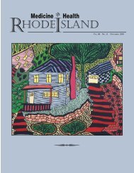
Cover: “Turbulence,” by Arides Pichardo, Originally<br />
from the Dominican Republic, the artist obtained a<br />
bachelor’s degree in publicity, with a major in graphic<br />
design, from the Universidad Autonoma de Santo<br />
Domingo. In 2003 he immigrated to the United<br />
States. In Providence, he assisted in the creation of<br />
Vision Moderna Magazine and worked in the reconstruction<br />
of antique pictures. He has exhibited at<br />
Gail Cahalan Gallery, URI Feinstein Providence<br />
Campus, Gallery Z, and the <strong>Rhode</strong> <strong>Island</strong> Home<br />
Show. http://aridesfinearts.blogspot.com/.<br />
339 Cardiac Manifestations of Lyme Disease<br />
Thomas J. Earl, MD<br />
342 Evaluation and Management of Vesicoureteral Reflux: A Decade of Change<br />
Zachary N. Gordon, Anthony Caldamone, MD, FACS, FAAP,<br />
Pamela Ellsworth, MD, FACS, FAAP<br />
349 A Pilot Retrospective Comparison of Fondaparinux and Enoxaparin for the<br />
Prevention of Venous Thromboembolism (VTE) In Patients With Stroke<br />
Jon A. Mukand, MD, PhD, and Nita H. Mukand<br />
COLUMNS<br />
354 HEALTH BY NUMBERS: Injury Visits to Emergency Departments and Hospital<br />
Discharges in <strong>Rhode</strong> <strong>Island</strong>, 2005-2009: Focus on Falls<br />
Patricia M. Burbank, DNSc, RN, and Edward F. Donnelly, RN, MPH<br />
356 GERIATRICS FOR THE PRACTICING PHYSICIAN: Fecal Incontinence<br />
Leslie Roth, MD<br />
359 Vital Statistics<br />
359 PHYSICIAN’S LEXICON: Ten You Can Count On<br />
James T. McIlwain, MD<br />
360 November Heritage<br />
Medicine and Health/<strong>Rhode</strong> <strong>Island</strong> (USPS 464-820), a monthly publication, is owned and published by the <strong>Rhode</strong> <strong>Island</strong> <strong>Medical</strong> <strong>Society</strong>, 235<br />
Promenade St., Suite 500, Providence, RI 02908, Phone: (401) 331-3207. Single copies $5.00, individual subscriptions $50.00 per year, and $100<br />
per year for institutional subscriptions. Published articles represent opinions of the authors and do not necessarily reflect the official policy of the <strong>Rhode</strong><br />
<strong>Island</strong> <strong>Medical</strong> <strong>Society</strong>, unless clearly specified. Advertisements do not imply sponsorship or endorsement by the <strong>Rhode</strong> <strong>Island</strong> <strong>Medical</strong> <strong>Society</strong>. Periodicals<br />
postage paid at Providence, <strong>Rhode</strong> <strong>Island</strong>. ISSN 1086-5462. POSTMASTER: Send address changes to Medicine and Health/<strong>Rhode</strong> <strong>Island</strong>, 235<br />
Promenade St., Suite 500, Providence, RI 02908. Classified Information: Cheryl Turcotte/<strong>Rhode</strong> <strong>Island</strong> <strong>Medical</strong> <strong>Society</strong>, phone: (401) 331-3207, fax:<br />
(401) 751-8050, e-mail: cturcotte@rimed.org. Production/Layout Design: John Teehan, e-mail: jdteehan@sff.net.<br />
Note: Medicine & Health/<strong>Rhode</strong> <strong>Island</strong> appears on www.rimed.org, under Publications.<br />
VOLUME 93 NO. 11 NOVEMBER 2010<br />
329
330<br />
MEDICINE & HEALTH/RHODE ISLAND<br />
Commentaries<br />
Continuing <strong>Medical</strong> Education<br />
(CME): Goals and Objectives<br />
<br />
Many changes have occurred in the<br />
accreditation process for continuing medical<br />
education (CME) for doctors in the<br />
last 10 years, always under the guise of reform.<br />
For one thing, drug companies are not<br />
allowed to pay speakers or their expenses directly.<br />
This change occurred about 10 years<br />
ago. The money must be laundered through<br />
the hospital. I recently discussed this with a<br />
colleague within the academic administration,<br />
who took offense at my use of “laundered.”<br />
“That term is used for drug money<br />
and other illegal uses. Laundered money is<br />
made to look like it comes from another<br />
source. It’s a pejorative term.” I agreed. I<br />
used it purposely as a pejorative term. Under<br />
current guidelines, drug companies continue<br />
to pay the speaker and the speaker’s<br />
expenses but must give the money to the<br />
hospital to give to the speaker. The money<br />
is given as an “unrestricted educational grant,”<br />
but this unrestricted grant requires an application<br />
to the company, naming the speaker<br />
and the objectives, so that if the speaker or<br />
the objectives are not in keeping with the<br />
marketing goals of the company, the speaker<br />
is not funded. Although there is an “absolute<br />
fire wall” between the marketing and<br />
educational divisions of each company, companies<br />
seem less inclined to fund speakers<br />
who are not enamored of their drugs. When<br />
companies have been asked to give truly<br />
unrestricted educational grants, say to donate<br />
money to a general pool, the answer<br />
has been 100% negative in my department,<br />
although not so in some others. So what we<br />
have is a “restricted” educational grant, given<br />
as “unrestricted,” using laundered money,<br />
with which the CME agencies are entirely<br />
complicit.<br />
This is not to say that all companies<br />
behave badly. Some companies do, in fact,<br />
separate the marketing and educational parts<br />
of their companies. In Neurology, we have<br />
two or three corporate-funded talks per year,<br />
in which the majority are theoretically chosen<br />
from anywhere and in fact are chosen<br />
from lists approved by the corporation. The<br />
topics, however, are not, and the talks are<br />
not funded without review of the objectives.<br />
The ACCME cannot guarantee quality.<br />
Its central mission is reducing bias. But<br />
it plays a role in quality assurance. One attempt<br />
to do this is by establishing standards<br />
for all talks. For example, all talks must list<br />
objectives before a talk is approved for CME<br />
credit. For my department this is an unnecessary<br />
requirement since all our speakers have<br />
been vetted in some way, either by virtue of<br />
their having academic appointments at wellknown<br />
medical centers, because they are<br />
known in our community, or they have established<br />
reputations (especially for those<br />
sponsored by drug companies) and generally<br />
all three. They have been selected by the<br />
department.<br />
I recently submitted a set of objectives<br />
for an unsponsored talk I was giving myself<br />
and was told that my objectives were<br />
not acceptable because I did not use language<br />
approved by the ACCME. I asked<br />
to review the documents that provided the<br />
language that was acceptable to the<br />
ACCME and was given the list of the acceptable<br />
descriptors. I was struck first by<br />
their length, a three-page document. I<br />
would have thought that two or three sentences<br />
would do. I was next struck by the<br />
apparently important distinction between<br />
“goals” and “objectives.” Not having formal<br />
training in epistemology, I was unaware<br />
of the distinction. In case you aren’t either,<br />
“Objectives should not be confused with<br />
goals, which are more general or global.<br />
Objectives are the action statements that<br />
operationalize a goal. For example, a goal<br />
for a CME activity may be “to help physicians<br />
provide the very best possible care to<br />
patients through improved communications.”<br />
It turns out that objectives can only<br />
be met if they are introduced by particular<br />
verbs, 109 in number, for “communicating<br />
knowledge.” There are 15 acceptable<br />
verbs for “imparting skills,” four for “conveying<br />
attitudes,” specifically excluding “ap-<br />
preciate”, “understand” and “learn.” For arcane<br />
reasons, “acquire, consider, exemplify<br />
and realize” are more “measurable as the<br />
direct outcome of a CME activity” than<br />
“appreciate, understand and learn.” Thus I<br />
can have the audience “realize” the differences<br />
between A and B but I cannot plan<br />
to have them “learn” what the differences<br />
are.<br />
Since I have been giving CME talks for<br />
a few decades I felt transformed from the<br />
person who had been speaking in prose his<br />
whole life without knowing it to the person<br />
who discovered that he really was supposed<br />
to have been speaking in poetry. My<br />
objectives were rejected for using unacceptable<br />
verbs. I had thought my talk would allow<br />
the audience to “understand” the differences<br />
between two problems, when I should<br />
have been planning to lead them to “realize”<br />
the differences. Perhaps by realizing the differences<br />
they would be more likely to remember<br />
these differences, since realization<br />
carries the implication of self discovery, that<br />
is, my talk would lead the listener to come<br />
to certain deductions, achieving an epiphany<br />
that would seem to be his own, rather than<br />
mine, and therefore more likely to stick in<br />
his memory.<br />
I am reminded of a teaching rounds<br />
when I was a third-year student in internal<br />
medicine. We had a guest attending, an older<br />
distinguished doctor, who listened to a student<br />
case presentation and then proceeded<br />
to question us and discuss the case. At the<br />
end he said that when he was a student one<br />
of his professors had taught him that he<br />
should always learn at least one thing from<br />
every teaching session. He turned to a student<br />
and asked, “Can you tell me one thing<br />
you learned today?” The student was caught<br />
unawares. The discussion had been about a<br />
blood dyscrasia, and somewhere during the<br />
meandering discussion, probably having to<br />
do with lymph node enlargement, the professor<br />
had mentioned that in most people<br />
one foot was larger than the other, hypothesized,<br />
he thought, to be due to a venous<br />
asymmetry. So the student said that he<br />
learned that most people had one foot larger<br />
than the other. After his 90-minute discussion<br />
of blood dyscrasias the professor was<br />
temporarily speechless, but pulled himself<br />
together and replied, “Well, I guess that is<br />
one thing.”<br />
My first objective when I give a talk is<br />
keeping the audience awake. I rate my lectures<br />
by the number of people who stay<br />
awake. There are points deducted for myo-
clonic jerks from nodding heads. Imparting<br />
knowledge is my second objective, which I<br />
hope parallels the lack of nodding heads. I<br />
am unsure if “imparting knowledge” is an<br />
acceptable objective.<br />
– JOSEPH H. FRIEDMAN, MD<br />
Disclosure of Financial Interests<br />
Joseph Friedman, MD, and spouse/significant<br />
other. Consultant: Acadia Pharmacy,<br />
Ovation, Transoral; Grant Research Support:<br />
Cephalon, Teva, Novartis, Boehringer-<br />
Ingelheim, Sepracor, Glaxo; Speakers’ Bureau:<br />
Astra Zeneca, Teva, Novartis,<br />
Boehringer-Ingelheim, GlaxoAcadia,<br />
Sepracor, Glaxo Smith Kline, Neurogen, and<br />
EMD Serono.<br />
Conflicts: In addition to the potential<br />
conflicts posed by my ties to industry that<br />
are listed, during the years 2001-2009 I was<br />
a paid consultant for: Eli Lilly, Bristol Myers<br />
Squibb, Janssen, Ovation, Pfizer, makers of<br />
each of the atypicals in use or being tested.<br />
Superstition, Seizures and Science<br />
<br />
When facing a terrible sickness, despairing therapies<br />
have always been society’s response to the plea, “Do something!”<br />
But if you don’t know your destination, the likelihood of getting<br />
there becomes remote. Similarly, if the underlying mechanism of a<br />
disease such as epilepsy remains elusive, the chance of finding an<br />
effective therapy becomes equally remote.<br />
The history of the search for a meaningful therapy for those<br />
burdened by repeated convulsions has been a painful voyage through<br />
strange territories, a tale of failed interventions, desperate treatments<br />
and irrational measures. Indeed, most of those treatments resembled<br />
more the art of the fugue than exercises in intelligent reasoning.<br />
Despite the secular teachings of Hippocrates, the dominant<br />
thinking in the Classical Era had been that epilepsy resulted from<br />
supernatural, evil forces. Indeed, its very name, epilepsy, is a Greek<br />
word defining the condition of being seized, captured or overcome,<br />
with the implication that the grasping was undertaken by a nameless,<br />
outside entity.<br />
Effective therapy could only be achieved, then, by resorting to<br />
interventions that could overcome those unworldly, shadowy forces,<br />
forces that inevitably must have been evil. Thus appeals were made<br />
to such personages as St. Ignatius, who had driven the devils from<br />
many epileptic victims, St. Valentine (whose priory in Alsace was<br />
the goal of many pilgrimages undertaken by victims of epilepsy)<br />
and, of course, St. Vitus, whose very name defined a class of abnormal<br />
movement disorders in helpless humans. In general, people<br />
believed that evil could be vanquished solely by spiritual rather than<br />
material talent; therefore therapy, rendered with contriteness and<br />
humility, must be confined to prayer, instruction and fasting.<br />
Alternatively, there were those, particularly in primitive cultures,<br />
who believed that the roots of epilepsy lay in the victim’s head rather<br />
than in his spirit. Some early treatments were directed therefore to the<br />
victim’s head, through cauterization of the scalp and even by boring<br />
holes (trephining) in the living skull. Indeed, many a prehistoric skull<br />
shows evidence of trephination.<br />
If, on the other hand, epilepsy was caused by some ill-defined<br />
poison, a toxin perhaps, then efforts would be directed toward a search<br />
for some counteractive chemical. During the Middle Ages - and beyond<br />
the customary measures employed for the care of epileptics<br />
such as blood-letting, purging and the use of emetic agents - four<br />
botanicals were routinely prescribed in the vain treatment for epilepsy:<br />
mistletoe, garlic, peony and elderberries. The Scottish anthropologist<br />
J. G. Frazer (1854 – 1941) stated that many healers affirmed<br />
the value of mistletoe because it clung so resolutely to the branches of<br />
sturdy oak trees, did not fall to the ground and hence should obviously<br />
be used in epilepsy, the falling sickness. (Medieval therapies<br />
were often identified by seeking analogies in nature.)<br />
Other known substances to combat the unnamed toxins with<br />
the epileptic have included boar’s gall, powdered human skull,<br />
dragon’s blood and the intestinal stones of hawks.<br />
And when all else failed, there was always fresh human blood<br />
as a treatment. The blood of slain gladiators in the Coliseum of<br />
ancient Rome was routinely fed to epileptic children. Hans Christian<br />
Anderson, in his memoirs, recalled witnessing state executions<br />
in Copenhagen with parents making their epileptic children drink<br />
the shed blood.<br />
By 1850 epilepsy had been consigned to the category of those<br />
diseases which, in the words of one contemporary neurologist, were<br />
“cryptogenic, inscrutable, and alas, incurable.” In 1857, Dr. Charles<br />
Locock, obstetrician to Queen Victoria, published a brief commentary<br />
describing a trial with bromides that seemed to have suppressed<br />
the seizures in a group of young, epileptic women. And thus, gradually<br />
from an arena of vast ignorance, did earnest investigators gradually<br />
improvise effective, rational treatments for a disease previously<br />
thought to be incurable.<br />
In 1920, the German scientist Hans Berger (1873 – 1941)<br />
devised the electroencephalograph (EEG), which detected brain<br />
waves emanating from the living brain. These electrical waves were<br />
captured by electrodes placed on the scalp, conveyed the intracranial<br />
impulses by wires to the instrument and expressed as oscillating<br />
waves on strips of moving paper. By inspecting these EEG-generated<br />
squiggles one can arrive at an objective diagnosis of epilepsy<br />
since, by the 20 th Century, epilepsy was finally recognized as the<br />
systemic manifestations of abnormally discharging, anarchic, nerve<br />
cells. In the words of one neurologist, “What is greater magic than<br />
for the brain to write its own confession of wrongdoing on a sheet<br />
of moving paper?”<br />
Most patients with epilepsy today have their convulsions safely<br />
controlled by medications and can lead normal, productive lives<br />
unencumbered by social isolation, superstition, ignorant bias, dangerous<br />
medicines or societal fear.<br />
– STANLEY M. ARONSON, MD<br />
Stanley M. Aronson, MD is dean of medicine emeritus, Brown<br />
University.<br />
Disclosure of Financial Interests<br />
Stanley M. Aronson, MD, and spouse/significant other have<br />
no financial interests to disclose.<br />
CORRESPONDENCE<br />
e-mail: SMAMD@cox.net<br />
VOLUME 93 NO. 11 NOVEMBER 2010<br />
331
<strong>Rhode</strong> <strong>Island</strong> Chapter, American College of<br />
Physicians 2010 Annual Meeting Abstracts<br />
<br />
In May 2010, the <strong>Rhode</strong> <strong>Island</strong> Chapter, American College of Physicians, hosted its annual Associates’ Forum Competition at<br />
the Crowne Plaza Hotel in Warwick. More than 110 residents in <strong>Rhode</strong> <strong>Island</strong>’s teaching hospitals submitted entries. A<br />
committee of program directors chose the following six winners. These six podium presenters each received a plaque and a cash<br />
award from the College Chapter. The Chapter and program directors applaud this year’s Associates—they represent the future<br />
of medicine in the United States.<br />
– N. S. Damle, MD, FACP<br />
– Governor<br />
– <strong>Rhode</strong> <strong>Island</strong> Chapter, American College of Physicians<br />
– – E-mail: nsdamle@scim.necoxmail.com<br />
Outcome of Hepatitis C Patients Treated with Pegylated<br />
Interferon and Ribavirin at Roger Williams <strong>Medical</strong> Center<br />
Rita Semaan, MD, Adib R. Karam, MD, Nadia Aoun, MD, Alan Epstein, MD<br />
Roger Williams <strong>Medical</strong> Center<br />
Purpose: This study retrospectively evaluated the outcome<br />
of a diverse population of Hepatitis C patients treated with<br />
pegylated interferon and ribavirin and compared actual reallife<br />
results to published clinical trials.<br />
Materials and Methods: The medical records of a total of<br />
sixty seven Hepatitis C patients, treated by pegylated interferon<br />
and ribavirin, were retrospectively reviewed regarding their outcomes<br />
following treatment. The outcome variables considered<br />
in our analysis were: sustained virologic response (SVR), breakthrough,<br />
relapse, and no response. The different outcomes were<br />
plotted against the following variables: patients’ age, ethnicity,<br />
gender, Body Mass Index (BMI), hepatitis C genotype, Human<br />
Immunodeficiency Virus (HIV) status and stage. We also reviewed<br />
the treatment-related side effects and compliance to treatment.<br />
Results: SVR was achieved in forty one out of sixty seven<br />
patients (61.1%). The majority of the patients were in the fifth<br />
to sixth decade from whom nineteen out of twenty eight (67.8%)<br />
achieved SVR; three out of five patients (60%) were non-white;<br />
females had better SVR compared to males (75% versus 54.5%);<br />
seventeen out of thirty six patients (47.2%) had genotype 1, thirteen<br />
out of fourteen (92.8%) genotype 2, nine out of thirteen<br />
(69.2%) genotype 3, and two out of four (50%) genotype 4;<br />
one patient had S0 and achieved SVR, four out of twelve<br />
(33.3%)S1, eleven out of sixteen (68.7%) S2, eight out of thirteen<br />
(61.5%) S3, one out of seven (14.2%) S4 and sixteen out of<br />
eighteen (88.8%) had no biopsy. The patients who had no biopsy<br />
were mainly genotype 2 and 3; we did not notice a difference<br />
in the SVR according to the patients’ BMI. Ten out of sixty<br />
seven patients (14.9%) had relapse, two out of sixty seven (2.9%)<br />
had breakthrough, nine out of sixty seven (13.4%) did not respond,<br />
and twelve out of sixty seven (17.9%) did not complete<br />
the treatment. All patients developed side effects during their<br />
treatment, mainly fatigue (77.6%), flu like illness (32.8%), depression<br />
(26.3%) and insomnia (23.8%). Three patients out of<br />
the sixty seven (4.4%) developed serious side effects (pancytopenia,<br />
myocardial infarction, and suicidal attempt). Conclusion:<br />
Patients who have genotype 2 had better SVR compared to patients<br />
who have genotype 1. These results match with the statistical<br />
results shown in published clinical trials. BMI has no effect<br />
on SVR. The majority of nonresponders had genotype 1.<br />
E-mail: semaanrita@hotmail.com<br />
Effectiveness of Pulsed Electromagnetic Field Therapy on<br />
Reducing Proteinuria<br />
William E. Weber, MD, David A. Weinberg, MD, Sean Hagberg, PhD, Marc S. Weinberg, MD, FACP, FASN<br />
Roger Williams <strong>Medical</strong> Center<br />
As a major global health concern, proteinuria has been<br />
established as an important independent risk factor for cardiovascular<br />
disease and renal parenchymal damage leading to endstage<br />
kidney failure. We investigated the efficacy of pulsed electromagnetic<br />
field (PEMF) therapy in reducing urinary protein<br />
excretion over a 2 week period in subjects with overt nephropathy<br />
while continuing optimal mechanisms to reduce proteinuria<br />
by inhibition of the renin-angiotensin system (RAS)<br />
using supramaximal doses of angiotensin receptor blockading<br />
(ARB) agents. Electrotherapeutic technologies has been demonstrated<br />
to modulate the calcium calmodulin-dependent,<br />
cGMP induced nitric oxide signaling pathway which may con-<br />
332<br />
MEDICINE & HEALTH/RHODE ISLAND
fer anti-proteinuric and anti-fibrotic properties by working at<br />
the cellular level promoting nitric oxide release, creating a cascade<br />
of events including blood vessel dilation, growth factor<br />
secretion, angiogenesis and ultimately t<strong>issue</strong> remodeling.<br />
As a result of activated changes in nitric oxide and intracellular<br />
mechanisms following ARB agents, we investigated<br />
whether there was a further reduction in proteinuria in subjects<br />
on ARB agents when administered PEMF therapy.<br />
Methods: Four well controlled hypertensive volunteers with<br />
chronic macroalbuminuria applied low frequency PEMF<br />
therapy three times a day for 14 consecutive days, while continuing<br />
previously prescribed antihypertensive and RAS inhibitor<br />
medications.<br />
No changes were made to drug regimens during the 28-<br />
day observation. Proteinuria was expressed as the ratio of protein<br />
to creatinine, as determined on adequate twenty-four hour<br />
urine collections at baseline, 14 and 28 days after initiation of<br />
therapy. Office blood pressure measurements along with serum<br />
creatinine, potassium and urea nitrogen concentrations<br />
collected at one central location were monitored at 7 day intervals<br />
from baseline to study endpoint. The glomerular filtration<br />
rate was estimated by means of the Modified Diet of Renal<br />
Disease (MDRD) formula. Collected data was analyzed with<br />
the use of paired Student’s t-test.<br />
Results: After 14 days of PEMF therapy, all participants<br />
demonstrated an arithmetic but not statistically significant decrease<br />
in urinary protein excretion. Mean urinary protein ex-<br />
cretion was reduced by 36%, from 2.85 g of protein per gram<br />
of creatinine to 1.80 g of protein per gram of creatinine during<br />
the 2 week intervention period PEMF therapy was applied<br />
(p = 0.06). Proteinuria gradually increased again over time after<br />
discontinuation of PEMF therapy on day 15, resulting in a<br />
level of proteinuria that did not differ significantly from that at<br />
baseline. There were no significant changes in mean arterial<br />
pressures and serum creatinine concentrations during the 28<br />
day observation. No adverse events were reported.<br />
Conclusion: In this primary analysis, our observations demonstrate<br />
that additional and synergistic reductions in proteinuria<br />
were achieved with PEMF application in patients already receiving<br />
optimal pharmacological management to reduce proteinuria<br />
by RAS inhibition. Several limitations need to be taken<br />
into account such as small sample size and lack of long term<br />
follow-up. A larger study is currently in progress evaluating for<br />
persistent reductions in proteinuria, as well as the potential effect<br />
on possible preservation of renal function.<br />
While the emphasis of our laboratory as well as other researchers<br />
have focused on reducing proteinuria through pharmacological<br />
means, the advent of electromagnetic fields may<br />
confer a new modality which may act synergistically with RAS<br />
antagonists to maximally reduce urinary protein excretion in<br />
patients already receiving optimal drug therapy and with limited<br />
treatment options.<br />
E-mail:willweber8@aol.com<br />
High Prevalence of Bone Demineralization and Vitamin D<br />
Insufficiency in a Cohort of HIV-infected Postmenopausal Women<br />
Chia-ching Wang, MD, Geetha Gopalakrishnan, MD, Erna Kojic, MD, Susan Cu-Uvin, MD<br />
<strong>Rhode</strong> <strong>Island</strong> Hospital<br />
In the era of HAART, HIV-infected women will live longer<br />
and experience changes related to menopause. Osteopenia is<br />
prevalent in persons with HIV and is part of a normal sequence<br />
of aging in women. However, there are very little data on bone<br />
metabolism in HIV-infected postmenopausal women.<br />
HIV-infected women age > 45 were referred to the HIV<br />
Menopause Clinic at the Miriam Hospital (Providence, RI). A<br />
woman was considered postmenopausal if she was status-post<br />
bilateral salpingo-oophorectomy with or without hysterectomy,<br />
or if she had no menses for more than 1 year with elevated<br />
FSH and/or LH. Bone mineral density (BMD) was assessed by<br />
dual-energy X-ray absorptimetry (DEXA) in the lumbar spine<br />
and hip. We then calculated 10-year fracture risk for postmenopausal<br />
women with osteopenia using the FRAX tool, which<br />
was developed by WHO based on models that integrate the<br />
risks associated with clinical risk factors and BMD at the femoral<br />
neck.<br />
Thirty-five postmenopausal women were included. Median<br />
age was 52 years (range 38-72); 40% Caucasian, 34% African-<br />
American, 26% Latino. Median weight was 151 lb (range 99-<br />
261). Median follow-up since HIV diagnosis was 14 years. Median<br />
CD4 count was 373 cells/L (range 72-1260). 86% were<br />
on NRTI-based HAART: 40% with TDF, 28% with NNRTI,<br />
and 43% with PI. 63% of subjects had plasma viral loads (PVL)<br />
<strong>Medical</strong> Residents Behind Bars: A Unique Clinical Experience<br />
and Linkage Project<br />
Tony Trinh, MD, Joseph Frank, MD, Aaron Samuels, MD, Mike Poshkus, MD, Peter Friedman, MD<br />
<strong>Rhode</strong> <strong>Island</strong> Hospital/The Miriam Hospital<br />
Prisoners have a high burden of chronic medical conditions.<br />
While incarcerated, prisoners are entitled to medical care,<br />
but often experience a discontinuity of primary medical services<br />
upon release. This leads to poor health outcomes, including<br />
an increased risk of death.<br />
Background: In July 2008 a partnership was formed between<br />
the medical division of the <strong>Rhode</strong> <strong>Island</strong> Department of<br />
Corrections (RIDOC) and the Internal Medicine residency<br />
program of the Alpert School of Medicine of Brown University<br />
with the intention of exposing residents to correctional<br />
health, and promoting medical continuity for adult prisoners<br />
upon release into the community. In the men’s division of the<br />
Adult Correctional Institute (ACI), clinic sessions were developed<br />
to target inmates with chronic medical problems who<br />
were at risk of discontinuity of care upon release. Residents<br />
were precepted by RIDOC Board Certified internists at clinics<br />
sessions one half day a week and provided medical services<br />
to soon-to-be-released inmates. Those without identified plans<br />
for community medical follow up were seen by a social worker<br />
and referred to the medical residents’ clinics at <strong>Rhode</strong> <strong>Island</strong><br />
Hospital (RIH) and The Miriam Hospital (TMH) upon release.<br />
Methods: We conducted a review of all inmates seen at<br />
the medical residents’ clinic sessions held at the minimum security<br />
prison during Jan 1-Dec. 31, 2009. Baseline data were<br />
self-reported at initial pre-release visits. Patients were then<br />
searched against the data repository of RIH / TMH to identify<br />
clinical encounters occurring during the period following release.<br />
The RIDOC database was also searched for any activity<br />
of re-incarceration during the patients’ post-release period.<br />
Descriptive statistics were generated for demographic and clinical<br />
data.<br />
Results: A total of 146 patients were seen over the period.<br />
The mean age was 44 years. The most prevalent conditions<br />
were hepatitis C (21.2%), injection drug use (19.9%), depression<br />
(14.4%) and hypertension (13.0%). Patients were seen at<br />
a mean of 35 days prior to release from prison; 76% were referred<br />
to residents’ medical clinic. A search of the RIH / TMH<br />
/ RIDOC databases during the post-release period found 29<br />
(19.9%) patients had outpatient medical encounters at a mean<br />
of 38 days after release, 26 (18%) had Emergency Department<br />
(ED) encounters at 69.5 days, and 41 (26%) had reencounters<br />
with the ACI at 87 days.<br />
Conclusions: Early data suggest our patients are less likely<br />
to encounter the ED within 60 days of release than in the general<br />
population of released prisoners. Our project represents a<br />
novel collaboration incorporating correctional health with medical<br />
residents’ ambulatory experience and may serve as model of<br />
transitional care for adults released from prison. Further studies<br />
are needed to better understand the impact of this program<br />
on the health outcomes and resource utilization of its patients.<br />
E-mail: ttonytrinh@gmail.com<br />
Characteristics of Adults Hospitalized with Novel 2009<br />
influenza A (H1N1) in a Community Hospital<br />
Abdullah Chahin, MD, Denisa Hagau, MD, Aurora Pop-Vicas, MD<br />
Memorial Hospital of <strong>Rhode</strong> <strong>Island</strong><br />
Background: Results from studies of the novel 2009<br />
H1N1 influenza early on during the pandemic suggest that<br />
the majority of symptomatic patients are young, and clinical<br />
disease is mostly mild. Data on patients hospitalized with H1N1<br />
disease in community hospitals are scarce.<br />
Objective: To describe the epidemiological and clinical<br />
characteristics of adult patients hospitalized with pandemic influenza<br />
A (H1N1) during the peak of the pandemic in a community<br />
hospital.<br />
Methods: Review of medical records from adults admitted<br />
with laboratory-confirmed novel 2009 influenza A (H1N1)<br />
between October 31 and December 9, 2009 at Memorial<br />
Hospital of <strong>Rhode</strong> <strong>Island</strong> – a teaching community hospital.<br />
Results: A total of 23 (70%) of the laboratory-confirmed<br />
adult patients admitted with pandemic influenza H1N1 during<br />
this time period consented to study enrollment. Among these,<br />
74% were women, and 26% were men. The median age was 54<br />
(range 22 – 81), and the mean Charlson comorbidity index was<br />
3.7. The majority (91%) of these patients had underlying chronic<br />
lung disease, and 57% of the patients were obese (BMI = 30).<br />
The diagnosis of novel H1N1 influenza was confirmed by a positive<br />
RT-PCR in all cases. The sensitivity of our rapid nucleoprotein<br />
antigen test for detecting pandemic H1N1 influenza was<br />
only 32%. The median duration of clinical symptoms was 9 days<br />
(range 3-22 days), and the median length of hospital stay was 7<br />
days (range 2-19 days). Patients whose symptoms lasted = 10<br />
days were more likely to have a prolonged hospitalization (P =<br />
0.02), and tended to be = age 65 (P = 0.07). Radiological pulmonary<br />
infiltrates were present in 61% of the patients. These<br />
patients were more likely to have multiple underlying<br />
comorbidities (Charlson index = 5, P = 0.06) and hospital stays<br />
= 7 days (P = 0.02). The median duration between onset of clinical<br />
symptoms and laboratory confirmation of pandemic H1N1 influenza<br />
was 6 days (range 2-20 days).<br />
334<br />
MEDICINE & HEALTH/RHODE ISLAND
Conclusion: Our preliminary findings suggest that during<br />
the peak of the 2009 pandemic, novel H1N1 influenza<br />
was associated with significant morbidity in the hospitalized<br />
elderly and adult patients with underlying comorbid illnesses.<br />
Interestingly, in some hospitalized patients, nasopharyngeal viral<br />
samples were positive for influenza by RT-PCR for 6 or<br />
more days from clinical onset. Further research addressing risk<br />
factors associated with prolonged H1N1 viral shedding would<br />
have important infection control implications.<br />
E-mail: abdullah_shahin@hotmail.com<br />
Oseltamivir Resistant 2009-2010 Pandemic Influenza A<br />
(H1N1) in an Immunocompromised Patient<br />
Anne Gabonay Frank, MD, Philip A. Chan, MD, Nathan T. Connell, MD, Jerome M. Larkin, MD<br />
<strong>Rhode</strong> <strong>Island</strong> Hospital<br />
Although neuraminidase inhibitors are active against most<br />
2009-2010 pandemic influenza A (H1N1) swine-origin strains,<br />
since April of 2009, 54 total cases of oseltamivir-resistant H1N1<br />
swine-origin have been reported in the US (as of February 1st,<br />
2010). We report a patient with an underlying hematologic<br />
malignancy who was hospitalized with influenza A (H1N1)<br />
swine-origin and whose strain developed oseltamivir resistance<br />
during therapy.<br />
Case Report: A 26 year-old woman with ALL was admitted<br />
to our hospital on November 18, 2009 for re-induction<br />
chemotherapy. On admission, her absolute neutrophil count<br />
(ANC) was 800 cells/µL. On hospital day 3, she developed a<br />
non-productive cough and had a temperature of 102.6 °F with<br />
an ANC of 300 cells/µL. She was started on antibiotics and<br />
oseltamivir 75mg twice daily. A nasopharngyeal swab sent for<br />
a respiratory viral panel revealing probable influenza A (H1N1)<br />
swine-origin and rhinovirus. She continued to have fever so<br />
antifungal coverage was added, antibiotics were changed, and<br />
oseltamivir was continued. Fevers persisted, and bronchoscopy<br />
was performed. Viral culture derived from the bronchoscopy<br />
specimen grew influenza A (H1N1) swine-origin. The patient<br />
continued to have daily fever in the setting of prolonged neutropenia.<br />
On hospital day 17, a nasopharyngeal swab again<br />
revealed H1N1, as well as rhinovirus, despite 13 days of<br />
oseltamivir, and the concern for resistance was raised. Antiviral<br />
coverage was changed to zanamivir, and a nasopharyngeal swab<br />
culture was sent for resistance testing. A nasopharyngeal swab<br />
was used for respiratory viral panel testing on day 20 which<br />
was negative. On day 23, viral cultures from a nasopharyngeal<br />
swab done on day 17 returned positive for oseltamivir-resistant<br />
influenza A with the H275Y mutation. Despite aggressive<br />
therapy, she died on hospital day 40. After her death, we performed<br />
resistance testing on a stored sample of the initial influenza<br />
strain isolated on hospital day 3; it was sensitive to<br />
oseltamivir. Additional testing was also performed on the bronchoscopy<br />
specimen from hospital day 11, which demonstrated<br />
that resistance to oseltamivir had developed after eight days of<br />
oseltamivir therapy.<br />
Discussion: To our knowledge, this is the first case of<br />
oseltamivir-resistant, swine-origin influenza A (H1N1) that was<br />
associated with death. In the United States, 42 of 54 individuals<br />
with resistance had documented exposure to oseltamivir,<br />
suggesting that resistance develops under selective pressure to<br />
the drug and not as a natural variant. We have shown that<br />
resistance developed during oseltamivir treatment in our patient<br />
who was receiving the recommended dosing of 75mg twice<br />
a day. Recent literature suggests that an increased dose of 150mg<br />
twice a day may be preferable for critically ill patients.<br />
Oseltamivir resistance should be a consideration when treating<br />
critically ill or immunocompromised patients.<br />
E-mail: annegabonay@hotmail.com<br />
VOLUME 93 NO. 11 NOVEMBER 2010<br />
335
336<br />
MEDICINE & HEALTH/RHODE ISLAND<br />
Teen Pregnancy In <strong>Rhode</strong> <strong>Island</strong>:<br />
Policies To Improve Outcomes<br />
Susanna R. Magee, MD, MPH, Melissa Nothnagle, MD, MSc, Mary Beth Sutter, MD<br />
<br />
The United States has one of the highest<br />
rates of teen pregnancy among industrialized<br />
countries, with more than<br />
750,000 pregnancies each year among<br />
women less than 20 years of age. 1 Though<br />
teen pregnancy rates in the US had declined<br />
each year since 1991, the most<br />
recent national data show that rates of<br />
teen pregnancy, abortion, and birth are<br />
on the rise. 1 <strong>Rhode</strong> <strong>Island</strong> has 2,430 teen<br />
pregnancies per year, the highest prevalence<br />
among the New England states. In<br />
<strong>Rhode</strong> <strong>Island</strong>, 51% of these pregnancies<br />
result in live births and 35% in abortion. 2<br />
Rates of teen pregnancy in <strong>Rhode</strong> <strong>Island</strong><br />
vary by region: the highest rates from<br />
2003-2007 were seen in Central<br />
Falls (95.5 births per 1,000 teens), followed<br />
by Woonsocket (65.2 births per<br />
1,000 teens), Pawtucket (58.7 births per<br />
1,000 teens), and Providence (48.0 births<br />
per 1,000 teens). 3<br />
Births to teens have long been associated<br />
with adverse outcomes, including<br />
individual and familial poverty and reduced<br />
educational attainment. 4,5,6 Children<br />
born to adolescent parents experience<br />
higher rates of behavioral and developmental<br />
disorders, substance abuse,<br />
depression, early sexual activity, and teen<br />
pregnancy. 7 Nationally, about 20% of<br />
births to teens are repeat births, which<br />
place additional socioeconomic and<br />
health pressures on teen parents. 8 In<br />
<strong>Rhode</strong> <strong>Island</strong>, the repeat birth rate in<br />
2004 was 19% for women ages 15-19. 8<br />
PREVENTION OF TEENAGE<br />
PREGNANCY AND BIRTH<br />
Sexual education in schools<br />
Comprehensive sexual education is an<br />
important tool to prevent teen pregnancy.<br />
From 1996 to 2009, federal funding of<br />
sexual education programs was available<br />
only for abstinence-only programs, which<br />
exclusively teach the benefits of abstaining<br />
from sexual activity. 9 Comprehensive<br />
sexual education programs promote abstinence<br />
but also provide information on<br />
contraception for pregnancy prevention<br />
and condoms for prevention of sexually<br />
transmitted infections. Well-designed<br />
studies of abstinence-only sexual education<br />
programs have found no significant impact<br />
on teen sexual activity or rates of unprotected<br />
sex. 9, 10 However, a populationbased<br />
study of US sexual education programs<br />
found that teens who received comprehensive<br />
sex education were significantly<br />
less likely to become pregnant than those<br />
who received abstinence-only or no sex<br />
education. 11<br />
State law mandates that <strong>Rhode</strong> <strong>Island</strong><br />
schools offer sexual education, including<br />
instruction on sexually transmitted infections<br />
and HIV, but requires abstinence to<br />
be emphasized and permits parental optout<br />
from participation. 12 From 2003-2007,<br />
<strong>Rhode</strong> <strong>Island</strong> received federal money to<br />
support abstinence-only education; the<br />
majority of this funding was distributed to<br />
community-based organizations. In 2008,<br />
<strong>Rhode</strong> <strong>Island</strong> declined Title V federal funding<br />
for abstinence-only-until-marriage programs.<br />
13 Currently there is no standardized<br />
sexual education curriculum for<br />
<strong>Rhode</strong> <strong>Island</strong> schools and little teacher<br />
training or supervision.<br />
Access to Contraception<br />
<strong>Rhode</strong> <strong>Island</strong> is one of 27 states that<br />
require insurers to provide coverage of<br />
the full range of FDA approved contraceptive<br />
options. 14 However, approximately<br />
62,670 reproductive-age, sexually-active<br />
<strong>Rhode</strong> <strong>Island</strong> women are in<br />
need of publicly-funded contraceptive<br />
services. 15 Among these, 19,660 (31.4%)<br />
are teens. A sexually active teen not using<br />
contraception has a 90% chance of<br />
pregnancy within one year. 16 In 2005,<br />
only 23% of <strong>Rhode</strong> <strong>Island</strong> teenagers in<br />
need received care from publically<br />
funded clinics. 17 In 2006, <strong>Rhode</strong> <strong>Island</strong>’s<br />
family planning clinics received<br />
$3,778,000 from federal and state governments,<br />
or approximately $60 per<br />
woman in need of services. 18 These clinics<br />
are expected to deliver sexually transmitted<br />
infection screening and treatment,<br />
cervical cancer screening, education,<br />
contraceptive methods, and counseling<br />
to ensure consistent and correct use of<br />
contraception.<br />
In addition to funding barriers, <strong>issue</strong>s<br />
of consent may limit teens’ access to<br />
contraceptive services in <strong>Rhode</strong> <strong>Island</strong>.<br />
While Connecticut allows confidential<br />
contraceptive access to married minors,<br />
and Massachusetts funds a statewide program<br />
to give all minors access to confidential<br />
contraceptive care, <strong>Rhode</strong> <strong>Island</strong><br />
is one of only four states with no explicit<br />
policy on minors’ authority to consent to<br />
contraceptive services. 19 As a result minors<br />
in <strong>Rhode</strong> <strong>Island</strong> are not assured confidentiality<br />
regarding contraceptive care<br />
and may have difficulty obtaining contraception<br />
if they are afraid to inform<br />
their parents of their sexual activity. Furthermore,<br />
<strong>Rhode</strong> <strong>Island</strong> lacks an emancipated<br />
minor law for parenting teens,<br />
so even those who already have children<br />
may need parental consent to obtain contraception<br />
to prevent repeat pregnancies.<br />
Federal law requires access to confidential<br />
contraceptive services for all teens<br />
covered by Medicaid. While <strong>Rhode</strong> <strong>Island</strong><br />
law does not prohibit physicians from providing<br />
contraceptives to minors without<br />
parental consent, there is no law protecting<br />
those who prescribe contraception to<br />
teens. <strong>Rhode</strong> <strong>Island</strong>’s silence on this <strong>issue</strong><br />
may discourage physicians from providing<br />
teens with confidential access to contraception.<br />
Programs such as California’s<br />
comprehensive teen pregnancy prevention<br />
program which expand free confidential<br />
contraception and comprehensive sexual<br />
education for all teens are associated with<br />
reduced rates of teen sexual activity as well<br />
as substantially fewer births to teens. 20<br />
Another potential barrier to effective<br />
contraception for teens is lack of<br />
awareness among primary care providers<br />
about the safety of long-acting reversible<br />
contraceptive methods such as in-
trauterine contraception among teenagers.<br />
Historically, intrauterine devices were<br />
recommended only for monogamous<br />
women who had already given birth, but<br />
current evidence supports their safety<br />
and efficacy in nulliparous women. 21,22<br />
Intrauterine devices and contraceptive<br />
implants are the most effective methods<br />
of reversible contraception available and<br />
should be discussed with all sexually active<br />
adolescents.<br />
In addition, emergency contraception<br />
offers a chance to prevent pregnancy<br />
to women who have had unprotected<br />
intercourse or contraception failure or<br />
who have experienced sexual assault.<br />
Teens can use it correctly, and access to<br />
emergency contraception does not increase<br />
risky sexual behavior. 23<br />
Federal law recently made<br />
levonorgestrel-containing emergency<br />
contraception available to all women ages<br />
17 and older without a prescription, as it<br />
has no contraindications or drug interactions,<br />
does not cause birth defects, and<br />
is nontoxic. 23<br />
Ten states including Massachusetts<br />
have laws allowing pharmacists to dispense<br />
emergency contraception without<br />
a prescription through collaborativepractice<br />
agreements. 24 These laws now<br />
apply specifically to minors under age 17,<br />
given the recent federal legislation. As<br />
mentioned above, <strong>Rhode</strong> <strong>Island</strong> lacks any<br />
such policy regarding contraceptive access<br />
for minors.<br />
Access to abortion<br />
In addition to accessible and effective<br />
contraception, access to abortion is<br />
essential in reducing unwanted births to<br />
teens. In 2005 in <strong>Rhode</strong> <strong>Island</strong>, 5,290<br />
women obtained abortions including<br />
1,620 teens; in the same year, 22 of every<br />
1,000 teen pregnancies in <strong>Rhode</strong> <strong>Island</strong><br />
ended in abortion, compared with<br />
19 per 1,000 teen pregnancies nationally.<br />
25,26<br />
<strong>Rhode</strong> <strong>Island</strong> law creates multiple<br />
barriers for teens seeking to end unwanted<br />
pregnancies. First, <strong>Rhode</strong> <strong>Island</strong><br />
is one of 32 states that prohibit the use of<br />
public funds (including Medicaid) to pay<br />
for abortion, except in cases of rape, incest<br />
or life endangerment. 25 This restriction<br />
also applies to insurance policies for<br />
public employees in <strong>Rhode</strong> <strong>Island</strong>. The<br />
federal Hyde Amendment prohibits use<br />
of federal Medicaid funds for abortion,<br />
and allows states to determine whether<br />
state Medicaid funds will be used to pay<br />
for abortions for low income women.<br />
Second, <strong>Rhode</strong> <strong>Island</strong> is one of 35<br />
states that require parental consent or notification<br />
for abortion. 25 Parental consent<br />
laws have little effect on rates of abortion<br />
among minors; they do, however, result<br />
in delays (with increases in cost and associated<br />
risk) and increase the number of<br />
minors who travel to another state for<br />
abortions. 27-28 Massachusetts also requires<br />
parental consent for abortion, but allows<br />
an exception for medical emergencies,<br />
and Connecticut does not require parental<br />
consent for abortion. 29 The number<br />
of <strong>Rhode</strong> <strong>Island</strong> teens who travel out of<br />
state to avoid parental consent laws is not<br />
known.<br />
Intrauterine devices<br />
and contraceptive<br />
implants are the<br />
most effective<br />
methods of<br />
reversible<br />
contraception<br />
available and<br />
should be discussed<br />
with all sexually<br />
active adolescents.<br />
RECOMMENDATIONS<br />
We recommend the following actions:<br />
1.) Provide comprehensive sexual health<br />
education in schools.<br />
<strong>Rhode</strong> <strong>Island</strong> schools should provide<br />
a comprehensive, age-appropriate<br />
sexual health curriculum, including information<br />
on contraception and sexually<br />
transmitted infection prevention for<br />
middle and high school students. Our<br />
Commissioner on Education, district superintendents,<br />
and leadership at the Department<br />
of Health must implement a<br />
system of teacher training and oversight<br />
in order for this to be effective.<br />
2.) Improve access to contraception for adolescents.<br />
Legislative barriers prevent many<br />
<strong>Rhode</strong> <strong>Island</strong> teens from accessing effective<br />
contraceptive services. Minors in<br />
<strong>Rhode</strong> <strong>Island</strong> should be guaranteed confidential<br />
access to contraceptive services<br />
through primary care providers and family<br />
planning clinics. Education for primary<br />
care providers should emphasize the safety<br />
and efficacy of long-acting reversible contraceptive<br />
methods, including intrauterine<br />
contraception, in teenage women.<br />
Given its safety and potential to reduce<br />
unplanned pregnancy when used in a<br />
timely manner, emergency contraception<br />
should be made available to women under<br />
17 without a prescription, as it is currently<br />
in 10 other states including Massachusetts.<br />
3.) Improve minors’ access to abortion.<br />
As parental consent requirements<br />
have little impact on abortion rates, but<br />
result in delays in care and increase the<br />
number of teens who travel to other states<br />
to get abortions, parental consent for<br />
abortion should not be required for minors.<br />
Use of state funds such as Medicaid<br />
to pay for abortions for low-income<br />
women should also be permitted.<br />
4.) Provide educational opportunities to<br />
pregnant and parenting teens.<br />
Teens who become pregnant are<br />
less likely to graduate high school and<br />
more likely to live in poverty. Likewise,<br />
the children of these teen moms are<br />
more likely to grow up in homes where<br />
the income is significantly below the<br />
national poverty level. Pregnant teens<br />
should be encouraged to remain in<br />
school as long as possible and return to<br />
school after delivery as soon as possible.<br />
Day care programs based on site at<br />
schools have had success, yet in recent<br />
years these programs have been cut<br />
rather than expanded. 30 State funding<br />
should support programs that help teens<br />
complete their education.<br />
<strong>Rhode</strong> <strong>Island</strong>’s small size allows statelevel<br />
initiatives to make great differences<br />
in the lives of its citizens. We must promote<br />
healthy teens; young people are the<br />
future of our state and their success or<br />
failure is in our hands.<br />
VOLUME 93 NO. 11 NOVEMBER 2010<br />
337
REFERENCES<br />
1. Kost K, Henshaw S, Carlin L. U.S. Teenage Pregnancies,<br />
Births and Abortions: National and State<br />
Trends and Trends by Race and Ethnicity. 2010.<br />
http://www.guttmacher.org/pubs/<br />
USTPtrends.pdf<br />
2. Guttmacher Institute. Contraception Counts:<br />
<strong>Rhode</strong> <strong>Island</strong>. 2006. http://www.guttmacher.org/<br />
pubs/state_data/states/rhode_island.pdf<br />
3. <strong>Rhode</strong> <strong>Island</strong> KIDS COUNT. 2010 <strong>Rhode</strong> <strong>Island</strong><br />
Kids Count Factbook: Births to teens. http:/<br />
/www.rikidscount.org/matriarch/documents/<br />
10_Factbook_Indicator_30.pdf<br />
4. Hofferth SL, Moore KA. Amer Sociolog Review<br />
1979;44:784-815.<br />
5. Klepinger DH, Lundberg S, Plotnick RD. Fam<br />
Plann Persp 1995;27:23-8.<br />
6. Moore KA, Myers DE, et al. J Res Adolescence<br />
1993;3:393-422.<br />
7. Klein JD. Pediatrics 2005; 116:281-6.<br />
8. Schelar E, Franzetta K, Manlove J. Repeat teen childbearing:<br />
Differences across states and by race and<br />
ethnicity. 2007. Washington, DC: Child Trends.<br />
http://www.childtrends.org/Files//Child_Trends-<br />
2007_11_27_RB_RepeatCB.pdf<br />
9. Santelli J, Ott MA, et al. J Adolesc Health<br />
2006;38:72-81.<br />
10. Trenholm C, Devaney B, et al. J Policy Anal Manag<br />
2008;27:255-76.<br />
11. Kohler PK, Manhart LE, Lafferty WE. J Adolesc<br />
Health. 2008;42:344-51.<br />
12. <strong>Rhode</strong> <strong>Island</strong> General Law 16-22-18: Health and<br />
family life courses. http://www.rilin.state.ri.us/<br />
Statutes/TITLE16/16-22/16-22-18.HTM<br />
13. Sexuality Information and Education Council of<br />
the United States. A Portrait of Sexuality Education<br />
and Abstinence-Only-Until-Marriage Programs<br />
in the States. http://www.siecus.org/<br />
index.cfm?fuseaction=Page.viewPage<br />
&pageId=487&parentID=478<br />
14. Guttmacher Institute. State Policies in Brief. Insurance<br />
Coverage of Contraceptives. 2010. http://<br />
www.guttmacher.org/statecenter/spibs/spib_ICC.pdf<br />
15. Frost JJ, Henshaw SK, Sonfield A. Contraceptive<br />
Needs and Services: National and State Data, 2008<br />
Update. May 2010. http://www.guttmacher.org/<br />
pubs/win/contraceptive-needs-2008.pdf<br />
16. Harlap S, Kost K, Forrest JD. Preventing pregnancy,<br />
Protecting Health: A New Look at Birth<br />
Control Choices in the Unites States. New York:<br />
Alan Guttmacher Institute; 1991.<br />
17. Guttmacher Institute. Contraception Counts <strong>Rhode</strong><br />
<strong>Island</strong>. 2006. http://www.guttmacher.org/pubs/<br />
state_data/states/rhode_island.pdf<br />
18. Sonfield A, Alrich C, Gold RB. Public Funding<br />
for Family Planning, Sterilization and Abortion<br />
Services, FY 1980-2006. January 2008. http://<br />
www.guttmacher.org/pubs/2008/01/28/<br />
or38.pdf<br />
19. Guttmacher Institute. State Policies in Brief. Minors’<br />
Access to Contraception Services. 2010.<br />
http://www.guttmacher.org/statecenter/spibs/<br />
spib_MACS.pdf<br />
20. Brindis CD, Geierstanger SP, Faxio A. Health Educ<br />
Behav 2009;36:1095-108.<br />
21. Prager S, Darney PD. Contraception 2007;75(6<br />
Suppl):S12-5.<br />
22. Allen RH, Goldberg AB, Grimes DA. Am J Obstet<br />
Gynecol 2009;201:456.e1-5.<br />
23. Harper CC, Weiss DC, et al. Contraception<br />
2008;77:230-3.<br />
24. Guttmacher Institute. State Policies in Brief: Emergency<br />
Contraception. 2010. http://<br />
www.guttmacher.org/statecenter/spibs/spib_EC.pdf<br />
25. Guttmacher Institute. State Facts on Abortion:<br />
<strong>Rhode</strong> <strong>Island</strong>. 2008. http://www.guttmacher.org/<br />
pubs/sfaa/pdf/rhode_island.pdf<br />
26. Guttmacher Institute. US Teenage Pregnancies,<br />
Births and Abortions: National and State Trends<br />
by Race and Ethnicity. January 2010. http://<br />
www.guttmacher.org/pubs/USTPtrends.pdf<br />
27. Colman S, Joyce T. Perspect Sex Reprod Health<br />
2009;41:119-26.<br />
28. Dennis A, Henshaw SK, et al. The Impact of Laws<br />
Requiring Parental Involvement for Abortion: A Literature<br />
Review. New York: Guttmacher Institute, 2009.<br />
29. Guttmacher Institute. State policies in Brief: Parental<br />
Involvement in Minors’ Abortions. 2010.<br />
http://www.guttmacher.org/statecenter/spibs/<br />
spib_PIMA.pdf<br />
30. Sadler LS, Swartz MK, et al. J Sch Health<br />
2007;77:121-30.<br />
Susanna R, Magee, MD, MPH, is<br />
Assistant Professor of Family Medicine, The<br />
Warren Alpert <strong>Medical</strong> School of Brown<br />
University.<br />
Melissa Nothnagle, MD, MSc, is Assistant<br />
Professor of Family Medicine, The<br />
Warren Alpert <strong>Medical</strong> School of Brown<br />
University.<br />
Mary Beth Sutter, MD, is a resident in<br />
Family Medicine at Memorial Hospital/The<br />
Warren Alpert <strong>Medical</strong> School of Brown<br />
University.<br />
Disclosure of Financial Interests<br />
The authors and spouses/significant<br />
others have no financial interests to disclose.<br />
CORRESPONDENCE<br />
Susanna R, Magee, MD, MPH<br />
Memorial Hospital of <strong>Rhode</strong> <strong>Island</strong><br />
111 Brewster Street<br />
Pawtucket, RI 02860<br />
Phone: (401) 729 2237<br />
e-mail: Susanna_Magee@mhri.org<br />
338<br />
MEDICINE & HEALTH/RHODE ISLAND
Cardiac Manifestations of Lyme Disease<br />
CASE 1<br />
A 15-year old man with no significant<br />
medical history presented to the<br />
emergency department following two<br />
episodes of syncope. He was in his usual<br />
state of good health until earlier on the<br />
day of admission when he awoke with a<br />
severe headache, shortly followed by a<br />
syncopal episode with brief loss of consciousness.<br />
Later that same day, he experienced<br />
a second syncopal episode and<br />
subsequently sought medical attention.<br />
On arrival to the emergency department,<br />
he was afebrile and hemodynamically<br />
stable. Physical examination was notable<br />
for irregular and bradycardic heart<br />
sounds without murmurs, rubs, or gallops.<br />
Electrocardiogram (ECG) (Figure 1) revealed<br />
complete heart block with a narrow<br />
QRS complex. Transthoracic<br />
echocardiogram showed normal<br />
biventricular size and function and mild<br />
mitral regurgitation. There was no pericardial<br />
effusion visualized. He was given intravenous<br />
ceftriaxone for presumed Lyme<br />
carditis and transferred to the Coronary<br />
Care Unit for monitoring and treatment.<br />
Shortly after admission, telemetry<br />
monitoring revealed progressively lower<br />
heart rates requiring transcutaneous and<br />
eventual transvenous pacing. He was continued<br />
on ceftriaxone. Subsequent ECGs<br />
showed progression from complete heart<br />
block, to 2:1 atrioventricular (AV) block,<br />
and eventually sinus rhythm with first<br />
degree AV block. (Figure 2) Subsequent<br />
laboratory data revealed elevated Lyme<br />
IgM and IgG antibodies.<br />
With improvement in his native<br />
conduction the transvenous pacing wire<br />
was removed. He was discharged home<br />
to complete a twenty-eight day course<br />
of ceftriaxone and remained well in follow-up.<br />
<br />
Thomas J. Earl, MD<br />
was notable for elevated Lyme antibody<br />
titres. He was prescribed doxycycline but<br />
discontinued it after 3 days after experiencing<br />
nausea and vomiting.<br />
On presentation to our institution,<br />
initial workup was notable for an ECG<br />
(Figure 3) showing diffuse, upsloping STsegment<br />
elevations without reciprocal<br />
depressions along with subtle PR-segment<br />
depressions. Serial cardiac<br />
biomarkers were negative. Transthoracic<br />
echocardiogram revealed normal<br />
biventricular size and systolic function<br />
without a pericardial effusion.<br />
Figure 1. 12-lead ECG showing complete heart block with a narrow QRS complex.<br />
CASE 2<br />
A 34-year old man with no significant<br />
medical history presented to the<br />
emergency department with pleuritic<br />
chest pain. Approximately five weeks<br />
prior to presentation he developed fevers,<br />
a “bull’s-eye” type rash, and Bell’s palsy.<br />
Serologic workup at an outside hospital<br />
Figure 2. 12-lead ECG showing sinus rhythm with a first-degree AV block.<br />
VOLUME 93 NO. 11 NOVEMBER 2010<br />
339
340<br />
Figure 3. 12-lead ECG showing sinus rhythm with diffuse, upsloping ST-segment<br />
elevations as well PR-segment depressions in the inferior leads.<br />
Given his clinical presentation and<br />
recent serologic data he was started on<br />
intravenous ceftriaxone for a diagnosis of<br />
Lyme pericarditis. Treatment was continued<br />
for a total of twenty-eight days. He<br />
was treated with NSAIDs and opiate analgesics<br />
with eventual relief of his pain.<br />
At the time of completion of antibiotic<br />
therapy he was without chest pain.<br />
DISCUSSION<br />
Lyme disease, a tick-borne illness first<br />
described in Connecticut in 1977, is currently<br />
the most commonly reported vector-borne<br />
illness in the United States. In<br />
this country, Lyme disease is caused by<br />
the spirochete Borrelia burgdorferi,<br />
which is transmitted by the bite of Ixodes<br />
tick. According to the Centers for Disease<br />
Control and Prevention (CDC), the<br />
incidence of Lyme disease in <strong>Rhode</strong> <strong>Island</strong><br />
in 2008 was 17.7 (confirmed cases<br />
per 100,000 persons), with peak reporting<br />
in the mid and late summer<br />
months.1,2 The majority of patients<br />
present with the classic rash of Lyme disease,<br />
erythema migrans, and/or arthritis,<br />
while a small minority experience cardiac<br />
manifestations of the disease.3<br />
Cardiac manifestations of Lyme disease<br />
are typically seen in the early-disseminated<br />
phase (Stage 2) of the illness, with<br />
approximately 5% of untreated patients<br />
having cardiac involvement within the<br />
MEDICINE & HEALTH/RHODE ISLAND<br />
first few weeks after disease onset.4 Despite<br />
a slightly higher incidence of Lyme<br />
disease in females, there is a 3:1 male-tofemale<br />
predominance of Lyme carditis.<br />
The spectrum of cardiac involvement in<br />
Lyme disease is highly variable, ranging<br />
from asymptomatic to severe manifestations.<br />
Notably, early recognition of infection<br />
and prompt administration of<br />
antibiotic therapy is thought to decrease<br />
the likelihood of cardiac involvement,<br />
although to my knowledge there are no<br />
randomized trials demonstrating a clinical<br />
benefit of antibiotics in terms of duration<br />
or severity of cardiac symptoms.5<br />
Clinical manifestations of Lyme carditis<br />
include syncope or pre-syncope, palpitations,<br />
dyspnea, and/or chest pain. Cardiac<br />
abnormalities in Lyme disease most<br />
commonly include varying degrees of AV<br />
block and/or myopericarditis.<br />
Steere et al reported twenty patients<br />
with cardiac manifestations of Lyme disease<br />
in 1980. Within this group, eighteen<br />
patients developed fluctuating degrees<br />
of AV block, with eight patients<br />
developing complete heart block. Interestingly,<br />
the degree of AV block was noted<br />
to fluctuate even over the course of minutes.6<br />
Electrophysiology studies performed<br />
on select patients with Lyme<br />
carditis from van der Linde’s series revealed<br />
diffuse conduction system involvement,<br />
with the majority of patients dem-<br />
onstrating prolonged A-H intervals suggestive<br />
of AV nodal involvement.7 There<br />
is typically no response to atropine in acquired<br />
AV block attributed to Lyme disease,<br />
suggesting direct involvement of the<br />
conduction system as opposed to increased<br />
vagal tone.<br />
Fortunately, acquired AV block in<br />
Lyme carditis is typically transient and<br />
resolves with antibiotic therapy. This is<br />
demonstrated in Case 1 in which a rapid<br />
improvement in native conduction was<br />
observed. The need for temporary pacemaking<br />
is not uncommon, but persistent<br />
AV block necessitating permanent pacemaker<br />
placement is rare. In a retrospective<br />
study of 105 patients with documented<br />
Lyme carditis, 35% of patients<br />
required temporary pacing, while only<br />
five patients went on to receive a permanent<br />
pacemaker. Among these patients<br />
receiving a permanent pacemaker, four<br />
were noted to have resolution of their<br />
conduction system disease, while one remained<br />
pacer-dependent.7<br />
Following conduction system disease,<br />
Lyme carditis most commonly manifests<br />
as myopericarditis. Steere et al described<br />
ECG abnormalities suggestive of myocardial<br />
involvement such as ST-segment depressions,<br />
T-wave inversions, and/or intraventricular<br />
conduction delays in thirteen<br />
of twenty patients. Of note, all of the observed<br />
abnormalities resolved over time.<br />
Furthermore, three patients were observed<br />
to have mild left ventricular (LV)<br />
systolic dysfunction by radionuclide imaging<br />
during the active phase of the disease;<br />
all had normalization of LV systolic<br />
function on repeat imaging when the disease<br />
was in remission.6 Several case reports<br />
and small cohort studies have additionally<br />
described patients with clinical<br />
and ECG evidence of pericarditis.8-11<br />
Finally, Lyme disease has been purported<br />
to play a causative role in the development<br />
of a dilated cardiomyopathy and/or<br />
chronic congestive heart failure following<br />
small European studies showing higher<br />
rates of seropositivity in patients with an<br />
idiopathic dilated cardiomyopathy as compared<br />
to controls as well as the ability to<br />
grow B burgdorferi from endomyocardial<br />
biopsy specimens taken from a small cohort<br />
of patients with an idiopathic dilated<br />
cardiomyopathy.12,13 To date, similar<br />
findings have not been replicated in the<br />
United States.
The diagnosis of Lyme carditis is typically<br />
established through a combination<br />
of clinical presentation, serologic data, and<br />
non-invasive cardiac testing. A directed<br />
history should focus on potential tick exposures<br />
as well as current or antecedent<br />
erythema migrans. In Steere’s cohort, eighteen<br />
of the twenty patients described<br />
erythema migrans at some point in the<br />
disease course, while fifteen had the classic<br />
skin manifestation at the time that cardiac<br />
involvement was recognized.6 Of<br />
note, cardiac disease as the sole manifestation<br />
of Lyme disease has also been reported.14<br />
Suspicion of Lyme carditis is<br />
confirmed with serologic testing.<br />
Guidelines from the Infectious Diseases<br />
<strong>Society</strong> of America suggest that patients<br />
with Lyme disease and AV block<br />
and/or myopericarditis be treated with<br />
either oral or parenteral antibiotics for<br />
fourteen days. For hospitalized patients,<br />
a parenteral antibiotic is the preferred<br />
initial choice. Preferred oral antibiotics<br />
include amoxicillin, doxycycline, or<br />
cefuroxime axetil, while ceftriaxone is the<br />
suggested parenteral antibiotic of<br />
choice.15<br />
In summary, Lyme disease is an uncommon<br />
but readily reversible cause of a<br />
variety of cardiac complications, most<br />
notably AV block. The diagnosis of Lyme<br />
carditis requires a high index of suspicion,<br />
especially in the absence of the antecedent<br />
skin rash typically seen in Lyme disease.<br />
For clinicians practicing in an endemic<br />
area, the differential diagnosis of<br />
newly recognized AV block should include<br />
Lyme disease. As demonstrated by<br />
our two patients, cardiac manifestations<br />
of Lyme disease are typically reversible<br />
and patients recover completely without<br />
any long-term sequelae.<br />
REFERENCES<br />
1. Reported Lyme disease cases by state, 1999-2008.<br />
Division of Vector-Borne Infectious Diseases.<br />
www.cdc.gov.<br />
2. Reported Cases of Lyme Disease by Month of<br />
Illness Onset United States, 1992-2004. Division<br />
of Vector-Borne Infectious Diseases.<br />
www.cdc.gov.<br />
3. Reported Clinical Findings Among Lyme Disease<br />
Patients, 1992-2004. Division of Vector-<br />
Borne Infectious Diseases. www.cdc.gov.<br />
4. Steere AC. Lyme disease. NEJM 1989;321:586-<br />
96.<br />
5. Sangha O, Phillips CB, et al. Lack of cardiac manifestations<br />
among patients with previously treated<br />
Lyme disease. Ann Intern Med 1998;128:346-<br />
53.<br />
6. Steere AC, Batsford WP, et al. Lyme carditis. Ann<br />
Intern Med 1980;93:8-16.<br />
7. van der Linde MR. Lyme carditis. Scan J Infect<br />
Dis Suppl 1991;77:81-4.<br />
8. Horowitz HW, Belkin RN. Acute myopericarditis<br />
resulting from Lyme disease. Am Heart J<br />
1995;130:176-8.<br />
9. Lorcerie B, Boutron MC, et al. Pericardial manifestations<br />
of Lyme disease. Ann Med Interne (Paris)<br />
1987;138:601-3.<br />
10. Bruyn GA, De Koning J, et al. Lyme pericarditis<br />
leading to tamponade. Br J Rheumatol<br />
1994;33:862-6.<br />
11. Veyssier P, Davous N, Kaloustian E, et al. Cardiac<br />
involvement in Lyme disease. 2 cases. Rev Med<br />
Interne 1987;8:357-60.<br />
12. Stanek G, Klein J, et al. Borrelia burgdorferi as an<br />
etiologic agent in chronic heart failure? Scand J<br />
Infect Dis Suppl 1991;77:85-7.<br />
13. Lardieri G, Salvi A, et al. Isolation of Borrelia<br />
burgdorferi from myocardium. Lancet<br />
1993;342:490.<br />
14. Kimball SA, Janson PA, LaRaia PJ. <strong>Complete</strong> heart<br />
block as the sole presentation of Lyme disease.<br />
Arch Intern Med 1989;149:1897-8.<br />
15. Wormser GP, Dattwyler RJ, et al. The Clinical<br />
Assessment, Treatment, and Prevention of Lyme<br />
Disease, Human Granulocytic Anaplasmosis, and<br />
Babesiosis: Clinical Practice Guidelines by the<br />
Infectious Diseases <strong>Society</strong> of America. Clin Infect<br />
Dis 2006;4:1089-134.<br />
Thomas J. Earl, MD, is a Fellow in<br />
Cardiovascular Medicine, <strong>Rhode</strong> <strong>Island</strong><br />
Hospital and The Miriam Hospital/The<br />
Warren Alpert <strong>Medical</strong> School of Brown<br />
University.<br />
Disclosure of Financial Interests<br />
The author and/or spouse/significant<br />
other has no financial interests to report.<br />
CORRESPONDENCE<br />
Thomas J. Earl, MD<br />
<strong>Rhode</strong> <strong>Island</strong> Hospital<br />
593 Eddy St, room 209<br />
Main Building, Room 209<br />
Providence, RI 02903<br />
Phone: (401) 862-6179<br />
E-mail: tearl@lifespan.org<br />
VOLUME 93 NO. 11 NOVEMBER 2010<br />
341
342<br />
Evaluation and Management of Vesicoureteral Reflux:<br />
A Decade of Change<br />
Zachary N. Gordon, Anthony Caldamone, MD, FACS, FAAP, Pamela Ellsworth, MD, FACS, FAAP<br />
Vesicoureteral reflux (VUR) is the most<br />
common urinary tract abnormality in<br />
children, 1 yet the optimal diagnostic and<br />
therapeutic approach remains controversial.<br />
Studies over the past decade have<br />
raised significant questions regarding all<br />
aspects of VUR management, including<br />
the approach to the evaluation of childhood<br />
urinary tract infection (UTI). Determining<br />
which children will actually<br />
benefit from diagnosis and treatment is<br />
the greatest challenge to VUR management.<br />
DEFINITION, ETIOLOGY, AND<br />
INCIDENCE<br />
VUR, the retrograde flow of urine<br />
from the bladder up the ureter toward<br />
the kidney, is the result of an incompetent<br />
antireflux mechanism at the ureterovesical<br />
junction (UVJ). VUR is considered<br />
primary or secondary depending<br />
on the main etiology. Primary VUR, the<br />
most common, is caused by a congenital<br />
maldevelopment of the UVJ antireflux<br />
mechanism. 2 Secondary VUR results<br />
when abnormally increased bladder pressures,<br />
as seen in posterior urethral valves,<br />
neuropathic bladder, or voiding dysfunction,<br />
overwhelm and/or destabilize the<br />
normal UVJ. 3<br />
VUR is estimated to occur in ~1-3%<br />
of otherwise healthy children. In children<br />
with a febrile UTI, the incidence increases<br />
to ~30-40% and there is a female predominance<br />
of ~4:1. 1 Nearly 80% of VUR<br />
cases are diagnosed after UTI. 3 Approximately<br />
10-20% of infants with a history<br />
of prenatal hydronephrosis have VUR. 4<br />
A strong inheritance pattern exists for<br />
primary VUR, with an incidence of<br />
~32% in siblings 5 and ~65% in offspring<br />
of a patient with a history of VUR. 6<br />
MEDICINE & HEALTH/RHODE ISLAND<br />
<br />
REFLUX NEPHROPATHY<br />
The clinical significance of VUR is<br />
its association with renal parenchymal<br />
scarring, also referred to as reflux nephropathy<br />
(RN). 7 Historically, RN was postulated<br />
to be due to a “water-hammer”<br />
effect, resulting from the direct transmission<br />
of bladder pressures to the renal pelvis<br />
via sterile refluxing urine. 8 While<br />
VUR associated with abnormally elevated<br />
bladder pressures may cause renal damage,<br />
in 1975 Ransley and Risdon demonstrated<br />
that, at physiologic bladder<br />
pressures, renal scarring occurs only in<br />
the presence of UTI. 9 In the presence of<br />
UTI, reflux facilitates the transport of<br />
infected urine from the bladder to the<br />
kidney, potentially leading to bacterial<br />
invasion of the renal parenchyma (i.e.,<br />
pyelonephritis). The inflammatory response,<br />
in turn, leads to focal ischemia,<br />
interstitial damage, fibrosis, and potentially<br />
irreversible renal scarring. 10<br />
The primary concern regarding RN<br />
is the potential for serious long-term sequelae,<br />
including hypertension, chronic<br />
kidney disease, and end-stage renal disease.<br />
The risk of developing such complications<br />
may vary with age, degree of<br />
renal scarring, and unilaterality/bilaterality<br />
of damage. 11 Retrospective studies<br />
have demonstrated that the incidence of<br />
hypertension in the setting of RN is ~15-<br />
20% in children, but ~30-40% in<br />
adults. 12 In the United States and Canada<br />
in 2008, RN was the fourth leading diagnosis<br />
in pediatric transplant (5.2%),<br />
dialysis (3.5%), and chronic kidney disease<br />
(8.5%) patients, and, in Italy, VUR<br />
remains a leading cause of end-stage renal<br />
disease in children and young adults,<br />
accounting for 25% of all cases. 11 Few<br />
prospective, longitudinal studies of RNassociated<br />
complications exist; thus, clear<br />
incidences and actual risks of the late<br />
clinical sequelae remain poorly defined.<br />
HISTORICAL APPROACH TO<br />
EVALUATION AND TREATMENT<br />
The American Academy of Pediatrics<br />
recommends both renal ultrasonography<br />
(US) and voiding cystourethrography<br />
(VCUG) following first febrile UTI in all<br />
children between 2 months and 2 years<br />
of age, as the prevalence of VUR and risk<br />
of renal scarring following pyelonephritis<br />
is highest in this age group. 1 US is a safe,<br />
noninvasive, highly sensitive screening test<br />
for collecting system dilatation. However,<br />
because its sensitivity for detecting VUR<br />
or renal scarring is low, it should primarily<br />
be regarded as a screening tool to detect<br />
patients at risk of these or other abnormalities.<br />
11 The traditional gold standard<br />
diagnostic study for detecting VUR is fluoroscopic<br />
VCUG, in which contrast material<br />
is instilled into the bladder through a<br />
catheter, and intermittent fluoroscopy is<br />
utilized during filling and voiding. VCUG<br />
allows for both visualization of urethral<br />
and bladder anatomy, as well as grading<br />
of VUR severity if present. 13<br />
The initial grade of VUR is correlated<br />
with both the likelihood of spontaneous<br />
reflux resolution as well as the risk<br />
of renal scarring. 1 Grades I and II VUR,<br />
non-dilating “low-grade” reflux, are<br />
found in over half of children diagnosed<br />
with VUR after UTI, and, regardless of<br />
age at presentation, spontaneously resolves<br />
within 5 years in 92% and 81%,<br />
respectively. 14 In contrast, grades IV and<br />
V, “high-grade” reflux, involve moderate<br />
(IV) to severe (V) dilation of the collecting<br />
system, blunting of the calyces, and<br />
tortuosity of the ureter, 15 and are unlikely<br />
to spontaneously resolve. 14<br />
VCUG requires urethral catheterization,<br />
in most cases of the non-sedated<br />
child, and ionizing radiation exposure.<br />
Direct radionuclide cystography (RNC)<br />
is an alternative to VCUG in which a radionuclide,<br />
rather than contrast material,<br />
is instilled into the bladder under a<br />
gamma camera detector. RNC involves<br />
100 times less ionizing radiation than traditional<br />
VCUG and is highly sensitive for<br />
detecting VUR; however, it does not allow<br />
for anatomical assessment or precise<br />
VUR grading, and still requires catheterization.<br />
13 Thus RNC is not recommended<br />
as the initial diagnostic study in<br />
a child with suspected VUR, but may be<br />
used in follow-up studies. 1<br />
The primary goal of VUR treatment<br />
is prevention of reflux-related febrile<br />
UTIs to reduce the risk of renal scarring<br />
and long-term consequences. 14 Historically,<br />
the initial approach to VUR man-
agement was via open surgical correction<br />
of the UVJ abnormality, “ureteral<br />
reimplantation,” performed using either<br />
an intravesical, extravesical, or combined<br />
approach. 2 Intravesical approaches have<br />
a 98-100% success rate; however, these<br />
procedures are associated with transient<br />
postoperative hematuria and bladder<br />
spasm. In contrast, extravesical approaches<br />
have similar success rates, but<br />
avoid opening the bladder, and thus, are<br />
associated with less postoperative morbidity.<br />
However, due to an increased risk of<br />
acute urinary retention after bilateral<br />
extravesical procedures, this approach is<br />
more commonly performed in children<br />
with unilateral VUR. 16<br />
In 1979 Smellie et al. 17 challenged<br />
the concept that surgery is necessary in<br />
all children with VUR. Their seminal<br />
study demonstrated the role of medical<br />
therapy via continuous low-dose antibiotic<br />
prophylaxis in reducing the rate of<br />
UTI while awaiting reflux resolution or<br />
surgical correction. This led to widely<br />
divergent opinions regarding the optimal<br />
initial management of VUR, and<br />
two randomized, controlled trials<br />
(RCTs), the International Reflux Study 18<br />
and the Birmingham Study, 19 compared<br />
the outcome of surgical versus medical<br />
treatment of grade III-IV VUR. A 50%<br />
decrease in the incidence of clinical<br />
pyelonephritis in the surgical group was<br />
noted; 18 however, there were no differences<br />
in the incidence of cystitis or renal<br />
scars between the two management<br />
18, 19<br />
arms.<br />
Based on these findings and the high<br />
resolution rates in low-grade reflux, in<br />
1997 the American Urological Association<br />
recommended the initial management<br />
of children with grades I-IV VUR<br />
consist of antibiotic prophylaxis until either<br />
spontaneous reflux resolution, or<br />
surgery is indicated. Indications for surgical<br />
correction included: (1) recurrent<br />
UTIs despite prophylaxis (i.e., breakthrough<br />
UTIs); (2) persistent VUR after<br />
a variable period of observation; (3) poor<br />
compliance with prophylaxis; and (4)<br />
development of new renal scarring. 14 A<br />
relative indication for surgery is if the<br />
parents are felt to be unreliable in terms<br />
of seeking treatment immediately at the<br />
first sign of infection, and thus, placing<br />
the child at risk for pyelonephritis and<br />
renal scarring.<br />
Technetium-99m labeled dimercaptosuccinic<br />
acid scintigraphy (DMSA<br />
scan) is the gold standard technique for<br />
the detection and evaluation of acute<br />
pyelonephritis and renal scarring. When<br />
performed at the time of UTI, sensitivity<br />
and specificity for detecting pyelonephritis<br />
are both 92-95%, 20 and, when performed<br />
at 6-month follow-up, are 96%<br />
and 98%, respectively, for detection of renal<br />
scarring. 21 Although follow-up DMSA<br />
scan may help identify those at risk for<br />
long-term sequelae, its routine use is controversial,<br />
as the incidence of scarring after<br />
first febrile UTI is only ~15%. 22<br />
Children without<br />
pyelonephritis are<br />
not at risk for<br />
scarring, regardless<br />
of the presence of<br />
reflux.<br />
CHANGES IN EVALUATION<br />
Until recently a common assumption<br />
was that VUR is an absolute prerequisite<br />
for new or acquired renal scarring following<br />
UTI; however, over the past decade<br />
this assumption has been questioned.<br />
DMSA scintigraphy has demonstrated<br />
that pyelonephritis, rather than VUR, is<br />
the prerequisite for acquired renal scarring,<br />
21 and that low-grade VUR is of low<br />
clinical significance. 23, 24 Evidence supporting<br />
these conclusions include: (1)<br />
only about two-thirds of children with a<br />
febrile UTI actually have acute pyelonephritis,<br />
and only about one-third of those<br />
have VUR; 23 (2) there is no significant<br />
difference in the risk of pyelonephritis or<br />
acquired renal scarring between children<br />
with low-grade VUR and those without<br />
VUR, whereas children with high-grade<br />
VUR have a significantly increased risk<br />
of pyelonephritis as well as renal scarring;<br />
23 and (3) once pyelonephritis occurs,<br />
the rate of subsequent renal scarring<br />
(~30-60%) 23, 25 is independent of the<br />
presence of reflux; children without<br />
pyelonephritis are not at risk for scarring,<br />
21, 26<br />
regardless of the presence of reflux.<br />
Recently, a new diagnostic strategy for<br />
the evaluation of childhood UTI, the<br />
“top-down” approach (TDA), has been<br />
proposed. 27 In contrast to the traditional<br />
“bottom-up” approach, in which the initial<br />
diagnostic concern is detection of VUR<br />
via VCUG, the TDA focuses first on detecting<br />
pyelonephritis via a DMSA scan<br />
performed at the time of infection. 28 Since<br />
children with pyelonephritis are more<br />
likely to have high-grade VUR, the TDA<br />
recommends a VCUG be performed only<br />
in those with an abnormal DMSA. 27<br />
Both retrospective and prospective<br />
studies have confirmed the validity of the<br />
TDA. Hansson et al. 29 and Preda et al. 30<br />
found that the sensitivity and negative<br />
predictive value (NPV) of initial DMSA<br />
scan after febrile UTI to predict VUR<br />
were 73% and 87%, respectively. The<br />
incidence of VUR missed with this strategy<br />
was ~10%, all of which were lowgrade,<br />
and both the sensitivity and NPV<br />
of DMSA to predict high-grade VUR<br />
were 100%. Thus, if VCUG is only performed<br />
in those children with DMSAconfirmed<br />
pyelonephritis, then all cases<br />
of clinically significant high-grade VUR<br />
would be detected and ~40% of VCUGs<br />
could be avoided. 27<br />
CHANGES IN MANAGEMENT<br />
Increasing concerns regarding antibiotic-resistant<br />
bacteria, poor patient<br />
compliance with prophylaxis (reported to<br />
be as low as 40% 31 ), and recent challenges<br />
to the clinical benefit of prophylaxis<br />
have questioned the role of antibiotic<br />
prophylaxis as the initial management<br />
in all VUR patients. 32 A major limitation<br />
of prior RCTs was the lack of a placebo<br />
or “observation-only” arm with which to<br />
compare the efficacy of antibiotic prophylaxis;<br />
however, four recently published<br />
RCTs, all of which included a control<br />
group, 24, 33-35 failed to demonstrate a<br />
reduction in the rate of UTI in children<br />
with low-grade reflux treated with prophylaxis.<br />
As these studies were limited by<br />
insufficient statistical power, enrollment<br />
primarily of children with grades I-III<br />
VUR, and lack of categorization regarding<br />
voiding patterns, the question remains<br />
whether antibiotic prophylaxis is indeed<br />
an effective treatment for reflux, particularly<br />
in children with grades III-V.<br />
Despite the high success rate of<br />
antireflux surgery, concerns regarding the<br />
invasive nature and morbidity of these<br />
procedures have led to less invasive alternatives<br />
for VUR correction. 36 Both intra-<br />
VOLUME 93 NO. 11 NOVEMBER 2010<br />
343
344<br />
MEDICINE & HEALTH/RHODE ISLAND
THE IMAGING INSTITUTE<br />
OPEN MRI • MEDICAL IMAGING<br />
• Offering both 1.5T High Field &<br />
Higher Field OPEN MRI Systems<br />
High Field MRI<br />
• Advanced CT with multi-slice<br />
technology, 3D reconstruction<br />
• Digital Ultrasound with enhanced<br />
3D/4D technology<br />
MRA<br />
• Digital Mammography with CAD<br />
(computer assisted diagnosis)<br />
CT • 3D CT<br />
CTA<br />
3D Ultrasound<br />
Digital Mammography<br />
Digital X-Ray & DEXA<br />
• Electronic <strong>Medical</strong> Record (EMR) Interfaces now available<br />
• Preauthorization Department for obtaining<br />
all insurance preauthorizations<br />
• Fellowship, sub-specialty trained radiologists<br />
• Friendly, efficient staff and convenient,<br />
beautiful office settings<br />
• Transportation Service for patients<br />
Higher Field OPEN MRI<br />
WARWICK<br />
250 Toll Gate Rd.<br />
TEL 401.921.2900<br />
CRANSTON<br />
1301 Reservoir Ave.<br />
TEL 401.490.0040<br />
CRANSTON<br />
1500 Pontiac Ave.<br />
TEL 401.228.7901<br />
N. PROVIDENCE<br />
1500 Mineral Spring<br />
TEL 401.533.9300<br />
E. PROVIDENCE<br />
450 Vets. Mem. Pkwy. # 8<br />
TEL 401.431.0080<br />
A Clearer Vision of Health <br />
theimaginginstitute.com
346<br />
MEDICINE & HEALTH/RHODE ISLAND
vesical and extravesical procedures have<br />
been approached laparoscopically, which<br />
offers the benefits of improved cosmesis<br />
due to smaller incision(s), shorter hospital<br />
stay, and decreased postoperative bladder<br />
spasm and analgesia requirements. 37<br />
Although success rates are comparable to<br />
open surgical correction, 38 a steep learning<br />
curve, increased postoperative complications,<br />
and increased operating time<br />
have led few to embrace the laparoscopic<br />
approach.<br />
In 2001, dextanomer/hyaluronic<br />
acid (Dx/HA) (Deflux ® , [Oceana Therapeutics<br />
Ltd, USA]) was approved as an<br />
injectable gel for endoscopic correction<br />
of grades II-IV VUR. Comprised of cystoscopy<br />
and subureteric injection of Dx/<br />
HA under general anesthesia, endoscopic<br />
treatment is a minimally invasive outpatient<br />
procedure, generally lasting less than<br />
20 minutes, and the child may resume<br />
preoperative activities immediately after.<br />
The likelihood of initial success after Dx/<br />
HA injection, in terms of VUR resolution,<br />
is correlated with preoperative VUR<br />
grade. 39 On average, success rates for lowgrade<br />
reflux are ~85%, and ~75% and<br />
~60% for grade III and IV, respectively. 40<br />
The long-term durability of Dx/HA is<br />
unclear, with long-term success rates ranging<br />
from 74-87% after 1-5 years. 41 Nevertheless,<br />
some have begun to recommend<br />
it as a first-line treatment alternative<br />
to prophylaxis or surgery. 39 In the<br />
absence of rigorous comparisons between<br />
the different treatment modalities, however,<br />
the indications for endoscopic treatment<br />
should currently remain the same<br />
as open surgery. 14<br />
Over the past decade, the evaluation<br />
of childhood UTI and nearly all<br />
aspects of VUR management have experienced<br />
a large paradigm shift. In<br />
some centers, the initial diagnostic study<br />
in children presenting with febrile UTI<br />
has changed from VCUG for detection<br />
of VUR to DMSA scan to assess for<br />
pyelonephritis. Under this new “topdown”<br />
approach, a VCUG is only ordered<br />
in those with DMSA-confirmed<br />
pyelonephritis. Antibiotic prophylaxis is<br />
currently the mainstay of initial VUR<br />
management, with surgical correction<br />
being reserved for select cases. Traditional<br />
viewpoints regarding the clinical<br />
significance of low-grade VUR as well<br />
as its management with prophylactic<br />
antibiotics have also recently been challenged.<br />
Many authorities now consider<br />
low-grade VUR clinically insignificant,<br />
and recent RCTs strongly suggest that<br />
antibiotic prophylaxis is ineffective at<br />
reducing the rate of febrile UTI in lowgrade<br />
VUR. Given the unclear effectiveness<br />
of antibiotic prophylaxis, a longterm,<br />
multicenter, double-blind, randomized,<br />
placebo-controlled trial was<br />
designed in 2005 and is currently underway:<br />
the Randomized Intervention<br />
for Children with Vesicoureteral Reflux<br />
(RIVUR) study. This large trial of 600<br />
children should have the necessary statistical<br />
power to assess the efficacy of antibiotics<br />
in reducing the rate of febrile<br />
UTI and renal scarring. 42<br />
In the meantime, the recently published<br />
results from the Swedish Reflux<br />
Study, a prospective, multicenter RCT,<br />
have addressed some of the questions regarding<br />
the management of grade III-<br />
IV reflux. In this study, 203 children between<br />
the ages of 1 and 2 years with<br />
grade III-IV VUR were randomized to<br />
treatment with either antibiotic prophylaxis,<br />
endoscopic treatment with Dx/<br />
HA, or surveillance with antibiotics only<br />
for symptomatic UTI. 32 After 2 years of<br />
follow-up they demonstrated: (1) reflux<br />
resolution or downgrading to low-grade<br />
was significantly more common in the<br />
endoscopic group (~70%) compared to<br />
the prophylaxis and surveillance groups<br />
(~40-45%); (2) recurrent dilating reflux<br />
occurred in 20% of those initially treated<br />
successfully with Dx/HA; (3) when compared<br />
to the control group, prophylaxis<br />
and endoscopic treatment both decreased<br />
the rate of recurrent febrile UTI<br />
in females by ~60%; neither treatment<br />
reduced the rate of febrile UTI in males;<br />
and (4) the rate of new scarring in females<br />
was significantly less in the prophylaxis<br />
group compared to the control<br />
group, whereas in males the rate of new<br />
scarring was low in all groups. 43-45 While<br />
these results must be validated, hopefully<br />
by the RIVUR study, this is the largest<br />
RCT to date investigating children with<br />
dilating reflux, and the first to provide<br />
convincing evidence that antibiotic prophylaxis<br />
is effective in reducing the rate<br />
of febrile UTI and renal scarring in children<br />
with dilating reflux, albeit only in<br />
females.<br />
CONCLUSION<br />
The evaluation and management of<br />
VUR is evolving. The historical philosophy<br />
of evaluating for VUR in all children<br />
presenting with UTI and managing all<br />
children with VUR with antibiotic prophylaxis<br />
or surgical correction has evolved<br />
to identifying those children at greatest<br />
risk for renal scarring via the use of<br />
DMSA scintigraphy and the “top-down”<br />
approach. Endoscopic treatment has<br />
emerged as a new promising management<br />
option and results of the RIVUR<br />
study are likely to lead to further modification<br />
of VUR management in the coming<br />
decade.<br />
REFERENCES<br />
1. American Academy of Pediatrics, Committee on<br />
Quality Improvement, Subcommittee on Urinary<br />
Tract Infection. Pediatrics. 1999;103(4 Pt 1):843-<br />
52. Erratum in: Pediatrics 1999 May;103(5 Pt<br />
1):1052, 1999 Jul;104(1 Pt 1):118. 2000<br />
Jan;105(1 Pt 1):141.<br />
2. Hutch JA. J Urol 2002;167:1410-4.<br />
3. Elder JS. Curr Urol Rep 2008;9:143-50.<br />
4. Nguyen HT, Herndon CD, et al. J Pediatr Urol<br />
2010;6:212-31.<br />
5. Hollowell JG, Greenfield SP. J Urol.<br />
2002;168:2138-41.<br />
6. Noe HN, Wyatt RJ, et al. J Urol 1992;148:1869-<br />
71.<br />
7. Bailey RR. Clin Nephrol 1973;1:132-41.<br />
8. Hodson CJ, Edwards D. Clin Radiol<br />
1960;11:219-31.<br />
9. Ransley PG, Risdon RA. Urol Res 1975;3:105-9.<br />
10. Roberts JA. J Urol. 1992;148(5 Pt 2):1721-5.<br />
11. Ismaili K, Avni FE, et al. EAU-EBU Update Series<br />
2006;4:129-40.<br />
12. Farnham SB, Adams MC, et al. J Urol<br />
2005;173:697-704.<br />
13. Lee RS, Diamond DA, Chow JS. Pediatr Radiol<br />
2006;36 Suppl 2:185-91.<br />
14. Elder JS, Peters CA, et al. Pediatric J Urol<br />
1997;157:1846-51.<br />
15. Lebowitz RL, Olbing H, et al. Pediatr Radiol<br />
1985;15:105-9.<br />
16. Palmer JS. Urol 2009;73:285-8.<br />
17. Smellie J, Normand IC. Reflux nephropathy in<br />
childhood. In: Hodson J, Kincaid-Smith P, eds.<br />
Reflux nephropathy. New York: Masson Publishing<br />
USA; 1979:14-20.<br />
18. Jodal U, Smellie JM, et al. Pediatr Nephrol<br />
2006;21:785-92.<br />
19. Birmingham Reflux Study Group. Br Med J (Clin<br />
Res Ed). 1987;295:237-41.<br />
20. Majd M, Nussbaum Blask AR, et al. Radiol<br />
2001;218:101-8.<br />
21. Rushton HG. Pediatr Nephrol 1997;11:108-20.<br />
22. Montini G, Zucchetta P, et al. Pediatrics<br />
2009;123(2):e239-46.<br />
23. Merguerian P, Sverrisson E, et al. Current Urol<br />
Reports 2010;11:98-108.<br />
24. Garin EH, Olavarria F, et al. Pediatrics<br />
2006;117:626-32.<br />
25. Peters C, Rushton HG. J Urol 2010;184:265-73.<br />
26. Hoberman A, Charron M, et al. NEJM<br />
2003;348:195-202.<br />
VOLUME 93 NO. 11 NOVEMBER 2010<br />
347
27. Pohl HG, Belman AB. Adv Urol 2009;783409.<br />
28. Herz DB. The top-down approach: an expanded<br />
methodology. J Urol. 2010;183(3):856-857.<br />
29. Hansson S, Dhamey M, et al. J Urol.<br />
2004;172:1071-3; discussion 1073-4.<br />
30. Preda I, Jodal U, et al. J Pediatr 2007;151:581-4,<br />
584.e1.<br />
31. Copp HL, Nelson CP, et al. J Urol 2010; 183:1994-9.<br />
32. Brandstrom P, Esbjorner E, et al. J Urol<br />
2010;184:274-9.<br />
33. Montini G, Rigon L, et al. Pediatrics<br />
2008;122:1064-71.<br />
34. Pennesi M, Travan L, et al. Pediatrics<br />
2008;121:e1489-94.<br />
35. Roussey-Kesler G, Gadjos V, et al. J Urol<br />
2008;179:674-9; discussion 679.<br />
36. Simforoosh N, Radfar MH. Adv Urol 2008;536428.<br />
37. Kutikov A, Guzzo TJ, et al. J Urol<br />
2006;176:2222-5; discussion 2225-6.<br />
38. Capolicchio JP. Adv Urol 2008;567980.<br />
39. Cerwinka WH, Scherz HC, Kirsch AJ. Adv Urol<br />
2008;513854.<br />
40. Routh JC, Inman BA, Reinberg Y. Pediatrics 2010;<br />
183:1994-9.<br />
41. Lackgren G, Wahlin N, et al. J Urol<br />
2001;166:1887-92.<br />
42. Keren R, Carpenter MA, et al. Pediatrics 2008;122<br />
Suppl 5:S240-50.<br />
43. Holmdahl G, Brandstrom P, et al. J Urol<br />
2010;184:280-5.<br />
44. Brandstrom P, Esbjorner E, et al. J Urol<br />
2010;184:286-91.<br />
45. Brandstrom P, Neveus T, et al. J Urol<br />
2010;184:292-7.<br />
Zachary N. Gordon is a medical student<br />
at The Warren Alpert School of Medicine/Brown<br />
University.<br />
Anthony Caldamone, MD, FACS,<br />
FAAP, is Professor of Urology, The Warren<br />
Alpert School of Medicine/Brown University.<br />
Pamela Ellsworth, MD, FACS, FAAP,<br />
is Associate Professor of Urology, The Warren<br />
Alpert School of Medicine/Brown University.<br />
Disclosure of Financial Interests<br />
The authors and/or spouses/significant<br />
others have no financial interests to<br />
disclose.<br />
CORRESPONDENCE<br />
Pamela Ellsworth, MD<br />
University Urological Associates<br />
2 Dudley Street, Suite 185<br />
Providence, RI 02905<br />
E-mail: pamelaellsworth@aol.com<br />
348<br />
MEDICINE & HEALTH/RHODE ISLAND
A Pilot Retrospective Comparison of Fondaparinux and<br />
Enoxaparin For the Prevention of Venous<br />
Thromboembolism (VTE) In Patients With Stroke<br />
Stroke is a leading cause of morbidity and<br />
mortality in the world, with an annual<br />
incidence of fifteen million cases each<br />
year, of which 5 million people die and<br />
5 million are permanently disabled. 1 In<br />
addition to myriad neurologic deficits,<br />
people with stroke are susceptible to a<br />
variety of medical complications, including<br />
venous thromboembolic disease<br />
(VTE). The analysis of pro-thrombotic<br />
factors in a small population of<br />
patients with stroke revealed elevated<br />
levels of biochemical markers of coagulation<br />
activity or fibrinolysis, in comparison<br />
to the levels of healthy control<br />
adults. 2 Clinical risk factors for VTE in<br />
patients with stroke include age, paralysis,<br />
immobility, and infections such as<br />
urinary tract infections and pneumonia.<br />
3 As a major source of morbidity and<br />
mortality after stroke, VTE has received<br />
a great deal of attention in terms of<br />
screening, prophylaxis, and treatment. In<br />
the absence of preventive measures, the<br />
incidence of deep venous thrombosis<br />
(DVT) after a stroke is 24 to 55%. 4-6 If<br />
the DVT is untreated, the mortality rate<br />
resulting from a pulmonary embolus<br />
(PE) is as high as 25%. 7,8 Even with treatment<br />
of a DVT, the mortality rate is still<br />
6%. 9 Pulmonary embolus is the leading<br />
cause of mortality in the first 2-4 weeks<br />
after a stroke, based on autopsy studies. 10<br />
Although the highest incidence of VTE<br />
diagnosis after a stroke is in the first four<br />
weeks, there is a significant continued risk<br />
of these events until the eighth week, and<br />
some patients at high risk will develop<br />
VTE more than three months after the<br />
stroke. 2<br />
In the setting of stroke rehabilitation<br />
units, the prevalence of DVT is almost<br />
20%. 11 A recent cost-effectiveness study<br />
of routine Doppler ultrasound screening<br />
for DVT in patients with ischemic stroke<br />
was in favor of routine screening for only<br />
certain subgroups on admission for rehabilitation.<br />
12<br />
<br />
Jon A. Mukand, MD, PhD, and Nita H. Mukand<br />
Patients with stroke due to cardiac<br />
emboli are given warfarin for secondary<br />
stroke prevention, which reduces the risk<br />
of VTE. Warfarin is contraindicated for<br />
patients with intracranial hemorrhage,<br />
and they receive pneumatic compression<br />
sleeves to improve blood flow and reduce<br />
the risk of VTE. 2 Patients with ischemic<br />
strokes remain at high risk of VTE, in<br />
spite of treatment with anti-platelet agents<br />
such as aspirin and clopidogrel. All patients<br />
with acute neurologic conditions<br />
should receive prophylaxis for VTE, according<br />
to the American College of<br />
Chest Physicians (ACCP) consensus. 13<br />
A study of DVT prevention, in<br />
which 360 rehabilitation patients with<br />
stroke were randomized into four groups<br />
(heparin, intermittent pneumatic compression,<br />
functional electrical stimulation,<br />
or placebo), found no significant difference<br />
in the development of DVT. 14 In<br />
contrast, other studies of prophylaxis with<br />
unfractionated heparin (UFH) have<br />
Table 1: Patient Demographics by Drug Group<br />
VOLUME 93 NO. 11 NOVEMBER 2010<br />
349
350<br />
shown effectiveness in reducing the rates<br />
of DVT/PE by about 60%. 4-5 In comparison<br />
with UFH, low-molecularweight<br />
heparin (LMWH) appears more<br />
effective in preventing VTE among patients<br />
with stroke. 5,15 A Cochrane database<br />
analysis (2005) suggested that<br />
LMWH decreases the incidence of DVT,<br />
when compared to UFH, but there were<br />
insufficient data to comment on other<br />
outcomes including intracranial hemorrhage<br />
and death. 16<br />
Most recently, a randomized controlled<br />
trial of almost eighteen hundred<br />
patients with acute ischemic stroke found<br />
that daily enoxaparin reduced the risk of<br />
VTE by 43% in comparison with UFH<br />
(68 vs. 121 patients, P = 0.0001). The<br />
bleeding rates, a composite of major extra-cranial<br />
and clinically significant intracranial<br />
hemorrhage, were slightly<br />
higher in the enoxaparin group (11 vs.<br />
6, P = 0.23). 17<br />
Fondaparinux (Arixtra), made by<br />
GlaxoSmith Kline, is the latest therapeutic<br />
option for DVT/PE prevention and<br />
treatment. This small, synthetic<br />
pentasaccharide is a potent inhibitor of<br />
Factor Xa, via its action on Antithrombin<br />
III. It does not significantly bind to<br />
plasma proteins or affect platelet function,<br />
and has a predictable pharmacokinetic<br />
profile that allows for daily dosing<br />
without routine monitoring of levels. 18-20<br />
Fondaparinux has been shown to<br />
be superior to enoxaparin, with comparable<br />
safety, in preventing VTE in patients<br />
after hip fracture, hip replacement,<br />
and knee replacement surgery. 21-<br />
23<br />
Fondaparinux is also approved for the<br />
prevention of VTE in patients after abdominal<br />
surgery, who are at risk of<br />
VTE. 24 Finally, fondaparinux is approved<br />
for the treatment of VTE, in<br />
conjunction with warfarin; a comparative<br />
study with enoxaparin (for DVT)<br />
showed similar efficacy and safety. 25 Due<br />
to its efficacy and safety in comparison<br />
with current therapies, fondaparinux<br />
appeared to be a potentially valuable<br />
option for preventing VTE in patients<br />
with stroke. Therefore we compared it<br />
to enoxaparin.<br />
MEDICINE & HEALTH/RHODE ISLAND<br />
Table 2: Clinical Characteristics by Drug Group<br />
METHODS<br />
After approval by the rehabilitation<br />
center’s Research Oversight Committee,<br />
we conducted a retrospective chart review<br />
of adults with strokes admitted over<br />
three years who received enoxaparin (40<br />
mg subcutaneously daily) or<br />
fondaparinux (2.5 mg subcutaneously<br />
daily) for VTE prevention. The first<br />
group included 30 consecutive admissions<br />
until 2005, when our rehabilitation<br />
center started using fondaparinux as the<br />
preferred agent for VTE prophylaxis.<br />
Thirty patients in the second group were<br />
also consecutive admissions. In effect,<br />
these patients were randomly assigned to<br />
the two drugs as there was no selection<br />
process. Both groups included only patients<br />
with non-hemorrhagic strokes, no<br />
active bleeding, and a calculated creatinine<br />
clearance of 30 ml/min or above,<br />
since both drugs are cleared by the kidneys.<br />
Patients received either drug until<br />
they consistently walked a total of 100-<br />
150 feet each day. All patients were assessed<br />
on a daily basis for DVT by examination<br />
of the legs and for PE by clinical<br />
signs such as chest pain and dyspnea; an<br />
ultrasound was obtained if indicated to<br />
diagnose a DVT.<br />
The data set included demographics<br />
(age, sex, race), length of stay, weight,<br />
location of stroke (right vs. left), admission<br />
Functional Independence Measure<br />
(FIM) scores for transfers and<br />
ambulation; distance of ambulation on<br />
admission; lower extremity strength (hip<br />
flexors, knee extensors, ankle dorsiflexors<br />
graded on a scale of 0 - 5); lower extremity<br />
tone; presence of leg edema and calf<br />
tenderness on admission; risk factors for<br />
DVT/PE based on the discharge summary<br />
and rehabilitation medicine evaluation;<br />
duration of enoxaparin or<br />
fondaparinux; concomitant use of antiplatelet<br />
agents including aspirin,<br />
clopidogrel, or aspirin/dipyridamole; calculated<br />
creatinine clearance; initial hemoglobin<br />
and platelets; hemoglobin and<br />
platelets obtained closest to the discharge<br />
date; presence of an ultrasound study to<br />
diagnose a DVT; and discharge disposition.<br />
Patients admitted with concomitant<br />
anti-platelet agents received them<br />
throughout the study. The outcome measures<br />
were occurrence of DVT and PE,
Table 3: Lab Characteristics by Drug Group<br />
Table 4: Presence of Risk Factors by Drug Group<br />
as well as bleeding complications. All patients<br />
were closely monitored for bleeding<br />
complications. Bleeding was classified<br />
as either minor or major; the latter included<br />
bleeding that was fatal, in a critical<br />
organ, or with a bleeding index (decline<br />
in hemoglobin + units transfused)<br />
greater than 2.<br />
The rationale for selecting these variables<br />
was as follows. In order for an unbiased<br />
comparison of the two drugs, we<br />
wished to ensure that the two groups<br />
were similar in terms of risk factors for<br />
VTE: age (over 60), obesity (weight ><br />
120% of ideal body weight), paralysis<br />
(strength of = 1/5 at lower extremity<br />
muscles), immobility (FIM scores of 1 for<br />
transfers and ambulation, or initial<br />
ambulation < 5 feet), leg edema, and calf<br />
tenderness. In addition, other risk factors<br />
such as inflammatory bowel disease and<br />
hormone replacement therapy were<br />
noted, to obtain the total number of risk<br />
factors. The length of stay and discharge<br />
dispositions were indicators of stroke severity.<br />
Duration of enoxaparin or<br />
fondaparinux, creatinine clearance, initial<br />
platelet count, and the use of antiplatelet<br />
agents could all increase the risk<br />
of bleeding complications. Initial and final<br />
platelets and hemoglobin were laboratory<br />
indicators of bleeding complications,<br />
the outcome measure for safety.<br />
Statistical analysis using cross tabulation<br />
was used to determine whether<br />
there was a significant difference in the<br />
frequency of PEs, DVTs, and bleeding<br />
complications between those patients taking<br />
fondaparinux versus those taking<br />
enoxaparin. Bivariate analysis, including<br />
difference of means test and Chi-square,<br />
was used to examine whether there were<br />
significant differences between groups<br />
that may affect the statistical significance<br />
of DVT/PE and bleeding complications<br />
after stroke.<br />
RESULTS<br />
There were no significant (NS) differences<br />
between the groups in almost all demographic<br />
characteristics (Table 1); clinical<br />
features, anti-platelet agents, and level<br />
of disability related to the stroke (Table 2);<br />
lab characteristics (Table 3); and risk factors<br />
for DVT/PE (Table 4). The mean duration<br />
of treatment was 21.1 ± 14.1 days<br />
with enoxaparin and 20.6 ± 15.0 days with<br />
fondaparinux. Significant differences occurred<br />
only with FIM scores and platelets.<br />
Mean FIM scores for ambulation were<br />
higher among the enoxaparin group (2.6<br />
± 0.3, 95% CI, 2.05 - 3.1) vs.1.7 ± 1.2<br />
(95% CI, 1.2-2.1) with fondaparinux<br />
(T=2.7, P=0.01). The enoxaparin group<br />
had higher mean admission platelets than<br />
the fondaparinux patients (293.2 ± 111.9<br />
k/cumm, 95% CI, 251.5 - 335.0) vs. 232.5<br />
± 96.0 k/cumm (95% CI, 196.7 - 268.4;<br />
P = 0.03). There was also a greater mean<br />
decline in the platelet count at discharge<br />
with enoxaparin (- 35.1, 95% CI, -69.5 to<br />
-0.6) vs. + 0.5 k/cumm (95% CI, -20.8 -<br />
21.8; T = 1.8, P = 0.07, NS).<br />
Among the fondaparinux patients,<br />
the greatest decline in platelets was from<br />
622 to 371 k/cumm and with<br />
enoxaparin the worst decline was from<br />
474 to 301 k/cumm. The major bleed<br />
with fondaparinux was a decline in hemoglobin<br />
that required transfusion, and<br />
the one with enoxaparin required transfer<br />
to the acute care hospital.<br />
The number of patients on antiplatelet<br />
agents was higher in the<br />
VOLUME 93 NO. 11 NOVEMBER 2010<br />
351
352<br />
fondaparinux group (29, or 96.7% vs.<br />
26, or 86.7%, P = 0.16), but the combination<br />
of aspirin and clopidogrel was<br />
higher in the enoxaparin group (8, or<br />
26.7 % vs. 4, or 13.3 %, P = 0.16); neither<br />
of these findings was significant. The<br />
fondaparinux group had a decline in<br />
hemoglobin of 0.49 (from a mean of 12.8<br />
gm/dl ± 1.8), but the enoxaparin patients<br />
had a decrease of 1.1 (from a mean of<br />
13.0 gm/dl ± 1.7, P =0.28, NS).<br />
More patients in the fondaparinux<br />
group had leg edema (5 vs. 3), but this<br />
was not a significant finding (P=0.58).<br />
The mean total number of risk factors<br />
for DVT/PE was higher in the enoxaparin<br />
patients (2.7 vs. 2.6), and this was also<br />
not significant (P=0.53).<br />
There were no significant differences<br />
between the two drugs in the safety and<br />
efficacy outcomes (Table 5). Neither<br />
group had a PE. The incidence of DVTs<br />
was 3.3% (1) with enoxaparin and no<br />
patient on fondaparinux had a DVT (P=<br />
0.31, NS). There were 3 (10%) minor<br />
and one major bleeding incidents 1<br />
(3.3%) among the fondaparinux group,<br />
and 4 (13.3%) minor and 1 (3.3%) major<br />
bleeding incidents among the patients<br />
on enoxaparin. With the minor bleeding<br />
episodes, the fondaparinux group had<br />
two instances of hematuria and one of<br />
epistaxis. With enoxaparin, the patients<br />
had one subconjunctival hemorrhage,<br />
one knee effusion, and two instances of<br />
gastrointestinal bleeding.<br />
Bivariate analysis of the impact of<br />
those traits that significantly differed by<br />
group showed no significant relationship<br />
to the outcome measures. For instance,<br />
the ambulation FIM did not change the<br />
statistical significance of the difference in<br />
DVT incidence.<br />
MEDICINE & HEALTH/RHODE ISLAND<br />
DISCUSSION<br />
There was no DVT in the<br />
fondaparinux group and one occurred<br />
in the enoxaparin group, but this difference<br />
was not statistically significant. The<br />
two groups were similar in terms of age,<br />
gender, race, weight and risk factors for<br />
VTE, except for the ambulation FIM<br />
score at admission, which was significantly<br />
lower in the fondaparinux group.<br />
Safety assessment in both groups revealed<br />
no significant differences. Major<br />
bleeding in the fondaparinux group involved<br />
a patient who was on two medications<br />
that significantly increase the risk<br />
of gastrointestinal bleeding: aspirin and<br />
clopidogrel. The patient on enoxaparin<br />
with a major bleeding episode was on aspirin.<br />
Both the creatinine clearance and<br />
platelet count were normal in these two<br />
patients.<br />
Platelets declined among the<br />
enoxaparin group by a mean of 35.1 k/<br />
cumm, a difference that approached significance<br />
(P = .07, NS). This finding may<br />
be consistent with the pharmacology of<br />
these two drugs in relation to heparininduced<br />
thrombocytopenia (HIT). An<br />
in vitro study (platelet aggregation test)<br />
of 25 patients with HIT showed that 19<br />
(76%) samples of serum cross-reacted<br />
with enoxaparin, but none interacted<br />
with fondaparinux. 26<br />
A randomized controlled trial of<br />
enoxaparin and heparin in 212 patients<br />
revealed that VTE, major bleeding, and<br />
death within three months of stroke occurred<br />
in 37.7 % of patients on daily<br />
enoxaparin and 49.1 % of patients on<br />
thrice daily heparin (P = 0.127). 15 A<br />
larger study of almost 1,800 patients<br />
showed a 10.2 % incidence of VTE with<br />
enoxaparin and 18.1 % for those given<br />
heparin (P = 0.0001), although bleeding<br />
occurred more often with enoxaparin.<br />
17<br />
Enoxaparin seems to be superior to heparin<br />
for VTE prevention in the stroke<br />
population.<br />
The ACCP guidelines for VTE prevention<br />
were updated in 2008.<br />
Unfractionated heparin, low molecular<br />
weight heparin, and fondaparinux all<br />
received the highest recommendation of<br />
Table 5: Outcomes by Drug Group<br />
Grade 1A for acutely ill medical patients<br />
who are confined to bed and have an<br />
acute neurological disease. 27<br />
Limitations of our study include the<br />
small sample size and lack of randomization.<br />
In addition, mandatory venography<br />
was not performed (as was done for the<br />
orthopedic studies comparing the two<br />
drugs), so there was the possibility of undiagnosed<br />
DVTs.<br />
CONCLUSION<br />
Based on this retrospective chart review,<br />
fondaparinux appears as effective<br />
and safe as enoxaparin in preventing<br />
DVTs and PEs in patients with stroke.<br />
There were no DVTs with fondaparinux,<br />
but one occurred in the enoxaparin, and<br />
minor bleeding was slightly higher with<br />
enoxaparin. These differences in efficacy<br />
and safety were not significant. A randomized<br />
controlled trial with a sufficient<br />
number of patients is needed to determine<br />
if one of these medications is superior.<br />
REFERENCES<br />
1. Mackay J, Mensah G. The Atlas of Heart Disease<br />
and Stroke. Part three: The burden. World Health<br />
Organization. http://www.who.int/<br />
cardiovascular_diseases/resources/atlas/en/.<br />
2. Harvey RL. Topics Stroke Rehabil 2003;10:61- 9.<br />
3. Wolozinsky M, Yavin YY, Cohen AT. Am J<br />
Cardiovasc Drugs 2005;5:409-15.<br />
4. Geerts WH, Heit JA, et al. Chest 2001;119(1<br />
Suppl):132S-175S.<br />
5. Turpie AG, Gent M, et al. Ann Intern Med<br />
1992;117:353-7.<br />
6. Gitter MJ, Jaeger TM, et al. Mayo Clin Proc<br />
1995;70:725-33.<br />
7. Kelly J, Rudd A, et al. Stroke 2001;32:262-7.<br />
8. Byrne JJ. NEJM 1995;253:579-86.
9. Salzman EW, Davies GC. Ann Surg<br />
1980;191:207-18.<br />
10. Vitanen M, Winblad B, Asplund K. Acta Med<br />
Scand 1987;222:401-8.<br />
11. Desmukh M, Bisignani M, et al. Am J Phys Med<br />
Rehabil 1991;70:313-6.<br />
12. Wilson RD, Murray PK. Arch Phys Med Rehabil<br />
2005; 86:1941-8.<br />
13. Geerts WH, Pineo GF, et al. Chest 2004;126(3<br />
Suppl):338S-400S.<br />
14. Pambianco G, Orchard T, Landau P. Arch Phys<br />
Med Rehabil 1995;76:324-30.<br />
15. Hillbom M, Erila T, et al. Acta Neurol Scand 2002;<br />
106:84-92.<br />
16. Sandercock P, Counsell C, Stobbs SL. Update of:<br />
Cochrane Database Syst Rev.<br />
2001;(4):CD000119, Cochrane Database Syst<br />
Rev. 2005;(2):CD000119.<br />
17. Sherman DG, Albers GW, et al. Lancet<br />
2007;369:1347-55.<br />
18. Prescribing information, Arixtra.<br />
19. Donat F, Duret JP, et al. Clin Pharmacokinet<br />
2002;41 Suppl 2:1-9.<br />
20. Nutescu EA, Helgason CM. Pharmacotherapy<br />
2004;24(7 Pt 2):82S-87S.<br />
21. Eriksson BI, Bauer KA, et al. NEJM<br />
2001;345:1298-304.<br />
22. Lassen MR, Bauer KA, et al. Lancet<br />
2002;359:1715-20.<br />
23. Bauer KA, Eriksson BI, et al. NEJM<br />
2001;345:1305-10.<br />
24. Agnelli G, Bergqvist D, et al. Br J Surg<br />
2005;92:1212-20.<br />
25. Buller HR, Davidson BL, et al. Ann Intern Med<br />
2004;140:867-73.<br />
26. Elalamy I, Lecrubier C, et al. Thromb Haemost<br />
1995;74:1384-5.<br />
27. Hirsh J, Gordon G, et al. Chest 2008;133:71S-<br />
105S.<br />
Acknowledgements and<br />
Funding<br />
The authors are grateful to Kathleen<br />
Reilly for her expert statistical analysis.<br />
Part of this manuscript was presented<br />
at the Association of Academic Physiatrists<br />
meeting in Anaheim, CA, in February<br />
2008.<br />
“A pilot retrospective comparison of<br />
fondaparinux and enoxaparin for the prevention<br />
of venous thromboembolism (VTE)<br />
in patients with stroke”<br />
Disclosure of Financial<br />
Interests<br />
Jon Mukand, MD, PhD. Speakers’<br />
Bureau and Grant support for this study:<br />
GlaxoSmithKline.<br />
Jon Mukand, MD, PhD, is <strong>Medical</strong><br />
Director, Southern New England Rehabilitation<br />
Center, and Clinical Assistant Professor,<br />
Rehabilitation Medicine, The Warren<br />
Alpert <strong>Medical</strong> School of Brown University,<br />
Boston University <strong>Medical</strong> School,<br />
and Tufts University <strong>Medical</strong> School.<br />
Nita H. Mukand is a student at Classical<br />
High School, Providence, RI.<br />
CORRESPONDENCE<br />
Jon A. Mukand, MD, PhD<br />
Southern New England Rehabilitation<br />
Center<br />
200 High Service Avenue<br />
North Providence, RI 02904<br />
Phone: (401) 456-3825<br />
E-mail: jmukand@saintjosephri.com<br />
VOLUME 93 NO. 11 NOVEMBER 2010<br />
353
RHODE ISLAND DEPARTMENT OF HEALTH • DAVID GIFFORD, MD, MPH, DIRECTOR OF HEALTH<br />
EDITED BY SAMARA VINER-BROWN, MS<br />
Injury Visits To Emergency Departments and Hospital<br />
Discharges In <strong>Rhode</strong> <strong>Island</strong>, 2005–2009: Focus On Falls<br />
Patricia M. Burbank, DNSc, RN, and Edward F. Donnelly, RN, MPH<br />
The number of emergency department (ED) visits in the<br />
United States reached 119.2 million visits in 2007. 1 The largest<br />
percentage of these visits was for people with injury-related diagnoses<br />
(42.4 million visits), 1 with falls accounting for 76% of<br />
these among adults age 65 and older. 2 Hospital discharge data<br />
for injury-related admissions reflects those patients visiting the<br />
ED whose injuries were so severe that they warranted admission.<br />
Beginning January 1, 2005, hospitals in <strong>Rhode</strong> <strong>Island</strong> have<br />
reported patient-level data on visits to EDs to the <strong>Rhode</strong> <strong>Island</strong><br />
Department of Health. 3 This report presents summary information<br />
for 2005 - 2009 on hospital ED visits and hospital in-patient<br />
discharges in <strong>Rhode</strong> <strong>Island</strong> for injuries and poisonings, together<br />
referred to as “injuries,” with emphasis on falls.<br />
injury the most frequently occurring diagnosis for ED visits.<br />
Annual ED visits from all diagnoses steadily increased in number<br />
from 473,847 in 2005 to 507,331 in 2009. Of the total<br />
723,380 hospital discharges during the five years, 58,895(8%)<br />
of these involved an injury.<br />
The highest rates of injury ED visits occurred in persons 85+<br />
years of age with slightly higher rates among females. Many of<br />
these injuries were severe and resulted in hospital admissions for<br />
those in age groups 65 years and older; those ages 85+ had the<br />
highest actual number and the highest rate of injury discharges<br />
from hospitals. (Figure 2) Falls were the most commonly reported<br />
injury among patients seen in hospital EDs, which resulted in 26%<br />
METHODS<br />
Under licensure regulations, the eleven acutecare<br />
general hospitals and two psychiatric facilities<br />
in <strong>Rhode</strong> <strong>Island</strong> report to the Department of<br />
Health’s Center for Health Data and Analysis a<br />
defined set of data items on each ED visit beginning<br />
with visits occurring January 1, 2005. Submission<br />
of similar records of hospital discharges<br />
began in 1992. The data include patient-level demographic<br />
and clinical information. This analysis<br />
covers five years of ED visits occurring January<br />
1, 2005 - December 31, 2009, including those<br />
where the patient received treatment only in the<br />
ED, was held for observation, and was admitted<br />
as an inpatient. Principal diagnosis and cause of<br />
injury for each patient were extracted from the<br />
ED record where available, otherwise from the<br />
inpatient record or observation stay record. Diagnoses<br />
are coded in ICD-9-CM 4 and were<br />
grouped as for published national data. 3 ICD-9-<br />
CM external cause of injury codes (“E-codes”)<br />
used to record the mechanism of injury were<br />
grouped according to national standards. 5 Denominators<br />
used in the calculation of rates were<br />
derived from US Census estimates of the state<br />
population for each of the five event-years.<br />
Table 1. Sex and Age group-specific average annual rates of injury ED visits<br />
per 100 population, <strong>Rhode</strong> <strong>Island</strong>, 2005-2009<br />
354<br />
RESULTS<br />
During this five-year period, 2005 – 2009,<br />
there were 2,466,757 total visits to hospital EDs<br />
in <strong>Rhode</strong> <strong>Island</strong>. Of these, 665,773 visits<br />
(28%) suffered an injury or poisoning, making<br />
MEDICINE & HEALTH/RHODE ISLAND<br />
Table 2. Sex and Age group-specific average annual rates of injury discharges<br />
per 1,000 population, <strong>Rhode</strong> <strong>Island</strong>, 2005-2009
shown fall prevention measures such as exercise<br />
and Vitamin D are effective in reducing<br />
falls by as much as 30%. 6,7,8 It is hoped that<br />
future analyses will show a reduction of injury-related<br />
visits to EDs and hospital discharges,<br />
reflecting statewide success at reducing<br />
falls and preventing injuries.<br />
Table 3. Specific rates of fall discharges in older persons per 100 population<br />
by sex by age group, <strong>Rhode</strong> <strong>Island</strong>, 2005-2009<br />
Patricia M. Burbank, DNSc, RN, is a<br />
Professor in the College of Nursing, University<br />
of <strong>Rhode</strong> <strong>Island</strong>; Coordinator for the<br />
Gerontological Clinical Nurse Specialist and<br />
Doctor of Nursing Practice programs; and<br />
Chair of the Fall Injury Prevention Committee<br />
at the RI Department of Health.<br />
Edward F. Donnelly, RN, MPH, is Senior<br />
Public Health Epidemiologist in the Center for<br />
Health Data and Analysis.<br />
of all visits in <strong>Rhode</strong> <strong>Island</strong> from 2005 - 2009 and 43% of hospital<br />
discharges due to injury. Next most common were poisoning<br />
(10%) and motor vehicle and other transport injuries (7%). [Children<br />
are a major part of poisonings seen in the ED but not in<br />
discharges. Over 25% of poison visits and about 22% of poison<br />
discharges are in males 25-64. The poisoning category includes<br />
drug overdoses regardless of the source of the drug, thus the high<br />
numbers in middle age males. If an older person takes too many<br />
pills, it’s an overdose. If the pills are taken as intended, resulting<br />
adverse effects are not included in poisonings in this analysis.]<br />
Specific rates of ED visits for falls and fall hospital discharges<br />
increase dramatically with age, both following the same pattern.<br />
Figure 3 illustrates the increased rate of fall discharge with slightly<br />
higher rates among females in all the older age groups.<br />
DISCUSSION<br />
The availability of statewide patient-level records on hospital<br />
ED visits and hospital discharges in <strong>Rhode</strong> <strong>Island</strong> has broad<br />
implications for public health efforts in our state. Studies have<br />
REFERENCES<br />
1. CDC FASTSTATS – Emergency Department Visits. 2010. www.cdc.gov/<br />
nchs/fastats/ervisits.htm<br />
2. National center for health statistics, Health, United States, 2009: With special<br />
feature on medical technology. Hyattsville, MD, 2010. www.cdc.gov.nchs/<br />
data/hus/hus90.pdf#090<br />
3. Williams KA, Buechner JS. Utilization of hospital emergency departments,<br />
<strong>Rhode</strong> <strong>Island</strong>, 2005. Medicine & Health / RI 2006; 89:415-6.<br />
4. Public Health Service and Health Care Financing Administration. International<br />
Classification of Diseases, 9 th Revision, Clinical Modification, 6 th ed.<br />
Washington: Public Health Service, 1996.<br />
5. National Center for Injury Prevention and Control, Centers for Disease Control<br />
and Prevention. Recommended framework of E-code groupings for presenting<br />
injury mortality and morbidity data (February 1, 2007). http://<br />
www.cdc.gov/ncipc/whatsnew/matrix2.htm.<br />
6. Tinetti ME, Baker DI, et al. A multifactorial intervention to reduce the risk of<br />
falling among elderly people living in the community. NEJM 1994; 331:821-7.<br />
7. Day L, Finch CF, et al. Modelling the population –level impact of tai-chi on<br />
falls and fall-related injury among community –dwelling older people. Injury<br />
Prevention 2010;July 19.<br />
8. Kalyani RR, Stein B, et al. Vitamin D treatment for the prevention of falls in<br />
older adults. J American Geriatrics Soc 2010; 1299–310.<br />
Disclosure of Financial Interests<br />
The authors and/or spouses/significant others have no financial<br />
interests to disclose.<br />
<strong>Medical</strong> Office Space For Lease<br />
Providence, RI<br />
1,400 sq. ft. in a medical office building,<br />
walking distance to three major hospitals.<br />
Many referral opportunities, on-site parking.<br />
For more information, contact Filomena<br />
daSilva at 421-1710 ext. 7022.<br />
VOLUME 93 NO. 11 NOVEMBER 2010<br />
355
THE WARREN ALPERT MEDICAL SCHOOL<br />
OF BROWN UNIVERSITY<br />
Division of Geriatrics<br />
Department of Medicine<br />
GERIATRICS FOR THE<br />
PRACTICING PHYSICIAN<br />
Fecal Incontinence<br />
Leslie Roth, MD<br />
Quality Partners of RI<br />
EDITED BY ANA TUYA FULTON, MD<br />
356<br />
A 74-year old woman with a medical history of hypertension,<br />
diabetes, and urinary incontinence comes for an annual exam.<br />
She feels well, has no physical complaints, and has been healthy<br />
since her last visit. She reports her urinary incontinence has<br />
improved, but mentions that she sometimes has “accidents” with<br />
stool. Her obstetrical history is significant for 3 vaginal deliveries,<br />
one with a 3 rd degree tear. She had no difficulties with<br />
fecal control when she was younger. She reluctantly admits<br />
this has been going on for many years but she has been too<br />
embarrassed to talk about it. When asked how often this happens,<br />
she says often enough that she has adjusted her lifestyle.<br />
She tries to stay home as much as she can, has her friends come<br />
to her, and avoids going any place where a bathroom is not<br />
readily accessible.<br />
INTRODUCTION<br />
Fecal incontinence affects an estimated 2-20% of the general<br />
population, and up to 50% of the elderly and institutionalized<br />
population. 1 Patients with incontinence tend to suffer in<br />
silence; they often do not seek help because of embarrassment<br />
and stigma. They often become confined to their homes because<br />
they are afraid of having an “accident”. Although this is<br />
not a life-threatening condition, the psychological, emotional,<br />
and social impact can be devastating.<br />
BACKGROUND & ANAL PHYSIOLOGY<br />
Normal bowel control involves the coordinated interaction<br />
among multiple different neuronal pathways and the pelvic<br />
and perineal musculature. Normal defecation is started by<br />
distention of the rectum which stimulates pressure receptors<br />
located on the puborectalis and pelvic floor muscles. This stimulates<br />
the rectoanal inhibitory reflex (RAIR) which causes relaxation<br />
of the internal anal sphincter. Defecation occurs unless<br />
there is voluntary contraction of the external anal sphincter<br />
and levator ani muscles.<br />
ETIOLOGY & DIFFERENTIAL DIAGNOSIS<br />
Decreased ability to control bowel movements may be related<br />
to: alterations in bowel motility; stool volume and consistency;<br />
compliance of the rectum; mental awareness; neural<br />
pathways; pelvic floor muscles; and anal sphincters. Incontinence<br />
occurs when one or more of these are altered without<br />
adequate compensation.<br />
There are numerous etiologies of fecal incontinence,<br />
some of which may be reversible. It is important to determine<br />
all possible sources in order to offer appropriate management.<br />
2,3<br />
MEDICINE & HEALTH/RHODE ISLAND<br />
Common causes include:<br />
1. Medications: Many medications can alter bowel motility<br />
causing diarrhea or constipation, resulting in incontinence.<br />
2. Obstetrical injury: Direct tear of the sphincters occur<br />
in 0.6% of deliveries, but occult injuries can be seen<br />
on ultrasound in 20-35% of all deliveries. These patients<br />
can compensate when they are younger, but as<br />
they age, continence decreases.<br />
3. Trauma: Pelvic fractures, insertion of foreign bodies,<br />
spinal injuries, or perineal lacerations.<br />
4. Diabetes<br />
5. Radiation<br />
6. Stroke/Brain tumors<br />
7. Dementia<br />
8. Multiple sclerosis/Muscular dystrophies/Myasthenia<br />
gravis<br />
9. Amyloidosis<br />
EVALUATION AND WORK-UP<br />
A thorough history should include:<br />
1. Incontinence to gas, liquid, and/or solid stool<br />
2. Frequency of stools<br />
3. Frequency of incontinent episodes<br />
4. Consistency of stools<br />
5. Awareness of the incontinent events<br />
6. Obstetrical history (episiotomies, forceps, multiparity)<br />
7. Previous surgery (prolapse, urinary incontinence, hemorrhoids,<br />
fissure, bowel resection)<br />
8. Sexual history (anal sex, causing dilation or injury of<br />
internal anal sphincter)<br />
9. CNS disorders (peripheral neuropathy, back injury)<br />
10. Chronic diseases (Diabetes, Crohn’s, Ulcerative Colitis,<br />
Irritable bowel syndrome)<br />
11. Medications<br />
The physical exam should include a detailed inspection of<br />
the anus. The exam should seek evidence of scarring, trauma,<br />
fistulas, or prolapse. During the digital rectal exam, the patient<br />
should be asked to squeeze and relax during exam to allow<br />
assessment of squeezing and resting sphincter tone. The<br />
work-up should begin with evaluation of the rectal mucosa<br />
with a rigid or flexible procto-sigmoidoscope to look for tumors,<br />
inflammation, prolapse, hemorrhoids, or infectious coli-
tis. Once these are ruled out, a specialist can do further testing<br />
to delineate other causes of fecal incontinence.<br />
DIAGNOSTIC TESTING<br />
Anal Manometry: A small catheter is inserted in the anus<br />
to measure the resting and squeeze pressures of the sphincters.<br />
A small balloon on the end of the catheter can test for the RAIR<br />
and compliance of the rectum.<br />
Defecography: Radiologic imaging of the act of defecation,<br />
allowing visualization of the anorectal angle; degree of evacuation;<br />
and the presence or absence of rectal prolapse, rectoceles,<br />
enteroceles, and internal intussusception.<br />
Ultrasound: Allows 3-dimentional visualization of the anal<br />
sphincters. This is a useful test which can demonstrate defects<br />
or scars in the anal sphincters, as well as the thickness of the<br />
perineal body.<br />
Pudendal Nerve Latency Time: Used to evaluate nerve damage<br />
to the pelvic floor. Measures the time from the electrical<br />
stimulus of the pudendal nerve to the onset of the electrical<br />
response of the pelvic floor muscles.<br />
TREATMENT<br />
The choice of therapy depends on the cause of incontinence,<br />
any anatomical defects, and the degree of neurologic<br />
damage. Treatment should be tailored for each patient and<br />
realistic goals should be set. 1<br />
Initial steps should usually include dietary modification.<br />
Patients should be encouraged to keep a food and bowel movement<br />
diary, looking for connections between foods and accidents.<br />
Patients should avoid foods that cause diarrhea or accidents.<br />
All patients should be encouraged to avoid caffeine,<br />
which increases colonic motility and augments fluid secretion<br />
in the small bowel. 1<br />
Next, patients can increase their stool consistency and decrease<br />
stool frequency by eating more fiber, which helps bulk<br />
up stool, making it firmer and easier to control. Patients with<br />
diarrhea should consume fiber with limited water to increase<br />
stool consistency. Additionally, anti-diarrheal agents can help<br />
slow intestinal transit time, allowing for increased fluid absorption<br />
and increased stool consistency. Loperamide (Imodium)<br />
in low dose (one every other day) has been shown to increase<br />
the resting internal anal sphincter pressure and can help with<br />
minor fecal incontinence. 4<br />
Biofeedback, or pelvic floor physical therapy, can give the<br />
patient better information about physiologic activities that are<br />
under the control of the nervous system but not always clearly<br />
or accurately perceived by the patient. The three components<br />
of biofeedback include: exercising the external sphincter<br />
muscle, discrimination training of rectal sensation, and training<br />
synchrony of the internal and external sphincter responses<br />
during rectal distension. Beneficial effects of biofeedback can<br />
be seen in up to 75% of patients and the therapy is noninvasive.<br />
However, biofeedback requires a dedicated therapist and a<br />
competent, motivated patient for optimal outcomes. 5<br />
Surgical treatment can be an option for many patients who<br />
are not satisfactorily helped by medical therapy, or for whom<br />
specific indications exist. For example, patients with anterior<br />
sphincter defects and adequate residual muscle mass (usually<br />
caused by obstetrical injury) can be offered an overlapping<br />
sphincteroplasty, a low-cost operation with a relatively short<br />
hospital stay. The most commonly noted adverse reaction is<br />
pain from the perineal wound. Sphincteroplasty has an immediate<br />
post-operative 50-80% success rate for solid and liquid<br />
stool control, but deteriorates to 26-57% at 3-4 year followup.<br />
The declining function may be attributed to degeneration<br />
with aging, stretching of the scar, or progressive pudendal nerve<br />
deterioration. Prior to choosing surgical intervention, patients<br />
need to be aware that the resulting control will never equal the<br />
level prior to injury. 6<br />
Another option is an artificial anal sphincter, an implantable<br />
silicone cuff balloon filled with fluid that encircles the<br />
anus. A pump is placed in the scrotum or labia that can be<br />
deflated for defecation. This is the best option for patients<br />
with substantial sphincter injury for whom other repairs are<br />
not possible. The success rate is 49-82% but there is a removal<br />
rate of 19-38%. The intention-to-treat success rate is 53%. 7<br />
Sacral Nerve Stimulator is used for urinary incontinence<br />
and was found to be helpful in fecal incontinence as well. An<br />
electrode is placed through S2, S3, S4 sacral foramina to stimulate<br />
the pelvic floor muscles. Numerous studies have shown its<br />
effectiveness, and the Federal Drug Administration (FDA)<br />
approval for use in fecal incontinence is anticipated in the near<br />
future. Patients are first evaluated, using a temporary electrode<br />
that is left in place for 3 weeks. If incontinence has significantly<br />
improved during this period, a permanent stimulator<br />
is placed subcutaneously in the gluteal area. The mechanism<br />
of action is unclear as it not only works on the pelvic floor<br />
but also on the entire colorectum and anus. An advantage over<br />
the external anal sphincter is that this device does not have to<br />
be turned “off” to defecate. There is a 70-90% success rate<br />
with minimal morbidity although long term follow-up is not<br />
yet known. This may be a very good option in the near future.<br />
8 For patients who are not suitable candidates for surgery or<br />
have failed other options, a trial of daily suppositories or enemas<br />
is reasonable to clean out the lower colon and rectum to<br />
help avoid or eliminate accidents. This simple solution may<br />
allow a patient to resume activities without the fear of an accident.<br />
A permanent stoma is an option for patients who have<br />
experience failed attempts at sphincter-preserving procedures,<br />
radiation, or who have major comorbidities. A permanent<br />
stoma can provide patients with relief from the symptoms associated<br />
with fecal incontinence. It can drastically improve quality<br />
of life if patients are accepting of a stoma. Some patients<br />
with a permanent stoma can irrigate their colostomy and have<br />
scheduled function, allowing them to wear a colostomy cap<br />
instead of an appliance. Eighty-three percent of patients with<br />
permanent colostomy report substantial improvement in<br />
lifestyle, and 84% of patients would choose to have the stoma<br />
again.<br />
CONCLUSION<br />
This 74-year old woman underwent testing and was found<br />
to have a sphincter tear. She was started on fiber supplements<br />
and experienced improved control, but continued to have ac-<br />
VOLUME 93 NO. 11 NOVEMBER 2010<br />
357
cidents. She underwent a successful sphincteroplasty and has<br />
not had an accident of solid or liquid stool in the past 2 years.<br />
She is happy that she spoke with her doctor, and her life has<br />
improved greatly.<br />
Most patients will avoid discussing fecal incontinence. It<br />
is imperative to put patients at ease to elicit open, honest communication<br />
about this under-reported condition. Asking about<br />
fecal and urinary incontinence routinely as part of the annual<br />
review of systems is strongly encouraged. A careful and<br />
thoughtful history is the important first step in the evaluation<br />
of fecal incontinence. The key to helping patients lies in eliciting<br />
the symptoms and educating patients that fecal incontinence<br />
is treatable, that diagnostic tests exist, and that treatment<br />
strategies (other than a permanent stoma) are available.<br />
REFERENCES<br />
1. Tan, JJY, Chan, M, Tjandra, JJ. Evolving therapy for fecal incontinence. Dis<br />
Colon Rectum 2007; 50:1950-67.<br />
2. Damon H, Bretones S, et al. Long-term consequences of first vaginal deliveryinduced<br />
anal sphincter defect. Dis Colon Rectum 2005; 48: 1772-6.<br />
3. Galandiuk S, Roth LA, Greene QJ. Anal incontinence - Sphincter ani erepair.<br />
Langenbeck Arch Surg 2009; 394:425-33.<br />
4. Ehrenpres ED, Chang BA, Eichenwald BS. Pharmacotherapy for fecal incontinence.<br />
Dis Colon Rectum 2006; 50:641-9.<br />
5. Heymen S, Scarlett Y, et al. Randomized controlled trial shows biofeedback to<br />
be superior to pelvic floor exercises for fecal incontinence. Dis Colon Rectum<br />
2009; 52: 1730-7.<br />
6. Bravo Gutierrez A, Madoff RD, et al. Long term results of anterior<br />
sphincteroplasty. Dis Colon Rectum 2004; 47: 727-32.<br />
7. Wong DW, Congliosi SM, et al. The safety and efficacy of the artificial bowel<br />
sphincter for fecal incontinence. Dis Colon Rectum; 45:1139-53.<br />
8. Michelsen HB, Thompson-Fawcett M, et al. Six years of experience with<br />
sacral nerve stimulation for fecal incontinence. Dis Colon Rectum 2010; 53:414-<br />
21.<br />
9. Jorge JMN, Wexner SD. Etiology and management of fecal incontinence. Dis<br />
Colon Rectum 1993; 36: 77-97.<br />
10. Matsuoka H, Mavrantonis C, et al. Postanal repair for fecal incontinence. Dis<br />
Colon Rectum 2000;43:1561-1567.<br />
11. Devesa JM, Rey A, et al. Artificial anal sphincter. Dis Colon Rectum<br />
2002;45:1154-63.<br />
12. Osterberg A, Graf W, et al. Persults of neurophysiologic evaluation in fecal<br />
incontinence. Dis Colon Rectum 2000;43:1256-61.<br />
13. Gooneratne ML, Scott SM, Lunniss PJ. Unilateral pudendal neuropathy is common<br />
in patients with fecal incontinence. Dis Colon Rectum 2007; 50:449-85.<br />
14. Fang DT, Nivatvongs S, et al. Overlapping sphincteroplasty for acquired anal<br />
incontinence. Dis Colon Rectum 1984;27:720-2.<br />
15. Varma MG, Brown JS, et al. Fecal incontinence in females older than aged 40<br />
years. Dis Colon Rectum 2006; 49:841-51.<br />
16. Venkatesh KS, Ramanujam PS, et al. Anorectal complications of vaginal delivery.<br />
Dis Colon Rectum 1989;32:1039-41.<br />
17. Rasmussen OO, Buntzen S, et al. Sacral nerve stimulation in fecal incontinence.<br />
Dis Colon Rectum 2004;47:1158-63.<br />
18. Belmonte-Montes C, Hagerman G, et al. Anal sphincter injury After vaginal<br />
delivery in primiparous females. Dis Colon Rectum 2001;44:1244-8.<br />
Leslie Roth, MD, is is Assistant Professor of Surgery, The Warren<br />
Alpert <strong>Medical</strong> School of Brown University.<br />
Disclosure of Financial Interests<br />
The authors and/or spouse/significant other has no financial<br />
interests to disclose.<br />
Discussion of product not labeled for use under<br />
discussion or investigational:<br />
Sacral nerve stimulator<br />
9SOW-RI-GERIATRICS-112010<br />
THE ANALYSES UPON WHICH THIS PUBLICATION IS BASED were performed<br />
under Contract Number 500-02-RI02, funded by the<br />
Centers for Medicare & Medicaid Services, an agency of the<br />
U.S. Department of Health and Human Services. The content<br />
of this publication does not necessarily reflect the views or policies<br />
of the Department of Health and Human Services, nor does<br />
mention of trade names, commercial products, or organizations<br />
imply endorsement by the U.S. Government. The author assumes<br />
full responsibility for the accuracy and completeness of<br />
the ideas presented.<br />
358<br />
MEDICINE & HEALTH/RHODE ISLAND
As unlikely as it may seem, the digital<br />
examination and the digital computer share<br />
a linguistic ancestor: digitus, the Latin word<br />
for finger. It is the descendent of an Indo-<br />
European root meaning ‘to show’, from<br />
which we also get such words as indicate and<br />
index. Time and technology have obscured<br />
the ancestral line from the ten fingers to the<br />
decimal counting system and ultimately to<br />
the ones and zeros forming a binary number,<br />
which employs only one of the nine<br />
Arabic numerals but is nevertheless said to<br />
comprise a series of digits. The word, finger,<br />
comes from a root meaning five, which also<br />
begat penta in Greek and quintus in Latin.<br />
Students of anatomy know the little<br />
finger as digitus quintus, but tradition has<br />
given to the fingers names richer in meaning<br />
than their numerical designations. The<br />
little finger was known to the Romans as<br />
the auricular finger because of its utility in<br />
Physician’s Lexicon<br />
Ten You Can Count On<br />
<br />
cleaning the ear. In Old English it is the<br />
earclænsend finger. The Dutch word for this<br />
finger, pinkje, found its way into the Scottish<br />
dialect as pinkie, a term used to indicate<br />
something very small.<br />
A Roman belief held that the heart<br />
receives blood directly from a vena amoris<br />
arising from the fourth finger on which one<br />
traditionally places a wedding band. In his<br />
Etymologies, Isidore of Seville observes that<br />
physicians use this ring finger or gold finger<br />
to apply salves, hence digitus medicinalis<br />
or, in Old English, læce or leech finger.<br />
The middle finger, the digitus summus<br />
or tallest finger in Latin, has acquired a colorful<br />
reputation from its use in gestures of<br />
derision, hence the nickname digitus<br />
impudicus, the shameless finger. Borrowed<br />
by the Anglo-Saxons, the name appears in<br />
Old English as the æwiscberend finger, the<br />
shame-bearer.<br />
The index finger; so named from its<br />
use in pointing (L. indicare), is the scytefinger<br />
or shooting finger in Old English, presumably<br />
from its role in pulling the bow-string.<br />
The name might well apply today when<br />
the fingers form a make-believe pistol with<br />
the scytefinger extended.<br />
The word, thumb, can be traced to<br />
an Indoeuropean root meaning ‘to swell’,<br />
from which we also get thigh, tumor, and<br />
thousand. The Romans called it digitus<br />
pollex, the powerful finger (from pollere, to<br />
be powerful), figuratively deployed to subjugate<br />
those under it.<br />
James T. McIlwain, MD, is Professor of<br />
Neurosciences Emeritus, Brown University.<br />
– JAMES T. MCILWAIN, MD<br />
e-mail: james_mcilwain@brown.edu<br />
RHODE ISLAND DEPARTMENT OF HEALTH<br />
DAVID GIFFORD, MD, MPH<br />
DIRECTOR OF HEALTH<br />
VITAL STATISTICS<br />
EDITED BY COLLEEN FONTANA, STATE REGISTRAR<br />
<strong>Rhode</strong> <strong>Island</strong> Monthly<br />
Vital Statistics Report<br />
Provisional Occurrence<br />
Data from the<br />
Division of Vital Records<br />
Underlying<br />
Cause of Death<br />
Diseases of the Heart<br />
Malignant Neoplasms<br />
Cerebrovascular Diseases<br />
Injuries (Accidents/Suicide/Homicde)<br />
COPD<br />
November<br />
2009<br />
Number (a)<br />
200<br />
179<br />
41<br />
54<br />
30<br />
Reporting Period<br />
12 Months Ending with November 2009<br />
Number (a) Rates (b) YPLL (c)<br />
2,356 224.2 3,324.5<br />
2,238 213.0 6,317.5<br />
433 41.2 834.5<br />
586 55.8 9,873.5<br />
521 49.6 312.5<br />
Vital Events<br />
Live Births<br />
Deaths<br />
Infant Deaths<br />
Neonatal Deaths<br />
Marriages<br />
Divorces<br />
Induced Terminations<br />
Spontaneous Fetal Deaths<br />
Under 20 weeks gestation<br />
20+ weeks gestation<br />
May<br />
2010<br />
Reporting Period<br />
12 Months Ending with<br />
May 2010<br />
Number Number Rates<br />
1,010 12,137 11.4*<br />
805 9,123 8.5*<br />
(6) (77) 6.3#<br />
(4) (63) 5.3#<br />
549 6,030 5.6*<br />
160 3,153 3.0*<br />
363 4,225 348.1#<br />
42 693 57.1#<br />
(38) (620) 51.1#<br />
(4) (73) 6.0#<br />
(a) Cause of death statistics were derived from<br />
the underlying cause of death reported by<br />
physicians on death certificates.<br />
(b) Rates per 100,000 estimated population of<br />
1,067,610. (US Census: July 1, 2007)<br />
(c) Years of Potential Life Lost (YPLL).<br />
Note: Totals represent vital events which occurred in<br />
<strong>Rhode</strong> <strong>Island</strong> for the reporting periods listed above.<br />
Monthly provisional totals should be analyzed with caution<br />
because the numbers may be small and subject to<br />
seasonal variation.<br />
* Rates per 1,000 estimated population<br />
# Rates per 1,000 live births<br />
VOLUME 93 NO. 11 NOVEMBER 2010<br />
359
The Official Organ of the <strong>Rhode</strong> <strong>Island</strong> <strong>Medical</strong> <strong>Society</strong><br />
Issued Monthly under the direction of the Publications Committee<br />
VOLUME 1 PER YEAR $2.00<br />
NUMBER 1 PROVIDENCE, R.I., JANUARY, 1917 SINGLE COPY, 25 CENTS<br />
<br />
NINETY YEARS AGO, NOVEMBER 1920<br />
The year 1920 marked an era before malpractice suits. In<br />
“Diagnosis and Treatment of Gall Bladder Disease,” Charles<br />
O. Cooke, AM, MD, advised readers to conduct routine physical<br />
examinations. “I recall one patient whom I anaesthetized<br />
for another surgeon in which failure to examine the heart led<br />
to a needless exploratory operation on a normal gall bladder.”<br />
In “Case Report: Salivary Calculus,” James W. Leech, MD,<br />
described a 52-year old woman who complained of “swelling<br />
and sores in the right neck of a few days’ duration.” When,<br />
eighteen months before, she had had swelling in her jaw, a<br />
physician in another state advised “extraction of teeth, followed<br />
by vaccine injections, the nature not known.” The swelling subsided<br />
in 2 to 3 weeks. This time, “with lachrymal probes the<br />
opening was enlarged and a Eustachian whale-bone bougee<br />
introduced the entire length of the duct into the gland where<br />
a grating sensation made certain the presence of a salivary calculus.”<br />
A few days later, healing was complete.<br />
Harvey B. Sanborn, MD, in “Analysis of Wasserman Reactions,”<br />
discussed the interpretation of partial readings.<br />
FIFTY YEARS AGO, NOVEMBER 1960<br />
Alfred A. Argrist, MD, Professor and Chair, Department<br />
of Pathology, Albert Einstein College of Medicine, presented<br />
the 13 th Annual Doctor Isaac Gerber Oration: A Pathologist’s<br />
Experience with Death.” He concluded: “Let us foster a rational<br />
satisfying equanimity for the event of death” and strive to<br />
“live a creative life and do some good which will live after us.”<br />
Hyman Goldman, MD, Orthopedic Surgeon, Poriah<br />
Government Hospital, Israel, presented at the 149 th Annual<br />
Meeting of the <strong>Rhode</strong> <strong>Island</strong> <strong>Medical</strong> <strong>Society</strong>: “Report of an<br />
Orthopedic Survey of 5,000 Israeli children.” Discussing<br />
scoliosis, he noted the U shape of Israeli classrooms: no child<br />
has a front seat. But children who habitually sat to the right of<br />
the teacher showed scoliosis to the right; children who sat to<br />
the left showed scoliosis to the left. As to whether the curve<br />
followed a political bent, Dr. Goldman concluded: “..we must<br />
confess that we did not discover more curves to the left in the<br />
pink-tinged kibbutzim.”<br />
J. Merrill Gibson, Jr, MD, described “Spontaneous Rupture<br />
of an Umbilical Hernia Manifested by Severe Hemorrhage”<br />
in a 40-year old man.<br />
Robert A. Brogan, MD, and Howard J. Morrison, MD,<br />
in “Fanconi-De Toni-Delire Syndrome,” described a boy who<br />
died at 4 year, 5 months. The autopsy “revealed crystalline deposits<br />
in liver, spleen, kidney and lymph areas.”<br />
TWENTY-FIVE YEARS AGO, NOVEMBER 1985<br />
Seebert J. Goldowsky, MD, Editor, discussed “The Next<br />
Great Need;” specifically, the need for long-term care insurance.<br />
President Reagan had just announced a plan to spur private<br />
insurers to enter this market.<br />
In “Case Records: <strong>Rhode</strong> <strong>Island</strong> Hospital Clinicopathological<br />
Case,” Tom J. Wachtel, MD, George F. Meissner, MD,<br />
and David O. Williams, MD, Editors, presented the case of a<br />
77-year old retired painter, admitted with fractures of the left<br />
hip. He had received radiation therapy on his face for basal<br />
cell carcinomas, and had developed right facial palsy. Three<br />
months before, he developed hoarseness, dysphagia with solids,<br />
and impaired vision in his right eye. After 106 days in the<br />
hospital, he died. The diagnosis after autopsy: “poorly differentiated<br />
squamous cell carcinoma, probably of nasopharyngeal<br />
or nasal sinus origin…”<br />
Toussaint A. Leclerq, MD, and Rosalie Bolton, MD, in<br />
“Indications for CT Scanning in Benign Head trauma,” urged<br />
a liberal policy toward CT evaluations of head trauma.<br />
Michael L. Linenberger, MD, and Robert E. Knisley, MD,<br />
discussed the “rare occurrence” of “IgD Myeloma with Plasma<br />
Cell Leukemia.”<br />
360<br />
MEDICINE & HEALTH/RHODE ISLAND
The Name of Choice in MRI<br />
Open MRI<br />
of New England, Inc.<br />
‘OASIS’ 1.2 Tesla open-sided scanner<br />
• High Field Open-Sided and<br />
Short-Bore Systems<br />
• Fast appointments and reports<br />
• Insurance authorization services,<br />
physician web portal and EMR<br />
system interfaces<br />
ADVANCED<br />
Radiology, Inc.<br />
• New, lower dose CT systems<br />
• Digital xray, bone density<br />
and ultrasound<br />
• Insurance authorization services,<br />
physician web portal and EMR<br />
system interfaces<br />
BrightSpeed lower dose CT system<br />
Multislice CT system<br />
525 Broad St • Cumberland<br />
T 401-725-OPEN (6736) F 726-2536<br />
501 Great Road • North Smithfield<br />
T 401-766-3900 F 766-3906<br />
1002 Waterman Ave • East Providence<br />
T 401-431-5200 F 431-5205<br />
335 Centerville Rd • Warwick<br />
T 401-732-3205 • F 766-3906<br />
148 West River St • Providence<br />
T 401-621-5800 F 621-8300<br />
101 Airport Rd • Westerly<br />
P 401-315-0095 F 732-3276
Mutualprotection<br />
You provide superior care.<br />
We provide superior protection.<br />
Many personal injury lawyers have learned the hard way not to bring nonmeritorious<br />
claims against NORCAL Mutual policyholders. Drawing on 35<br />
years of experience, we defend in the strongest possible way.<br />
Our passion protects<br />
your practice<br />
Call RIMS Insurance Brokerage Corporation at 401.272.1050 to purchase your<br />
NORCAL Mutual coverage. Or, visit www.norcalmutual.com.


