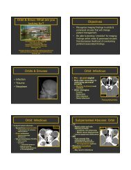
Critical Imaging Diagnoses: - Radiology
Critical Imaging Diagnoses: - Radiology
Critical Imaging Diagnoses: - Radiology

Create successful ePaper yourself
Turn your PDF publications into a flip-book with our unique Google optimized e-Paper software.
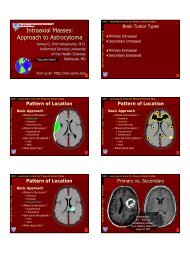
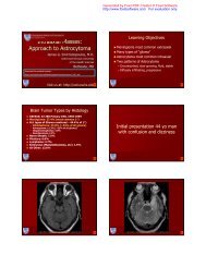
Abscess<br />
Brain Abscess<br />
AA<br />
Peaks<br />
Inverted<br />
AA Peaks<br />
DWI<br />
Short TE MRS<br />
Long TE MRS<br />
Viscous Pus and Coagulation Necrosis<br />
MRS Courtesy of Mauricio Castillo - UNC<br />
DWI: Necrosis vs. PUS<br />
Ring Lesion Differences<br />
GBM<br />
Abscess<br />
“We conclude that viable cell density is the main biological parameter<br />
responsible for restricted diffusion in brain abscess, and it is not<br />
influenced by the etiological agents responsible for its causation.”<br />
Magn. reson. med. 2005, vol. 54, no4, pp. 878-885<br />
GBM<br />
Abscess - Toxo<br />
MR and CT <strong>Imaging</strong> Checklists<br />
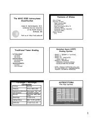
Fluid Secreting Pilocytic Astrocytoma<br />
Nodule<br />
“Cyst”<br />
• Morphologic Features<br />
– Mass Effect<br />
• Yes, proportional<br />
• Less than expected<br />
• No mass effect<br />
– Abnormal WM Signal<br />
• Vasogenic Edema<br />
• Demyelination<br />
• Infiltrating neoplasm<br />
– Ring Lesion<br />
• Necrotic Neoplasm<br />
• Reactive (e.g. abscess)<br />
• Fluid or Inflammatory<br />
5 min.<br />
Neoplasm + thin<br />
rim of enhancing<br />
gliosis