Critical Imaging Diagnoses: - Radiology
Critical Imaging Diagnoses: - Radiology Critical Imaging Diagnoses: - Radiology
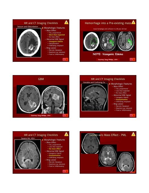
MR and CT Imaging Checklists Seizure and Obtundation • Morphologic Features – Mass Effect • Yes, proportional • More than expected • No mass effect – Abnormal WM Signal • Vasogenic Edema • Demyelination • Infiltrating neoplasm – Ring Lesion • Necrotic Neoplasm • Reactive (e.g. abscess) • Fluid or Inflammatory Hemorrhage into a Pre-existing existing mass Acute hemiplegia and confusion in a 68 year old man NOTE: Vasogenic Edema Courtesy Doug Phillips, UVA GBM MR and CT Imaging Checklists Variable and Confusing Sx • Morphologic Features – Mass Effect • Yes, proportional • Less than expected • No mass effect – Abnormal WM Signal • Vasogenic Edema • Demyelination • Infiltrating neoplasm – Ring Lesion • Necrotic Neoplasm • Reactive (e.g. abscess) • Fluid or Inflammatory Courtesy Doug Phillips, UVA MR and CT Imaging Checklists Severe HA, HIV+ • Morphologic Features – Mass Effect • Yes, proportional • Less than expected • No mass effect – Abnormal WM Signal • Vasogenic Edema • Demyelination • Infiltrating neoplasm – Ring Lesion • Necrotic Neoplasm • Reactive (e.g. abscess) • Fluid or Inflammatory Lesion w/o Mass Effect - PML
T2W – Geographic hyperintensity FLAIR Geographic hyperintensity, no mass Looks like vasogenic edema Courtesy Jacqueline Bello, M.D. Looks like vasogenic edema … but, affects the corpus callosum Courtesy Jacqueline Bello, M.D. T1 w/Gd – No enhancement PML Looks like vasogenic edema … but, no enhancement ! Courtesy Jacqueline Bello, M.D. No Mass, No Enhancement Courtesy Jacqueline Bello, M.D. Progressive Multifocal Leukoencephalopathy • WM Disease - JC Papova/Polyoma Virus – Initials of first patient cultured (1) • John Cunningham • Tx for Hodgkins, , died from PML in 1971 • Lysis of Oligodendrocytes • Demyelination • Geographic and Peripheral – Little or No Mass Effect – Little or No Enhancement • Poor Survival of 2-62 6 months reported • Improved survival w/ HAART - up to 3-43 4 years MR and CT Imaging Checklists Severe HA, HIV+ • Morphologic Features – Mass Effect • Yes, proportional • Less than expected • No mass effect – Abnormal WM Signal • Vasogenic Edema • Demyelination • Infiltrating neoplasm – Ring Lesion • Necrotic Neoplasm • Reactive (e.g. abscess) • Fluid or Inflammatory
- Page 1 and 2: Disclosures Critical Imaging Diagno
- Page 3 and 4: Lymphoma: hyperdense PCNSL FLAIR Lo
- Page 5 and 6: Met Hemoglobin in Sella Region Macr
- Page 7 and 8: MR and CT Imaging Checklists • An
- Page 9 and 10: MR and CT Imaging Checklists • An
- Page 11 and 12: MR and CT Imaging Checklists • An
- Page 13 and 14: 1° CNS Lymphoma 71 yo AA man with
- Page 15 and 16: MR and CT Imaging Checklists MR and
- Page 17: MR and CT Imaging Checklists • Mo
- Page 21 and 22: T1W Gd+ + in Two Astrocytomas Witho
- Page 23 and 24: Fluid Secreting Tumor: Pilocytic As
- Page 25: T1W Gd+ + in Two Astrocytomas Witho
MR and CT <strong>Imaging</strong> Checklists<br />
Seizure and Obtundation<br />
• Morphologic Features<br />
– Mass Effect<br />
• Yes, proportional<br />
• More than expected<br />
• No mass effect<br />
– Abnormal WM Signal<br />
• Vasogenic Edema<br />
• Demyelination<br />
• Infiltrating neoplasm<br />
– Ring Lesion<br />
• Necrotic Neoplasm<br />
• Reactive (e.g. abscess)<br />
• Fluid or Inflammatory<br />
Hemorrhage into a Pre-existing existing mass<br />
Acute hemiplegia and confusion in a 68 year old man<br />
NOTE: Vasogenic Edema<br />
Courtesy Doug Phillips, UVA<br />
GBM<br />
MR and CT <strong>Imaging</strong> Checklists<br />
Variable and Confusing Sx<br />
• Morphologic Features<br />
– Mass Effect<br />
• Yes, proportional<br />
• Less than expected<br />
• No mass effect<br />
– Abnormal WM Signal<br />
• Vasogenic Edema<br />
• Demyelination<br />
• Infiltrating neoplasm<br />
– Ring Lesion<br />
• Necrotic Neoplasm<br />
• Reactive (e.g. abscess)<br />
• Fluid or Inflammatory<br />
Courtesy Doug Phillips, UVA<br />
MR and CT <strong>Imaging</strong> Checklists<br />
Severe HA, HIV+<br />
• Morphologic Features<br />
– Mass Effect<br />
• Yes, proportional<br />
• Less than expected<br />
• No mass effect<br />
– Abnormal WM Signal<br />
• Vasogenic Edema<br />
• Demyelination<br />
• Infiltrating neoplasm<br />
– Ring Lesion<br />
• Necrotic Neoplasm<br />
• Reactive (e.g. abscess)<br />
• Fluid or Inflammatory<br />
Lesion w/o Mass Effect - PML


