Richtlijn: Otitis Externa - Kwaliteitskoepel
Richtlijn: Otitis Externa - Kwaliteitskoepel
Richtlijn: Otitis Externa - Kwaliteitskoepel

Create successful ePaper yourself
Turn your PDF publications into a flip-book with our unique Google optimized e-Paper software.
<strong>Richtlijn</strong>: <strong>Otitis</strong> <strong>Externa</strong><br />
INITIATIEF<br />
Nederlandse Vereniging voor Keel-Neus-Oorheelkunde en Heelkunde van het Hoofd-Halsgebied<br />
MET ONDERSTEUNING VAN<br />
Orde van Medisch Specialisten<br />
FINANCIERING<br />
De richtlijnontwikkeling werd gefinancierd uit de Stichting Kwaliteitsgelden Medisch Specialisten<br />
(SKMS)<br />
1 <strong>Richtlijn</strong> <strong>Otitis</strong> <strong>Externa</strong> 2010<br />
Nederlandse Vereniging voor Keel-Neus-Oorheelkunde en Heelkunde van het Hoofd-Halsgebied
COLOFON<br />
<strong>Richtlijn</strong> <strong>Otitis</strong> <strong>Externa</strong><br />
© 2010 Nederlandse Vereniging voor Keel-Neus-Oorheelkunde en Heelkunde van het Hoofd-<br />
Halsgebied<br />
Mercatorlaan 1200<br />
Domus Medica - Kamer 4C-18<br />
3528 BL Utrecht<br />
kno@kno.nl<br />
Alle rechten voorbehouden. De tekst uit deze publicatie mag worden verveelvoudigd, opgeslagen in<br />
een geautomatiseerd gegevensbestand, of openbaar gemaakt in enige vorm of op enige wijze, hetzij<br />
elektronisch, mechanisch door fotokopieën of enige andere manier, echter uitsluitend na<br />
voorafgaande toestemming van de uitgever. Toestemming voor gebruik van tekst(gedeelten) kunt u<br />
schriftelijk of per e-mail en uitsluitend bij de uitgever aanvragen. Adres en e-mailadres: zie boven.<br />
2 <strong>Richtlijn</strong> <strong>Otitis</strong> <strong>Externa</strong> 2010<br />
Nederlandse Vereniging voor Keel-Neus-Oorheelkunde en Heelkunde van het Hoofd-Halsgebied
SAMENSTELLING VAN DE WERKGROEP<br />
Dr. E.A.M. Mylanus, voorzitter<br />
Prof. R.J. Stokroos<br />
Dr. P. Merkus<br />
Dr. R.J.H. Ensink<br />
De werkgroep werd methodologisch ondersteund door de afdeling Ondersteuning Professionele<br />
Kwaliteit van de Orde van Medisch Specialisten in de personen van ir. T.A. van Barneveld<br />
(afdelingshoofd) en M.M.J. Ploegmakers, MSc (junior adviseur).<br />
De richtlijn betreft een adaptatie van:<br />
Clinical practice guideline: Acute otitis externa. Richard M. Rosenfeld, MD, MPH, Lance Brown, MD,<br />
MPH, C. Ron Cannon, MD, Rowena J. Dolor, MD, MHS, Theodore G. Ganiats, MD, Maureen<br />
Hannley, PhD, Phillip Kokemueller, MS, CAE, S. Michael Marcy, MD, Peter S. Roland, MD, Richard<br />
N. Shiffman, MD, MCIS, Sandra S. Stinnett, DrPH and David L. Witsell, MD, MHS,<br />
Brooklyn, New York; Loma Linda, California; Jackson, Mississippi; Durham, North Carolina; San Diego,<br />
California; Dallas, Texas; New Haven, Connecticut; and Alexandria, Virginia<br />
2006 American Academy of Otolaryngology–Head and Neck Surgery Foundation, Inc.<br />
Otolaryngology–Head and Neck Surgery (2006) 134, S4-S23<br />
3 <strong>Richtlijn</strong> <strong>Otitis</strong> <strong>Externa</strong> 2010<br />
Nederlandse Vereniging voor Keel-Neus-Oorheelkunde en Heelkunde van het Hoofd-Halsgebied
Samenvatting aanbevelingen<br />
Onderstaande is een samenvatting van de aanbevelingen uit de multidisciplinaire evidence-based<br />
klinische richtlijn ‘<strong>Otitis</strong> <strong>Externa</strong>’. In deze samenvatting ontbreken het wetenschappelijk bewijs en de<br />
overwegingen die tot de aanbevelingen geleid hebben. Lezers van deze samenvatting worden voor<br />
deze informatie verwezen naar de volledige richtlijn. Deze samenvatting van aanbevelingen staat niet<br />
op zichzelf. Bij medische besluitvorming dient rekening te worden gehouden met de<br />
omstandigheden en voorkeuren van de patiënt. Behandeling en procedures met betrekking tot de<br />
individuele patiënt berusten op wederzijdse communicatie tussen patiënt, arts en andere<br />
zorgverleners.<br />
Acute otitis externa (AOE) wordt gedefinieerd als roodheid of zwelling van de gehoorgang of debris in<br />
de gehoorgang dat gepaard gaat met pijn, jeuk en/of loopoor en soms met gehoorverlies en een ‘vol’<br />
gevoel in het oor, gedurende minder dan drie weken. Na drie weken persisterende symptomen<br />
noemen we het therapieresistente/persisterende otitis externa. Na drie maanden spreken we van<br />
chronische otitis externa.<br />
Acute otitis externa, Differentiaal diagnose<br />
De diagnose acute otitis externa wordt gesteld op basis van anamnese, inspectie en otoscopie. Het is<br />
belangrijk om onderscheid te maken tussen acute otitis externa en otitis media acuta,<br />
contactdermatitis, huidziekten, furunculose en virale infecties.<br />
Factoren van invloed op ziektebeloop en consequenties voor de behandeling<br />
De behandelend arts dient bij de patiënt met een AOE factoren die de behandeling kunnen<br />
beïnvloeden in kaart te brengen. Er moet worden gedacht aan: radiotherapie in de voorgeschiedenis,<br />
stoornissen in de afweer zoals bij Diabetes Mellitus en een open middenoor. Indien deze factoren<br />
aanwezig zijn is alleen topische behandeling mogelijk onvoldoende en moet er gedacht worden aan<br />
het toevoegen van systemisch antibiotica of in sommige gevallen chirurgie. Afsluiting, manipulatie en<br />
overmatige ontvetting van de gehoorgang dienen vermeden te worden.<br />
Acute otitis externa, pijnbehandeling<br />
De intensiteit van pijn kan het beste anamnestisch bepaald worden. Voor objectivering van de pijn<br />
kan gebruik worden gemaakt van de visual analogue scale (VAS).<br />
Systemische pijnbestrijding, direct starten met paracetamol in een frequente dosering. Zonodig<br />
NSAID’s toevoegen. Bij zeer ernstige pijn kunnen opioiden overwogen worden.<br />
Behandeling<br />
Voor de behandeling van acute otitis externa wordt gekozen voor een topisch preparaat. De topische<br />
medicatie moet ofwel antibiotisch, ofwel antiseptisch zijn, met of zonder steroïden.<br />
Systemische antibiotica worden afgeraden bij patiënten met een goede algemene gezondheid. Bij<br />
(verdenking op) uitbreiding buiten de gehoorgang (osteiitis of abces in middenoor of oorschelp) of bij<br />
patiënten met een onderliggend lijden is aanvullende behandeling met systemische antibiotica wel<br />
geïndiceerd. De (systemische) antibiotica moeten bij nog onbekende verwekker gericht zijn tegen<br />
met name P. Aeruginosa en S. Aureus.<br />
Naast de medische behandeling van acute otitis externa moet de patiënt ook geadviseerd worden<br />
over het vermijden van watercontact en manipulatie van de gehoorgang.<br />
4 <strong>Richtlijn</strong> <strong>Otitis</strong> <strong>Externa</strong> 2010<br />
Nederlandse Vereniging voor Keel-Neus-Oorheelkunde en Heelkunde van het Hoofd-Halsgebied
Keuze oordruppels<br />
De topische behandeling kan antibiotisch of antiseptisch zijn, met of zonder steroïd. Bij een<br />
langdurige laag gedoseerde topische toepassing van een antibioticum moet men zich bewust zijn van<br />
een risico op resistentievorming. Resistentievorming van P.aeruginosa voor quinolonen kan een<br />
ernstig probleem opleveren bij de behamdeling van een ontsteking van het os petrosum.<br />
Bij de keuze voor het type topische behandeling moet rekening worden gehouden met factoren zoals<br />
werkzaamheid tegen de waarschijnlijke verwekkers P. Aeruginosa en S. Aureus, beschikbaarheid,<br />
kosten, compliantie, sterkte van het corticosteroid bij behandeling van kinderen, ototoxiciteit en<br />
contactallergie. Een overzicht van topische preparaten wordt gegeven in bijlage 2, hierbij dient te<br />
worden opgemerkt dat niet alle middelen in bijlage 2 ook zijn geregistreerd voor de behandeling van<br />
otitis externa (otitis media oordruppels en oogdruppels).<br />
Duur van de behandeling<br />
De duur van de behandeling moet zo lang zijn als de symptomen voortbestaan, tenminste 7 dagen.<br />
De initiële behandeling voorschrijven langer dan 14 dagen wordt niet geadviseerd.<br />
Toediening<br />
De patiënt dient goed geïnstrueerd te worden over het toedienen van de topische behandeling. De<br />
patiënt moet een liggende houding aannemen, met het aangedane oor naar boven. Deze houding<br />
dient na toediening nog 3-5 minuten te worden gecontinueerd. De druppels worden bij voorkeur<br />
door iemand anders dan de patiënt toegediend.<br />
Wanneer de gehoorgang geobstrueerd is dient een oortoilet plaats te vinden, eventueel kan er ook<br />
een oortampon worden geplaatst, of beide. Indien een tampon geplaatst wordt, ligt het in de lijn der<br />
verwachting dat ontzwelling binnen 24 tot 48 is opgetreden.<br />
Therapieresistentie<br />
In het geval een acute otitis externa langer dan drie weken aanhoudt, ondanks therapie, dienen de<br />
differentiaaldiagnose (uitgangsvraag 1a) en factoren van invloed op het ziektebeloop/behandeling<br />
(uitgangsvraag 1b) opnieuw in overweging genomen te worden. Daarnaast dienen een aantal<br />
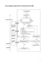
misdiagnosen (uitgangsvraag 7) uitgesloten te worden (zie tabel 1), de aanbevolen strategie wordt<br />
weergegeven in het stroomschema op pagina 6.<br />
Tabel 1. Heroverwegingen betreffende de diagnose en behandeling van OE<br />
Differentiaal diagnose<br />
Acute <strong>Otitis</strong> Media<br />
Contact dermatitis<br />
Dermatose<br />
Furunculose<br />
Virale infectie<br />
Factoren van invloed op<br />
ziektebeloop/behandeling<br />
Radiotherapie in<br />
voorgeschiedenis<br />
Immunogecompromiteerde<br />
patiënt<br />
Open middenoor of buisjes<br />
Misdiagnose<br />
Dermatologische afwijkingen:<br />
- seborrhoїsche dermatitis<br />
- psoriasis<br />
- dermatomycose<br />
(schimmel/gistinfecties van<br />
de huid)<br />
- acne<br />
5 <strong>Richtlijn</strong> <strong>Otitis</strong> <strong>Externa</strong> 2010<br />
Nederlandse Vereniging voor Keel-Neus-Oorheelkunde en Heelkunde van het Hoofd-Halsgebied
- folliculitis<br />
Cholesteatoom van de gehoorgang<br />
of middenoor; chronische<br />
suppuratieve otitis media<br />
Maligne/necrotiserende otitis<br />
externa<br />
Gehoorgangcarcinoom<br />
Chirurgie<br />
Bij een patiënt met een chronische otitis externa is chirurgie, op geleide van inspectie en otoscopie,<br />
de behandeling van eerste keus.<br />
6 <strong>Richtlijn</strong> <strong>Otitis</strong> <strong>Externa</strong> 2010<br />
Nederlandse Vereniging voor Keel-Neus-Oorheelkunde en Heelkunde van het Hoofd-Halsgebied
Figuur 1. Flowdiagram persisterende OE<br />
7 <strong>Richtlijn</strong> <strong>Otitis</strong> <strong>Externa</strong> 2010<br />
Nederlandse Vereniging voor Keel-Neus-Oorheelkunde en Heelkunde van het Hoofd-Halsgebied
Inhoud<br />
Samenvatting aanbevelingen .................................................................................................................. 4<br />
Inhoud ..................................................................................................................................................... 8<br />
Achtergrond otitis externa .................................................................................................................. 9<br />
Doelstelling .......................................................................................................................................... 9<br />
Acute <strong>Otitis</strong> <strong>Externa</strong>, Differentiaal diagnose ......................................................................................... 13<br />
Factoren van invloed op ziektebeloop en consequenties voor de behandeling ................................... 16<br />
Pijnbehandeling ..................................................................................................................................... 18<br />
Behandeling ........................................................................................................................................... 21<br />
Keuze oordruppels ................................................................................................................................ 25<br />
Duur van de behandeling ...................................................................................................................... 29<br />
Toediening ............................................................................................................................................. 30<br />
Therapieresistentie................................................................................................................................ 33<br />
Chirurgie ................................................................................................................................................ 38<br />
Bijlage 1 Zoekverantwoording ............................................................................................................... 41<br />
Bijlage 2 Tabel topische druppels .......................................................................................................... 42<br />
8 <strong>Richtlijn</strong> <strong>Otitis</strong> <strong>Externa</strong> 2010<br />
Nederlandse Vereniging voor Keel-Neus-Oorheelkunde en Heelkunde van het Hoofd-Halsgebied
Achtergrond otitis externa<br />
Definitie:<br />
<strong>Otitis</strong> externa is een diffuse ontsteking van de huid van de gehoorgang die gepaard kan gaan met<br />
pijn, jeuk, afscheiding, schilfering, roodheid, zwelling en eventueel gehoorverlies. <strong>Otitis</strong> externa<br />
wordt in deze richtlijn onderverdeeld in drie fases; (1) acute otitis externa, (2) persisterende en (3)<br />
chronische otitis externa. Uitgangsvraag 1 tot en met 6 gaan over de acute fase, vraag 7 betreft<br />
persisterende otitis externa en de laatste vraag gaat over chronische otitis externa.<br />
Aandoeningen van het trommelvlies, zoals myringitis bullosa en myringitis granulomatosa, die<br />
eenzelfde klachtenpatroon met zich mee kunnen brengenworden niet in deze richtlijn meegenomen.<br />
Daarnaast wordt secundaire otitis externa, als gevolg van chronische suppuratieve otitis media<br />
buiten beschouwing gelaten.<br />
De huisarts kan otitis externa meestal zelf diagnosticeren en behandelen; slechts een klein aantal<br />
patiënten met otitis externa wordt verwezen. De prognose van otitis externa is doorgaans goed:<br />
meer dan driekwart van de patiënten is na drie weken behandeling klachtenvrij.<br />
De incidentie van acute otitis externa in de huisartsenpraktijk is 14 per 1000 patiënten per jaar 2 . In de<br />
zomer is de incidentie het hoogst, hetgeen voornamelijk aan zwemmen wordt geweten. Ook door de<br />
KNO-artst wordt de diagnose otitis externa frequent gesteld. Tussen de 3 en 10% van de patiënten in<br />
de KNO-praktijk presenteert zich met een otitis externa 1 . De indicaties voor een verwijzing zijn in de<br />
NHG-standaard 2 duidelijk beschreven. Doorgaans betreft het patiënten met diabetes mellitus of<br />
verminderde weerstand met een ernstige acute otitis externa, patiënten met een otitis externa met<br />
uitbreiding naar de omgeving of andere complicaties of patiënten met een chronische, therapieresistente<br />
otitis externa.<br />
Doelstelling<br />
De Amerikaanse multidisciplinair ontwikkelde richtlijn ‘Clinical practice guideline: Acute <strong>Otitis</strong><br />
<strong>Externa</strong>’ van de American Association of Otolaryngology – Head and Neck Surgery Foundation (AAO-<br />
HNSF) vormde de basis voor deze richtlijn. Het Nederlands Huisartsen Genootschap (NHG) bracht in<br />
2005 de herziene NHG-standaard ‘<strong>Otitis</strong> <strong>Externa</strong>’ uit, hieruit zijn gegevens over de Nederlandse<br />
situatie geëxtraheerd. Onze doelstelling was om de Amerikaanse multidisciplinaire richtlijn te<br />
adapteren aan de Nederlandse situatie met behulp van de kennis van Nederlandse experts. Bij het<br />
formuleren van de aanbevelingen is uitgegaan van wetenschappelijk bewijs waarbij specifieke<br />
aandacht is besteed aan benefit-harm balans. Waar onvoldoende wetenschappelijk bewijs<br />
beschikbaar was, zijn aanbevelingen gebaseerd op consensus tussen de experts . De aanbevelingen<br />
uit deze richtlijn kunnen gebruikt worden om indicatoren te ontwikkelen ten behoeve van<br />
kwaliteitsverbetering.<br />
Doelgroep<br />
Deze richtlijn is bedoeld voor medisch specialisten in de tweede lijn die in hun klinische praktijk in<br />
aanraking komen met otitis externa: KNO-artsen, kinderartsen, internisten, SEH-artsen, physicianassistants<br />
en nurse-practitioners. De richtlijn is van toepassing op alle settings waar otitis externa<br />
gediagnostiseerd en behandeld wordt, bij kinderen, adolescenten en volwassenen. De richtlijn werd<br />
1 Bojrab DI, Bruderly T, Abdulrazzak Y. <strong>Otitis</strong> externa. Otolaryngol Clin North Am 1996;29:761-82.<br />
2 Rooijackers-Lemmens E, Balen FAM van, Opstelten W, Wiersma Tj. NHG-standaard <strong>Otitis</strong> externa. Eerste<br />
herziening. In: Wiersma Tj, Goudswaard AN, redacteuren. NHG-standaarden voor de huisarts. Versie 2006.<br />
Houten: Bohn Stafleu van Loghum; 2006. p. 885-93.<br />
9 <strong>Richtlijn</strong> <strong>Otitis</strong> <strong>Externa</strong> 2010<br />
Nederlandse Vereniging voor Keel-Neus-Oorheelkunde en Heelkunde van het Hoofd-Halsgebied
niet ontwikkeld voor eerstelijns zorg. Echter, het is waarschijnlijk dat de aanbevelingen ook voor de<br />
huisartsenpraktijk van toepassing zijn.<br />
Werkwijze werkgroep<br />
De Amerikaanse richtlijn ‘Clinical practice guideline: Acute otitis externa’ vormde het uitgangspunt<br />
van de onderhavige richtlijn. Dit betekent dat voor wat betreft de wetenschappelijke onderbouwing<br />
de Nederlandse richtlijncommissie de studies, de beoordeling en gradering ervan en de begeleidende<br />
tekst heeft overgenomen. Studies die nadien werden gepubliceerd konden in de richtlijncommissie<br />
worden ingebracht, waarbij de volgende selectiecriteria werden gehanteerd: reviews en RCT’s<br />
betreffende otitis externa.<br />
De richtlijncommissie is voor elke aanbeveling in de Amerikaanse richtlijn nagegaan welke<br />
overwegingen naast het wetenschappelijk bewijs zijn gebruikt en of de door de commissie<br />
aangedragen studies de aanbeveling zouden kunnen veranderen. Wanneer er consensus was over<br />
deze overwegingen en door de commissie aangedragen studies geen ander inzicht opleverden, zijn<br />
de aanbevelingen overgenomen. Indien de commissie andere overwegingen (ook) van belang achtte<br />
of meende dat de door haar aangedragen studies een (iets) ander licht wierpen op de in de<br />
Amerikaanse richtlijn vermelde aanbeveling, zijn de aanbevelingen gemodificeerd. De<br />
wetenschappelijke onderbouwing wordt in deze richtlijn in het Engels weergegeven, de<br />
uitgangsvragen, overwegingen en aanbevelingen zijn in het Nederlands geformuleerd.<br />
De gradering van de studies in de Amerikaanse richtlijn wijkt af van wat hier te lande gangbaar is.<br />
Vanuit het oogpunt van uniformiteit achtte de Nederlandse commissie het wenselijk de classificatie<br />
van bewijs c.q. gradering te converteren naar de Nederlandse classificatie. De Amerikaanse<br />
classificatie is hieronder afgebeeld. De corresponderende “Nederlandse” classificatie is in tabel 1<br />
opgenomen.<br />
Tabel 1. Relatie tussen Evidence quality for grades of evidence en Niveau van conclusie op basis van<br />
kwaliteit van bewijs.<br />
Evidence Evidence Quality - Niveau Niveau van conclusie omschrijving<br />
Quality<br />
- symbool<br />
omschrijving<br />
van<br />
conclusie<br />
–<br />
symbool<br />
A Well-designed randomized 1 Meerdere gerandomiseerde dubbelblinde ver-<br />
10 <strong>Richtlijn</strong> <strong>Otitis</strong> <strong>Externa</strong> 2010<br />
Nederlandse Vereniging voor Keel-Neus-Oorheelkunde en Heelkunde van het Hoofd-Halsgebied
B<br />
C<br />
D<br />
controlled trials or<br />
diagnostic studies<br />
performed on a population<br />
similar to the guideline’s<br />
target population<br />
Randomized controlled trials<br />
or diagnostic studies with<br />
minor limitations;<br />
overwhelmingly consistent<br />
evidence from observational<br />
studies<br />
Observational studies (casecontrol<br />
and cohort design)<br />
Expert opinion, case reports,<br />
reasoning from first<br />
principles (bench research<br />
or animal studies)<br />
gelijkende klinisch onderzoeken van goede<br />
kwaliteit van voldoende omvang, of<br />
Meerdere onderzoeken ten opzichte van een<br />
referentietest (een ‘gouden standaard’) met<br />
tevoren gedefinieerde afkapwaarden en<br />
onafhankelijke beoordeling van de resultaten<br />
van test en gouden standaard, betreffende een<br />
voldoende grote serie van opeenvolgende<br />
patiënten die allen de index- en referentietest<br />
hebben gehad<br />
2 Meerdere vergelijkende onderzoeken, maar niet<br />
met alle kenmerken als genoemd onder 1<br />
(hieronder valt ook patiënt-controle onderzoek,<br />
cohort-onderzoek), of<br />
Meerdere onderzoeken ten opzichte van een<br />
referentietest, maar niet met alle kenmerken die<br />
onder 1 zijn genoemd.<br />
3 en 4 Niet vergelijkend-onderzoek of mening van<br />
deskundigen<br />
In de Amerikaanse richtlijn worden ook de aanbevelingen gegradeerd in termen van ‘strong<br />
recommendation’, ‘recommendation’, ‘option’. Hier te lande is graderen van aanbevelingen niet<br />
gebruikelijk. Om deze reden zijn in de Nederlandse richtlijn de aanbevelingen niet gegradeerd.<br />
Juridische betekenis van richtlijnen<br />
<strong>Richtlijn</strong>en zijn geen wettelijke voorschriften, maar bevatten expliciete, zo veel mogelijk op evidence<br />
gebaseerde aanbevelingen en inzichten waaraan zorgverleners zouden moeten voldoen om<br />
kwalitatief optimale zorg te verlenen. Aangezien deze aanbevelingen hoofdzakelijk gericht zijn op de<br />
‘gemiddelde patiënt’, kunnen zorgverleners op basis van individuele patiëntkenmerken zo nodig<br />
afwijken van de richtlijn. Afwijken van richtlijnen is, als de situatie van de individuele patiënt dat<br />
vereist, soms zelfs noodzakelijk. Een richtlijn kan worden gezien als een papieren weergave van een<br />
best practice. Als van de richtlijn wordt afgeweken, is het raadzaam dit gedocumenteerd en<br />
beargumenteerd te doen.<br />
Financiën en belangenverstrengeling<br />
De kosten voor de ontwikkeling van deze richtlijn, zijn betaald uit de Stichting Kwaliteitsgelden<br />
Medisch Specialisten. De industrie werd op geen enkele wijze bij het ontwikkelingsproces betrokken.<br />
Belangenverklaringen werden door alle werkgroepleden ingevuld. Er zijn geen bijzondere vormen<br />
van belangenverstrengeling gemeld. Een map met verklaringen van werkgroepleden over mogelijke<br />
financiële belangenverstrengeling ligt ter inzage bij de afdeling Ondersteuning Professionele Kwaliteit<br />
van de Orde van Medisch Specialisten.<br />
Herziening<br />
De richtlijn zal periodiek worden getoetst aan de wetenschappelijke ontwikkelingen door een (nog<br />
samen te stellen) commissie. De commissie draagt de verantwoordelijkheid om tussentijdse<br />
peilingen bij de beroepsgroepen te verrichten naar behoefte voor herziening(en) van de huidige<br />
richtlijn. Bij essentiële ontwikkelingen kan er besloten worden tussentijdse amendementen te maken<br />
11 <strong>Richtlijn</strong> <strong>Otitis</strong> <strong>Externa</strong> 2010<br />
Nederlandse Vereniging voor Keel-Neus-Oorheelkunde en Heelkunde van het Hoofd-Halsgebied
en deze digitaal onder de verschillende beroepsgroepen te verspreiden. Zo nodig wordt een nieuwe<br />
werkgroep geïnstalleerd om (delen van) de richtlijn te herzien. Uiterlijk in 2015 zal een nieuwe<br />
werkgroep worden geïnstalleerd die de richtlijn volledig zal herzien.<br />
12 <strong>Richtlijn</strong> <strong>Otitis</strong> <strong>Externa</strong> 2010<br />
Nederlandse Vereniging voor Keel-Neus-Oorheelkunde en Heelkunde van het Hoofd-Halsgebied
Acute <strong>Otitis</strong> <strong>Externa</strong>, Differentiaal diagnose<br />
Uitgangsvraag 1a<br />
Hoe wordt de diagnose acute otitis externa gesteld?<br />
Onderbouwing<br />
A diagnosis of diffuse AOE requires rapid onset with signs and symptoms of ear canal inflammation.<br />
Symptoms of AOE include otalgia (70%), itching (60%), or fullness (22%), with or without hearing loss<br />
(32%) or ear canal pain on chewing. A hallmark sign of diffuse AOE is tenderness of the tragus (when<br />
pushed), pinna (when pulled up and back), or both. The tenderness is often intense and<br />
disproportionate to what might be expected based on visual inspection. Otoscopy will reveal diffuse<br />
ear canal edema, erythema, or both, either with or without otorrhea or material in the ear canal.<br />
Regional lymphadenitis or cellulitis of the pinna and adjacent skin may be present in some patients<br />
(Agius 1992, Lucente 1995).<br />
Anything that disrupts the epithelium of the ear canal can permit invasion by bacteria that cause<br />
diffuse AOE. Common predisposing factors for AOE (Hirsch 1992) are humidity or prolonged<br />
exposure to water, dermatologic conditions (eczema, seborrhea, psoriasis, folliculitis), anatomic<br />
abnormalities (narrow canal, exostoses), trauma or external devices (wax removal, insertion of<br />
earplugs, use of hearing aids), and otorrhea caused by middle-ear disease. AOE may also occur as a<br />
result of ear canal obstruction by impacted cerumen, a foreign object, or a dermoid or sebaceous<br />
cyst. Clinical history should identify predisposing factors and assess swimming behavior. Other causes<br />
of otalgia, otorrhea, and inflammation should be distinguished from diffuse AOE because<br />
management will differ. The most important differential diagnoses are summed up below.<br />
Acute <strong>Otitis</strong> Media<br />
AOE can mimic the appearance of acute otitis media (AOM) because of erythema that involve the<br />
tympanic membrane. Distinguishing AOE from AOM is important because the latter may require<br />
systemic antimicrobials (Lieberthal 2004).<br />
Pneumatic otoscopy will demonstrate good tympanic membrane mobility with AOE but will show<br />
absent or limited mobility with AOM and associated middle-ear effusion. Similarly, tympanometry<br />
will show a normal peaked curve (type A) with AOE, but a flat tracing (type B) with AOM. The validity<br />
of acoustic reflectometry with AOE is unknown.<br />
Contact dermatitis<br />
Contact dermatitis of the ear canal is an allergic reaction to antigens such as metals (nickel, silver),<br />
chemicals (cosmetics, soaps, detergents, shampoo, hairspray), plastics, rubber, leather, or drugs.<br />
Fragrance additives may also cause similar reactions. Finally, contact sensitivity may be caused by<br />
silicone ear plugs or by hearing-aid molds that contain silicone or methyl-methacrylate. Nickel is the<br />
most common contact allergen, affecting around 10% of women with pierced ears (Peltonen 1981,<br />
Rudner 1973, Larsson-Styme 1985). Contact allergy also occurs in some patients who wear hearing<br />
aids as a reaction to the plastics and other chemicals used in hearing aid molds (Meding 1992,<br />
Cockerill 1987).<br />
13 <strong>Richtlijn</strong> <strong>Otitis</strong> <strong>Externa</strong> 2010<br />
Nederlandse Vereniging voor Keel-Neus-Oorheelkunde en Heelkunde van het Hoofd-Halsgebied
Contact sensitivity of the external auditory canal can result in refractory AOE in some patients.<br />
Delayed-type hypersensitivity reactions to topical antiseptic otic preparations are characterized by<br />
severe pruritus, skin inflammation, edema of the external auditory canal, and persistent otorrhea;<br />
blisters and vesicles may be present. The allergic reaction can extend beyond the ear canal to involve<br />
the skin around the ear and the neck. Neomycin-containing eardrops are most commonly noted to<br />
cause contact sensitivity, which has a 13% to 30% prevalence on patch testing of patients with<br />
chronic otitis externa (Sood 2002, Devos 2000, Rutka 2004). Contact sensitivity of the ear canal may<br />
also result from other topical antimicrobials (bacitracin, quinolones, gentian violet, polymyxin B<br />
sulfate), topical steroid preparations (hydrocortisone, triamcinolone), or topical anesthetics<br />
(benzocaine alone, or combined with dibucaine and tetracaine [caine mix]). Preservatives in topical<br />
otic preparations associated with at least a 1% incidence of contact sensitivity include propylene<br />
glycol, thimerosal, benzalkonium chloride, benzethonium chloride, and methyl-p-oxybenzoate (Sood<br />
2002, Devos 2000, Rutka 2004).<br />
Dermatose<br />
Eczema, seborrhea, and other inflammatory dermatoses that involve the ear canal and surrounding<br />
tissues are relatively common and may predispose to acute infection.<br />
Furunculosis<br />
Furunculosis is the presence of an infected hair follicle on the outer third of the ear canal, sometimes<br />
referred to as localized otitis externa. Clinical findings include otalgia, otorrhea, and localized<br />
tenderness. Treatment may include local heat, incision and drainage, or systemic antibiotics that<br />
cover S aureus, the most common causative agent.<br />
Viral infections<br />
Viral infections of the external ear, caused by varicella, measles, or herpesvirus, are rare. Herpes<br />
zoster oticus (Ramsay Hunt syndrome) causes vesicles on the external ear canal and posterior surface<br />
of the auricle, severe otalgia, facial paralysis or paresis, loss of taste on the anterior two-thirds of the<br />
tongue, and decreased lacrimation on the involved side (Kuhweide 2002). Management involves<br />
antiviral therapy, with or without systemic steroid.<br />
Conclusie<br />
Level of Evidence 3<br />
Observational studies<br />
Elements of the diagnosis of diffuse acute otitis externa<br />
1. Rapid onset (generally within 48 hours) in the past 3 weeks<br />
AND<br />
2. Symptoms of ear canal inflammation that include:<br />
• otalgia (often severe), itching, or fullness,<br />
• WITH OR WITHOUT hearing loss or jaw pain*<br />
AND<br />
3. Signs of ear canal inflammation that include:<br />
• tenderness of the tragus, pinna, or both<br />
• OR diffuse ear canal edema, erythema, or both<br />
• WITH OR WITHOUT otorrhea, regional lymphadenitis,<br />
tympanic membrane erythema, or cellulitis of the pinna and<br />
adjacent skin<br />
*Pain in the ear canal and temporomandibular joint region intensified by jaw motion.<br />
14 <strong>Richtlijn</strong> <strong>Otitis</strong> <strong>Externa</strong> 2010<br />
Nederlandse Vereniging voor Keel-Neus-Oorheelkunde en Heelkunde van het Hoofd-Halsgebied
Overwegingen<br />
Het komt wel eens voor dat patiënten een dusdanig gezwollen gehoorgang hebben dat het niet<br />
mogelijk is om de diagnose direct te stellen. Het plaatsen van een tampon doordrenkt met<br />
corticosteroïd en azijnzuuroplossing en herevaluatie binnen enkele dagen wordt geadviseerd.<br />
Aanbeveling<br />
De diagnose acute otitis externa wordt gesteld op basis van anamnese, inspectie en otoscopie. Het is<br />
belangrijk om onderscheid te maken tussen acute otitis externa en otitis media acuta,<br />
contactdermatitis, huidziekten, furunculose en virale infecties.<br />
Referenties<br />
• Agius AM, Pickles JM, Burch KL. A prospective study of otitis externa. Clin Otolaryngol<br />
1992;17:150–4.<br />
• Cockerill D. Allergies to ear moulds. Br J Audiol 1987;21:143–5.<br />
• Devos SA, Mulder JJ, van der Valk PG. The relevance of positive patch test reactions in<br />
chronic otitis externa. Contact Dermatitis 2000;42:354 –5.<br />
• Hirsch BE. Infections of the external ear. Am J Otolaryngol 1992;13:145–55.<br />
• Kuhweide R, Van de Steene V, Vlaminck S et al. Ramsay Hunt syndrome: pathophysiology of<br />
cochleovestibular symptoms. J Laryngol Otol 2002;116:844–8.<br />
• Larsson-Styme B, Widstrom L. Ear piercing: a cause of nickel allergy in schoolgirls? Contact<br />
Dermatitis 1985;13:268 –93.<br />
• Lieberthal AS, Ganiats TG, Cox EO, et al. Clinical practice guideline: American Academy of<br />
Pediatrics Subcommittee on Management of Acute <strong>Otitis</strong> Media: diagnosis and management<br />
of acute otitis media. Pediatrics 2004;113:1451– 65.<br />
• Lucente FE, Lawson W, Novick NL. <strong>Externa</strong>l ear. Philadelphia: WB Saunders Co; 1995.<br />
• Meding B, Ringdahl A. Allergic contact dermatitis from the earmoulds of hearing aids. Ear<br />
Hear 1992;13:122– 4.<br />
• Peltonen L. Nickel sensitivity: an actual problem. Int J Dermatol 1981;20:352–3.<br />
• Rudner EF, Clendenning WE, Epstein E. Epidemiology of contact dermatitis in North America:<br />
1972. Arch Dermatol 1973;108:537– 40.<br />
• Schapowal A. Contact dermatitis to antibiotic ear drops is due to neomycin but not to<br />
ciprofloxacin [abstract]. Allergy 2001;56(suppl 68):148.<br />
• Smith IM, Kaey DG, Buxton PK. Chronic hypersensitivity in patients with chronic otitis<br />
externa. Clin Otolaryngol 1990;15:155– 8.<br />
• Sood S, Strachan DR, Tsikoudis A, et al. Allergic otitis externa. Clin Otolaryngol Allied Sci<br />
2002;27:233–36.<br />
• Rutka J. Acute otitis externa: treatment perspectives. Ear Nose Throat J 2004;83(suppl 4):20 –<br />
2.<br />
15 <strong>Richtlijn</strong> <strong>Otitis</strong> <strong>Externa</strong> 2010<br />
Nederlandse Vereniging voor Keel-Neus-Oorheelkunde en Heelkunde van het Hoofd-Halsgebied
Factoren van invloed op ziektebeloop en consequenties voor de<br />
behandeling<br />
Uitgangsvraag 1b<br />
Welke factoren kunnen het ziektebeloop en de behandeling van acute otitis externa beïnvloeden?<br />
Onderbouwing<br />
Key components of the clinical history that can modify management of diffuse AOE include:<br />
History of radiotherapy<br />
Radiotherapy can damage the external ear by causing acute and late skin reactions that involve the<br />
pinna, external canal, and periauricular region (Jereczek-Fossa 2004). Acute events include erythema,<br />
desquamation, or ulceration of the auricle and ear canal, thus leading to pain and otorrhea. Late skin<br />
changes include atrophy, necrosis or ulceration, external otitis, and external canal stenosis. Damage<br />
to the epithelium of sebaceous and apocrine glands can diminish cerumen secretion. Management of<br />
AOE in patients after radiotherapy may require systemic antimicrobials.<br />
Immunocompromised state<br />
In patients with a disease which may have an effect on their immune system, such as diabetes and<br />
HIV infection, infections of the ear may have a protracted course. Otomycosis or fungal infection of<br />
the external ear canal is seen more frequently in those patients. Aspergillus species (60% to 90%) and<br />
Candida species (10% to 40%) are often cultured (Kaur 2000). Symptoms include pruritus and<br />
thickened otorrhea, which may be black, gray, bluish green, yellow, or white. Candidal otitis externa<br />
results in white debris sprouting hyphae, best seen with an otologic microscope. Aspergillus niger<br />
appears as a moist white plug dotted with black debris (“wet newspaper”) (Ruckenstein 2005).<br />
Management may include debridement plus topical antifungal therapy, systemic antifungal therapy,<br />
or both.<br />
Open middle ear cavity and the presence of tympanostomy tubes<br />
Middle-ear disease can modify treatment of AOE. Patients with a tympanostomy tube or tympanic<br />
membrane perforation may develop diffuse AOE because of purulent middle-ear secretions that<br />
enter the ear canal. Clinicians should prescribe a non-ototoxic topical preparation when the tympanic<br />
membrane is not intact. Management of the underlying middle ear disease may also require systemic<br />
antimicrobials, imaging studies, or surgery.<br />
Conclusies<br />
Level of Evidence 3<br />
Jereczek-Fossa 2004, Kaur<br />
2000, Ruckenstein 2005<br />
Treatment can be influenced by radiotherapy, an<br />
immunocompromised state and an open middle ear cavity<br />
16 <strong>Richtlijn</strong> <strong>Otitis</strong> <strong>Externa</strong> 2010<br />
Nederlandse Vereniging voor Keel-Neus-Oorheelkunde en Heelkunde van het Hoofd-Halsgebied
Overwegingen<br />
Het is belangrijk dat de behandelend arts zich realiseert dat andere patiëntgerelateerde factoren<br />
zoals afsluiting van de gehoorgang door een hoortoestel, manipulatie van de gehoorgang<br />
(rechtstreeks, dan wel wrijven van de oorschelp of tragus), overmatige ontvetting van de gehoorgang<br />
met zeep en shampoo en zwemmen, de behandeling eveneens kunnen beïnvloeden.<br />
Aanbeveling<br />
De behandelend arts dient bij de patiënt met een AOE factoren die de behandeling kunnen<br />
beïnvloeden in kaart te brengen. Er moet worden gedacht aan: radiotherapie in de voorgeschiedenis,<br />
een afweerstoornis en een open middenoor. Indien deze factoren aanwezig zijn is alleen topische<br />
behandeling mogelijk onvoldoende en moet er gedacht worden aan het toevoegen van systemisch<br />
antibiotica of in sommige gevallen chirurgie. In het algemeen moeten afsluiting, manipulatie en<br />
overmatige ontvetting van de gehoorgang vermeden worden.<br />
Referenties<br />
• Grandis Rubin J, Branstetter BF 4th, Yu VL. The changing face of malignant (necrotizing)<br />
external otitis: clinical, radiological, and anatomic correlations. Lancet Infect Dis 2004;4:34 –<br />
9.<br />
• Hellier Ismail H. WP, Batty V. Use of magnetic resonance imaging as the primary imaging<br />
modality in the diagnosis and follow-up of malignant external otitis. J Laryngol Otol<br />
2004;18:576 –9.<br />
• Jereczek-Fossa BA, Zarowski A, Milani F, et al. Radiotherapy-induced ear toxicity. Cancer<br />
Treat Rev 2003;29:417–30.<br />
• Kaur R. Mittal N, Kakkar M, et al. Otomycosis: a clinicomycologic study. Ear Nose Throat J<br />
2000;79:606 –9.<br />
• Lucente FE, Lawson W, Novick NL. <strong>Externa</strong>l ear. Philadelphia: WB Saunders Co; 1995.<br />
• Ruckenstein MJ. Infections of the external ear. In Cummings CW Jr (ed). Otolaryngology:<br />
Head and Neck Surgery, 4th ed. Philadelphia: Mosby; 2005: p. 2979-87.<br />
17 <strong>Richtlijn</strong> <strong>Otitis</strong> <strong>Externa</strong> 2010<br />
Nederlandse Vereniging voor Keel-Neus-Oorheelkunde en Heelkunde van het Hoofd-Halsgebied
Pijnbehandeling<br />
Uitgangsvraag 2<br />
Wat is de optimale pijnbehandeling van patiënten met een acute otitis externa?<br />
Onderbouwing<br />
Pain relief is a major goal in the management of AOE. Frequent use of analgesics is often necessary to<br />
permit patients to achieve comfort, rest, and to resume normal activities (Schechter 1993, Joint<br />
Commission on Accreditation of Health Care Organizations 2001, American Academy of Pediatrics<br />
2001). Ongoing assessment of the severity of discomfort is essential for proper management. Use of<br />
a faces (Bieri 1990), Oucher (Beyer 1998), or visual analog (Powell 2001) scale may help determine<br />
the level of pain, particularly for children and non-English speaking patients.<br />
Systemic<br />
Adequate pain control requires knowing the dose, timing, routes of delivery, and possible adverse<br />
effects of an analgesic (Schechter 1993, Joint Commission on Accreditation of Health Care<br />
Organizations 2001, American Academy of Pediatrics 2001, Loesser 2001). Mild to moderate pain<br />
usually responds to paracetamol or nonsteroidal anti-inflammatory drugs (NSAID’s) given alone or in<br />
fixed combination with an opioid. Administering a nonsteroidal anti-inflammatory drug during the<br />
acute phase of diffuse AOE significantly reduces pain compared with placebo (Valencia 1987).<br />
Topical<br />
There are no clinical trials that show efficacy on pain relief of topical opioids in AOE, and the use of<br />
these drops may mask progression of underlying disease while pain is being suppressed. Topical<br />
benzocaine may cause contact dermatitis that can worsen or prolong AOE (Lucente 1995). If a topical<br />
anesthetic drop is prescribed for temporary pain relief, the patient should be reexamined within 48<br />
hours to ensure that AOE has responded appropriately to primary therapy. The addition of a topical<br />
steroid to topical antimicrobial drops has been shown to hasten pain relief in some randomized trials<br />
(Pistorius 1999, van Balen 1993, Roland 2007), but others have shown no significant benefits (Slack<br />
1987, Pfifidis 2005, Schwartz 2006).<br />
Nonpharmacologic therapies such as heat or cold, relaxation, and distraction are unproven.<br />
Conclusies<br />
Level of evidence 2<br />
Valencia 1987<br />
Level of evidence 4<br />
Expert opinion<br />
Level 1<br />
Pistorius 1999, van Balen<br />
1993, Roland 2007 Slack 1987,<br />
Pfifidis 2005, Schwartz 2006<br />
Mild to moderate pain usually responds to paracetamol or<br />
nonsteroidal anti-inflammatory drugs (NSAID’s).<br />
It is plausible that paracetamol and NSAID’s relieve pain caused by<br />
otitis externa.<br />
There is conflicting evidence regarding the effects of the addition<br />
of a topical steroid to topical antimicrobial drops on pain relief.<br />
18 <strong>Richtlijn</strong> <strong>Otitis</strong> <strong>Externa</strong> 2010<br />
Nederlandse Vereniging voor Keel-Neus-Oorheelkunde en Heelkunde van het Hoofd-Halsgebied
Overwegingen<br />
Het starten van de behandeling van otitis externa op zich draagt bij aan de pijnbestrijding.<br />
Vanuit het oogpunt van pijnbestrijding is de werkgroep van mening dat een steroïd als bestanddeel<br />
van de topische behandeling een zinvolle bijdrage levert.<br />
Topische analgetica kunnen een onderliggende aandoening maskeren. Daarnaast geeft het<br />
toedienen van een lokaal analgeticum bij een open trommelvlies het risico op een tijdelijke<br />
facialisparese en ototoxiciteit.<br />
Een goed beleid voor pijnbestrijding wordt beschreven in de richtlijn Postoperatieve pijn van de<br />
Nederlandse Vereniging voor Anesthesiologie en de <strong>Richtlijn</strong> Pijnbehandeling van kinderen van de<br />
Nederlandse Vereniging voor Kindergeneeskunde.<br />
Aanbeveling<br />
De intensiteit van pijn kan het beste anamnestisch bepaald worden. Voor objectivering van de pijn<br />
kan gebruik worden gemaakt van de visual analogue scale (VAS).<br />
Systemische pijnbestrijding, direct starten met paracetamol in een frequente dosering. Zonodig<br />
NSAID’s toevoegen. Bij zeer ernstige pijn kunnen opioiden overwogen worden.<br />
Referenties<br />
• American Academy of Pediatrics/American Pain Society. The assessment and management of<br />
acute pain in infants, children, and adolescents. Pediatrics 2001;108:793–7.<br />
• American Academy of Pediatrics. Report of the subcommittee on the management of pain<br />
associated with procedures in children with cancer. Pediatrics 1990;86:826 –31.<br />
• Balen, van FAM, Smit WM, Zuithoff NPA, et al. Clinical efficacy of three common treatments<br />
in acute otitis externa in primary care: randomised controlled trial. BMJ 2003; 327:1201–3.<br />
• Beyer JE, Knott CB. Construct validity estimation for the African-American and Hispanic<br />
versions of the Oucher scale. J Pediatr Nurs 1998;13:20 –31.<br />
• Bieri D, Reeve RA, Champion G D, et al The Faces Pain Scale for the self-assessment of the<br />
severity of pain experienced by children: development, initial validation, and preliminary<br />
investigation for ratio scale properties. Pain 1990;41:139 –50.<br />
• Joint Commission on Accreditation of Health Care Organizations. Pain: current understanding<br />
of assessment, management and treatments. National Pharmaceutical Council & JCAHO,<br />
2001. Accessed 8/22/2005 at: www.JCAHO.org/.<br />
• Loesser JD, ed. Bonica’s management of pain, 3rd ed. Baltimore, MD: Lippincott Williams and<br />
Wilkins, 2001.<br />
• Lucente FE, Lawson W, Novick NL. <strong>Externa</strong>l ear. Philadelphia: WB Saunders Co; 1995.<br />
• Pistorius B, Westberry K, Drehobl, et al. Prospective, randomized, comparative trial of<br />
ciprofloxacin otic drops, with or without hydrocortisone, vs. polymyxin B-neomycinhydrocortisone<br />
otic suspension in the treatment of acute diffuse otitis externa. Infect Dis Clin<br />
Pract 1999;8:387–95.<br />
• Powell CV, Kelly A M, Williams A. Determining the minimum clinically significant difference in<br />
visual analog pain score for children. Ann Emerg Med 2001;37:28 –31.<br />
• Premachandra DJ. Use of EMLA cream as an analgesic in the management of painful otitis<br />
externa. J Laryngol Otol 1990;104: 887–8.<br />
• Psifidis A, Nikolaidis P, Tsona A, et al. The efficacy and safety of local ciprofloxacin in patients<br />
with external otitis : a randomized comparative study. Mediterranean J Otol Audiol 2005; 1.<br />
Accessed 7/27/2005 at: www.mediotol.org/mjo.htm.<br />
19 <strong>Richtlijn</strong> <strong>Otitis</strong> <strong>Externa</strong> 2010<br />
Nederlandse Vereniging voor Keel-Neus-Oorheelkunde en Heelkunde van het Hoofd-Halsgebied
• Roland PS, Younis R, Wall GM. A comparison of ciprofloxacin/ dexamethasone with<br />
neomycin/polymyxin /hydrocortisone for otitis externa pain. Advances in Therapy<br />
2007;24(3):671–5.<br />
• Schechter N L, Berde CM, Yaster M, eds. Pain in infants, children, and adolescents. Baltimore,<br />
MD: Williams and Wilkins; 1993.<br />
• Schwartz RH. Once-daily ofloxacin otic solution versus neomycin sulfate/polymyxin B<br />
sulfate/hydrocortisone otic suspension four times a day: a multicenter, randomized,<br />
evaluator-blinded trial to compare the efficacy, safety, and pain relief in pediatric patients<br />
with otitis externa. Current Medical Research and Opinion 2006;22 (9):1725–36.<br />
• Slack RWT. A study of three preparations in the treatment of otitis externa. J Laryngol Otol<br />
1987;101:533–5.<br />
• Valencia CG, Valencia PG. Potassium diclofenac vs placebo in acute otitis externa: a doubleblind,<br />
comparative study [Spanish]. Invest Med Int 1987;14:56–60.<br />
20 <strong>Richtlijn</strong> <strong>Otitis</strong> <strong>Externa</strong> 2010<br />
Nederlandse Vereniging voor Keel-Neus-Oorheelkunde en Heelkunde van het Hoofd-Halsgebied
Behandeling<br />
Uitgangsvraag 3<br />
Wat is de geïndiceerde behandeling van acute otitis externa?<br />
Onderbouwing<br />
The recommendation for initial topical therapy applies to the otherwise healthy patient with diffuse<br />
AOE that is not complicated by osteitis, abscess formation, middle ear disease, or recurrent episodes<br />
of infection. Topical therapy should be supplemented by systemic antibiotics if the affected<br />
individual has a condition, especially diabetes that is associated with markedly increased morbidity,<br />
or HIV infection/ AIDS or other conditions with immune deficiency that could impair host defenses; if<br />
the infection has spread beyond the confines of the ear canal into the pinna, skin of the neck or face,<br />
or into deeper tissues such as occurs with malignant/necrotizing external otitis; or if there is good<br />
reason to believe that topical therapy cannot be delivered effectively (see section 6, application)<br />
(Rowlands 2001, Zikk 1991). There is no trial to evaluate the efficacy of cleaning of the ear canal,<br />
whereas cleaning and placebo drops only achieves a cure rate of 10% (Kaushik 2010).<br />
Topical preparations are recommended as initial therapy for diffuse, uncomplicated AOE because of<br />
safety, efficacy over placebo in randomized trials, and excellent clinical and bacteriologic outcomes in<br />
comparative studies. There are no data on the efficacy of systemic therapy alone with the use of<br />
appropriate antibacterials and stratified by severity of the infection. Moreover, orally administered<br />
antibiotics have significant adverse effects that include rashes, vomiting, diarrhea, allergic reactions,<br />
altered nasopharyngeal flora, and development of bacterial resistance (Doern 2000, McCormick<br />
2003, Schrag 2004, Pottumarthy 2005). Societal consequences include direct transmission of<br />
resistant bacterial pathogens in homes and child care centers (Levy 2002).<br />
Topical antimicrobial treatment vs placebo<br />
Three randomized trials have compared topical antimicrobial vs placebo for treating diffuse AOE<br />
(Cannon 1967, Cannon 1970, Freedman 1978). Meta-analysis of the 2 trials with similar methods<br />
(Cannon 1967, Cannon 1970) yields a combined absolute rate difference (RD) of 0.46 based on 89<br />
patients (95% CI, 0.28 to 0.63), which suggests that only 2 patients needed to be treated (NNT) with<br />
topical antimicrobial to achieve 1 additional cure. Bacteriologic efficacy (RD, 0.61) was higher than<br />
clinical efficacy. Another trial (Freedman 1978) reported significantly less edema and itching 3 days<br />
after therapy was initiated, and less edema, itching, redness, scaling, and weeping 7 days after<br />
therapy was initiated.<br />
Topical vs systemic antibiotics<br />
No randomized, controlled trials have directly compared oral antibiotic therapy alone, with topical<br />
therapy. Reviews of survey data, however, show that about 20% to 40% of subjects with AOE receive<br />
oral antibiotics, often in addition to topical antimicrobials (Rowlands 2001, Halpern 1999, McCoy<br />
2004). Many of the oral antibiotics selected are inactive against P aeruginosa and S aureus, the most<br />
common pathogens identified in cases of AOE. Further, treatment with penicillins, macrolides, or<br />
cephalosporins increases disease persistence (rate ratios, 1.56 to 1.91), and treatment with<br />
cephalosporins also increases recurrence (rate ratio, 1.28; 95% CI, 1.03 to 1.58) (Rowlands 2001).<br />
Topical + systemic antibiotics vs topical antimicrobial + placebo<br />
One study is comparing oral antibiotics (amoxicillin) and topical (non-quinolone) antibiotic/ steroid<br />
drops with topical (quinolone)/ steroid drops (Roland 2008), and found no difference between<br />
groups. In an additional study (Yelland 1993) patients were randomized to topical ointment plus oral<br />
antibiotic (trimethoprim-sulfamethoxazole) vs topical ointment plus placebo; there was no significant<br />
21 <strong>Richtlijn</strong> <strong>Otitis</strong> <strong>Externa</strong> 2010<br />
Nederlandse Vereniging voor Keel-Neus-Oorheelkunde en Heelkunde van het Hoofd-Halsgebied
difference in cure rates at 2 to 4 days (RD, – 0.01; 95% CI, – 0.21 to 0.18) or at 5 to 6 days (RD 0.08;<br />
95% CI, – 0.15 to 0.30).<br />
Resistance and Therapeutic concentration<br />
An advantage of topical therapy is the very high concentration of antimicrobial that can be delivered<br />
to infected tissue, often 100 to 1000 times higher than can be achieved with systemic therapy. For<br />
example a 0.3% solution of antibiotic (a typical concentration in commercial otic drops) has a<br />
concentration of 3000 mcg/mL. Any organisms known to cause AOE, even those considered<br />
“resistant,” will be unlikely to survive contact with this antibiotic concentration. Because there are<br />
between 10 to 20 drops/mL, depending on the nature of the liquid (solution vs suspension, viscosity,<br />
etc), each dose of 3 to 5 drops contains about 0.5 to 1.5 mg of antibiotic.<br />
Topical therapy avoids prolonged exposure of bacteria to subtherapeutic concentrations of<br />
antibiotic, and may therefore be less likely than systemic therapy to result in selective pressure for<br />
resistant organisms (Roland 2002, Weber 2004). The avoidance of antibiotic exposure of host<br />
bacteria resident outside the ear canal, as occurs with systemic therapy, provides a further<br />
advantage to the reduction of the selection of resistant microorganisms. Restrictive use of oral<br />
antibiotics for AOE is important because of the increased resistance among common AOE pathogens,<br />
especially S aureus and P aeruginosa (Walshe 2001, Cantrell 2004).<br />
Topical treatment without antibiotics<br />
Effective topical treatments include acetic acid (Balen van 1993, Cannon 1970, Kime 1978, Ordonez<br />
1978), boric acid (Slack 1978), aluminum acetate (Clayton 1990, Lamber 1981), silver nitrate<br />
(Smathers 1977, Hasselt van 2004), and an endogenous antiseptic N-chlorotaurine (Neher 2004).<br />
Topical steroids are also effective, as a single agent (Ruth 1990, Tsikoudas 2002, Emgard 2005), or in<br />
combination with acetic acid (Balen van 1993, Kime 1978, Ordonez 1978) or an antifungal<br />
preparation (Bak 1983). When the success of these nonantibiotic therapies is considered, it is likely<br />
that for cases of uncomplicated AOE, oral antibiotics, particularly those with no activity against P<br />
aeruginosa or S aureus, are unnecessary. From the Cochrane review becomes apparent that most<br />
topical treatments are equally effective (Kaushik 2010).<br />
Lifestyle recommendations during treatment<br />
Along with prescribing topical antimicrobials, clinicians should advise patients to resist manipulation<br />
to minimize ear trauma and should discuss issues that pertain to water restrictions during treatment.<br />
The insertion of earplugs or cotton (with petroleum jelly) before showering or swimming can reduce<br />
the introduction of moisture into the ear. The external auditory canal can be dried after swimming or<br />
bathing with a hair dryer on the lowest setting. Patients with hearing aids or ear phones should limit<br />
insertion until pain and discharge (if present) have subsided.<br />
Conclusies<br />
Level of evidence 1<br />
Roland 2008<br />
Yelland 2003<br />
Level of evidence 1<br />
There is no evidence that systemic antibiotic treatment has an<br />
additional value compared to topical treatment alone in otherwise<br />
healthy individuals with acute otitis externa.<br />
Most topical treatments are equally effective.<br />
Kaushik 2010<br />
22 <strong>Richtlijn</strong> <strong>Otitis</strong> <strong>Externa</strong> 2010<br />
Nederlandse Vereniging voor Keel-Neus-Oorheelkunde en Heelkunde van het Hoofd-Halsgebied
Overwegingen<br />
Het meest overtuigende argument tegen het geven van orale antibiotica bij de behandeling van<br />
acute otitis externa is de bevinding dat de combinatie van topische medicatie met orale antibiotica<br />
even effectief is als topische medicatie zonder orale antibiotica.<br />
Als een patiënt extreme pijn heeft, koorts of als er algemene malaise is dan moet men bedacht zijn<br />
op een uitbreiding van de ontsteking buiten de gehoorgang.<br />
Bij immuungecompromitteerde patiënten is een aanvullende behandeling met systemische<br />
antibiotica aanbevolen, aangezien zij een hoger risico hebben op een gecompliceerd beloop.<br />
Aanbevelingen<br />
Voor de behandeling van acute otitis externa wordt gekozen voor een topisch preparaat. De topische<br />
medicatie moet ofwel antibiotisch, dan wel antiseptisch zijn, met of zonder steroïden.<br />
Systemische antibiotica worden afgeraden bij patiënten met een goede algemene gezondheid. Bij<br />
(verdenking op) uitbreiding buiten de gehoorgang (osteïtis of abces in middenoor of oorschelp) of bij<br />
patiënten met een onderliggend lijden is aanvullende behandeling met systemische antibiotica wel<br />
geïndiceerd. De (systemische) antibiotica moeten bij nog onbekende verwekker gericht zijn tegen<br />
met name P. aeruginosa en S. aureus.<br />
Naast de medische behandeling van acute otitis externa moet de patiënt ook geadviseerd worden<br />
over het vermijden van watercontact en van manipulatie van de gehoorgang.<br />
Referenties<br />
• Bak JP, Wagenfeld DJ. Treatment of otitis externa with miconazole nitrate: a comparative<br />
study involving 85 cases. S Afr Med J 1983; 63:562–3.<br />
• Balen, van FAM, Smit WM, Zuithoff NPA, et al. Clinical efficacy of three common treatments<br />
in acute otitis externa in primary care: randomised controlled trial. BMJ 2003; 327:1201–3.<br />
• Cannon S. <strong>Externa</strong>l otitis: controlled therapeutic trial. Eye Ear Nose Throat Monthly<br />
1970;49:186 –9.<br />
• Cannon SJ, Grunwaldt E. Treatment of otitis externa with a topical steroid-antibiotic<br />
combination: a controlled clinical trial. Eye Ear Nose Throat Monthly 1967;46:1296 –302.<br />
• Cantrell HF, Lumbardy CE, Duncanson FP, et al. Declining susceptibility to neomycin and<br />
polymyxin B of pathogens in otitis externa in clinical trials. So Med J 2004;95:465–71.<br />
• Clayton MI, Osborne JE, Rutherford D, et al. A double-blind, randomized, prospective trial of<br />
a topical antiseptic versus a topical antibiotic in the treatment of otorrhoea. Clin Otolaryngol<br />
Allied Sci 1990;15:7–10.<br />
• Doern GV. Antimicrobial resistance with Streptococcus pneumonia in the United States.<br />
Semin Respir Crit Care Med 2000;21:273– 84.<br />
• Emgard P, Hellstrom S. A group III steroid solution without antibiotic components: an<br />
effective cure for external otitis. J Laryngol Otol 2005;119:342–7.<br />
• Freedman R. Versus placebo in treatment of acute otitis externa. Ear Nose Throat J<br />
1978;57:198 –204.<br />
• Halpern MT, Palmer CS, Seidlen M. Treatment patterns for otitis externa. J Am Board Fam<br />
Pract 1999;12:1–7.<br />
• Hasselt van P, Gudde H. Randomized controlled trial on the treatment of otitis externa with<br />
one percent silver nitrate gel. J Laryngol Otol 2004;118:93– 6.<br />
23 <strong>Richtlijn</strong> <strong>Otitis</strong> <strong>Externa</strong> 2010<br />
Nederlandse Vereniging voor Keel-Neus-Oorheelkunde en Heelkunde van het Hoofd-Halsgebied
• Kaushik V, Malik T, Saeed SR. Interventions for Acute <strong>Otitis</strong> <strong>Externa</strong>. Cochrane Database of<br />
Systematic Reviews Art. No.: CD004740. DOI 10.1002/14651858. CD004740.pub2.<br />
• Kime CE, Ordonez GE, Updegraff WR, et al. Effective treatment of acute diffuse otitis externa:<br />
II. a controlled comparison of hydrocortisone- acetic acid, nonaqueous and hydrocortisoneneomycin-colistin<br />
otic solutions. Curr Ther Res Clin Exp 1978;23(suppl 5):ss15–ss28.<br />
• Lambert IJ. A comparison of the treatment of otitis externa with Otosporin and aluminium<br />
acetate: a report from a services practice in Cyprus. J Royal Col Gen Pract 1981;31:291– 4.<br />
• Levy SB. The antibiotic paradox. how the misuse of antibiotic destroys their curative powers.<br />
Cambridge, MA: Perseus Publishing; 2002.<br />
• McCormick AW, Whitney CG, Farley MM, et al. Geographic diversity and temporal trends of<br />
antimicrobial resistance in Streptococcus pneumoniae in the United States. Nat Med<br />
2003;9:424 –30.<br />
• McCoy SI, Zell ER, Besser RE. Antimicrobial prescribing for otitis externa in children. Pediatr<br />
Infect Dis J 2004;23:181–3.<br />
• Neher A, Nagl M, Appenroth E, et al. Acute otitis externa: efficacy and tolerability of N-<br />
chlorotaurine, a novel endogenous antiseptic agent. Laryngoscope 2004;114:850–4.<br />
• Ordonez GE, Kime CE, Updegraff WR, et al. Effective treatment of acute diffuse otitis externa:<br />
I. a controlled comparison of hydrocortisone- acetic acid, non-aqueous and hydrocortisoneneomycin-polymyxin<br />
B otic solutions. Curr Ther Res Clin Exp 1978;23(suppl 5):ss3–ss14.<br />
• Pottumarthy S, Fritsche TR, Sader HS, et al. Susceptibility patterns of Streptococcus<br />
pneumoniae isolates in North America (2002-2003): contemporary in vitro activities of<br />
amoxicillin/clavulanate and 15 other antimicrobial agents. Int J Antimicrob Agents<br />
2005;25:282–9.<br />
• Roland PS, Belcher BP, Bettis R, Makabale RL, Conroy PJ, Wall GM, et al. A single topical agent<br />
is clinically equivalent to the combination of topical and oral antibiotic treatment for otitis<br />
externa. American Journal of tolaryngology 2008;29(4):255–61.<br />
• Roland PS, Stroman DW. Microbiology of acute otitis externa. Laryngoscope 2002;112:1166 –<br />
77.<br />
• Rowlands S, Devalia H, Smith C, et al. <strong>Otitis</strong> externa in UK general practice: a survey using the<br />
UK General Practice Research Database. Br J Gen Pract 2001;51:533– 8.<br />
• Ruth M, Ekstrom T, Aberg B, et al. A clinical comparison of hydrocortisone butyrate with<br />
oxytetracycline/hydrocortisone acetate-polymyxin B in the local treatment of acute external<br />
otitis. Eur Arch Otorhinolaryngol 1990;247:77– 80.<br />
• Schrag SJ, McGee L, Whitney CG, et al. Emergence of Streptococcus pneumoniae with veryhigh-level<br />
resistance to penicillin. Antimicrob Agents Chemother 2004;48:3016 –23.<br />
• Slack RWT. A study of three preparations in the treatment of otitis externa. J Laryngol Otol<br />
1987;101:533–5.<br />
• Smathers CR. Chemical treatment of external otitis. South Med J 1977;70:543–5.<br />
• Tsikoudas A, Jasser P, England RJ. Are topical antibiotics necessary in the management of<br />
otitis externa? Clin Otolaryngol Allied Sci 2002;27:260 –2.<br />
• Walshe P, Rowley H, Timon C. A worrying development of otitis externa. Clin Otolaryngol<br />
2001;26:218 –20.<br />
• Weber PC, Roland PS, Hannley M, et al. The development of antibiotic resistant organisms<br />
with the use of ototopical medications. Otolaryngol Head Neck Surg 2004;130(suppl):S89 –<br />
94.<br />
• Yelland MJ. The efficacy of oral cotrimoxazole in the treatment of otitis externa in general<br />
practice. Med J Aust 1993;158:697–9.<br />
• Zikk D, Rapoport Y, Redianu C, et al. Oral ofloxacin therapy for invasive external otitis. Ann<br />
Otol Rhinol Laryngol 1991;100:632–7.<br />
24 <strong>Richtlijn</strong> <strong>Otitis</strong> <strong>Externa</strong> 2010<br />
Nederlandse Vereniging voor Keel-Neus-Oorheelkunde en Heelkunde van het Hoofd-Halsgebied
Keuze oordruppels<br />
Uitgangsvraag 4<br />
Welk preparaat moet gekozen voor de initiële topische behandeling?<br />
Onderbouwing<br />
A variety of topical preparations are approved in the Netherlands for treating AOE. Also there are<br />
some preparations (mostly ophtalmological approved treatments) on the market which could be<br />
effective in the treatment of AOE (Attachment 2). Most of those currently available in the<br />
Netherlands provide antimicrobial activity or reduction of imflammation through:<br />
1) an antibiotic, such as an aminoglycoside, polymyxin B, a quinolone<br />
2) a steroid, such as hydrocortisone or dexamethasone<br />
3) a low pH antiseptic, such as aluminum acetate solution or acetic acid.<br />
Two methodologically strong systematic reviews were conducted recently; Kaushik 2010 (Cochrane)<br />
and Rosenfeld 2006 (American Association of Otolaryngology – Head and Neck Surgery Foundation<br />
(AAO-HNSF)). We used both to validate the conclusions of this guideline.<br />
Differences of meta analyses<br />
The Cochrane review detected three additional studies conducted prior to 2005. Furthermore, since<br />
the Cochrane review is of later date than the AAO-HNSF, they included six more recent studies.<br />
Following from differences in methodology, the Cochrane study excluded 11 studies compared to the<br />
AAO-HNSF practice guideline. There are subtle but important differences in the way in which the<br />
results were analysed. The “antiseptic versus antibiotic” comparison category in the AAO-HNSF<br />
review included treatments containing steroids. The Cochrane review chose to analyse trials<br />
containing steroid components separately as the steroid component will have had its own<br />
therapeutic effect and is likely to have confounded the result. This same applies to their “quinolone<br />
versus non-quinolone” comparison, which also included treatments containing steroids. These<br />
differences resulted in the AAO-HNSF review having 13 meta analyses compared to only three in the<br />
Cochrane review.<br />
Conclusions meta analyses<br />
Despite these differences the conclusions were similar. The Cochrane review conclusions are<br />
presented in the next section.<br />
High quality level 1 evidence regarding interventions for acute otitis externa is sparse. The<br />
comparison categories studied in the Cochrane review mostly contain single trials. Only three metaanalyses<br />
were possible. Results are largely based on odds ratios calculated from single trials, most of<br />
which have very broad 95% confidence intervals because of small to modest sample sizes. A number<br />
of significant results have 95% confidence intervals whose limits approach 1.0, suggesting that<br />
negligible differences cannot be excluded. A number of recent trials report results using P values that<br />
do not allow the magnitude or precision of the results to be evaluated, and as a result any findings<br />
merit cautious interpretation. Having said all this, a few salient points can be made from the<br />
evidence available: topical treatments alone are effective for uncomplicated acute otitis externa.<br />
25 <strong>Richtlijn</strong> <strong>Otitis</strong> <strong>Externa</strong> 2010<br />
Nederlandse Vereniging voor Keel-Neus-Oorheelkunde en Heelkunde van het Hoofd-Halsgebied
Additional oral antibiotics are not required. In most cases the choice of topical intervention does not<br />
appear to influence the therapeutic outcome significantly. Any observable differences in efficacy<br />
were minor and not consistently present at every assessment point.<br />
Evidence from one trial (Sabater 1996) of low quality found no difference in clinical efficacy between<br />
quinolone and non-quinolone drops. Quinolones are more expensive than non-quinolones. This<br />
finding may influence their use in cost-driven and resource-poor settings. If treatment needs to be<br />
extended beyond one week acetic acid alone appears to perform less well when compared against<br />
other topical treatments. One high quality trial (van Balen 2003) compared acetic acid with<br />
antibiotic/steroid drops; although the cure rate was comparable at day seven to nine it was poorer in<br />
the acetic acid group at weeks two and three. A separate trial, of low quality, showed that acetic acid<br />
spray had a poorer cure rate than acetic acid/antibiotic/steroid spray at two and four weeks (Slack<br />
1987). Acetic acid is available in many countries as a non-prescription remedy at low-cost, in both<br />
drop and spray form. The manufacturer recommends using it for a maximum of seven days. The<br />
results from van Balen 2003 support their use for this duration. However, their study also showed<br />
that symptoms were more prolonged in the acetic acid group (eight days versus six days in the<br />
antibiotic/steroid group); this may influence the decision to use acetic acid in primary care.<br />
There is some evidence which indicates that patients treated with topical antibiotics containing<br />
steroid benefit from a reduction of swelling (Mosges 2008), severe redness, secretion and analgesic<br />
consumption (Mosges 2007) compared to their non-steroid counterpart.<br />
There is a suggestion that high-potency steroids may be more effective than low-potency steroids (in<br />
terms of severe pain, inflammation and swelling) (Roland 2007). Further investigation is required.<br />
Evidence from one low quality trial (Masood 2008) suggests a glycerine-ichthammolmedicated wick<br />
may provide better pain relief in early severe acute otitis externa than a<br />
triamcinolone/gramicidin/neomycin/nystatin medicated wick, but the magnitude or precision of<br />
effect has yet to be established.<br />
In general, given the apparent parity in clinical efficacy of topical interventions used to treat acute<br />
otitis externa, other factors such as cost, availability, dosing regimen, risk of contact sensitivity, risk<br />
of resistance and risk of ototoxicity may determine the choice of therapy. Parameters such as speed<br />
of healing and pain relief are yet to be determined for many topical treatments and may also<br />
influence this decision. Clinicians should use a topical drop that is efficacious for diffuse AOE.<br />
Conclusies<br />
Level of evidence 1<br />
Rosenfeld 2006<br />
Kaushik 2010<br />
Level of evidence 1<br />
Rosenfeld 2006<br />
Level of evidence 1<br />
Kaushik 2010<br />
There is no difference in effectiveness between quinolone and non-quinolone<br />
for the treatment of AOE.<br />
There is no difference in effectiveness between antiseptic and antibiotic<br />
topical treatment for the treatment of AOE.<br />
There is no difference in effectiveness between antibiotic/steroid vs<br />
antiseptic for the treatment of AOE.<br />
26 <strong>Richtlijn</strong> <strong>Otitis</strong> <strong>Externa</strong> 2010<br />
Nederlandse Vereniging voor Keel-Neus-Oorheelkunde en Heelkunde van het Hoofd-Halsgebied
Level of evidence 1<br />
Kaushik 2010<br />
Level of evidence 1<br />
Kaushik 2010<br />
There is no difference in effectiveness between antibiotic/steroid vs<br />
antiseptic/steroid for the treatment of AOE.<br />
There is no difference in effectiveness between antibiotic/steroid vs antibiotic<br />
for the treatment of AOE.<br />
Rosenfeld 2006<br />
Level of evidence 1<br />
Kaushik 2010<br />
There is no difference in effectiveness between steroid/antibiotic vs steroid<br />
for the treatment of AOE.<br />
Rosenfeld 2006<br />
Level of evidence 1<br />
Kaushik 2010<br />
There is no difference in clinical effectiveness between antiseptic/steroid vs<br />
antiseptic for the treatment of AOE.*<br />
* although adding triamcinolone improves cure rate at 3 wks. The confidence interval and duration of treatment results in a non clinical<br />
significant difference (van Balen 2003)<br />
Overwegingen<br />
De kans op resistentievorming neemt toe bij langdurig laag gedoseerd topisch gebruik van eenzelfde<br />
antibioticum, dit beperkt de (poli)klinische behandelopties. Bij het gebruik van topische quinolonen is<br />
resistentievorming door P. Aeruginosa beschreven (Knauf 1996). De werkgroep is van mening dat<br />
quinolonen niet de eerste keus voor de behandeling van acute otitis externa zouden moeten zijn.<br />
Quinolonen zijn het middel van eerste keus bij de behandeling van ernstige ontstekingen van het<br />
rotsbeen.<br />
In het algemeen worden bij zuigelingen corticosteroïden uit klasse I voorgeschreven, bij oudere<br />
kinderen (>2 jaar) kan een klasse II corticosteroïd worden gebruikt. Voor ernstige aandoeningen met<br />
een gering huidoppervlak, kunnen bij uitzondering klasse III en IV corticosteroïden worden<br />
voorgeschreven. Voor de doseringen bij kinderen verwijzen wij naar de richtlijn<br />
Dermatocorticorticosteroïden (2001, NVDV), waarin voor kinderen de hoeveelheid corticosteroïd<br />
klasse I en II voor verschillende leeftijdsgroepen zijn beschreven rekening houdend met het<br />
vehiculum. Bij het voorschrijven van zure druppels in combinatie met een corticosteroïd moet<br />
rekening worden gehouden met het etsende effect van de zure druppel en daardoor een verhoogde<br />
opname van het corticosteroïd. Daardoor kan een klasse II corticosteroid in combinatie met een zure<br />
druppel geclassificeerd worden als klasse III.<br />
Aanbevelingen<br />
De topische behandeling kan antibiotisch of antiseptisch zijn, met of zonder steroïd. Bij een<br />
langdurige laag gedoseerde topisch toepassing van een antibioticum moet men zich bewust zijn van<br />
een risico op resistentievorming. Resistentievorming van P. Aeruginosa voor quinolonen kan een<br />
ernstig probleem opleveren bij de behandeling van het os petrosum.<br />
Bij de keuze voor het type topische behandeling moet rekening worden gehouden met factoren zoals<br />
werkzaamheid tegen waarschijnlijke verwekkers P. Aeruginosa en S. Aureus, beschikbaarheid,<br />
kosten, compliantie, sterkte van het corticosteroïd bij behandeling van kinderen, ototoxiciteit en<br />
contact-allergie. Een overzicht van topische middelen wordt gegeven in bijlage 2, hierbij dient te<br />
27 <strong>Richtlijn</strong> <strong>Otitis</strong> <strong>Externa</strong> 2010<br />
Nederlandse Vereniging voor Keel-Neus-Oorheelkunde en Heelkunde van het Hoofd-Halsgebied
worden opgemerkt dat niet alle middelen in bijlage 2 ook zijn geregistreerd voor de behandeling van<br />
otitis externa (otitis media oordruppels en oogdruppels).<br />
Referenties<br />
• Rosenfeld, 2006 Rosenfeld 2006 Rosenfeld RM, Singer M, Wasserman JM, Stinnett SS.<br />
Systematic review of topical antimicrobial therapy for acute otitis externa. Otolaryngology -<br />
Head and Neck Surgery 2006;134:S24–S48.<br />
• Kaushik V, Malik T, Saeed SR. Interventions for Acute <strong>Otitis</strong> <strong>Externa</strong>. Cochrane Database of<br />
Systematic Reviews Art. No.: CD004740. DOI 10.1002/14651858. CD004740.pub2.<br />
• Knauf HP, Sylvany R, Southern PM, Risser Jr RC, Wilson SE. Susceptibility of corneal and<br />
conjunctival pathogens to ciprofloxacin. Cornea 1996;15:66-71.<br />
Referenties genoemd in resultaten:<br />
• Balen van FAM, Smit WM, Zuithoff NPA, et al. Clinical efficacy of three common treatments<br />
in acute otitis externa in primary care: randomised controlled trial. BMJ 2003; 327:1201–3.<br />
• Masood A, Moumoulidis I, Ray S, Chawla O, Panesar J. A randomised controlled trial<br />
comparing Triadcortyl(R) with 10% glycerine-ichthammol in the initial treatment of severe<br />
acute otitis externa. European Archives of Otorhinolaryngology 2008;265(8): 881–5.<br />
• Mosges R, Schroder T, Baues CM, Sahin K. Dexamethasone phosphate in antibiotic ear drops<br />
for the treatment of acute bacterial otitis externa. Current Medical Research and Opinion<br />
2008; 24(8):2339–47.<br />
• Mosges R, Domrose CM, Loffler J. Topical treatment of acute otitis externa: clinical<br />
comparison of an antibiotics ointment alone or in combination with hydrocortisone acetate.<br />
European Archives of Oto-rhino-laryngology 2007;264(9):1087–94.<br />
• Roland PS, Younis R, Wall GM. A comparison of ciprofloxacin/ dexamethasone with<br />
neomycin/polymyxin /hydrocortisone for otitis externa pain. Advances in Therapy<br />
2007;24(3):671–5.<br />
• Roland PS, Belcher BP, Bettis R, Makabale RL, Conroy PJ, Wall GM, et al.A single topical agent<br />
is clinically equivalent to the combination of topical and oral antibiotic treatment for otitis<br />
externa. American Journal of Otolaryngology 2008;29(4):255–61.<br />
• Sabater F, Maristany M, Mensa J, et al. Prospective double-blind randomized study of the<br />
efficacy and tolerance of topical ciprofloxacin vs topical gentamicin in the treatment of<br />
simple chronic otitis media and diffuse external otitis [Spanish]. Acta Otorrinolaryngol Esp<br />
1996;47:217–20.<br />
• Slack RWT. A study of three preparations in the treatment of otitis externa. J Laryngol Otol<br />
1987;101:533–5.<br />
28 <strong>Richtlijn</strong> <strong>Otitis</strong> <strong>Externa</strong> 2010<br />
Nederlandse Vereniging voor Keel-Neus-Oorheelkunde en Heelkunde van het Hoofd-Halsgebied
Duur van de behandeling<br />
Uitgangsvraag 5<br />
Hoelang moet de behandeling van acute otitis externa duren?<br />
Onderbouwing:<br />
Patients prescribed antibiotic/steroid drops can expect their symptoms to last for approximately six<br />
days after treatment has begun (95% CI : 5.1-6.9 days) (van Balen, 2003). Patients are usually treated<br />
for seven to 10 days, although it is apparent that they are cured at different time points. Cure rate at<br />
7, 14 and 21 days: 42, 82 and 86% respectively (van Balen, 2003). Patients with symptoms beyond<br />
two weeks should be considered treatment failures and alternative management initiated (Kaushik<br />
2010). Management of AOE if treatment fails is addressed in question 7.<br />
Conclusies<br />
Level of Evidence 2<br />
van Balen 2003<br />
Cure rates for AOE with topical treatment for 7, 14 and 21 days are<br />
respectively 42, 82 and 86%<br />
Overweging<br />
De kans op resistentie bij korte behandelduur (7-14 dagen) en in de gebruikelijke dosering van 3x<br />
daags 3 oordruppels is klein.<br />
Aanbeveling<br />
De duur van de behandeling moet zo lang zijn als de symptomen voortbestaan, tenminste 7 dagen.<br />
De initiële behandeling voorschrijven langer dan 14 dagen wordt niet geadviseerd.<br />
Referenties<br />
• Balen van FAM, Smit WM, Zuithoff NPA, et al. Clinical efficacy of three common treatments<br />
in acute otitis externa in primary care: randomised controlled trial. BMJ 2003; 327:1201–3.<br />
• Kaushik V, Malik T, Saeed SR. Interventions for Acute <strong>Otitis</strong> <strong>Externa</strong>. Cochrane Database of<br />
Systematic Reviews Art. No.: CD004740. DOI 10.1002/14651858. CD004740.pub2.<br />
29 <strong>Richtlijn</strong> <strong>Otitis</strong> <strong>Externa</strong> 2010<br />
Nederlandse Vereniging voor Keel-Neus-Oorheelkunde en Heelkunde van het Hoofd-Halsgebied
Toediening<br />
Uitgangsvraag 6<br />
Wat is de beste methode van toediening van topische medicatie bij een acute otitis externa?<br />
Onderbouwing<br />
For topical treatment to be effective, the drug must be delivered to infected tissues. Drug delivery<br />
may be impaired by poor adherence to therapy or poor application (ie, “missing” the ear canal). Clear<br />
instructions should solve these problems. Moreover, drug delivery may be hampered by debris filling<br />
the canal, or edema closing the canal. The ear canal should be cleared of inflammatory debris,<br />
obstructing cerumen, or any foreign object. There are no randomized studies of the use of aural<br />
toilet in AOE, but some investigators have proposed that aural toilet by itself (without antimicrobials)<br />
is therapeutic (Tsikoudas 2002).<br />
Self administration<br />
Self-administration of eardrops is difficult because it must be done by feel. Only 40% of patients who<br />
self-medicate do so appropriately during the first 3 days (England 2000), often tending to undermedicate.<br />
Adherence to therapy increases significantly when someone other than the patient applies<br />
the drops (Agius 1994), which makes this the preferred method of administration when feasible.<br />
Ototopical drops should be applied with the patient lying down and the affected ear upward. Drops<br />
should be administered either three drops three times daily, or 5 drops twice a day (according to<br />
Dutch usual practice). The amount required will vary with the age and size of the patient. Gentle toand-fro<br />
movement of the pinna is often necessary to eliminate trapped air and to assure filling,<br />
particularly when a viscous solution is used. The patient should remain in this position for about 3 to<br />
5 minutes. Use of a timer to mark the minutes is often helpful to facilitate the cooperation of young<br />
children. After the placement of drops, the canal is best left open to dry to avoid trapped moisture<br />
and infected debris.<br />
Aural toilet<br />
Aural toilet may be done with a gentle lavage using body-temperature water, saline solution, or<br />
hydrogen peroxide. Alternative methods of aural toilet include physically removing the obstructing<br />
debris with suction or dry mop (blotting with cotton). Adequate visualization for suctioning may be<br />
facilitated by using an otoscope with an open head or a binocular otologic microscope. There is no<br />
trial to evaluate the efficacy of cleaning of the ear canal, whereas cleaning and placebo drops only<br />
achieves a cure rate of 10% (Kaushik 2010).<br />
There are no randomized trials that address the safety of aural lavage in diabetics or<br />
immunocompromised patients with AOE. Lavage of the ear canal for cerumen impaction in elderly or<br />
diabetic patients, however, has been suggested as a contributing factor in malignant/necrotizing<br />
otitis externa (Grandis 2004, Rubin 1988, Ford 1990, Zikk 1991).<br />
Wick<br />
Clinicians may place a wick in the ear canal if there is edema that prevents drop entry (Ruth 1990) or<br />
if most of the tympanic membrane cannot be visualized (Balen van 1993). The wick should preferably<br />
be made of compressed cellulose because it expands when exposed to moisture, facilitates drug<br />
delivery, and reduces ear canal edema. Alternatively, ribbon gauze can be used (Pond 2002). Once a<br />
dry wick is placed in the ear canal, some experts recommend moistening the wick with an aqueous<br />
solution (water, saline solution, aluminum acetate) before the first application of an otic suspension<br />
30 <strong>Richtlijn</strong> <strong>Otitis</strong> <strong>Externa</strong> 2010<br />
Nederlandse Vereniging voor Keel-Neus-Oorheelkunde en Heelkunde van het Hoofd-Halsgebied
or a nonaqueous viscous medication. A wick should not be made of a simple cotton ball since the<br />
cotton can fall apart and become lodged in the ear canal.<br />
Many treatment studies uniformly use a wick to improve drug delivery, but there are no trials of wick<br />
efficacy. Consequently, the benefit of a wick is questioned by some clinicians, especially in managing<br />
uncomplicated AOE. However, following first principles, if the anatomy (narrow or edematous canal)<br />
makes delivery of the topical medicine problematic, the use of a wick seems prudent. A wick is<br />
unnecessary once the ear canal edema subsides, which may occur within 24 hours (Lucente 1995) or<br />
a few days of topical therapy. The wick may fall out spontaneously, may be removed by the patient,<br />
or may be removed by a clinician at a scheduled follow-up visit.<br />
Conclusies<br />
Level of evidence 4<br />
Expert opinion<br />
Level of evidence 4<br />
Expert opinion<br />
Level of evidence 4<br />
Expert opinion<br />
Level of evidence 3<br />
Agius 1994<br />
Level of evidence 3<br />
England 2000<br />
Ototopical drops should be applied with the patient lying down and the<br />
affected ear upward. Drops should be run along the side of the canal until it<br />
is filled. The amount required will vary with the age and size of the patient.<br />
Gentle to-and-fro movement of the pinna is often necessary to eliminate<br />
trapped air and to assure filling. The patient should remain in this position<br />
for about 3 to 5 minutes.<br />
A wick can be placed when the ear canal is blocked as a result of oedema.<br />
The wick is preferably made of cellulose (Merocel) or ribbon gauze.<br />
When the edema subsides the wick can be removed.<br />
The aural toilet contributes to the topical treatment of acute otitis externa.<br />
Topical treatment should preferably be administered by someone other than<br />
the patient.<br />
Patients tend to undermedicate themselves at the end of the treatment<br />
period.<br />
Overwegingen<br />
Een tampon wordt geplaatst teneinde bij een afgesloten gehoorgang als gevolg van oedeem<br />
medicatie toch ter plaatse te krijgen. Daarnaast is het soms pas na ontzwelling mogelijk goed<br />
otoscopisch onderzoek te verrichten en de juiste diagnose te stellen.<br />
Aanbeveling<br />
De patiënt dient goed geïnstrueerd te worden over het toedienen van de topische behandeling. De<br />
patiënt moet een liggende houding aannemen, met het aangedane oor naar boven. Deze houding<br />
dient na toediening nog 3-5 minuten te worden gecontinueerd. De druppels worden bij voorkeur<br />
door iemand anders dan de patiënt toegediend.<br />
Wanneer de gehoorgang geobstrueerd is dient een oortoilet plaats te vinden, eventueel kan er ook<br />
een oortampon worden geplaatst, of beide. Indien een tampon geplaatst wordt, ligt het in de lijn der<br />
verwachting dat ontzwelling binnen 24 tot 48 is opgetreden.<br />
Referenties<br />
• Agius AM, Reid AP, Hamilton C. Patient compliance with short-term topical aural antibiotic<br />
therapy. Clin Otolaryngol 1994;19:138–41.<br />
• Balen van FAM, Smit WM, Zuithoff NPA, et al. Clinical efficacy of three common treatments<br />
in acute otitis externa in primary care: randomised controlled trial. BMJ 2003; 327:1201–3.<br />
31 <strong>Richtlijn</strong> <strong>Otitis</strong> <strong>Externa</strong> 2010<br />
Nederlandse Vereniging voor Keel-Neus-Oorheelkunde en Heelkunde van het Hoofd-Halsgebied
• England RJ, Homer JJ, Jasser P, et al. Accuracy of patient self-medication with topical<br />
eardrops. J Laryngol Otol 2000;114:24–5.<br />
• Ford GR, Courteney-Harris RG. Another hazard of ear syringing: malignant external otitis. J<br />
Laryngol Otol 1990;104:709 –10. 124. Zikk D, Rapoport Y, Himelfarb MZ. Invasive external<br />
otitis after removal of impacted cerumen by irrigation. N Engl J Med 1991;325: 969–70.<br />
• Grandis Rubin J, Branstetter BF 4th, Yu VL. The changing face of malignant (necrotizing)<br />
external otitis: clinical, radiological, and anatomic correlations. Lancet Infect Dis 2004;4:34 –<br />
9.<br />
• Lucente FE, Lawson W, Novick NL. <strong>Externa</strong>l ear. Philadelphia: WB Saunders Co; 1995.<br />
• Kaushik V, Malik T, Saeed SR. Interventions for Acute <strong>Otitis</strong> <strong>Externa</strong>. Cochrane Database of<br />
Systematic Reviews Art. No.: CD004740. DOI 10.1002/14651858. CD004740.pub2.<br />
• Pond F, McCarthy D, O’Leary S. Randomized trial on the treatment of oedematous acute<br />
otitis externa using ear wicks or ribbon gauze: clinical outcome and cost. J Laryngol Otol<br />
2002;116:415–9.<br />
• Rubin J, Yu YL. Malignant external otitis: insight into pathogenesis, clinical manifestations,<br />
diagnosis and therapy. Am J Med 1988;85: 391–8.<br />
• Ruth M, Ekstrom T, Aberg B, et al. A clinical comparison of hydrocortisone butyrate with<br />
oxytetracycline/hydrocortisone acetate-polymyxin B in the local treatment of acute external<br />
otitis. Eur Arch Otorhinolaryngol 1990;247:77– 80.<br />
• Tsikoudas A, Jasser P, England RJ. Are topical antibiotics necessary in the management of<br />
otitis externa? Clin Otolaryngol Allied Sci 2002;27:260 –2.<br />
32 <strong>Richtlijn</strong> <strong>Otitis</strong> <strong>Externa</strong> 2010<br />
Nederlandse Vereniging voor Keel-Neus-Oorheelkunde en Heelkunde van het Hoofd-Halsgebied
Therapieresistentie<br />
Uitgangsvraag 7<br />
Wat is de aanbevolen strategie bij therapieresistente OE?<br />
Onderbouwing<br />
Definitie<br />
In this guidelines three fases of otitis externa are distinguished; (1) acute otitis externa, (2)<br />
persistent and (3) chronic otitis externa. In this section treatment of persistent OE is discussed,<br />
in section 8 treatment of chronic OE is addressed.<br />
Acute otitis externa was defined as redness or swelling of the external auditory canal or debris<br />
within the canal, accompanied by pain, itchiness, otorrhoea, often accompanied by hearing loss,<br />
or aural fullness, for less than three weeks. After three weeks of persisting symptoms it is called<br />
persistent/therapy resistent otitis externa. After three months we speak of chronic otitis<br />
externa.<br />
Causes of treatment failure<br />
If the clinical symptoms fail to respond to therapy a reassessment of the probable cause of the<br />
external otitis should be made. Reinspection; otoscopy and thorough cleaning of the ear canal is<br />
necessary. Also a culture of debris in the ear canal should than be considered.<br />
There are several causes of treatment failure:<br />
Obstructed ear canal<br />
If topical antimicrobial therapy was prescribed, the clinician should reassess the patency of the ear<br />
canal to ensure that edema, debris or a corpus alienum are not impeding drug delivery. Any<br />
obstruction should be addressed with aural toilet, wick placement, or both (see preceding section,<br />
question 6), or, if the obstruction cannot be relieved, systemic therapy is begun with an oral<br />
antibiotic that covers P aeruginosa and S aureus.<br />
Poor adherence to therapy<br />
Assessment of adherence with therapy is important (compliance).<br />
Alternative causes of ear pain and associated otorrhea should be considered if the patient fails to<br />
respond to treatment (Rowlands, 2001).<br />
Misdiagnosis<br />
In section 1a the differential diagnosis of acute otitis externa is presented. If acute otitis externa<br />
persists, this differential diagnosis should be reassessed (question 1a) and modifying factors should<br />
be reconsidered (question 1b). Several other causes of misdiagnosis can be present (question 7). An<br />
overview is presented in Table 2. Persistent symptoms can be caused by dermatologic disorders that<br />
include seborrheic dermatitis, psoriasis, dermatomycosis, folliculitis or acne that involves the<br />
33 <strong>Richtlijn</strong> <strong>Otitis</strong> <strong>Externa</strong> 2010<br />
Nederlandse Vereniging voor Keel-Neus-Oorheelkunde en Heelkunde van het Hoofd-Halsgebied
external auditory canal. Consultation of a dermatologist can be helpful; the treatment of<br />
dermatologic disorders is not discussed in this guideline. The ear canal and tympanic membrane<br />
should be reexamined to detect cholesteatoma of the external ear canal or middle ear or chronic<br />
suppurative otitis media. Also, malignant otitis externa (necrotizing) or carcinoma of the external<br />
auditory canal should be excluded, especially if granulation tissue is present (Lucente 1995, Marzo<br />
2003). If any response to extensive therapies fail (pathologic) automutilation should be considered.<br />
Below, a table is presented giving an overview of factors discussed in question 1a, 1b and 7. The<br />
flowchart on page 37 (in Dutch) describes the steps to follow when a patient presents with persistent<br />
otitis externa.<br />
Table 2. Reconsideration OE diagnosis and treatment<br />
Differential Diagnosis Modifying factors Misdiagnosis<br />
Acute <strong>Otitis</strong> Media<br />
Contact dermatitis<br />
Dermatose<br />
Furunculosis<br />
Viral infections<br />
History of radiotherapy<br />
Immunocompromised state<br />
Open middle ear cavity and the<br />
presence of tympanostomy tubes<br />
Dermatologic disorders that involve<br />
the external auditory canal<br />
- seborrheic dermatitis<br />
- psoriasis<br />
- dermatomycosis<br />
- acne<br />
- folliculitis<br />
Cholesteatoma of the external ear<br />
canal or middle ear; chronic<br />
suppurative otitis media.<br />
Malignant/necrotizing otitis externa<br />
Carcinoma of the external auditory<br />
canal<br />
Automutilation<br />
Microbiologic factors:<br />
Fungi may be present as a copathogen in some patients with AOE, and cause persistent infection<br />
from overgrowth in the ear canal if the flora is altered after topical antibacterial therapy (Dibb 1991).<br />
A culture of the ear canal can identify fungi, resistant bacteria, or unusual causes of infection that<br />
require targeted topical or systemic therapy.<br />
Persistent otitis externa<br />
The literature provides us with three guidelines concerning this topic (Rosenfeld, Kaushik, NHG). The<br />
period of time after which AOE is called persistent differs.<br />
34 <strong>Richtlijn</strong> <strong>Otitis</strong> <strong>Externa</strong> 2010<br />
Nederlandse Vereniging voor Keel-Neus-Oorheelkunde en Heelkunde van het Hoofd-Halsgebied
• AAO-HNSF 2006: The guideline of the AAO-HNSF discusses persisting AOE, meaning AOE that<br />
does not dissolve despite topic treatment. If initial therapy does not respond within 48-72<br />
hours, the ear should be reassessed. The clinician should inspect the ear, perform otoscopy<br />
and or cleaning of the ear. A culture is advised.<br />
• NHG-Standaard 2005: In case an ear wick soaked in topical medication is used, the acute<br />
symptoms of AOE should dissolve within 24 hours. When the symptoms do not dissolve<br />
within a week using eardrops , the ear should be reassessed and cleaned. This should be<br />
repeated after another week of topical treatment. If however symptoms persist after three<br />
weeks despite therapy, a culture is advised together with active screening for contact allergy.<br />
After six weeks of continuing complaints of AOE a chronic middle ear disease should be<br />
considered. Patients should be referred to an otorhinolaryngologist in case of fever or if they<br />
have a compromised immune system or a combination of diabetes mellitus and general<br />
weakness. When symptoms persist for more than five to six weeks or frequent episodes of<br />
OE occur, referral to an otorhinolaryngologist is also advised.<br />
• Cochrane review 2010: The review discusses treatment of acute otitis externa, persistent or<br />
chronic otitis externa is not subject of the review. It is stated that symptoms of AOE last for<br />
approximately six days once treatment has commenced. When symptoms do not resolve<br />
within two weeks of treatment, the treatment is considered a failure and an alternative<br />
therapy should be considered. The options for alternative therapy are not described. The<br />
review mentions a specialist referral percentage of 3%. Surgical treatment for chronic<br />
(recurrent) OE is not described.<br />
Conclusies<br />
Level of evidence 3<br />
Observational<br />
studies<br />
Causes for persistent AOE are obstructed ear canal, poor patient adherence<br />
to therapy and misdiagnosis (see table 2). In these cases the differential<br />
diagnosis and possible modifying factors should be reassessed.<br />
Overwegingen<br />
Uit literatuur blijkt dat de meeste patiënten met AOE na twee weken hersteld zijn. In de derde week<br />
treedt er weinig additioneel herstel op, daarom is een termijn van 3 weken gerechtvaardigd voordat<br />
een AOE persisterend wordt genoemd (Van Balen 2003).<br />
Aanbeveling<br />
In het geval een acute otitis externa langer dan drie weken aanhoudt, ondanks therapie, dient de<br />
differentiaal diagnose (1a) en factoren van invloed op het ziektebeloop/behandeling (1b) opnieuw in<br />
overweging genomen te worden. Daarnaast dient een aantal misdiagnosen uitgesloten te worden<br />
(zie tabel), de strategie wordt weergegeven in het onderstaande stroomschema.<br />
35 <strong>Richtlijn</strong> <strong>Otitis</strong> <strong>Externa</strong> 2010<br />
Nederlandse Vereniging voor Keel-Neus-Oorheelkunde en Heelkunde van het Hoofd-Halsgebied
Figuur 2. Flowdiagram Persisterende OE<br />
36 <strong>Richtlijn</strong> <strong>Otitis</strong> <strong>Externa</strong> 2010<br />
Nederlandse Vereniging voor Keel-Neus-Oorheelkunde en Heelkunde van het Hoofd-Halsgebied
Referenties<br />
• Balen van FAM, Smit WM, Zuithoff NPA, et al. Clinical efficacy of three common treatments<br />
in acute otitis externa in primary care: randomised controlled trial. BMJ 2003; 327:1201–3.<br />
• Dibb WL. Microbial aetiology of otitis externa. J Infect 1991;22:233–9.<br />
• Kaushik V, Malik T, Saeed SR. Interventions for Acute <strong>Otitis</strong> <strong>Externa</strong>. Cochrane Database of<br />
Systematic Reviews Art. No.: CD004740. DOI 10.1002/14651858. CD004740.pub2.<br />
• Lucente FE, Lawson W, Novick NL. <strong>Externa</strong>l ear. Philadelphia: WB Saunders Co; 1995.<br />
• Marzo SJ, Leonetti JP. Invasive fungal and bacterial infections of the temporal bone.<br />
Laryngoscope 2003;113:1503–7.<br />
• Rooijackers-Lemmens E, Balen FAM van, Opstelten W, Wiersma Tj. NHG-standaard <strong>Otitis</strong><br />
externa. Eerste herziening. In: Wiersma Tj, Goudswaard AN, redacteuren. NHG-standaarden<br />
voor de huisarts. Versie 2006. Houten: Bohn Stafleu van Loghum; 2006. p. 885-93.<br />
• Rowlands S, Devalia H, Smith C, et al. <strong>Otitis</strong> externa in UK general practice: a survey using the<br />
UK General Practice Research Database. Br J Gen Pract 2001;51:533– 8.<br />
37 <strong>Richtlijn</strong> <strong>Otitis</strong> <strong>Externa</strong> 2010<br />
Nederlandse Vereniging voor Keel-Neus-Oorheelkunde en Heelkunde van het Hoofd-Halsgebied
Chirurgie<br />
Uitgangsvraag 8:<br />
Wat is de effectiviteit van een chirurgische behandeling van patiënten met chronische otitis externa?<br />
Onderbouwing:<br />
In chronic and recurrent otitis externa swelling and hypertrophy of the skin of the earcanal are<br />
persisting and topical treatment becomes ineffective. In these patients surgical management is<br />
indicated. With respect to the surgical treatment of chronic otitis externa and its clinical outcome<br />
conclusions are based on observational studies as randomized trials are lacking. Studies regarding<br />
myringitis bullosa and myringitis granulomatosa were excluded as these conditions are outside the<br />
scope of this guideline.<br />
Depending on the condition of the skin and subcutaneous tissue of the medial part of the earcanal,<br />
surgical treatment is directed at the meatus or at the entire ear canal, including the lateral epithelial<br />
layer of the tympanic membrane.<br />
In chronic pathology, like chronic otitis externa, the humidity in the ear canal is significantly higher<br />
than under normal conditions (Gray 2005). Goal of the surgical procedure is to establish an open,<br />
ventilating ear canal and to facilitate reoccurrence of normal physiology (Parisier 1996). The<br />
procedure is mainly aimed at widening the meatus and/or ear canal.<br />
Meatoplasty<br />
The surgical procedure might entail the excision of pathological skin, subcutaneous tissue and<br />
underlying cartilage. If the cause of the chronic or recurrent otitis externa is identified at the level of<br />
the meatus or lateral part of the earcanal, for example in the case of impingement of the conchal<br />
cartilage over the meatus, surgery may be limited merely to a meatoplasty. In these cases, only skin,<br />
subcutis and conchal cartilage is removed through an endaural approach (Hunsaker 1980, Rombout<br />
2001, Kumar 2007).<br />
Canalplasty<br />
In patients with pathology of the medial part of the ear canal, often including the tympanic<br />
membrane, performing a canalplasty may be considered. In most canalplasty techniques, pathologic<br />
skin and subcutaneous tissue is excised and the bony ear canal is widened using a drill. Sometimes it<br />
is necessary to remove the lateral epithelial layer of the tympanic membrane as well (Paparella<br />
1966). Some have described a technique which refrains from drilling the ear canal (Proud 1966) but<br />
which may lead to a higher rate of recurrence (Cremers 1991). Canal plasty is also performed in cases<br />
of an acquired atresia of the ear canal, observed equally frequent as a result of chronic otitis externa<br />
(37%) as of chronic otitis media (43%), and in patients with cholesteatoma of the ear canal (Jacobson<br />
2006, Becker 1998,).<br />
Canalplasty can be performed with an endaural approach (Banerjee 1995, Sharp 2003) or combined<br />
with a retro-auricular approach (Parisier 1996, Cremers 1991). In case more than half of the skin is<br />
missing after the widening of the earcanal it is advisable to use skin transplants to provide enough<br />
38 <strong>Richtlijn</strong> <strong>Otitis</strong> <strong>Externa</strong> 2010<br />
Nederlandse Vereniging voor Keel-Neus-Oorheelkunde en Heelkunde van het Hoofd-Halsgebied
lining of the newly formed earcanal (Parisier 1996, Cremers 1991) to prevent restenosis with scar<br />
tissue. A split skin graft or Thiersch may be suitable and can be harvested retro-auricularly. Full<br />
thickness grafts may give rise to contraction and restenosis. Preserving as much healthy skin as<br />
possible is of importance in reinstalling normal physiology including normal production of earwax<br />
and lateral migration of debris. In some patients, the use of regional pediculed skin flaps is effective.<br />
Efficacy of surgical treatment<br />
Both the meatoplasty as the more complex canalplasty have been reported to give good results with<br />
patients free of recurrence in 85% (Proud 1966), 93% (Kumar 2007) to 100% (Cremers 1991, Banerjee<br />
1995) in 10 to a maximum of 118 operated ears with a mean follow up of 15 to 56 months (Kumar<br />
2007, Paparella 1966, Proud 1966, Cremers 1991, Jacobson 2006, Becker 1998, Banjeree 1995, Sharp<br />
2003).<br />
Conclusies<br />
Level of evidence 3<br />
Kumar 2007, Paparella<br />
1966, Proud 1966,<br />
Cremers 1991, Jacobson<br />
2006, Becker 1998,<br />
Banjeree 1995, Sharp<br />
2003<br />
Meatoplasty and canalplasty are effective procedures for patients with<br />
chronic AOE to prevent reccurence.<br />
Overwegingen<br />
Alvorens over te gaan tot chirurgische behandeling dient de indicatie voor een meatusplastiek of een<br />
plastiek van de kraakbenige en benige gehoorgang gesteld te worden op basis van inspectie en<br />
otoscopie daar de locatie van de pathologie hierin van belang is. Indien de stenose beperkt is tot het<br />
laterale gedeelte van de gehoorgang kan met een meatusplastiek worden volstaan.<br />
Aanbeveling<br />
Bij een patiënt met een chronische otitis externa is chirurgie, op geleide van inspectie en otoscopie,<br />
de behandeling van eerste keus.<br />
Referenties<br />
• Banerjee AR, Moir AA, Jervis P. Narula AA. A canalplasty technique for the surgical treatment<br />
of chonic otitis externa. Clin Otolaryngol Allied Sci 1995;20:150-2.<br />
• Becker BC, Tos M. Postinflammatory acquired atresia of the external auditory canal:<br />
treatment and results of surgery over 27 years. Laryngoscope 1998;108:903-7.<br />
• Cremers CWRJ, Smeets JHJM. Chirurgische behandeling wegens otitis externa. Ned Tijdschr<br />
Geneeskd 1991;135:236-7.<br />
• Gray RF, Sharma A, Vowler SL. Relative humidity of the external auditory canal in normal and<br />
abnormal ears, and its pathogenic effects. Clin Otolaryngol 2005;30:105-11.<br />
• Hunsaker DH. Conchomeatoplasty for chronic otitis externa. Arch Otolaryngol Head Neck<br />
Surg 1988;114:395-8.<br />
• Jacobson N, Mills R. Management of stenosis and acquired atresia of the external auditory<br />
meatus. J Laryngol Otol 2006;120:266-71.<br />
39 <strong>Richtlijn</strong> <strong>Otitis</strong> <strong>Externa</strong> 2010<br />
Nederlandse Vereniging voor Keel-Neus-Oorheelkunde en Heelkunde van het Hoofd-Halsgebied
• Kumar PJ, Smelt GJ. A long term follow up of conchal flap meatoplasty in chronic otitis<br />
externa. J Laryngol Otol 2007;121:1-4.<br />
• Paparella MM. Surgical management for intractable external otitis. Laryngoscope<br />
1966;76:1136-47.<br />
• Parisier SC, Levenson MJ, Hanson MB. Canalplasty. Otolaryngol Clin North Am 1996;29:867-<br />
86.<br />
• Proud GD. Surgery for chronic refractory otitis externa. Arch Otolaryngol 1966;83:54-6.<br />
• Rombout J, Rijn PM van. M-Meatoplasty: results and patient satisfaction in 125 patients (199<br />
ears). Otol Neurotol 2001;22:457-60.<br />
• Sharp HR, Oakley RJ, Padgham ND. The Canterbury technique for canalplasty via an endaural<br />
approach in the surgical management of chronic refractory otitis externa. J Laryngol Otol<br />
2003;117:195-7.<br />
40 <strong>Richtlijn</strong> <strong>Otitis</strong> <strong>Externa</strong> 2010<br />
Nederlandse Vereniging voor Keel-Neus-Oorheelkunde en Heelkunde van het Hoofd-Halsgebied
Bijlage 1 Zoekverantwoording<br />
A MEDLINE search from 1966 through July 2005 was performed on PubMed with the terms “otitis<br />
externa” (MeSH term) and “swimmer’s ear.” Titles and abstracts unrelated to AOE were excluded,<br />
leaving 240 articles that were collated under these headings: risk factors (n _ 30), microbiology (n _<br />
24), pharmacologic intervention (n _ 118), other interventions (n _ 17), epidemiology and practice<br />
patterns (n _ 14), potential harms (n _ 30), and otomycosis (n _ 9). Citations and abstracts were<br />
distributed to all group members to assist in formulating and prioritizing evidence-based statements.<br />
In January 2010 an additional targeted MEDLINE search was performed through 2006 to supplement<br />
the initial broad search. This resulted in 165 new articles, of which the RCT’s (18) and systematic<br />
reviews (5) were selected. Below the search strategy is presented:<br />
1 <strong>Otitis</strong> <strong>Externa</strong>/ (1887)<br />
2 exp <strong>Otitis</strong>/ (22249)<br />
3 (otitis or inflamm* or infect*).ab,ti. (1272877)<br />
4 exp Ear, <strong>Externa</strong>l/ (18451)<br />
5 ((ear or "auditory canal") and externa*).ab,ti. (5352)<br />
6 pinna.ab,ti. (1193)<br />
7 (2 or 3) and (4 or 5 or 6) (4352)<br />
8 ((swimmer* or tank or "hot weather" or tropical) adj ear).ab,ti. (19)<br />
9 "ear*".ab,ti. (1013293)<br />
10 8 and 9 (19)<br />
11 1 or 8 or 10 (1892)<br />
12 limit 11 to (yr="2006 -Current" and (dutch or english)) (165)<br />
13 Systematische review (zoekfilter) > 5 refs.<br />
14 randomized controlled trial (zoekfilter) > 18 refs.<br />
66 Overigen (142)<br />
41 <strong>Richtlijn</strong> <strong>Otitis</strong> <strong>Externa</strong> 2010<br />
Nederlandse Vereniging voor Keel-Neus-Oorheelkunde en Heelkunde van het Hoofd-Halsgebied
Bijlage 2 Tabel topische druppels<br />
Preparaat Bestanddelen Merknaam Steroid Werkzaamheid Commentaar<br />
OOR Aandoeningen van de uitwendige gehoorgang<br />
Aluminium<br />
acetotartraat<br />
azijnzuur<br />
hydrocortison/<br />
azijnzuur<br />
aluminiumacetotartraat 1,2% en 12%: Alucet oordruppels FNA Nee P. aeruginosa en S.<br />
aureus.<br />
Oordruppels; 10 g. Bevat per g:<br />
azijnzuur 98% (g/g) 7 mg, in<br />
propyleenglycol<br />
Oordruppels; 10 g. Bevat per g:<br />
hydrocortison 10 mg, azijnzuur 98%<br />
g/g 7,2 mg. Basis: propyleenglycol.<br />
Zure oordruppels FNA Nee P. aeruginosa en S.<br />
aureus.<br />
Zure oordruppels met<br />
Hydrocortison 1% FNA<br />
I P. aeruginosa en S.<br />
aureus.<br />
De 1,2%-oplossing geeft bij geperforeerd trommelvlies<br />
minder risico van ototoxiciteit.<br />
Ototoxiciteit (door propyleenglycol) bij geperforeerd<br />
trommelvlies.<br />
Hydrocortison tegen jeuk. Irritatie, ototoxiciteit (door<br />
propyleenglycol) bij geperforeerd trommelvlies.<br />
Zie voetnoot 4<br />
lidocaïne lidocaïne-hydrochloride 0,5% Otalgan (+overige fabrikanten) Nee Alleen pijnstillende werking.<br />
miconazol Miconazol 2% Miconazol 2% FNA Nee Schimmel en gisten en<br />
enkele gram<br />
positieven (gerichte<br />
kweek)<br />
Ototoxiciteit bij geperforeerd trommelvlies.<br />
Ototoxisch bij geperforeerd trommelvlies<br />
minder werkzaam bij Aspergillus<br />
polymyxine B Polymyxine B 0,1% Polymyxine B 0,1% FNA Nee Gram neg. mo.<br />
(Pseudomonas)<br />
Omvat alleen een antibioticum naast conserveermiddel<br />
triamcinolon/<br />
azijnzuur<br />
Oordruppels; 10 g. Bevat per g:<br />
triamcinolonacetonide 1 mg, azijnzuur<br />
98% g/g 7,2 mg. Basis:<br />
propyleenglycol.<br />
Zure Oordruppels met<br />
Triamcinolonacetonide 0,1% FNA<br />
II P. aeruginosa en S.<br />
aureus.<br />
Ototoxisch bij geperforeerd trommelvlies.<br />
Zie voetnoot 4<br />
OOR<br />
Aandoe<br />
dexamethason/<br />
chlooramfenicol/<br />
polymyxine B<br />
dexamethason (als natriumfosfaat)<br />
0,05%, chlooramfenicol 0,25% mg,<br />
polymyxine B<br />
dexamethason (als natriumfosfaat)<br />
0,05%, chlooramfenicol 0,25% mg,<br />
polymyxine B FNA<br />
I Breed Zie voetnoot 1<br />
42 <strong>Richtlijn</strong> <strong>Otitis</strong> <strong>Externa</strong> 2010<br />
Nederlandse Vereniging voor Keel-Neus-Oorheelkunde en Heelkunde van het Hoofd-Halsgebied
dexamethason/<br />
framycetine/<br />
gramicidine<br />
0,05% dexamethason-natrium-msulfobenzoaat,<br />
0,5% framycetinesulfaat<br />
en 0,005% gramicidine<br />
Sofradex I Breed Zie voetnoot 1<br />
Minder werkzaam tegen pseudomonas<br />
dexamethason/<br />
neomycine/<br />
polymyxine B<br />
hydrocortison/<br />
colistine/<br />
bacitracine<br />
hydrocortison/<br />
neomycine/<br />
polymyxine B<br />
flumetason/<br />
clioquinol<br />
Dexamethason/Neomycine/Polymyxin<br />
e B oordruppels FNA<br />
Hydrocortison (acetaat) 10 mg,<br />
colistimethaatnatrium 250.000 E (ca.<br />
20 mg), bacitracine 500 E (ca. 8 mg).<br />
neomycine(sulfaat) 0,5%, polymyxine<br />
B(sulfaat) 10.000 IE, hydrocortison<br />
0,1% en methylparahydroxybenzoaat.<br />
flumetason(pivilaat) 0,2 mg,<br />
clioquinol 10 mg en tevens<br />
macrogol.<br />
Dexaneopol FNA I Breed Zie voetnoot 1<br />
Minder werkzaam tegen streptokokken<br />
Bacicoline-B I Breed Zie voetnoot 1,2<br />
Ototoxisch bij trommelvliesperforatie<br />
Otosporin I Breed Zie voetnoot 1<br />
Minder werkzaam tegen streptokokken<br />
Locarten Vioform II en III Breed Ototoxisch bij geperforeerd trommelvlies<br />
Oogdruppels<br />
fludrocortison/<br />
neomycine/<br />
polymyxine B<br />
lidocaïne.<br />
fludrocortison(acetaat) 1 mg,<br />
neomycine(sulfaat) 10 mg,<br />
polymyxine B(sulfaat) 10.000 IE,<br />
lidocaïne(hydrochloride).<br />
ciprofloxacine Oogdruppels 0,3%,<br />
benzalkoniumchloride<br />
Panotile II Breed Zie voetnoot 3<br />
Ciloxan Nee Breed beschouwen reserve antibioticum<br />
polymyxine B/<br />
trimethoprim<br />
per ml: trimethoprim 1 mg,<br />
polymyxine B(sulfaat) 10.000 IE,<br />
benzalkoniumchloride<br />
Polytrim Nee Breed Keuze voor polytrim na niet werken enkelvoudig<br />
middel;<br />
ofloxacine 0,3% gefluorideerde chinolonen Trafloxal Nee Breed beschouwen reserve antibioticum<br />
dexamethason (di-Na-fosfaat) Oogdruppels (0,1%) Dexa-Pos, Dexamethason<br />
Monofree, FNA<br />
I Breed Zie voetnoot 1<br />
43 <strong>Richtlijn</strong> <strong>Otitis</strong> <strong>Externa</strong> 2010<br />
Nederlandse Vereniging voor Keel-Neus-Oorheelkunde en Heelkunde van het Hoofd-Halsgebied
tobramycine/dexam<br />
athason<br />
Dexamathason 1mg, tobramycine<br />
3mg, benzalkoniumchloride<br />
Tobradex I Breed Zie voetnoot 1, 2<br />
1: CFH: Deze combinatiepreparaten van een antimicrobieel middel met een corticosteroïd zijn niet geregistreerd voor de behandeling van otitis externa.<br />
2: Middelen zijn relatief duur, dwz tweemaal de gemiddelde prijs van oordruppels<br />
3: CFH: De combinatiepreparaten van een antimicrobieel middel met een corticosteroïd hebben geen plaats bij de behandeling van acute otitis externa. Bij chronisch recidiverende otitis media met otorroe<br />
is Panotile niet aangewezen vanwege de ototoxiciteit van lidocaïne bij open middenoor. Het advies is Panotile dan niet voor te schrijven<br />
4. Door gebruik van zure druppels kan de doorlaatbaarheid van de huid voor bestanddelen van oordruppels zoals corticosteroiden toenemen. Een klasse 2 corticosteroid kan hierdoor een klasse 3<br />
corticosteroid worden hetgeen implicaties kan hebben voor de toepassing bij kinderen<br />
44 <strong>Richtlijn</strong> <strong>Otitis</strong> <strong>Externa</strong> 2010<br />
Nederlandse Vereniging voor Keel-Neus-Oorheelkunde en Heelkunde van het Hoofd-Halsgebied
45 <strong>Richtlijn</strong> <strong>Otitis</strong> <strong>Externa</strong> 2010<br />
Nederlandse Vereniging voor Keel-Neus-Oorheelkunde en Heelkunde van het Hoofd-Halsgebied