PDF (806.41 KB) - Korean Journal of Ophthalmology
PDF (806.41 KB) - Korean Journal of Ophthalmology
PDF (806.41 KB) - Korean Journal of Ophthalmology

You also want an ePaper? Increase the reach of your titles
YUMPU automatically turns print PDFs into web optimized ePapers that Google loves.
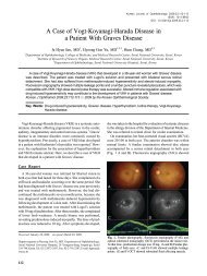
Visual Loss in One Eye after Spinal Surgery<br />
Min-Su Chung, MD, Jun-Hyuk Son, MD<br />
Department <strong>of</strong> <strong>Ophthalmology</strong>, Yeungnam University College <strong>of</strong> Medicine, Daegu, Korea<br />
Purpose: To report a patient who developed an unusual combination <strong>of</strong> central retinal artery occlusion with<br />
ophthalmoplegia following spinal surgery in the prone position.<br />
Methods: A 60-year-old man underwent a cervical spinal surgery in the prone position. Soon after recovery<br />
he could not open his right eye and had ocular pain due to the general anesthesia. Upon examination, we<br />
determined that he had a central retinal artery occlusion with total ophthalmoplegia.<br />
Results: Despite medical treatment, optic atrophy was still present at the following examination. Ptosis and<br />
the afferent pupillary defect disappeared and ocular motility was recovered, but visual loss persisted until the<br />
last follow-up.<br />
Conclusions: A prolonged prone position during spinal surgery can cause external compression <strong>of</strong> the eye,<br />
causing serious and irreversible injury to the orbital structures. Therefore, if the patient shows postoperative<br />
signs <strong>of</strong> orbital swelling after spinal surgery the condition should be immediately evaluated and treated.<br />
<strong>Korean</strong> <strong>Journal</strong> <strong>of</strong> <strong>Ophthalmology</strong> 20(2):139-142, 2006<br />
Key Words: Central retinal artery occlusion, Ophthalmoplegia, Prone position, Spinal surgery<br />
Sudden unilateral or bilateral visual loss occurring after<br />
general anesthesia has been reported and attributed to various<br />
causes including hemorrhagic shock, blood dyscrasia, hypotension,<br />
hypothermia, coagulopathic disorders, direct trauma,<br />
embolism, and prolonged compression <strong>of</strong> the eyes. 1,2<br />
However, visual loss with total ophthalmoplegia as a<br />
surgical complication has rarely been described as a consequence<br />
<strong>of</strong> prolonged compression <strong>of</strong> the eye. 2-5 There have<br />
been no case reports in the literature to date <strong>of</strong> visual loss<br />
following spinal surgery in Korea. We present the first case<br />
report <strong>of</strong> a <strong>Korean</strong> patient who developed an unusual<br />
combination <strong>of</strong> central retinal artery occlusion (CRAO) with<br />
ophthalmoplegia after spinal surgery in the prone position.<br />
Case Report<br />
A 60-year-old man came to the emergency department <strong>of</strong><br />
our hospital with an arm tingling sensation in both arms that<br />
he had experienced for seven months. Prior to the development<br />
<strong>of</strong> the sensation, he had injuried his neck by a fallen<br />
log but reported no pain at the cervical area at the time <strong>of</strong><br />
presentation at the emergency room. Radiographic views <strong>of</strong><br />
Received: December 2, 2005 Accepted: April 6, 2006<br />
Reprint requests to Jun-Hyuk Son, MD. Department <strong>of</strong> <strong>Ophthalmology</strong>,<br />
Yeungnam University College <strong>of</strong> Medicine, #317-1 Daemyungdong,<br />
Nam-gu, Daegu 705-717, Korea. Tel: 82-53-620-3444, Fax:<br />
82-53-626-5936, E-mail: junhyuk_son@hanmail.net<br />
*This study was presented as a poster at the 94th Annual Meeting<br />
<strong>of</strong> the <strong>Korean</strong> Ophthalmological Society, October 2005.<br />
the cervical spine showed osteophytosis at C4-C5, C5-C6 and<br />
at the narrow disc space from C4-C7. A magnetic resonance<br />
imaging (MRI) scan was performed and revealed a compressed<br />
dural sac from C3-C5 with spinal stenosis. The<br />
patient had a history <strong>of</strong> diabetes mellitus but had never<br />
received medical treatment for the disease. He was on a diet<br />
and his preoperative blood glucose was checked at 285<br />
mg/dL. The patient had no other diseases except diabetes<br />
mellitus. His preoperative blood count was normal, but he<br />
had a slightly low hemoglobin level at 13.5 mg/dL.<br />
A C3-C5 laminectomy was performed three days after the<br />
hospitalization. Under general anesthesia, the patient was put<br />
on the operating table in the prone position according to the<br />
standard procedure for spinal surgery. During surgery, his<br />
neck was kept in a mild flexion position using a head-holter.<br />
The total time for the intervention was 295 minutes, and a<br />
total <strong>of</strong> 60 additional minutes was required for the induction<br />
and cessation <strong>of</strong> anesthesia. During the operation, the<br />
patient’s blood pressure decreased from 120/80 mmHg to<br />
100/60 mmHg for 30 minutes because <strong>of</strong> some blood loss,<br />
and four pints <strong>of</strong> packed RBCs were transfused. There were<br />
no other general problems during surgery. The patient's<br />
postoperative hemoglobin concentration was 13.3 mg/dL,<br />
which recovered to within normal range the next day.<br />
Immediately after recovery from general anesthesia, the<br />
patient could not open his right eye because <strong>of</strong> swelling, and<br />
he also had ocular pain in the right eye. He showed visual<br />
loss when we examined his right eye on the second postoperative<br />
day. He had periorbital swelling, ptosis <strong>of</strong> the<br />
eyelid, chemosis, numbness <strong>of</strong> the first division <strong>of</strong> the right<br />
139
<strong>Korean</strong> J Ophthalmol Vol.20, No.2, 2006<br />
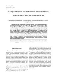
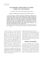
Fig. 1. Ocular fundus photography on the second postoperative<br />
day. The right eye shows a pale optic disc with an edematous<br />
retina and a cherry-red spot on the fovea.<br />
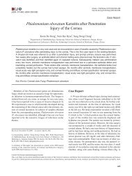
Fig. 3. T2-weighted image <strong>of</strong> orbital MRI shows mild periorbital<br />
swelling and edema <strong>of</strong> the extraocular muscles in the right eye,<br />
with the tendons spared.<br />
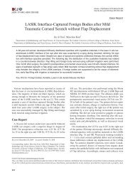
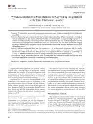
Fig. 2. Fluorescein angiogram on the second postoperative day.<br />
The right eye shows an extreme delay in arterial filling time.<br />
trigeminal nerve, afferent pupillary defect, and total ophthalmoplegia.<br />
Intraocular pressure was within the normal range.<br />
Anterior segments were normal in both eyes. He had a pale<br />
optic disc with an edematous retina and fundus examination<br />
revealed a cherry-red spot on the fovea (Fig. 1). His fluorescein<br />
angiographic examination revealed a delayed arterial<br />
filling time (Fig. 2). His brain MRI and magnetic resonance<br />
angiography (MRA) findings were normal with no evidence<br />
<strong>of</strong> embolic phenomena. However we noted mild swelling <strong>of</strong><br />
the right periorbital s<strong>of</strong>t tissue and edema <strong>of</strong> the extraocular<br />
muscles in the right eye, sparing their tendons (Fig. 3).<br />
Ptosis, the afferent pupillary defect, and ocular movement<br />
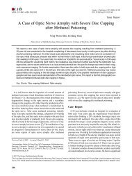
improved over three postoperative days, but despite medical<br />
treatment with carbonic anhydrase inhibitors and corticosteroids,<br />
the patient had optic atrophy on the following<br />
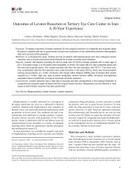
examination (Fig. 4). Eventually his vision deteriorated until<br />
he had no light perception after the last follow up at three<br />
months.<br />
Fig. 4. Ocular fundus photography after three months. The right<br />
eye shows optic atrophy.<br />
Discussion<br />
Postoperative visual loss following non-ocular surgery is<br />
an infrequent but disastrous complication with an estimated<br />
incidence ranging from 0.001 to 1% depending on the type<br />
<strong>of</strong> surgery. 6,7 A survey <strong>of</strong> vision loss after spinal surgery has<br />
suggested that possible intraoperative causes including patient<br />
positioning, blood loss, and intraoperative hypotension could<br />
be responsible for this complication. The three recognized<br />
causes <strong>of</strong> postoperative visual loss are ischemic optic neuropathy,<br />
central retinal artery or vein occlusion, and cerebral<br />
ischemia. 8,9<br />
In addition to speculation regarding the intraoperative<br />
140
MS Chung, et al. VISUAL LOSS AFTER SPINAL SURGERY<br />
causes <strong>of</strong> these problems, several patient risk factors have<br />
been suggested. These include chronic hypertension, diabetes<br />
mellitus, smoking, vascular disease, and other disorders that<br />
result in increased blood viscosity. 7,10,11 Because evidence for<br />
the role <strong>of</strong> these risk factors in the pathogenesis <strong>of</strong> visual loss<br />
has been determined from studies in diverse clinical settings,<br />
their exact influence on patients who undergo spinal surgery<br />
remains unknown.<br />
CRAO is a disease entity that occurs after trauma or<br />
embolic, thrombotic, or spasmodic episodes in both children<br />
and adults. 12 However, postoperative CRAO has rarely been<br />
reported as a consequence <strong>of</strong> direct pressure on the eye<br />
during surgery. Slocum et al 13 first observed this problem in<br />
a patient who underwent a neurosurgical procedure in the<br />
prone position using a Bailey headrest. He emphasized that<br />
CRAO was caused by the extraocular pressure due to the<br />
headrest, or because <strong>of</strong> anesthetic mask malposition in the<br />
presence <strong>of</strong> hypotension. Givner and Jaffe 14 reported four<br />
cases <strong>of</strong> CRAO occurring during abdominal operations in the<br />
supine position under mask anesthesia. They suggested that<br />
CRAO in these cases was due to a combination <strong>of</strong> pressure<br />
on the eye from the mask under hypotension and prolonged<br />
anesthesia. Gillan 15 reported two cases in which CRAO<br />
developed after general mask anesthesia with vascular<br />
hypotension in the supine position. The cause was believed<br />
to be similar to that in Givner and Jaffe's report. 14<br />
Hollenhorst et al 3 put forth the possibility that CRAO<br />
followed neurosurgical procedures which took place in the<br />
prone position and that used a padded headrest. He reported<br />
eight cases <strong>of</strong> CRAO in his clinical setting that resulted from<br />
the inadvertent application <strong>of</strong> pressure to the orbital contents<br />
by the headrest. Bradish and Flowers 16 reported the first case<br />
<strong>of</strong> CRAO in the prone position after an orthopedic procedure.<br />
They believed that CRAO was due to a combination <strong>of</strong><br />
hypotensive anesthesia and extrinsic pressure on the<br />
inherently susceptible eye <strong>of</strong> a patient with osteogensis<br />
imperfecta.<br />
The mechanism <strong>of</strong> visual loss after surgery is retinal<br />
ischemia secondary to external ocular pressure. The potential<br />
causes <strong>of</strong> this ischemia are venous congestion and arterial<br />
occlusion. 3,17 Hollenhorst et al 3 reproduced visual loss and<br />
ophthalmoplegia experimentally in seven rhesus monkeys<br />
using orbital compression for 60 minutes in the presence <strong>of</strong><br />
hypovolemia and hypotension. They proved them by<br />
suggesting a mechanism <strong>of</strong> partial or complete collapse <strong>of</strong> the<br />
arterial and venous channels <strong>of</strong> the orbit, which were<br />
produced by a temponade action <strong>of</strong> the ocular contents. When<br />
the external pressure is released, orbital edema, proptosis,<br />
paresis <strong>of</strong> ocular movement, and massive edema <strong>of</strong> the retina<br />
result from dilation <strong>of</strong> the ischemic vascular channels and<br />
from a transudation <strong>of</strong> fluid through the permeable walls into<br />
the tissue spaces. 3 Halfon et al 5 suggested that the reversibility<br />
<strong>of</strong> ophthalmoplegia probably depends on the degree <strong>of</strong><br />
ischemia suffered by both the extraocular muscles and the Ⅲ,<br />
Ⅳ, and Ⅵ cranial nerves. In our case, postoperative signs <strong>of</strong><br />
orbital swelling in the affected eye were appreciable evidence<br />
<strong>of</strong> external orbital compression. Our patinet's brain MRI and<br />
MRA showed no evidence <strong>of</strong> cavernous sinus thrombosis and<br />
revealed only mild periorbital swelling and enlargement with<br />
hyperintensity <strong>of</strong> the extraocular muscles in the right eye<br />
while sparing the tendons. Thus our case is believed to be<br />
similar to those reported by Halfon et al. 5<br />
CRAO with ophthalmoplegia has been more infrequently<br />
reported. 2-5 Hollenhorst et al 3 noted that the five most severe<br />
cases out <strong>of</strong> eight cases who had proptosis, ptosis, and<br />
paralysis <strong>of</strong> the extraocular muscles improved, even though<br />
there was no visual recovery. Halfon et al 5 reported two cases<br />
<strong>of</strong> palpebral edema, ocular pain, and total ophthalmoplegia<br />
which recovered partial or full ocular motility without the<br />
return <strong>of</strong> vision. Our patient, who exhibited vision loss <strong>of</strong> the<br />
right eye with swelling, ocular pain, and ophthalmoplegia<br />
recovered nearly full ocular movement without any improvement<br />
<strong>of</strong> vision, and is therefore believed to be identical to<br />
those in the two aforementioned reports.<br />
A prolonged prone position during spinal surgery and<br />
excessive pressure on the eye, especially as a result <strong>of</strong> patient<br />
malposition, can cause injury to the orbital structures and can<br />
lead to serious ocular complications. Although postoperative<br />
vision loss can occur in the absence <strong>of</strong> globe compression,<br />
avoidance <strong>of</strong> mechanical injury to the globe is very important<br />
for the surgeon and anesthetist to be aware <strong>of</strong>. The surgeon<br />
and anesthetist must pay particular attention to avoid the<br />
consequences <strong>of</strong> possible intraoperative movement without<br />
adequate eye protection during surgery. Special attention<br />
must be given to bradyarrhythmia or other heart conduction<br />
illnesses during the operation, as these may be signals <strong>of</strong><br />
vagal stimulation caused by increased intraorbital pressure. 2<br />
We believe this to be the first case report <strong>of</strong> CRAO with<br />
ophthalmoplegia after spinal surgery in the prone position in<br />
Korea. If patients show postoperative signs <strong>of</strong> orbital<br />
swelling, especially following surgery in the prone position,<br />
the condition must be evaluated and treated immediately.<br />
Ophthalmologists must be aware <strong>of</strong> the possibility <strong>of</strong> this<br />
complication, and always pay attention to avoid this<br />
devastating complication.<br />
References<br />
1. Brown GC, Magargal LE, Shields JA, et al. Retinal arterial<br />
obstruction in children and young adults. <strong>Ophthalmology</strong><br />
1981;88:18-25.<br />
2. Wolfe SW, Lospinuso MF, Burke SW. Unilateral blindness<br />
as a complication <strong>of</strong> patient positioning for spinal surgery.<br />
A case report. Spine 1992;17:600-5.<br />
3. Hollenhorst RW, Svien HJ, Benoit CF. Unilateral blindness<br />
occurring during anesthesia for neurosurgical operations.<br />
Arch Ophthalmol 1954;52:819-30.<br />
4. West J, Askin G, Clarke M, Vernon SA. Loss <strong>of</strong> vision in<br />
one eye following scoliosis surgery. Br J Ophthalmol 1990;<br />
74:243-4.<br />
5. Halfon MJ, Bonardo P, Valiensi S, et al. Central retinal<br />
artery occlusion and ophthalmoplegia following spinal<br />
141
<strong>Korean</strong> J Ophthalmol Vol.20, No.2, 2006<br />
surgery. Br J Ophthalmol 2004;88:1350-2.<br />
6. Warner ME, Warner MA, Garrity JA, et al. The frequency<br />
<strong>of</strong> perioperative vision loss. Anesth Analg 2001;93:1417-21.<br />
7. Williams EL, Hart WM Jr, Tempelh<strong>of</strong>f R. Postoperative<br />
ischemic optic neuropathy. Anesth Analg 1995;80:1018-29.<br />
8. Myers MA, Hamilton SR, Bogosian AJ, et al. Visual loss<br />
as a complication <strong>of</strong> spine surgery. A review <strong>of</strong> 37 cases.<br />
Spine 1997;22:1325-9.<br />
9. Stevens WR, Glazer PA, Kelley SD, et al. Ophthalmic<br />
complications after spinal surgery. Spine 1997;12:1319-24.<br />
10. Brown RH, Schauble JF, Miller NR. Anemia and hypotension<br />
as contributors to perioperative loss <strong>of</strong> vision.<br />
Anesthesiology 1994;80:222-6.<br />
11. Katz DM, Trobe JD, Cornblath WT, Kline LB. Ischemic<br />
optic neuropathy after lumbar spine surgery. Arch Ophthalmol<br />
1994;112:925-31.<br />
12. Grossman W, Ward WT. Central retinal artery occlusion<br />
after scoliosis surgery with a horseshoe headrest. Case<br />
report and literature review. Spine 1993;18:1226-8.<br />
13. Slocum HC, O'Neal KC, Allen CR. Neurovascular complications<br />
from malposition on the operating table. Surg<br />
Gynecol Obstet 1948;86:729-34.<br />
14. Givner I, Jaffe N. Occlusion <strong>of</strong> the central retinal artery<br />
following anesthesia. Arch Ophthalmol 1950;43:197-201.<br />
15. Gillan JG. Two cases <strong>of</strong> unilateral blindness following<br />
anaesthesia with vascular hypotension. Can Med Assoc J<br />
1953;69:294-6.<br />
16. Bradish CF, Flowers M. Central retinal artery occlusion in<br />
association with osteogenesis imperfecta. Spine 1987;12:<br />
193-4.<br />
17. Walkup HE, Murphy JD. Retinal ischemia with unilateral<br />
blindness-a complication occurring during pulmonary<br />
resection in the prone position; report <strong>of</strong> two cases. J<br />
Thorac Surg 1952;23:174-5.<br />
142