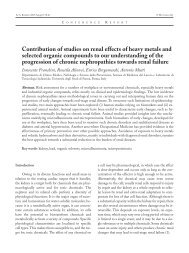
Mattioli - Acta Bio Medica Atenei Parmensis
Mattioli - Acta Bio Medica Atenei Parmensis
Mattioli - Acta Bio Medica Atenei Parmensis

Create successful ePaper yourself
Turn your PDF publications into a flip-book with our unique Google optimized e-Paper software.
ACTA BIOMED 2005; 76; 165-170 © <strong>Mattioli</strong> 1885<br />
O R I G I N A L A R T I C L E<br />
Electron microscopy as a reliable tool for rapid and<br />
conventional detection of enteric viral agents: a five-year<br />
experience report<br />
Maria Cristina Arcangeletti, Flora De Conto, Federica Pinardi, Maria Cristina Medici,<br />
Pierpaolo Valcavi, Francesca Ferraglia, Federica Motta, Silvia Covan, Adriana Calderaro,<br />
Carlo Chezzi, Giuseppe Dettori<br />
Section of Microbiology, Department of Pathology and Laboratory Medicine, University of Parma, Parma, Italy<br />
Abstract. Background and aim of the work: Since the introduction of the electron microscope and its subsequent<br />
development, virology has made a great step forward by the improvement of the basic knowledge on<br />
viral structure, as well as by broad application of electron microscopy (EM) to viral diagnosis. In this report,<br />
we describe a five-year experience in the use of EM for the diagnosis of enteric viral infections. Methods:<br />
Three thousand four hundred and ninety stool specimens were analyzed at the Virology Unit (Section of Microbiology,<br />
Department of Pathology and Laboratory Medicine, University of Parma, Italy) during a fiveyear<br />
period, from January 1999 to January 2004. The faecal extracts were subjected to EM after negative<br />
staining and were simultaneously cultured to evidence the presence of cytopathogenic agents. Results: EM<br />
directly applied to the above specimens allowed the detection of several enteric viral agents, particularly evidencing<br />
those normally hard to cultivate (thus easily lost with culture methods). It also enabled diagnosis of<br />
dual gut infections, such as those from rotavirus and calicivirus. On the other hand, EM-based identification<br />
of viral agents after cell culture and ultracentrifugation of cytopathogenic agent-containing cellular extracts,<br />
allowed the identification of cultivable agents, such as picornaviruses, which can escape the direct EM detection<br />
if low concentrated. Conclusions: A rationalized use of EM on selected samples, such as stool, appears<br />
suitable in epidemiological or clinical conditions when a very rapid diagnosis is required to save time, including<br />
cases of suspected emerging viral infections. (www.actabiomedica.it)<br />
Key words: Electron microscopy, rapid diagnosis, enteric viruses<br />
Introduction<br />
The introduction of the first functioning prototype<br />
of an electron microscope in the 1930s, and its subsequent<br />
development, was a milestone for basic and<br />
clinical virology. Particularly with regard to diagnostic<br />
virology, the use of electron microscopy (EM), initially<br />
limited to rapid diagnosis of the smallpox virus, was<br />
extended to «routine» diagnostics in the 1960s, thanks<br />
to the introduction of techniques such as negative<br />
staining (1-3). Initially used to identify cytopathogenic<br />
agents from cell cultures, EM has become even<br />
more widely used since its direct application to clinical<br />
samples (1, 4). With the implementation of automatic<br />
immunoenzymatic methods and, above all, the<br />
development of advanced molecular techniques, particularly<br />
those based on nucleic acid amplification, the<br />
use of EM has significantly decreased (5-7). Many<br />
factors have contributed to the marked reduction in<br />
EM application to viral diagnosis: its cost and, moreover,<br />
its relatively low sensitivity render it not suitable<br />
for the screening of a large number of clinical samples.<br />
The present study was carried out on stool specimens<br />
at the Virology Unit of the Section of Microbiology<br />
(Department of Pathology and Laboratory<br />
Medicine, University of Parma, Italy), a comprehensi-
166 M.C. Arcangeletti, F. De Conto, F. Pinardi, et al.<br />
ve centre, offering a combination of different diagnostic<br />
tools (EM, cell culture, fluorescence microscopy,<br />
nucleic acid amplification methods, antigen and antiviral<br />
antibody detection in serum). Its aim was chiefly<br />
to underscore the advantages of EM application to the<br />
diagnosis of enteric viral infections.<br />
Virological investigations carried out at the Virology<br />
Unit (Section of Microbiology Department of<br />
Pathology and Laboratory Medicine, University of<br />
Parma, Italy) involved 3490 stool samples, analyzed<br />
over a 5-year period, from January 1999 to January<br />
2004.<br />
After the stool sample arrived at the Virology laboratory,<br />
a faecal suspension was obtained by diluting<br />
specimens to 10% weight/volume in PBS (phosphate<br />
buffered saline); the faecal extract was thoroughly<br />
emulsified, then centrifuged at low speed (2500 g up<br />
to 20 minutes), in order to remove large debris and<br />
bacteria. Then the clarified faecal suspension was concentrated<br />
by adding polyacrylamide absorbent gel<br />
(0.02 gr/550 ml) for about 20 minutes, and subsequently<br />
used to prepare a standard drop to be put in<br />
contact with a 400-mesh, carbon-reiforced, plastic<br />
(formvar)-coated grid. After negative staining using<br />
an aqueous solution of phosphotungstic acid (2%, pH<br />
6.4), sample examination was performed using a transmission<br />
electron microscope (EM 208S Philips) with<br />
a 44,000x magnification. Faecal extracts were simultaneously<br />
inoculated into a wide range of cell lines and<br />
observed daily under an inverted optical microscope,<br />
in order to evidence cytopathogenic agents (conventional<br />
cell culture, CC). Positive cell extracts (324) were<br />
subjected to EM after ultracentrifugation and negative<br />
staining, for final identification steps.<br />
Methods<br />
Results<br />
EM and conventional cell culture are routinarily<br />
employed in our laboratory for the diagnosis of enteric<br />
viral infections. Specifically, EM is used as a rapid<br />
method directly on stool samples (which are, in parallel,<br />
cultured), as well as on ultracentrifuged cell extracts,<br />
which evidenced one or more cytopathogenic<br />
agents.<br />
In this study, carried out on 3490 stool specimens<br />
analyzed over a five-year period (1999-2004), we evaluated<br />
the efficacy of EM in revealing the presence of<br />
viral particles, also comparing it to conventional cell<br />
culture.<br />
Eight hundred ninety-one (25.5%) of the 3490<br />
studied stool samples were positive for enteric viruses.<br />
Positivity for one or more viral agents was determined<br />
only by EM in 567 samples (16.2%) and only by CC<br />
in 230 samples (6.6%). In 94 samples (2.7%), both<br />
methods were able to determine positivity (Table 1).<br />
These results clearly outline that EM is the most<br />
performant method in evidencing the presence of enteric<br />
viruses in stool samples, as summarized in figure<br />
1 (63.6% of the total positive samples, compared to<br />
Table 1. Number and annual distribution of total and positive samples, whose positivity was assessed by the application of electron<br />
microscopy (EM) and conventional culture (CC) on stool samples analyzed at the Virology Unit (Section of Microbiology, University<br />
of Parma), from January 1999 to January 2004<br />
Year Total Positive samples Positive EM* Positive CC** Positive [EM + CC]***<br />
samples N. % N. % N. % N. %<br />
1999 582 139 23.9 86 14.8 48 8.2 5 0.9<br />
2000 706 174 24.6 90 12.7 50 7.1 34 4.8<br />
2001 743 222 29.9 142 19.1 59 7.9 21 2.8<br />
2002 687 160 23.3 106 15.4 38 5.5 16 2.3<br />
2003 689 165 23.9 115 16.7 34 4.9 16 2.3<br />
2004 § 83 31 37.3 28 33.7 1 1.2 2 2.4<br />
Total 3490 891 25.5 567 16.2 230 6.6 94 2.7<br />
*: Samples whose positivity was assessed only by EM; **: Samples whose positivity was assessed only by CC; ***: Samples whose positivity<br />
was assessed by EM and CC; § : Only January 2004 has been included in this study
Electron microscopy and viral diagnosis<br />
167<br />
25.8% by CC). Indeed, EM allowed us to reveal several<br />
cultivable and non-cultivable viral agents, such as<br />
rotavirus, adenovirus, picornavirus and calicivirus<br />
(Fig. 2).<br />
Specifically, direct electron microscope examination<br />
of faecal extracts was very advantageous in detecting,<br />
within a few hours after sample arrival, the presence<br />
of those viral agents particularly difficult to cultivate<br />
(e.g. rotavirus), that would escape identification<br />
under parallel CC examination (Table 2, EM). As it<br />
can be noted from table 2, rotavirus corresponds to the<br />
viral genus most frequently detected in stool (95.6%),<br />
as it was expected, taken into account that it represents<br />
one of the major cause of paediatric enteric viral infections<br />
(8-15).<br />
EM was also useful to evidence the simultaneous<br />
presence of two viruses from different families/genera,<br />
such as rotavirus and calicivirus (Table 2, EM, 2004).<br />
Particularly concerning caliciviruses (then identified<br />
as belonging to norovirus genus by molecular<br />
methods; see table 2, 2001, 2002, 2004), it is noteworthy<br />
that they have “attracted attention” progressively<br />
during the last years, being now considered as a<br />
relevant cause of outbreaks of gastroenteritis in humans<br />
(5, 16), and also potentially involved in nosocomial<br />
infections (16-21). The presence of identifiable<br />
viral particles in sufficient number to be seen under<br />
the electron microscope suggests that, most likely, a significant<br />
virus replication has occurred, thus signaling,<br />
albeit preliminarly, a possible epidemic wave and allows<br />
us to finally identify the viral agent by the appropriate<br />
techniques.<br />
On the other hand, the use of conventional culturing<br />
(and the subsequent identification of the cytopathogenic<br />
agent by means of EM), enabled us to reveal<br />
the presence of cultivable adenoviruses, often<br />
missed under direct electron microscope observation<br />
(Table 2: 64.3% adenovirus by CC, vs 2.9% by EM),<br />
probably due to a too low concentration in the stool<br />
specimen. The parallel use of the conventional culture<br />
method also allowed us to confirm the presence of viruses<br />
such as picornaviruses, that might have escaped<br />
detection from EM observation of faecal extracts,<br />
both because of their poor structural definition and/or<br />
a too low concentration in the sample (Table 2: 33.5%<br />
picornavirus by CC, vs 0.2% by EM).<br />
Finally, the combined use of EM directly on stool<br />
samples and parallel cell culturing (Table 2, EM+CC)<br />
allowed us to detect Reoviridae viral strains, most<br />
likely belonging to the cultivable Orthoreovirus<br />
(rather than Rotavirus) genus (1.1%); furthermore, it<br />
evidenced the presence of dual gut infections, such as<br />
those from rotavirus and adenovirus (18.1%), or rotavirus<br />
and picornavirus (6.4%).<br />
Discussion and conclusions<br />
Although EM is not always the method of choice<br />
to diagnose viral infections, it has proven to be very<br />
Figure 1. The highest percentage of positive samples obtained by using electron microscopy (EM) directly on faecal extracts (“Positive<br />
EM”) demonstrates that EM is the most efficient method (vs conventional culture, CC) in revealing viral agents in stool samples<br />
analyzed from January 1999 to January 2004
168 M.C. Arcangeletti, F. De Conto, F. Pinardi, et al.<br />
A) B)<br />
C) D)<br />
Figure 2. Electron micrographs of enteric viruses after negative staining: A. Caliciviridae (Norovirus); B. Rotavirus; C. Picornaviridae<br />
(Enterovirus); D. Adenovirus. Bar = 50 nm<br />
effective in case of enteric viral infections (22-24). As<br />
it clearly appears from this study and is generally<br />
agreed, EM offers undoubted advantages linked to the<br />
possibility of an easy and very rapid examination of<br />
clinical samples and to the direct visualization of viral<br />
particles, also allowing us to evidence the concomitant<br />
presence of two or more, possibly unexpected, viral<br />
agents.<br />
Nevertheless, as already mentioned in the introductive<br />
section, the inherent economical limitations<br />
and the need for high particle concentration in the<br />
sample, as well as the introduction of performant molecular<br />
methods, often prevent - or at least strongly reduce<br />
- the use of EM as a diagnostic tool.<br />
Indeed, up to date many methodologies are available<br />
to detect viruses in clinical specimens, which generally<br />
differ in their objectives and principles, indicating<br />
the extent to which the choice between one and<br />
the other can be at times a matter of opinion and comparison<br />
of results and quite hard to make. In fact, very
Electron microscopy and viral diagnosis<br />
169<br />
Table 2. Annual distribution of viral families/genera detected in stool specimens (analyzed from January 1999 to January 2004) by<br />
using electron microscopy (EM) and/or conventional culture (CC)<br />
Year<br />
1999 2000 2001 2002 2003 2004 §<br />
EM* N. N. N. N. N. N. N. %<br />
Rota 83 86 138 98 112 25 542 95.6<br />
AdV 3 4 2 4 3 1 17 2.9<br />
PrV 0 0 1 0 0 0 1 0.2<br />
CV 0 0 1 4 0 1 6 1.1<br />
Rota/CV 0 0 0 0 0 1 1 0.2<br />
CC**<br />
Reo 1 2 2 0 0 0 5 2.2<br />
AdV 31 33 38 23 22 1 148 64.3<br />
PrV 16 15 19 15 12 0 77 33.5<br />
[EM+CC]***<br />
Reo 0 0 0 1 0 0 1 1.1<br />
AdV 2 25 15 11 15 2 70 74.4<br />
Rota/Adv 1 6 6 3 1 0 17 18.1<br />
Rota/PrV 2 3 0 1 0 0 6 6.4<br />
AdV: adenovirus; CV: calicivirus; PrV: picornavirus; Reo: reovirus; Rota: rotavirus.<br />
*: Samples whose positivity was assessed only by EM; **: Samples whose positivity was assessed only by CC; ***: Samples whose positivity<br />
was assessed by EM and CC; § : Only January 2004 has been included in this study<br />
Total<br />
often different methods provide complementary<br />
rather than “overlapping” information for a comprehensive<br />
laboratory diagnosis.<br />
Molecular methods, in particular, are undoubtedly<br />
characterized by a greater sensitivity in evidencing<br />
the presence of a virus through detection of specific<br />
nucleotidic sequences, if compared to EM (1, 5).<br />
This latter, however, due to its undirected “open view”,<br />
is really performant in picking up also new viral particles,<br />
as it doesn’t need the programmed use of specific<br />
probes. Furthermore, a morphological diagnosis, which<br />
uncovers the involved viral family (like that performed<br />
through EM), often fulfills the physician’s immediate<br />
needs and can be precious for first emergency<br />
measures, until a more precise diagnosis is achieved<br />
(25-28).<br />
Thus, the rationalized use of EM on selected<br />
samples, i.e. stool specimens, can be advantageous in<br />
epidemiological or clinical conditions when very rapid<br />
diagnosis is required to save time and avoid complex<br />
diagnostic efforts, such as in cases of suspected emerging<br />
viral infections and/or when alternative standard<br />
diagnostic tools (e.g. cell culture) fail to produce satisfactory<br />
results.<br />
References<br />
1. Hazelton PR, Gelderblom HR. Electron microscopy for rapid<br />
diagnosis of infectious agents in emergent situations.<br />
Emerg Infect Dis 2003; 9 (3): 294-303.<br />
2. Biel SS, Madeley D. Diagnostic virology – the need for electron<br />
microscopy: a discussion paper. J Clin Virol 2000; 22: 1-<br />
9.<br />
3. Curry A, Bryden A, Morgan-Capner P, et al. A rationalised<br />
virological electron microscope specimen testing policy.<br />
PHLS North West Viral Gastroenteritis and Electron Microscopy<br />
Subcommittee. J Clin Pathol 1999; 52 (6): 471-4.<br />
4. Gelderblom HR, Hazelton PR. Specimen collection for<br />
electron microscopy. Emerg Infect Dis 2000; 6 (4): 433-4.<br />
5. Rabenau HF, Sturmer M, Buxbaum S, Walczok A, Preiser<br />
W, Doerr HW. Laboratory diagnosis of norovirus: which<br />
method is the best? Intervirology 2003; 46 (4): 232-8.<br />
6. Otsu R, Ishikawa A, Mukae K. Detection of small round<br />
structured viruses in stool specimens from outbreaks of gastroenteritis<br />
by electron microscopy and reverse transcription-polymerase<br />
chain reaction. <strong>Acta</strong> Virol 2000; 44 (1): 53-<br />
5.<br />
7. Altindis M, Yavru S, Simsek A, Ozkul A, Ceri A, Koc H.<br />
Rotavirus Infection in Children with Acute Diarrhea as Detected<br />
by Latex Agglutination, ELISA and Polyacrylamide<br />
Gel Electrophoresis. Indian Pediatr 2004; 41 (7): 590-4.<br />
8. McIver CJ, Hansman G, White P, Doultree JC, Catton M,<br />
Rawlinson WD. Diagnosis of enteric pathogens in children<br />
with gastroenteritis. Pathology 2001; 33 (3): 353-8.
170 M.C. Arcangeletti, F. De Conto, F. Pinardi, et al.<br />
9. Dennehy PH, Nelson SM, Spangenberger S, Noel JS,<br />
Monroe SS, Glass RI. A prospective case-control study of<br />
the role of astrovirus in acute diarrhea among hospitalized<br />
young children. J Infect Dis 2001; 184 (1): 10-5.<br />
10. Kasule M, Sebunya TK, Gashe BA, Armah G, Steele AD.<br />
Detection and characterization of human rotavirus among<br />
children with diarrhoea in Botswana. Trop Med Int Health<br />
2003; 8 (12): 1137-42.<br />
11. Steele D, Reynecke E, de Beer M, Bos P, Smuts I. Characterization<br />
of rotavirus infection in a hospital neonatal unit in<br />
Pretoria, South Africa. J Trop Pediatr 2002; 48 (3): 167-71.<br />
12. Iturriza Gomara M, Kang G, Mammen A, Jana AK, et al.<br />
Characterization of G10P[11] rotaviruses causing acute gastroenteritis<br />
in neonates and infants in Vellore, India. J Clin<br />
Microbiol 2004; 42 (6): 2541-7.<br />
13. Piednoir E, Bessaci K, Bureau-Chalot F, et al. Economic<br />
impact of healthcare-associated rotavirus infection in a paediatric<br />
hospital. J Hosp Infect 2003; 55 (3): 190-5.<br />
14. Sharma R, Hudak ML, Premachandra BR, et al. Clinical<br />
manifestations of rotavirus infection in the neonatal intensive<br />
care unit. Pediatr Infect Dis J 2002; 21 (12): 1099-105.<br />
15. Rodriguez-Baez N, O’Brien R, Qiu SQ, Bass DM. Astrovirus,<br />
adenovirus, and rotavirus in hospitalized children:<br />
prevalence and association with gastroenteritis. J Pediatr<br />
Gastroenterol Nutr 2002; 35 (1): 64-8.<br />
16. Froggatt PC, Barry Vipond I, Ashley CR, Lambden PR,<br />
Clarke IN, Caul EO. Surveillance of norovirus infection in<br />
a study of sporadic childhood gastroenteritis in South West<br />
England and South Wales, during one winter season<br />
(1999-2000). J Med Virol 2004; 72 (2): 307-11.<br />
17. Lynn S, Toop J, Hanger C, Millar N. Norovirus outbreaks<br />
in a hospital setting: the role of infection control. N Z Med<br />
J 2004; 117(1189): U771.<br />
18. Meyer E, Ebner W, Scholz R, Dettenkofer M, Daschner<br />
FD. Nosocomial outbreak of norovirus gastroenteritis and<br />
investigation of ABO histo-blood group type in infected<br />
staff and patients. J Hosp Infect 2004; 56 (1): 64-6.<br />
19. McCall J, Smithson R. Rapid response and strict control<br />
measures can contain a hospital outbreak of Norwalk-like<br />
virus. Commun Dis Public Health 2002; 5 (3): 243-6.<br />
20. Gonzalez Moran F, Moreno Civantos A, Mateo Ontanon<br />
Sd S, et al. Nosocomial epidemic outbreak of acute gastroenteritis<br />
by Norwalk-like virus. Med Clin (Barc) 2002;<br />
118 (16): 611-5.<br />
21. Billgren M, Christenson B, Hedlund KO, Vinje J. Epidemiology<br />
of Norwalk-like human caliciviruses in hospital<br />
outbreaks of acute gastroenteritis in the Stockholm area in<br />
1996. J Infect 2002; 44 (1): 26-32.<br />
22. Leung WK, To KF, Chan PK, et al. Enteric involvement of<br />
severe acute respiratory syndrome-associated coronavirus<br />
infection. Gastroenterology 2003; 125 (4): 1011-7.<br />
23. Mori I, Matsumoto K, Sugimoto K, et al. Prolonged shedding<br />
of rotavirus in a geriatric patient. J Med Virol 2002; 67<br />
(4): 613-5.<br />
24. Marshall J, Botes J, Gorrie G, et al. Rotavirus detection and<br />
characterisation in outbreaks of gastroenteritis in aged-care<br />
facilities. J Clin Virol 2003; 28 (3): 331-40.<br />
25. Riordan FA, Quigley T: Estimating hospital admissions<br />
due to rotavirus gastroenteritis from hospital episode statistics.<br />
J Infect 2004; 49 (1): 13-6.<br />
26. Rivest P, Proulx M, Lonergan G, Lebel MH, Bedard L.<br />
Hospitalisations for gastroenteritis: the role of rotavirus.<br />
Vaccine 2004; 22 (15-16): 2013-7.<br />
27. Roman Riechmann E, Wilhelmi de Cal I, Cilleruelo Pascual<br />
ML, Calvo Rey C, Garcia Garcia ML, Sanchez-Fauquier<br />
A. Nosocomial gastroenteritis and asymptomatic rotavirus<br />
and astrovirus infection in hospitalized children. An<br />
Pediatr (Barc) 2004; 60(4): 337-43.<br />
28. Gelber SE, Ratner AJ. Hospital-acquired viral pathogens in<br />
the neonatal intensive care unit. Semin Perinatol 2002; 26<br />
(5): 346-56.<br />
Accepted: 13 October 2005<br />
Correspondence: Dr. M. Cristina Arcangeletti, M.D.<br />
Section of Microbiology<br />
Department of Pathology and Laboratory Medicine<br />
University of Parma<br />
Viale A. Gramsci, 14<br />
43100 Parma (Italy)<br />
Tel. +39 0521 988877<br />
Fax +39 0521 993620<br />
Email: mariacristina.arcangeletti@unipr.it