case study - Hartmann - Paul Hartmann AG
case study - Hartmann - Paul Hartmann AG
case study - Hartmann - Paul Hartmann AG

Create successful ePaper yourself
Turn your PDF publications into a flip-book with our unique Google optimized e-Paper software.
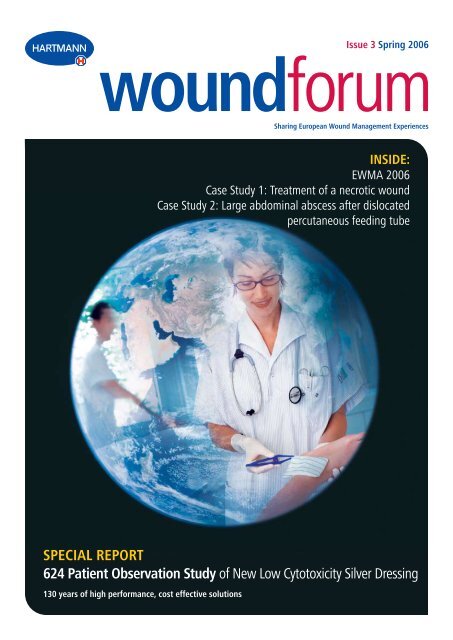
Issue 3 Spring 2006<br />
woundforum<br />
Sharing European Wound Management Experiences<br />
INSIDE:<br />
Ewma 2006<br />
Case Study 1: Treatment of a necrotic wound<br />
Case Study 2: Large abdominal abscess after dislocated<br />
percutaneous feeding tube<br />
SPECIAL REPORT<br />
624 Patient Observation Study of New Low Cytotoxicity Silver Dressing<br />
130 years of high performance, cost effective solutions
Welcome<br />
to this ‘new look’ issue of <strong>Hartmann</strong>’s Wound Forum journal.<br />
The Wound Forum journal has appeared in many guises over<br />
the years as some of the images below show.<br />
CASE STUDY<br />
The author:<br />
Frans Meuleneire, AZ St. Elisabeth, Heelkunde 2E,<br />
Godveerdegemstraat 69, 9620 Zottegem, Belgium<br />
F. Meuleneire, St. Elisabeth, Zottegem, Belgium<br />
Treatment of a necrotic<br />
wound due to<br />
subcutaneous haematoma<br />
This latest issue aims to draw on the experience and expertise of<br />
<strong>Hartmann</strong>’s technical specialists together with our end users to<br />
share with you examples of wound management practice in<br />
different settings within the UK and<br />
across Europe.<br />
You may have also seen the start of<br />
our series of advertising campaigns<br />
which are currently appearing in<br />
eminent UK and European journals.<br />
These highlight the history of<br />
<strong>Hartmann</strong> in the development of<br />
the first antiseptic dressings in 1870<br />
with Sir Joseph Lister – a story<br />
covered on page 12.<br />
Today, <strong>Hartmann</strong> continues to strive to use its up-to-date<br />
manufacturing processes and technical expertise to bring the NHS<br />
dressings that are clinically effective but which aim to provide the best<br />
value for money – a factor that is difficult to ignore in today’s climate.<br />
The latest example of this is our new product launch of Atrauman<br />
Ag - a silver primary dressing that combines efficacy with cost<br />
effectiveness. Now available on Drug Tariff and via NHS Logistics,<br />
Atrauman Ag has been successfully proven. The outcomes of a 624<br />
patient <strong>study</strong> using Atrauman Ag can be found on pages 8 – 11 and<br />
14, together with further <strong>case</strong> <strong>study</strong> information.<br />
<strong>Hartmann</strong>’s dressings are proven in use in over a billion instances<br />
each year, across 80% of the world’s countries, resulting in an<br />
extensive bank of knowledge relating to good wound management<br />
practices. I look forward to sharing further experiences of our users<br />
with you in future issues of Wound Forum.<br />
Hayley Summersgill<br />
Marketing Manager, Wound Management Division (Great Britain)<br />
The following <strong>case</strong> <strong>study</strong> demonstrates how treatment with phased<br />
use of different wound dressings, each with differentiated principles<br />
of action, can selectively stimulate and promote wound healing.<br />
Introduction<br />
An 84-year-old female patient had hit her lower leg on a chair, resulting<br />
in the formation of a haematoma of the left lower leg. The skin became<br />
necrotic relatively quickly and after ten days a small amount of wound<br />
exudate seeped through the absorbent dressing. Although the dressing was<br />
changed daily, there was still no improvement one week later (Fig. 1). The<br />
patient was in constant pain. Clinical signs of infection were also present:<br />
There was oedema and the peri-wound area felt hyperthermic to the touch.<br />
Since no positive development was apparent, the patient was referred to<br />
the wound centre for consultation. Here the patient’s vascular status was<br />
evaluated and a search was undertaken for possible systemic factors affecting<br />
wound healing. Despite her advanced age, only slight arterial insufficiency<br />
was detected. The venous circulation was normal. The patient was not taking<br />
medication that interfere with wound healing and no other diseases were<br />
present which could have caused a deterioration in wound healing.<br />
Local wound treatment / course<br />
After taking the medical history and inspecting the wound conditions,<br />
the treatment plan was established.<br />
Since the presence of necrotic tissue with an incipient infection was<br />
identified as the most serious interfering factor, the first therapeutic<br />
intervention consisted in removing the necrotic material as rapidly as<br />
possible. The following principle should be remembered: the more<br />
rapidly the necrosis can be eliminated, the sooner the wound can<br />
enter the granulation phase. Furthermore, necrotic tissue is a nutrient<br />
substrate for aetiologic agents that can cause wound infection.<br />
In the first step, the wound was thoroughly cleansed by thorough and<br />
extensive debridement and thereby prepared for further treatment. Since<br />
single debridement rarely provides sufficient wound cleansing in wounds<br />
of this type, the subsequently applied wound dressing must provide for<br />
continuous cleansing. This means that the wound dressing must be able<br />
to progress the wound into the granulation phase within a short time both<br />
by stimulating autolytic debridement and by active osmotic debridement.<br />
This patient had a dry, necrotic, oval wound bed measuring approximately<br />
7x 4 cm. Since the wound was not very deep, we chose an oval TenderWet<br />
2 <strong>Hartmann</strong> Woundcare Forum
24 wound pad of the same size (Fig. 3). With its special principle of action,<br />
TenderWet 24 is particularly effective in rapidly cleansing wounds.<br />
The wound pad has an absorbent core made of a super-absorbent<br />
polymer which is saturated in an amount of Ringer’s solution<br />
appropriate to the size of the wound before being applied. The<br />
Ringer’s solution is then released into the wound, actively softening<br />
and debriding the necrotic material. At the same time, microbially<br />
contaminated wound exudate is absorbed into the pad, reliably<br />
removing micro-organisms, debris and toxins from the wound.<br />
cells. After one full week we removed PermaFoam and observed a remarkable<br />
improvement in the wound condition. Both the depth and the surface of the<br />
wound had decreased considerably (Fig. 7).<br />
Considering the extremely positive development of the wound, a new<br />
PermaFoam dressing was applied.<br />
This dressing was also not removed until after one whole week and we<br />
again noted that the wound surface area had decreased by half. Because<br />
of this positive development of the wound and the fact that PermaFoam<br />
was not completely saturated, we were now able to leave the dressing<br />
in place on the wound for ten days. On removing the dressing, we<br />
observed that epithelisation was almost complete (Fig. 8).<br />
Since the patient resides at a distance of 90 km from our wound centre,<br />
we did not issue her a new appointment for a further assessment of her<br />
wound. After one week she informed us by telephone that the wound<br />
had closed completely.<br />
Fig. 1: Necrotic wound due to<br />
subcutaneous haematoma, status<br />
at start of treatment.<br />
Fig. 3: Start of treatment with<br />
TenderWet 24 for further<br />
Fig. 2: Extensive debridement from<br />
initial cleansing.<br />
Fig. 4: Wound cleansing with<br />
TenderWet 24 produces good results.<br />
wound cleansing.<br />
The TenderWet wound dressing was changed daily. On removing the dressing,<br />
we observed that it was moderately adhering to the surface of the wound.<br />
When the dressing was carefully removed, the patient experienced no<br />
appreciable pain. Moreover, the adhesion had the effect of mechanically<br />
cleansing the wound, making wound irrigation hardly necessary (Fig. 4).<br />
Conclusion<br />
A wound treatment plan is always based on contemporary principles<br />
of wound healing. In 2000, Falanga described it as a comprehensive<br />
program of management in which we have to include all aspects of<br />
wound care. Wound bed preparation is performed initially to eliminate<br />
all the necrotic material from the wound. The moist wound healing<br />
environment described by G. Winter more than 40 years ago is of great<br />
importance especially for wound conditioning and should be taken<br />
into account as appropriate in every <strong>case</strong>. Furthermore, the bacterial<br />
equilibrium has to be re-established and maintained. Finally, we have to<br />
promote the process of epithelization as effectively as possible.<br />
By using the resources offered by modern wound dressings, we are<br />
now in a position to influence the individual phases of wound healing<br />
by stimulating and promoting the overall process and applying the<br />
principles of wound healing in an optimal manner.<br />
Ten days later the wound bed had almost reached skin level. It was free from<br />
necrotic tissue and only small amounts of wound exudate were present.<br />
Epithelization was clearly starting from the wound margins. The TenderWet<br />
wound pad had now become too large for the regenerating wound and was<br />
causing moderate maceration of the wound margins (Fig. 5).<br />
Because of this changed wound situation, we were obliged to select<br />
a wound dressing with a different principle of action. We decided in<br />
favour of the PermaFoam foam dressing (Fig. 6). Its therapeutic efficacy<br />
derives from its special pore structure: Large pores become increasingly<br />
smaller towards the top layer, generating a strong vertical wicking effect<br />
and resulting in rapid absorption of exudate. The good fluid binding of<br />
PermaFoam is also of great practical importance.<br />
This protects wound edges against maceration and the foam dressing can be<br />
left on the wound for several days even with heavy exudation (in the absence of<br />
complications). In the <strong>case</strong> described here, the wound showed only moderate<br />
exudate production, allowing us to leave the PermaFoam foam dressing on the<br />
wound for a whole week. The wound healing process continued undisturbed<br />
during this period: detritus, wound exudate with toxins and bacteria are actively<br />
absorbed by this dressing, which in turn promotes the migration of epithelial<br />
Fig. 5: Because of the altered<br />
wound situation, time to change<br />
the wound dressing.<br />
Fig. 7: State of the wound after<br />
one week’s undisturbed healing<br />
under PermaFoam.<br />
Fig. 6: Change to PermaFoam<br />
foam dressing, which remains on<br />
the wound for a week.<br />
Fig. 8: Wound healing almost<br />
complete after another 10 days’<br />
PermaFoam treatment.<br />
<strong>Hartmann</strong> Woundcare Forum 3
CASE STUDY<br />
The Author:<br />
Friedhelm Lang, Head of Surgical Department, General Surgery Clinic Leonberg District Hospital, Rutesheimer Straße 50, D-71229 Leonberg<br />
F. Lang, Leonberg District Hospital<br />
Large abdominal abscess following dislocated<br />
percutaneous feeding tube<br />
PEG (percutaneous endoscopically controlled<br />
gastrostomy) is a patient sparing method<br />
of enteral nutrition with only a low rate of<br />
complications. If in rare <strong>case</strong>s the feeding<br />
tube becomes dislocated, immediate surgical<br />
intervention followed by adequate local wound<br />
treatment is indicated.<br />
PEG was developed in 1980 by Ponsky and<br />
Gauderer in the United States. In Germany,<br />
Fresenius introduced the tube in 1984 in a<br />
form modified by Keymling. The PEG is now<br />
the method of choice for long term enteral<br />
nutrition. For the patient it generally means an<br />
improvement in nutritional status and quality<br />
of life because enteral feeding resembles the<br />
physiological intake of food and the gastric<br />
and intestinal regions continue to be involved<br />
in digestion. Another benefit of the method<br />
is that swallowing and speaking are not<br />
compromised and the patient can move about<br />
freely with the tube if mobility is preserved.<br />
PEG is now regarded as a standard procedure with<br />
a low rate of complications. Complications can<br />
occur during placement (e.g. peritonitis) or during<br />
the indwelling period (e.g. incorrect administration<br />
of tube nutrition, ingrowth of the fixation plate<br />
into the stomach or abdominal wall, tube<br />
dislocation). Since PEG is an invasive therapeutic<br />
modality the permanent risk of infection should<br />
also be taken into account, which mandates<br />
a strictly aseptic procedure during care of the<br />
tube insertion site. In care of the tube insertion<br />
site however, products containing polyvidone<br />
should be avoided, since they can cause the tube<br />
material to swell and become brittle. Dressings<br />
with wound-friendly polyhexanide have proved<br />
more suitable. Attention is also explicitly drawn to<br />
the importance of secure retention of the external<br />
fixation plate with surgical adhesive tape to<br />
eliminate the risk of dislocation, for example when<br />
the patient changes position.<br />
Complication: dislocation<br />
Feeding tube dislocation which can occur in<br />
rare <strong>case</strong>s is a serious, often life-threatening<br />
complication. Disclocation of the internal<br />
fixation plate is usually the result of the tube<br />
being subjected to extreme tension. This<br />
tension forcibly tears the fixation plate out<br />
of the stomach wall, leaving behind a large<br />
perforation site. As a result, stomach contents<br />
can enter the abdominal cavity which can lead<br />
to peritonitis.<br />
The signs indicating that a feeding tube is not<br />
correctly in place are unequivocal:<br />
• The fluid diet can only be delivered through<br />
the tube with difficulty, if at all<br />
• Pain accompanies the administration<br />
of nutrition<br />
• Increasing pain in the upper abdomen, not<br />
necessarily around the stoma<br />
• Rigidly tense, tender abdominal wall<br />
• Elevated temperature to the point of fever<br />
Elderly or somnolent patients must be<br />
critically monitored, since these subjects often<br />
exhibit the symptoms at a late stage or in a<br />
masked form.<br />
If a feeding tube becomes dislocated, the<br />
patient requires immediate surgical treatment.<br />
The timing of the surgical intervention<br />
determines the patient’s prospects of survival.<br />
Case <strong>study</strong><br />
The following <strong>case</strong> report describes the<br />
dislocation of a feeding tube in which the<br />
internal fixation plate came to be positioned<br />
subcutaneously. The patient was 86 years old<br />
and was referred to our surgical outpatient<br />
department by a care institution due to<br />
“feeding tube infection”. The referral report<br />
contained the diagnoses Alzheimer dementia,<br />
aortic aneurysm, coronary heart disease<br />
(CHD) and hypertension. The patient was fed<br />
with 1,500 ml tube nutrition and additionally<br />
750 ml liquid. The patient’s extremities were<br />
completely contracted.<br />
On admission to hospital, the patient was<br />
found to have a hard swelling in the area of<br />
the feeding tube and a soft abdomen. Exertion<br />
of pressure at the tube insertion site resulted<br />
in the discharge of a large amount of pus.<br />
The fistula was exposed to reveal a dislocated<br />
feeding tube with the fixation plate in the<br />
subcutaneous tissue. A pronounced fistula<br />
system was also observed.<br />
Gastrointestinal transit with gastrografin<br />
administered through a stomach tube revealed<br />
a normally contoured stomach, with the status<br />
post tube feeding still apparent. There was no<br />
fistulation after complete filling of the stomach.<br />
The abdominal survey film taken after one<br />
hour with the patient supine showed that the<br />
contrast agent had already passed through the<br />
small intestine.<br />
In the ensuing operation the patient was<br />
difficult to position because of her contractures.<br />
The feeding tube was cut off to a length of two<br />
centimetres above the skin. After disinfection<br />
of the skin and waterproof draping, the<br />
feeding tube was excised in a spindle shaped,<br />
transverse manner. This revealed the base plate<br />
of the tube in an immediate subcutaneous<br />
position. From there, we proceeded to an<br />
epifascial cavity measuring two handbreadths<br />
and containing necrotic material, extending as<br />
far as the anterior axillary line on both sides.<br />
On applying compression to the abdominal<br />
wall, no fluid was discharged from the already<br />
granulated fistula channel of the feeding tube<br />
and therefore laparotomy was not performed<br />
since the abdomen was preoperatively<br />
completely soft. A counter-incision was made<br />
on the left side.<br />
4 <strong>Hartmann</strong> Woundcare Forum
Fig. 1: X-ray: Fistula filled with<br />
contrast agent, the subcutaneously<br />
located fixation plate and the full<br />
extent of the abscess are clearly seen.<br />
Fig. 3: PermaFoam cavity is placed<br />
loosely in the abscess cavity. The soft<br />
foam material and the special porous<br />
structure allow uncomplicated<br />
tamponading with good adaptation<br />
to the wound surfaces.<br />
Fig. 2: First dressing change after<br />
feeding tube removal and abscess<br />
removal, wound cavity with residual<br />
necroses and heavily exuding.<br />
Fig. 4: Wound cavity completely<br />
padded out with PermaFoam<br />
cavity.<br />
Wound management<br />
Dressing change was carried out once daily. After removal of the<br />
dressing and wound inspection, the wound cavity debrided on<br />
removing the feeding tube was irrigated with copious amounts<br />
of polyhexanide solution. This served to remove residual necrotic<br />
debris and fibrin layers. In addition, the wound cavity was cleansed<br />
mechanically for the first three days by drawing through cotton<br />
compresses. On the fourth day, the wound cavity already showed good<br />
blood supply with only residual fibrin layers. Mechanical cleansing<br />
with cotton compresses was therefore now unnecessary, allowing the<br />
initiation of wound conditioning while preserving the undisturbed state<br />
of the wound and without cell stripping.<br />
PermaFoam cavity completes the range of the foam dressing PermaFoam,<br />
which is available in a variety of presentations. Because of its soft<br />
material and special porous structure, PermaFoam cavity can be adapted<br />
individually to the wound and easily tamponaded into the cavity.<br />
Following the initial wound cleansing activities, the wound cavity was<br />
padded out very carefully and lightly with the foam dressing PermaFoam<br />
cavity to promote the formation of granulation tissue in the wound<br />
cavity (Fig. 3/4). Because of its special pore structure which generates a<br />
strong vertical wicking action, PermaFoam has pronounced absorbent<br />
and retention properties. In practical use, this means that excess,<br />
aggressive wound exudate, pus and cell debris are rapidly absorbed<br />
into and retained in the foam structure. This also has the effect of<br />
preventing maceration and irritation of the wound edges. On absorbing<br />
exudate, the foam dressing changes only slightly in size and the material<br />
therefore does not exert pressure inside the wound cavity.<br />
Fig. 5: PermaFoam cavity before<br />
the dressing change after 48 hours.<br />
The soft foam reliably stores excess,<br />
aggressive wound exudate, the wound<br />
edges are free from macerations.<br />
Fig. 7: PermaFoam cavity<br />
saturated with wound exudate and<br />
cell debris.<br />
Fig. 6: The wound is clean and<br />
moist, with increasing formation of<br />
granulation tissue.<br />
Fig. 8: On the day of discharge,<br />
the wound has decreased enough<br />
in size to allow loose tamponading<br />
with Sorbalgon calcium alginate<br />
compresses for the further treatment.<br />
This allowed the extensive necrotic cavity to be irrigated from both sides<br />
postoperatively. Preoperative laboratory parameters with a leukocyte<br />
count of 18, 6000 and C-reactive protein of 22 clearly demonstrated the<br />
presence of an infection. Rectally measured body temperature was 38<br />
°C. The patient was given 2.2 g Augmantan t.d.s. for three days.<br />
Thanks to the high absorption and retention capacity of PermaFoam<br />
cavity, a dressing change was only necessary every second day. This<br />
was performed in a cell-sparing manner and without administering<br />
analgesics, since PermaFoam has atraumatic properties and does<br />
not dry out the wound. We observed no adhesion to the wound even<br />
with longer dressing change intervals. Overall, we observed good<br />
conditioning when using PermaFoam cavity, with increasing formation<br />
of granulation tissue (Fig. 6).<br />
The tamponading properties of PermaFoam cavity were also good.<br />
The material remains compact under tensile stress and does not<br />
tear. Cutting to size beforehand to match the size of the wound is<br />
unnecessary, since the special porous structure of the foam material<br />
allows it to be adapted to the correct size before being placed in the<br />
wound. As a result, PermaFoam cavity can be placed in any wound<br />
and abscess cavity. Like all PermaFoam wound dressings, PermaFoam<br />
cavity is indicated for moderately to heavily exuding wounds in the<br />
inflammatory or proliferative phase of healing. This facilitated our<br />
continuous wound therapy activities.<br />
In our example of a large abdominal wall abscess, the wound steadily<br />
decreased in size during treatment with PermaFoam cavity, so that<br />
on the day of discharge from hospital, treatment was changed to<br />
Sorbalgon calcium alginate compresses for the patient’s further<br />
management in the care facility (Fig. 8).<br />
<strong>Hartmann</strong> Woundcare Forum 5
Clinical efficacy of the polyurethane foam dressing<br />
PermaFoam in the treatment of chronic wounds -<br />
a prospective and multicentre observational <strong>study</strong><br />
with 53 patients<br />
Treatment of patients with chronic wounds<br />
involves – besides treating the underlying<br />
disease – the stage-adapted use of hydroactive<br />
wound dressings, which maintain a moist wound<br />
condition (Choucair MM, Fivenson DP. Leg ulcer<br />
diagnosis and management. Dermatol Clin 2001;<br />
19: 659-678). A physiological moist environment<br />
encourages the migration of key cells, such as<br />
macrophages, keratinocytes, fibroblasts and<br />
endothelial cells and enhances granulation and<br />
reepithelialization (Mendez MV, Raffelto JD,<br />
Philipps T, Menzoian JO, Park HY. The proliferative<br />
capacity of neonatal skin fibroblast is reduced<br />
after exposure to venous ulcer wound fluid: a<br />
potential mechanism for senescence in venous<br />
ulcers. J Vasc Surg 1999; 30: 734-743). An<br />
appropriate wound dressing can remove excessive<br />
amounts of wound exudate while retaining a<br />
moist environment that supports the healing<br />
process (Reddy M, Kohr R, Quees D, Keast D,<br />
Sibbald RG: Practical treatment of wound pain<br />
and trauma: a patient-centred approach. An<br />
overview. Ostomy Wound Manage 2003; 49: 2-<br />
15; Schultz GS, Sibbald RG, Fallanga V, Ayello EA,<br />
Dowsett C, Harding K, Romanelli M, Stacey MC,<br />
Teot L, Vanscheidt W. Wound bed preparation:<br />
a systematic approach to wound management.<br />
Wound Rep Reg 2003; 11: 1-28). Polyurethane<br />
foam dressings such as PermaFoam have been<br />
introduced into clinical practice in order to<br />
promote a moist wound environment while, at<br />
the same time, being able to absorb excessive<br />
amounts of exudate and debris from the wound<br />
(Seaman S. Dressing selection in chronic wound<br />
management. J Am Podiatr Med Assoc 2002;<br />
92: 24-33). The fluid absorption and retention<br />
capacity of the foam dressing PermaFoam has<br />
been investigated in laboratory tests. Due to the<br />
high affinity of the foam material for exudate,<br />
PermaFoam binds it very efficiently. In comparison<br />
to competitor products, PermaFoam showed the<br />
highest residual absorption capacity.<br />
In a clinical <strong>study</strong>, conducted by Fernando<br />
Martinez Cuervo and his colleagues, the<br />
efficacy of PermaFoam in the treatment of<br />
chronic wounds of different aetiologies has<br />
been analysed (Cuervo FM, Gómez TS, Pérez<br />
FA, Soraiano JV, de con Ridondo J, García<br />
TL. Revista de Enfermería 2005; 28: 29-34).<br />
The investigators enrolled 53 patients in the<br />
prospective, randomised observational <strong>study</strong>,<br />
31 men (58%) and 22 women (42%) with a<br />
mean age of 76.1 years. Some of the patients<br />
suffered from comorbidities such as diabetes<br />
(13 patients), malnutrition (12) and lower limb<br />
arteriopathy (11). 37 patients took two or<br />
more drugs such as anxiolytics, neuroleptics,<br />
hypnotics, anticoagulants and antihypertensives.<br />
As Cuervo and colleagues report, the majority<br />
of the patients suffered from chronic, non/<br />
delayed healing wounds: Most frequently<br />
PermaFoam was used for the treatment<br />
of pressure ulcers (26), venous ulcers (11),<br />
mixed lesions (10) and 9 wounds with other<br />
aetiologies (4 surgical, 3 burns, 1 neurotrophic<br />
and 1 traumatic). Overall, 56 wounds were<br />
treated. The mean wound duration was 270<br />
days (± 604 days), the median 85 days.<br />
The maximum duration of the trial was 8 weeks<br />
for each patient, unless complete healing of<br />
the wound or withdrawal. Re-assessments of<br />
Tab. 1: Perilesional skin of wounds enrolled in the<br />
<strong>study</strong> before and after treatment with PermaFoam<br />
at admittance end of <strong>study</strong><br />
maceration 12 2<br />
erythematous 9 3<br />
lacerated 4 0<br />
Tab. 2: Opinion of professionals on the use of PermaFoam<br />
wound state were performed during the 1st,<br />
3rd, 5th and 7th weeks, and the final clinical<br />
examination was undertaken in the 8th week.<br />
According to the authors statement, 21 wounds<br />
(37.5%) completely healed in the course of the<br />
<strong>study</strong>, 6 patients withdrew (4 due to transfer and<br />
2 because of a fatal outcome unrelated to the<br />
treatment). The size of the wounds decreased<br />
from 26.44 cm 2 to 13.02 cm 2 (reduction 13.42<br />
cm 2 ; 95% CI: 7.39-19.45), at the end of the<br />
<strong>study</strong>. The mean relative reduction was 61.31%<br />
(95% CI: 50.66-71.97). The wound healing rate<br />
expressed as the percentage relative reduction of<br />
the wound over time is summarised in figure 1.<br />
The investigators further figured out that<br />
the numbers of exuding wounds decreased<br />
markedly. At the beginning of the <strong>study</strong>, 36<br />
wounds exhibited a moderate, 17 a heavy and<br />
3 a very heavy exudation. Of the wounds which<br />
had not healed after eight weeks, 9 showed a<br />
sparse, 15 moderate, 4 heavy and 1 very heavy<br />
exudation. At the same time less pathological<br />
findings in the perilesional skin has been<br />
diagnosed (table 1). The opinion of the treating<br />
physicians on the dressing and its performance<br />
during the <strong>study</strong> is given in table 2.<br />
As the investigators point out, PermaFoam<br />
demonstrated a very good performance profile<br />
in the course of the <strong>study</strong>. In an unselected<br />
panel of patients, mostly reflecting non-healing<br />
wounds encountered in daily practice, the<br />
wound size clearly decreased. The long mean<br />
very good good acceptable poor<br />
Ease of application 43.3% 53.3% 3.3% -<br />
Ease of removal 36.7% 36.7% 20% 6.7%<br />
Absorption capacity 50% 26.7% 23.3%<br />
Retention capacity 43.3% 30% 23.3% 3.3%<br />
Sparing of perilesional skin 56.7% 30% 13.3% -<br />
Pain due to dressing 53.6% 39.3% 7.1% -<br />
Flexibility/adaptability 60% 26.7% 13.3% -<br />
Opinion of dressing 23.3% 70% 3.6% -<br />
Acceptance of dressing by patient 50% 43.3% 6.7% -<br />
6 <strong>Hartmann</strong> Woundcare Forum
wound duration shows that physicians were<br />
confronted with long-term wounds, which have<br />
undergone multiple treatments and multiple<br />
medications. The positive assessment by the<br />
treating physicians revealed that PermaFoam<br />
was beneficial and the results indicated a<br />
significant reduction in the size of the wound<br />
over the <strong>study</strong> period, which was not affected<br />
either by the duration of the wounds or its<br />
aetiology. Most patients, who Cuervo and his<br />
colleagues enrolled in the <strong>study</strong>, suffered from<br />
chronic, non-healing wounds. The condition<br />
of the wound base (slough, exudation) and<br />
the surrounding skin at the time of inclusion<br />
into the <strong>study</strong> were correspondingly poor.<br />
PermaFoam was adapted to these conditions<br />
and enhanced the healing of the wounds.<br />
Furthermore the authors of the publication<br />
emphasise that maceration is usually the most<br />
common problem of the perilesional area<br />
(Cutting KF, White RJ. Maceration of skin and<br />
wound bed 1: its nature and causes. J Wound<br />
Care 2002; 11: 275-278), sometimes as a result<br />
of the use of moist wound dressings (Gago<br />
M, García RF, Gaztelu V, Romero J, Jiménez A.<br />
Problemas detectados en la piel perilesional al<br />
realizar cura húmeda con apósitos adhesivos<br />
hidrocoloides. Enf Cien 2000; 10: 36-41).<br />
Another common problem of the perilesional<br />
skin is erythema, which is usually caused due<br />
to the presence of exudate or the reaction<br />
between the skin and the adhesives in the<br />
dressings (Gago M, García F, Segovia T, Verdú<br />
J. Piel perilesional. En: Soldevilla JJ, Torra<br />
JE (Eds.). Atención integral de las heridas<br />
crónicas, 1a ed. Madrid: SPA 2004; Dykes PJ,<br />
Hill SA. Effects of adhesive dressings on the<br />
stratum corneum of the skin. J Wound Care<br />
2001; 10: 7-10). Appropriate monitoring of the<br />
exudate together with the characteristics of the<br />
adhesives has resulted in a smaller proportion<br />
of maceration and erythema of the perilesional<br />
skin. Maintaining adequate moisture conditions<br />
in the wound bed and sparing the perilesional<br />
skin is therefore one of the cardinal rules in<br />
the treatment of chronic wounds (Bergstrom<br />
N, Bennett MA; Carlson CE et al. Treatment of<br />
pressure ulcers: Clinical practise guideline, No<br />
15. Rockville, MD. AHCPR publication no 95-<br />
0652. December 1994). From the investigators<br />
point of view the <strong>study</strong> could demonstrate that<br />
PermaFoam promoted control of the exudate<br />
and care of the wound surrounding skin.<br />
Summarising the conclusion of the authors,<br />
PermaFoam occasioned the complete healing or<br />
improvement of the final size of all the lesions<br />
treated, regardless of aetiology,<br />
stage or duration. Treatment with<br />
the foam dressing enabled the<br />
physicians to control exudation,<br />
the retention time on the lesion<br />
prolonged and the number of<br />
changes of dressing reduced.<br />
By eliminating the exudate from<br />
the surrounding skin, it reduces<br />
changes in the perilesional area.<br />
The assessment of the patients<br />
and professionals was good or<br />
very good for practically all the<br />
aspects evaluated.<br />
PermaFoam is composed of three layers<br />
(Fig. 2). Wicking layer: The wound facing<br />
side is characterised by large-sized pores<br />
(1000 µm diameter) which rapidly absorb<br />
large quantities of exudate, cell residues<br />
and viscous exudates without the pores<br />
collapsing or clogging. Distribution layer: The<br />
second layer is composed of pores of smaller<br />
diameter (400-500 µm), which promotes good<br />
distribution and retention of the exudate<br />
Fig 1:<br />
Wound healing rate (relative percentage of wound healed) in<br />
the course of the <strong>study</strong>, n = 56 (p < 0.001)<br />
absorbed. Semipermeable top layer: The last layer is characterised by its pronounced elasticity<br />
and permeability to water vapour, endowing it with perfect adaptability and semipermeability.<br />
Fig. 3: Comparison of residual absorption capacity (retention)<br />
of five foam dressings under a 35 mmHg pressure.<br />
This difference in pore size<br />
is known as pore gradient.<br />
The high affinity of the<br />
foam material efficiently<br />
holds exudate and cell<br />
residues, preventing<br />
exposure of the wound<br />
to aggressive proteolytic<br />
enzymes and reducing<br />
the risk of maceration.<br />
In a laboratory test<br />
PermaFoam showed the<br />
highest residual absorption<br />
capacity under pressure compared with four competitor foam dressings (Fig. 3). For this reason<br />
PermaFoam is particularly efficient in combination with compression therapy.<br />
PermaFoam has been independently tested to<br />
provide higher fluid handling capacity than<br />
one of the current, UK market leading foam<br />
dressings*. Meaning reduced maceration and<br />
longer wear times with the subsequent cost<br />
savings and greater convenience for nurse<br />
and patient.<br />
Due to the vertical absorption mechanism<br />
and unique pore gradient, PermaFoam<br />
absorbs faster and is able to retain exudate<br />
at greater levels than other foam products<br />
even under compression.<br />
* SMTL 2006<br />
Fig. 2: Pore size decreasing from the bottom<br />
(wound-facing side) increases capillary<br />
strength, resulting in exudate absorbing into<br />
foam matrix and the back.<br />
Fig. 4: Vertical absorption and distribution<br />
by PermaFoam of a methylene blue solution.<br />
<strong>Hartmann</strong> Woundcare Forum 7
Treatment of infected and infection-prone wounds with Atrauman Ag - A new, low cytotoxicity silver dressing<br />
Clinical observation <strong>study</strong> in 624 patients confirms<br />
good efficacy and tolerability<br />
Summary<br />
More than 600 patients with predominantly<br />
chronic wounds – about two thirds of<br />
them infected – were treated with the<br />
silver-containing dressing Atrauman Ag<br />
in a multicentre clinical observation <strong>study</strong><br />
conducted in an outpatient setting. Wound<br />
condition improved markedly during the<br />
wound treatment which lasted for a mean<br />
period of 23 days.<br />
While at the beginning of the <strong>study</strong> 9% of<br />
the wounds were completely and 24.7%<br />
extensively covered with a coat of necrotic<br />
debris, at the final examination these<br />
proportions had decreased to 2% and 0.8%<br />
respectively. At the same time, the proportion<br />
of wounds without coatings increased from<br />
27% to 77%. Wound exudation was also<br />
reduced by treatment with Atrauman Ag.<br />
The marked decrease in wound coatings and<br />
exudation was accompanied by an increase in<br />
granulation and epithelial tissue.<br />
The number of wounds with moderate,<br />
pronounced or complete epithelization<br />
increased from 4.5% to just below 45%.<br />
Further parameters which improved during<br />
Atrauman Ag treatment included wound pain,<br />
number of infected wounds and condition<br />
of the peri-wound area. The treatment was<br />
tolerated very well by the patients. Both the<br />
physicians and patients therefore rated the<br />
tolerability of wound treatment with Atrauman<br />
Ag as good or very good.<br />
Conclusion: Atrauman Ag is an effective<br />
wound dressing for the management of<br />
infected and infection-prone wounds and can<br />
be used not only in the exudation phase but<br />
also in multiple phases of wound healing.<br />
Acute wounds and chronic ulcers are<br />
frequently colonized by faecal, oral and dermal<br />
microorganisms. Since wound flora can become<br />
colonized not only with bacteria but also with<br />
pathogenic and antibiotic resistant species,<br />
depending on the patient’s immune status<br />
colonization can lead – especially in therapy<br />
refractory chronic ulcers – to wound infection.<br />
In daily clinical practice, methicillin- resistant<br />
strains of Staphylococcus aureus (MRSA) present<br />
a logistic and therapeutic challenge.<br />
A wound infection prolongs the exudative<br />
healing phase, resulting in a delay in the overall<br />
wound healing process. This has far reaching<br />
consequences for the patient, who not only has<br />
to tolerate considerably more pain but also has to<br />
anticipate a prolonged treatment period of several<br />
months to years until the wound heals completely.<br />
Tab.1: Bacterial strains reliably killed by<br />
Atrauman Ag<br />
Staphylococcus aureus DSM 346<br />
Staphylococcus aureus (MRSA) ATCC 6538<br />
Staphylococcus epidermidis DSM 2134<br />
Klebsiella pneumoniae DSM 789<br />
Pseudomonas aeruginosa DSM 1117<br />
Escherichia coli DSM 1103<br />
Bacillus subtilis DSM 347<br />
The silver-containing dressing Atrauman Ag has<br />
proved successful both in the primary management<br />
of contaminated wounds and in the treatment<br />
of chronic ulcers with an elevated infectious risk.<br />
When applied to the wound, Atrauman Ag releases<br />
silver ions into its environment and acts against<br />
a broad spectrum of different bacteria (Tab.1),<br />
including microorganisms such as methicillinresistant<br />
strains of Staphylococcus aureus, which<br />
can cause major therapeutic problems in wound<br />
infections. Since only as many silver ions are<br />
released as are necessary to exert an efficient<br />
antimicrobial effect, Atrauman Ag – especially<br />
when compared to other silver-containing wound<br />
dressings – has only limited toxic effects on the<br />
cells in the wound area.<br />
Atrauman Ag has already demonstrated its<br />
clinical efficacy and tolerability in an observation<br />
<strong>study</strong> in 86 patients. These results have now been<br />
confirmed in a larger, prospective multicentre<br />
clinical observation <strong>study</strong> involving more than<br />
600 patients with acute and chronic wounds of<br />
differing aetiology.<br />
Structure and mode of action of<br />
Atrauman Ag<br />
The support material of Atrauman Ag is made<br />
from a water repellent polyamide textile<br />
coated with metallic silver firmly bound to the<br />
support material. This in turn is impregnated<br />
with a hydrophilic ointment consisting mainly<br />
of triglycerides. When Atrauman Ag is applied<br />
to the wound, the wound dressing releases<br />
silver ions from its metallic surface on contact<br />
with exudate. Most of these ions remain in the<br />
immediate environment of the wound dressing<br />
– only very few ions enter the wound itself<br />
– and adhere to the surface of the bacteria and<br />
reliably kill them. The wound exudate together<br />
with the killed bacteria and the endotoxins<br />
formed in the process are absorbed into the<br />
secondary wound dressing.<br />
624 patients treated with Atrauman Ag<br />
The prospective, multicentre clinical observation<br />
<strong>study</strong> was performed in the ambulatory setting<br />
by 211 physicians – general practitioners,<br />
dermatologists, surgeons and internal specialists<br />
– and 11 community nursing services, who<br />
treated 624 patients with Atrauman Ag and<br />
documented the course of wound healing over<br />
altogether 2352 visits.<br />
A standardized questionnaire was used to record<br />
the patients’ age, gender, general state of health<br />
and co-existing diseases, the age and size of the<br />
wound, previous treatments and concomitant<br />
medications. Wound status was evaluated at<br />
the initial examination and over the course of<br />
the three dressing changes on the basis of the<br />
following parameters:<br />
– smeary, purulent coat<br />
– exudation<br />
– wound pain<br />
– granulation tissue<br />
– epithelial tissue<br />
The size of the wound and the state of the<br />
peri-wound area were also documented at the<br />
beginning and end of the <strong>study</strong>.<br />
At the end of the observation <strong>study</strong>, physicians and<br />
nursing staff evaluated the efficacy and tolerability<br />
and the application characteristics of the silvercontaining<br />
impregnated tulle dressing. The patients<br />
were also questioned about their treatment with<br />
Atrauman Ag at the end of the <strong>study</strong>.<br />
8 <strong>Hartmann</strong> Woundcare Forum
Aetiology of wounds<br />
Tab. 2: Age of the wounds, listed according to indication<br />
Indication<br />
arithmetic<br />
mean<br />
median<br />
standard<br />
deviation<br />
Venous leg ulcer (n = 261) 1.6 years 4 months 4.0 years<br />
Arterial leg ulcer (n = 27) 1.2 years 6 months 1.5 years<br />
Mixed leg ulcer (n = 75) 3.0 years 1 year 7.4 years<br />
Diabetic foot (n = 57) 1.6 years 3 months 1.5 years<br />
Burn (n = 35) 13 days 2 days 1.6 months<br />
Pressure sore (n = 60) 6 months 2 months 1.1 years<br />
Other (n = 136) 11.3 months 1.5 months 5.2 years<br />
Almost two thirds of the wounds<br />
Concomitant therapies with Atrauman Ag<br />
Tab. 3: Microbial spectrum of infected wounds identified by swab test (multiple responses possible)<br />
Fig.1 Aetiology of wounds. The majority of the patients<br />
were suffering from chronic wounds.<br />
The data recorded on <strong>case</strong> report forms were<br />
analyzed using descriptive statistical methods<br />
and the mean values, standard deviations and<br />
medians were calculated. Because of missing<br />
data, not all the parameters elicited could be<br />
included in the analysis. The individual results<br />
were therefore calculated using different baseline<br />
values. The number of results included in the<br />
analysis is shown for each of the parameters.<br />
43% of the patients had chronic venous ulcers<br />
The patients – 61.4% were male, 38.6% female –<br />
had a mean age of 70 (±16) years. The physicians<br />
reported that just under 14% were in very good<br />
and 27.4% in a compromised general condition<br />
due to co-existing diseases. Just below 60% were<br />
in an age appropriate physical condition.<br />
The physicians and nursing staff treated patients<br />
with chronic and acute wounds of differing<br />
aetiology (Fig.1). At 43.3% (270 patients), venous<br />
leg ulcers were the wounds by far the most<br />
commonly treated with Atrauman Ag. This was<br />
followed by mixed ulcers (12.5%; 78 patients),<br />
pressure sores (9.8%; 61 patients), diabetic foot<br />
syndrome (9.1%; 57 patients), burns (5.6%; 35<br />
patients), arterial ulcers (4.5%; 28 patients) and<br />
other wounds (22.3%; 139 patients).<br />
Genus<br />
Number of<br />
selections<br />
Staphylococcus 60<br />
Pseudomonas 25<br />
Streptococcus 17<br />
E. coli 11<br />
Oxacillin-resistant Staphylococcus aureus (ORSA) 11<br />
Methicillin-resistant Staphylococcus aureus (MRSA) 9<br />
Proteus 9<br />
Enterococcus 7<br />
Acinetobacter 3<br />
Klebsiella 3<br />
Serratia liguefaciens 2<br />
Serratia marcescens 2<br />
Alcaligenes xylosoxidans 1<br />
Citrobacter 1<br />
Corynebacterium 1<br />
Gram-negative microorganisms 1<br />
Gram-positive chain cocci 1<br />
Haemophilus 1<br />
Moraxella 1<br />
Pyocyaneus 1<br />
Mixed infection 2<br />
No information on microbial spectrum 10<br />
The mean age of the wounds was 1.36 years<br />
(± 4.5 years). The longest mean age was 3 years<br />
for mixed leg ulcer and diabetic foot syndromes<br />
and venous leg ulcer at 1.6 years (Tab. 2).<br />
Wound dressings previously used<br />
for treatment<br />
The treating physicians documented for 463<br />
patients which wound dressings had been used<br />
prior to <strong>study</strong> enrolment. Most of the patients had<br />
been treated with impregnated tulle dressings<br />
(98 patients), foam dressings (68 patients),<br />
hydrocolloids (66 patients), silver- containing<br />
wound dressings (60 patients) and wound<br />
dressings with antibiotics (59).<br />
were infected<br />
The investigators diagnosed clinical symptoms<br />
of infection in more than 60% of the wounds<br />
(364 of 600).<br />
For every third infection the diagnosis was<br />
confirmed by taking swab samples from the<br />
wound. The most frequently detected aetiologic<br />
agents were staphylococci (in 60 patients),<br />
pseudomonads (25 patients) and streptococci<br />
(17 patients) (Tab. 3).<br />
Of the 364 infected wounds, 110 were treated<br />
with systemic antibiotics (ciprofloxacin, amoxicillin,<br />
fusidic acid, clindamycin, doxycyclin etc.).<br />
Two thirds of the patients (365 <strong>case</strong>s) received<br />
pharmacological treatments concurrently<br />
with Atrauman Ag; just under one-third<br />
(205 patients) received analgesics, 16.5%<br />
(103 patients) antibiotics, 6% (39 patients)<br />
anticoagulants and 10% (62 patients) other<br />
products such as diuretics, antidiabetics etc.<br />
The treating physicians also prescribed<br />
compression therapy for the 270 patients with<br />
venous ulcer and for another 46 patients with<br />
a different indication. Further supplementary<br />
measures - depending on the diagnosed<br />
indication - included pressure relief of the<br />
wound, elevation of the legs, special nutritional<br />
therapies, physiotherapy etc. (continued...)<br />
<strong>Hartmann</strong> Woundcare Forum 9
Secondary wound coverage<br />
Atrauman Ag, like all other primary dressings,<br />
is used in combination with a secondary,<br />
absorbent wound dressing. The physicians and<br />
nursing staff used a variety of products for the<br />
dressing, but predominantly traditional wound<br />
dressings such as wound dressing pads or<br />
gauze swabs (in 411 patients) were used for<br />
fixation of Atrauman Ag or wound coverage.<br />
Products such as foam dressings, hydrogels,<br />
TenderWet or calcium alginates were used, in<br />
some <strong>case</strong>s in combinations.<br />
Number of wounds covered completely<br />
or substantially with necrotic debris coats<br />
decreased from 35% to 3%<br />
The patients were treated for a mean 23 days<br />
with Atrauman Ag, with three dressing changes<br />
per patient. Documentation of the course of<br />
wound healing showed that the condition of the<br />
wounds improved considerably with Atrauman<br />
Ag (Fig. 2). While at the beginning of the <strong>study</strong><br />
9% of the wounds were covered completely<br />
and 24.7% substantially with a smeary coat,<br />
this incidence had decreased to only 2% and<br />
0.8% respectively at the final examination.<br />
At the same time, the proportion of wounds<br />
without coat increased from 27% to above<br />
77%. The degree of wound exudate formation<br />
also improved. The number of heavily and very<br />
heavily exudating wounds decreased from 39%<br />
to below 6% during the course of the <strong>study</strong>.<br />
The marked decrease in necrotic debris coatings<br />
and exudation was accompanied by an increase<br />
in granulation and epithelial tissue. The<br />
proportion of wounds in which no granulation<br />
tissue formed on the wound bed, or in which<br />
only one quarter of the wound bed was covered<br />
decreased from just below 90% to 37% by the<br />
end of the observation <strong>study</strong>. During the same<br />
period, the number of wounds with moderate,<br />
marked or complete epithelization increased<br />
from 4.5% to just below 45%.<br />
Wound pain and infections<br />
The patients also derived direct benefits from<br />
the wound healing promoting influence of<br />
the treatment. The proportion of patients with<br />
severe or very severe wound pain decreased<br />
from just below 40% to 6.5%. At the end<br />
of the <strong>study</strong> 53.5% of the patients reported<br />
having no pain. Before the start of Atrauman<br />
Ag treatment this was the <strong>case</strong> for 17%. The<br />
marked improvement in wound status and<br />
wound pain was accompanied by a decrease in<br />
the number of infected wounds. At the end of<br />
the <strong>study</strong>, 80% of the wounds were free from<br />
clinical signs of infection.<br />
10 <strong>Hartmann</strong> Woundcare Forum
Less oedema or dermal changes in the<br />
peri-wound area<br />
Not only the wound itself, but also the condition<br />
of the peri-wound area improved during the<br />
course of the <strong>study</strong> (Fig. 3). For example, the<br />
proportion of wounds with inconspicuous<br />
wound margins increased from 8.3% to 46.7%,<br />
and oedema decreased from 34.5% to 10%.<br />
At the initial examination, macerations were<br />
diagnosed in every fourth patient, and at the<br />
end of the <strong>study</strong> in only 4.3%.<br />
The average wound size decreased from 4.9<br />
(±3.8) cm in length and 3.3 (±2.6) cm in width<br />
to 3.5 (±3.6) cm und 2.4 (±2.6) cm, respectively.<br />
Physicians and patients rated the<br />
tolerability of Atrauman Ag as very good<br />
Both the treating physicians and the patients<br />
were very satisfied with the treatment<br />
outcome and the application characteristics of<br />
Atrauman Ag. Both emphasized the very good<br />
tolerability of the wound therapy, which was<br />
rated by over 90% as good or very good (Tab.<br />
4/5). Conformability to the wound bed, ease<br />
of removal and application characteristics<br />
were rated by more than 90% of users as<br />
very good or good. Furthermore, more than<br />
90% were of the opinion that the state of<br />
the wound had improved or even markedly<br />
improved compared to the initial examination<br />
(Fig. 4). Only 2.5% stated that the relevant<br />
wound parameters had deteriorated with<br />
Atrauman Ag treatment. 17.2% considered<br />
their expectations of the treatment had been<br />
exceeded and just under 60% regarded them<br />
as fulfilled (Fig. 5).<br />
Discussion<br />
Heavy microbial contamination of the wound<br />
can greatly influence the wound healing process.<br />
Besides an infection confined to the wound<br />
area, a growing bacterial burden resulting<br />
from a local wound infection can give rise to a<br />
systemic infection which may lead to sepsis (6).<br />
The development of silver-containing wound<br />
dressings has significantly improved the<br />
management of critically colonized and<br />
infected wounds (7). Especially for patients<br />
for whom systemic antibiotic therapy is not<br />
indicated but for whom topical antimicrobial<br />
therapy is beneficial, silver-containing wound<br />
dressings have a firmly established place.<br />
They also offer the possibility of providing<br />
additional topical therapy as a supplement to<br />
systemic antibiotic therapy.<br />
Compared to antibiotics, silver-containing<br />
wound dressings offer the advantage of having<br />
only a very slight tendency to induce bacterial<br />
resistance, even over long periods of use (8).<br />
The silver ions responsible for the bactericidal<br />
action, however, are also toxic for the cells in<br />
the wound area (9). Silver-containing wound<br />
dressings should therefore only release as<br />
many silver ions into their environment as are<br />
necessary to produce an effective bactericidal<br />
action. The silver-containing dressing Atrauman<br />
Ag was developed in accordance with this<br />
concept. Clinical studies and laboratory tests<br />
have already demonstrated the beneficial profile<br />
of action of low toxicity and potent antimicrobial<br />
action (Ziegler et al, Skin Pharmacology and<br />
Physiology), which showed keratinocyte survival<br />
to be 90% for Atrauman Ag compared with<br />
Acticoat’s 2%. These results have now been<br />
confirmed in the present large-scale, prospective<br />
multicentre clinical observation <strong>study</strong> conducted<br />
in the outpatient setting.<br />
Most of the patients were suffering from<br />
therapy refractory ulcerations – 60% of<br />
the wounds had no granulation tissue on<br />
<strong>study</strong> enrolment, almost 80% had formed<br />
no epithelial tissue – of the type frequently<br />
encountered in daily clinical practice.<br />
(continued on page 14 - back page)<br />
<strong>Hartmann</strong> Woundcare Forum 11
A history of ideas<br />
“Dear sir, I send you, according to your request,<br />
samples of the different materials which I use for<br />
antiseptic treatment…”<br />
This letter was written on 27 April 1874 by Sir Joseph<br />
Lister, recognised in history as the founder of antiseptic,<br />
to <strong>Paul</strong> <strong>Hartmann</strong> Senior in Heidenheim, Germany, the<br />
owner of a cotton spinning mill and bleaching works,<br />
which his father Ludwig von <strong>Hartmann</strong> had taken over<br />
in 1818.<br />
Fascinated by the advances<br />
in the fields of bacteriology<br />
and hygiene and also<br />
influenced by his son<br />
Arthur, who was a doctor,<br />
<strong>Paul</strong> <strong>Hartmann</strong> senior had<br />
attempted the industrial<br />
production of new kinds<br />
of wound dressings. The<br />
<strong>Hartmann</strong> company began<br />
production of impregnated<br />
absorbent cotton wool as early in 1870<br />
and thereby brought about a revolution in<br />
wound treatment. In 1874 the production<br />
programme was expanded through the socalled<br />
“Lister carbol gauze”. Both products<br />
were rightly regarded as significant advances<br />
in a medical as well as a humanitarian<br />
respect.<br />
New<br />
Atrauman ® Ag silver<br />
primary contact dressing<br />
Effective control of infection without the<br />
risk of bacterial resistance or cytotoxicity<br />
• Kills micro-organisms quickly to avoid risk<br />
of resistance<br />
• Proven low cytoxicity<br />
• Acts for up to 7 days to counter any recurring contaminants<br />
• Enables user to choose the most appropriate secondary dressing<br />
• Does not discolour the wound edges enabling better<br />
wound assessment<br />
• High conformability for difficult to dress areas<br />
PAUL HARTMANN LTD,<br />
Heywood 0L10 2TT,<br />
www.hartmann.co.uk
Do you want to investigate the value for money<br />
of your current dressing provider?<br />
If so and you would like to consider the added benefits that<br />
<strong>Hartmann</strong>’s proven range of modern wound dressings can bring,<br />
please use the fax back form below to request further data.<br />
Organisation / Trust<br />
Address<br />
Please send me via: Email<br />
Post<br />
Postcode<br />
Further product information<br />
Telephone<br />
Further clinical data<br />
Concerning<br />
PermaFoam Hydroactive<br />
foam dressings<br />
Atrauman Ag silver<br />
primary contact dressing<br />
All <strong>Hartmann</strong> dressings<br />
Email<br />
Professional Addresses Only<br />
Please fax this page to:<br />
01706 363 201<br />
Please arrange for a representative to contact me<br />
Name<br />
<strong>Hartmann</strong> continues to support EWMA<br />
at Prague 2006<br />
For those considering attendance at EWMA’s<br />
annual conference in Prague 18-20th May<br />
2006, <strong>Hartmann</strong> welcomes you to join our two<br />
English language workshops run by<br />
Dr Sabina Svestkova, University Hospital<br />
Brno, Czech Republic and Dr Jean-Francois,<br />
Department of Anatomy, University of Paris.<br />
Their workshops are both related to venous<br />
insufficiency and are respectively entitled<br />
“The most efficient therapy of venous leg<br />
ulcers - Hydroactive dressings and short stretch<br />
compression” and “Anatomy of the venous<br />
system and compression”. Details of time and<br />
locations can be found in your delegates’<br />
programme. <strong>Hartmann</strong> are also exhibiting in<br />
the main hall focusing on a range of newly<br />
launched hydroactive dressings. You are very<br />
welcome to join our staff to hear the latest<br />
<strong>Hartmann</strong> product developments.<br />
<strong>Hartmann</strong> Woundcare Forum 13
PAUL HARTMANN LTD<br />
Heywood Distribution Park<br />
Pilsworth Road<br />
Heywood<br />
Lancashire<br />
0L10 2TT<br />
Tel: 01706 363200<br />
Fax: 01706 363201<br />
Web: www.hartmann.co.uk<br />
E-mail: helpline@uk.hartmann.info<br />
(continued from page 11: Clinical observation <strong>study</strong> in 624 patients confirms good efficacy and tolerability)<br />
Tab. 4: Assessment of Atrauman Ag use by treating physicians<br />
very good<br />
%<br />
Tab. 5: Assessment of wound treatment with Atrauman Ag by patients<br />
very good<br />
%<br />
Furthermore, a large proportion of the wounds<br />
were infected and extensively covered by<br />
smeary purulent coats. Just under two thirds<br />
were producing large amounts of wound<br />
exudate. Treatment with Atrauman Ag markedly<br />
improved the condition of the wounds. During<br />
the course of the three dressing changes, the<br />
investigators diagnosed many fewer wound<br />
coats and much less exudate, while at the<br />
same time the proportion of granulation<br />
and epithelial tissue increased. The number<br />
of infected wounds also decreased. In the<br />
view of the treating physicians, 90% of the<br />
patients had experienced a marked or even<br />
very marked improvement in the condition of<br />
their chronic wound compared to the initial<br />
examination. Atrauman Ag, like all other<br />
impregnated tulle dressings, is combined<br />
with a secondary absorbent wound dressing.<br />
Both laboratory tests and clinical practice use<br />
have demonstrated the effective bactericidal<br />
and wound healing promoting properties in<br />
combination with numerous hydroactive and<br />
traditional wound dressings. The user can<br />
therefore select a suitable secondary dressing<br />
good<br />
%<br />
good<br />
%<br />
satisfactory<br />
%<br />
satisfactory<br />
%<br />
adequate<br />
%<br />
Conformability to wound bed (n = 613) 49.3 44.7 5.7 0.3 0<br />
Ease of removal (n = 612) 60.8 35.9 2.6 0.5 0.2<br />
Tolerability (n = 609) 56.5 38.3 2.3 0.8 2.1<br />
Application characteristics (n = 612) 51.5 45.6 2.6 0.3<br />
Overall impression (n = 609) 42.0 52.5 3.8 1.5 0.2<br />
adequate<br />
%<br />
Tolerability (n = 601) 60.9 32.8 3.3 0.3 2.7<br />
In-use comfort (n = 601) 54.9 39.1 4.3 0.8 0.8<br />
Overall impression (n = 599) 52.1 40.9 4.8 0.3 1.8<br />
poor<br />
%<br />
poor<br />
%<br />
depending on the state of the wound. It is also<br />
possible to continue using the previous wound<br />
dressing if supplementary treatment with<br />
Atrauman Ag is temporarily indicated.<br />
References<br />
1. Gillitzer R. Modernes Wundmanagement.<br />
Hautarzt 2002; 53: 130–147<br />
2. Hess CT, Kirsner RS. Orchestrating wound<br />
healing: Assessing and preparing the wound<br />
bed. Advances in Skin and Wound Care 2003;<br />
16: 246–259<br />
3. Mulligan S, Denman S, Bennett RG,<br />
Greenough WB, Lindsay J, Zelesnick LB.<br />
Methicillin-resistant Staphylococcus aureus<br />
colonization in a long-term care facility. Journal<br />
of the American Geriatrics Society 1992; 40:<br />
213–217.<br />
4. Bowler PG, Armstrong DG. Wound<br />
microbiology and associated approaches to<br />
wound management. Clinical Microbiology<br />
Reviews 2001; 14: 244–269<br />
Conclusion<br />
Atrauman Ag is an effective wound dressing<br />
for the management of infected and infectionprone,<br />
treatment refractory ulcers and can be<br />
used not only in the exudation phase but also in<br />
multiple phases of wound healing. Since only as<br />
many silver ions as are necessary to effectively<br />
kill bacteria are released into the dressing<br />
environment, Atrauman Ag offers an excellent<br />
benefit-risk ratio. Its cytotoxicity is low, its<br />
tolerability has proved to be very good, and the<br />
risk of resistance development is very low.<br />
5. Nishijima S, Kurokawa I, Nakaya H:<br />
Susceptibility change to antibiotics of<br />
Staphylococcus aureus strains isolated from skin<br />
infections between July 1994 and November<br />
2000. Journal of Infection and Chemotherapy<br />
2002; 8: 187–189<br />
6. Scheithauer M, Riechelmann H. Die<br />
gestörte kutane Wundheilung. Laryngo-Rhino-<br />
Otologie 2003; 82: 36–39<br />
7. Lansdown AB. A review of the use of silver<br />
in wound care: facts and fallacies. British<br />
Journal of Nursing 2004; 13: S6–19<br />
8. Percival SL, Bowler PG, Russel D. Bacterial<br />
resistance to silver in wound care. Journal of<br />
Hospital Infection 2005; 60: 1–7<br />
9. Hollinger MA. Toxicological aspects<br />
of topical silver pharmaceuticals. Critical<br />
Reviews in Toxicology 1996; 26: 255–260<br />
(Original published in German in: Aktuelle<br />
Dermatologie 2005; 31: 561– 565)<br />
Holger Kapp<br />
Department Clinical Studies<br />
PAUL HARTMANN <strong>AG</strong><br />
D-89504 Heidenheim, Germany