

Bone graft substitutes - The Podiatry Institute
Bone graft substitutes - The Podiatry Institute
Bone graft substitutes - The Podiatry Institute

You also want an ePaper? Increase the reach of your titles
YUMPU automatically turns print PDFs into web optimized ePapers that Google loves.
CHAPTER 6<br />
BONE, GR\I'T SUBSTITUTES<br />
D. Scot Malay, DPM, FACFAS<br />
\XI Aaron Broyles, DPM<br />
<strong>The</strong> purpose of this paper is to review the available bone<br />
<strong>graft</strong> <strong>substitutes</strong> that can be employed in podiatric<br />
reconstructive surgery. <strong>Bone</strong> <strong>graft</strong> <strong>substitutes</strong> (BGS) are<br />
readily available materials that can be used to eliminate or<br />
diminish the need to harvest aurogenous bone <strong>graft</strong>, while<br />
satisfactorily repairing osseous defects and enhancing bone<br />
healing related to the treatment of bone defects secondary<br />
to osteotomy, fracture, osteomyeiitis, and nonunion.<br />
FUNDAMENTAL PROCESSES<br />
OF BONE HEALING<br />
In order to better understand the use of BGS, it is<br />
necessary to review the basic processes involved in bone<br />
regeneration. <strong>The</strong> three fundamental processes of bone<br />
regeneration include: 1. osteogenesis, 2. osteoinduction,<br />
and 3. osteoconduction. Osteogenesis requires the presence<br />
of functional chondrocytes (for enchondral bone<br />
formation), osteocytes, and undifferentiated stems cells<br />
(osteoprogenitor cells) that have the ability to converr to<br />
chondrocytes and osteocytes. Osteoinduction effects<br />
differentiation of osteoprogenitors into chondrocytes<br />
and osteoblasts.<br />
Osteoconduction requires the presence of suitable<br />
mineralized scaffolds along which osteoprogenitor cells can<br />
produce new bone and osteoclasrs can remove dead bone<br />
remnants and remodel existing bone, and along which<br />
revascularization can occur. Of equal importance ro these<br />
fundamental processes, are the presence of satisfactory<br />
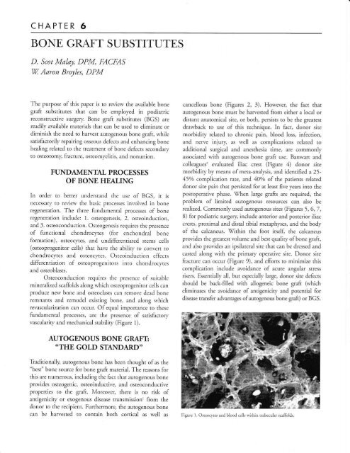
vascularity and mechanical stability (Figure 1).<br />
canceilous bone (Figures 2, 3). However, the fact that<br />
autogenous bone must be harvested from either a local or<br />
distant anatomical site, or both, persists to be the greatesr<br />
drawback to use of this technique. In fact, donor site<br />
morbidity related to chronic pain, blood loss, infection,<br />
and nerve injury as well as complications related to<br />
additional surgical and anesthesia time, are commonly<br />
associated with autogenous bone <strong>graft</strong> use. Banwart and<br />
colleagues' evaluated iliac crest (Figure 4) donor site<br />
morbidity by means of meta-analysis, and identified a 25-<br />
45o/o complication rate, and 40o/o of the patients related<br />
donor site pain that persisted for at least five years into the<br />
postoperative phase. \When large <strong>graft</strong>s are required, the<br />
problem of limited autogenous resources can also be<br />
realized. Commonly used autogenous sites (Figures 5, 6,7,<br />
8) for podiatric surgery, include anterior and posterior iliac<br />
crests, proxima.l and distal tibiai metaphyses, and the body<br />
of the calcaneus. Within the foot itself, the calcaneus<br />
provides the greatest volume and best quality of bone <strong>graft</strong>,<br />
and also provides an ipsilateral site that can be dressed and<br />
casted along with the primary operative site. Donor site<br />
fracture can occur (Figure 9), and efforts to minimize this<br />
complication include avoidance of acute angular stress<br />
risers. Essentially all, but especially large, donor site defects<br />
should be back-filled with allogeneic bone <strong>graft</strong> (which<br />
eliminates the avoidance of antigenicity and potential for<br />
disease transfer advantages of autogenous bone <strong>graft</strong>) or BGS.<br />
AUTOGENOUS BONE GRAFT<br />
*THE GOLD STANDARD''<br />
Tiaditionally, autogenous bone has been thought ofas the<br />
"best" bone source for bone <strong>graft</strong> material. <strong>The</strong> reasons for<br />
this are numerous, including the fact that autogenous bone<br />
provides osteogenic, osteoinducrive, and osteoconductive<br />
properties to the <strong>graft</strong>. Moreove! there is no risk of<br />
antigenicity or exogenous disease transmission' from the<br />
donor to the recipient. Furthermore, rhe autogenous bone<br />
can be harvested to contain both cortical as well as<br />
Figure 1. Osteocytes and blood cells within trabecular scaffolds
CHAPTER 6<br />
Figure 2. Autogenous cancellous bone chips<br />
Figure 3. Autogenous corticocancellous iliac crest<br />
ptr<br />
(lh<br />
\-.<br />
Figure,1. Tri-corrical iliac crest bon; sran<br />
,'-d-ll\<br />
T/j,r I-<br />
ll' /r+<br />
Figure 5. Commonly used autogenous bone <strong>graft</strong> sources.<br />
Laterat'Viex o{ Os eabls<br />
Figure 6. A. Target site fbr corticocalcellous calcmeal donor <strong>graft</strong>. B. Operative<br />
exposure. C. Harested <strong>graft</strong>.<br />
VA<br />
l-. ffib<br />
3W<br />
i'j"":.- we^<br />
Figure 7. Calcmeal donor sites. Avoid calaneofibular ligament,<br />
neutral triangle, peroneal tubercle, Achilles inserdon, STJ and<br />
CC].
CHAPTE,R 6 33<br />
Figure 9. (A) Immediate postoperative and, (B)13 months postoperative.<br />
Radiographs oflarge distal tibial cortjcocancellous donor site that fractured due<br />
to diaphyseal location and l:rrge cortical stress riser. Corraline hydroxyapatite<br />
backfilJ persists to be evident long after bone healing.<br />
Figure 8. Distal tibial metaphyseal, luge corticocancellous<br />
donor site.<br />
MOLECUIAR BIOLOGYAND<br />
BIOENGINEERING OF BGS<br />
BGS are engineered to meet the requirements of bone<br />
healing, namely osteogenesis, osteoinduction and<br />
osteoconduction. Osteogenic cells, including osteocltes,<br />
osteoblasts, and mesenchymal stem cells (MSC$, are found<br />
in autogenous bone as well as fresh bone marrow aspirares or<br />
cloned bone marrow. Osteoinduction is controlled by a<br />
variety of factors, including bone morphogenetic proteins<br />
(BMP$ 2 and7, transforming growth factors GGFs) derived<br />
from platelets and fibroblasts, and are responsible for<br />
recruitment and transformation of MSCs. Demineralized<br />
bone matrix,3'e either allogeneic or recombinant, and<br />
autogenous platelet rich plasma serve as sources for these<br />
osteoinductive agents. Osteoinductive agents include<br />
bioceramics,'n't such as tricalcium phosphate (TCP),<br />
calcium sulfate (CS), calcium carbonate (CC), and synthetic<br />
hydroxyapatite (HA); coralline hydroxyapatite (cHA)<br />
produced naturally by corals; extra cellular matrix scaffolds<br />
such as coliagen or glycosaminoglycan combined with<br />
F{A and TCP; as well as polymers such as poly-hydroxy<br />
acids, polylactide, and polyglycolide, and alloys such as<br />
titanium cages.<br />
BONE GRAFT SUBSTITUTES: OPTIONS<br />
Available options for BGS include: 1. demineraiized bone<br />
matrk, 2. bioceramics, 3. platelet concentrates, and 4. bone<br />
marrow aspirates. Each of these materials has advantages<br />
and disadvantages that should be taken into consideration<br />
prior to use.<br />
Demineralized bone matrix (DBM)<br />
1. Allogeneic (cadaver) demineralized and recombinant<br />
human cloned DBM<br />
a. Batch variabiliry<br />
b. Potential infection and immunogenicity<br />
2. Osteoinductive, containing bone growth factors<br />
a. BMPs 2 A7<br />
i. Regulate MSC and osteoblasts<br />
ii. Angiogenic<br />
3. Can be used in conjunction with allogeneic<br />
<strong>graft</strong>, and as a <strong>graft</strong> expander<br />
4. Available in various forms (Figure 10)<br />
a. Gel, putty, flexible sheet of demineralized<br />
bone collagen fibers<br />
b. Can combine with demineralized cortical<br />
cubes (inductive and conductive)<br />
c. Grafton*, DBX*, Osteofil, Allomatrix*,<br />
DynaGraft
34 CHAPTE,R 6<br />
Bioceramics<br />
Bioceramics provide osteoconductive scaffolds (see Figure<br />
14) that invite osteogenic and vascular ingrowth, and<br />
frequently used materials include: 1. tricalcium phosphate<br />
(TCP), 2. calcium sulfate (CS), 3. calcium carbonate (CC),<br />
and 4. hydroxyapatite (HA).<br />
1. ticalcium phosphate (TCP) scaffold<br />
a. Osteoconductive (pores)<br />
b. Mimics cancellous bone<br />
c. Pores 1-1000 microns<br />
d. Resorbable calcium phosphate<br />
i. Beta-TCP resorbed by 24 weeks<br />
e. Used for over 25 years<br />
f. Safe (sterile) and nonimmunogenic<br />
g. \ficks blood and cells into interstices<br />
h. Orthovita Vitoss (see Figure 15)<br />
i. Ideal for defect back-filI (see Figure 16)<br />
2. Calcium sulfate<br />
a. Semi-structural filler with porous<br />
J.<br />
4.<br />
i. conductive properties similar to TCP<br />
ii. Dense yet pliable<br />
iii. Injection system<br />
iv. Hardens in defect<br />
v. Can be drilled and fixated<br />
Resorbs completely (rapid)<br />
b. Support with internal and or external fixation<br />
c. 'Wright Medical AlloMatrix, miniMIIC (CS<br />
scaffold with DBM)<br />
d. CS with antibiotic impregnation<br />
Bioceramic combined with allogeneic collagen<br />
a. <strong>Bone</strong> <strong>graft</strong> expander<br />
b. Osteoconductive<br />
c. Non-structural paste, soft strip (sponge)<br />
d. Cryogenically deantigenated bovine fibrillar<br />
collagen & porous bioceramic (650/o HA,<br />
35%o TCP)<br />
e. Zimmer Colla<strong>graft</strong><br />
Hydroxyapatite<br />
a. Natural coralline hydroxyapatite and<br />
calcium carbonate (behaves like TCP)<br />
b. Osteoconductive porous scaffold for backfill<br />
(see Figure 18)<br />
c. Inorganic, sterile and nonimmunogenic<br />
d. Minimal structural strength<br />
e. Slow biodegradation (see Figure 9)<br />
f. Interpore International Interpore 200 and<br />
Pro Osteon 500 (see Figure 19)<br />
Platelet Concentrates<br />
Platelet concentrates, which provide growth factors and clot<br />
stabiliry are readily available in most podiatric cases wherein<br />
bone <strong>graft</strong>ing is necessary. Preoperative preparation should<br />
include notification of the anesthesiologist, since 55-60 ml<br />
of blood must be drawn for harvesting the platelets. <strong>The</strong><br />
blood can be drawn from either a peripheral access in the<br />
upper or lower extremiry or from a central venous line. <strong>The</strong><br />
blood is centrifuged and the platelets and supernatant are<br />
coliected. Platelet concentrates are used to augment bone<br />
<strong>graft</strong> stabi lizarion and incorporarion.<br />
Features of platelet concentrates:<br />
1. Osteoinduction, clot stabiliry and chemotaxis<br />
2. Requires at least 55 ml of autogenous blood<br />
drawn in the operating room<br />
3. Platelet rich plasma<br />
A. PDGF, TGF-B, EGF, VECF<br />
4. Point-of-care "mini lab" (centrifuge, efficient,<br />
takes about 15 minutes)<br />
5. DePuy Symphony (see Figure 20) and Smith &<br />
Nephew Magellan<br />
<strong>Bone</strong> Marrow Aspirate<br />
<strong>The</strong> best source of osteogenic cells is proximal bone marrow<br />
in young individuals. As humans age, the peripheral bone<br />
marrow, distal to the iliac crest, becomes less osteogenic.<br />
Nonetheless, tibial and calcaneal marrow aspirates in<br />
generally healthy individuals can enhance the osteogenic<br />
and osteoinductive capaciry of a bone <strong>graft</strong>.<br />
Features oI bone marrow aspirates:<br />
1. Osteogenic<br />
a. Osteoblasts<br />
b. MSCs<br />
2. Combine aspirate with scaffold (Vitoss TCP)<br />
3. Orthovita Imbibe <strong>Bone</strong> Marrow Aspiration<br />
Syringe (see Figure 21)<br />
Other Graft Enhancers and Extenders<br />
1. Acellular aliogeneic dermal periosteum replacement<br />
scaffold (\X/right Medical GraftJacket)<br />
a. Mimics periosteum<br />
b. Minimizes fibrous ingrowth<br />
c. Promotes revascularization<br />
2. Synthetic polymers and permanent bone<br />
growth guides<br />
a. Poly-hydroxy acids, polylactide, polyglycolide<br />
b. Titanium cages
CHAPTER 5 35<br />
Figure 10. Two available proprietary lbnns of demineralized bone matrix: (A)<br />
Grafton, and (B) DBX.<br />
Figure 11 A, B, & C. Repair of third metatars:Ll nonunion u'ing autogenoru<br />
calcaneal cancellous bone enhanced with DBX ancl stabilized with internal fixation.<br />
Figure 12 A, B, & C. Gralton DBM added to xlrrogenous canccllous<br />
chips to expand the volume ofthe <strong>graft</strong>.<br />
Figure 13 A, B, & C. Allogencic DBM added to freeze-dried cordcocancellous<br />
bone <strong>graft</strong> to add osteoindtrctive properties to the<br />
structurally stable and osteoconductive allogeneic <strong>graft</strong>.<br />
FigrLre 14. Structural similariry between femoral head trabecular bone (A) and<br />
manulactured calcium phosphate (B) scaffolding.<br />
Figure 15. Orthovita's Vitoss TCP, pellets and granules (A) and rnicrograph<br />
(B), 1000x, showing blood cells adhering to the scaffolding.
36 CHAPTE,R 6<br />
Figure 16. Solitary calcaneal cyst belore (A) and alter (B) TCP back-fil1.<br />
Figure 17 A & B. Collagrafr 6-strip kit provides bioceramic-bovine coilagen<br />
osteoconductive expandcr which, when combined u'ith autogenous b1ooc1 or<br />
marrorv aspirate, also provides osteoinduction.<br />
\rl!tt, ir{\J, UEUIU<br />
Dr:r:<br />
a r \v flarrnuB Al]<br />
ve r avri vvw<br />
frreus, 8*rx1&re{ l$q{ilf<br />
tttr iii'l<br />
Sterile - For $ingle Use Only<br />
n^^,^:^^<br />
^-^<br />
E^,"L.<br />
STERILE Block {1.0 oc)<br />
. ;1 O',rtn'i,,t {fxm,x::it*i*X<br />
Figure 18. Coral-osteocyte jnterface<br />
Figure 20. Symphonl. platelct rich plasma separated (A) and releaseate ready fbr<br />
implantation (B).<br />
Figure 21. Orthovica Imbibe <strong>Bone</strong> Marrorv Aspiration S1.ringe.<br />
SUMMARY<br />
Biosynthetic bone <strong>graft</strong> <strong>substitutes</strong> can be used to provide<br />
osteogenic, osteoinductive, and osteoconductive properties<br />
while decreasing the necessity of autogenous bone <strong>graft</strong>s and<br />
associated donor site morbidiry. <strong>Bone</strong> <strong>graft</strong> <strong>substitutes</strong><br />
include demineralized bone matrix, bioceramics, polymers<br />
and combinations of these materials. Adjunctive therapies<br />
used with BGS include stable bone fixation, vascular<br />
reconstruction, electrical bone growth stimulation,<br />
nutritional supplementation and anemia treatment,<br />
immobilization and non-weight bearing.
CHAPTER 6 37<br />
REFERENCES<br />
1. Validation of virus removal and inactivation. Biologicals 7997;<br />
1.9:247-51.<br />
2. Banwart et al. Iliac crest harvesr site morbidiry (meta-analysis). Spine<br />
1995;201055-60.<br />
3. Glowacki, Mulliken. Demineralized bone implants. Clin Plast Surg<br />
7987;72:233-41.<br />
4. Salyer et al. DBM in craniofacial surg. J Craniofac Surg 1992;3:55-62.<br />
5. Skoff. Allogenic cancellous plus autogenous marrow viable<br />
alternative to autogenous iliac crest. Am J Orthop 1995;24:40-7.<br />
6. Gazdag et a1. Alternatives to aurogenous bone <strong>graft</strong>s: efficacy &<br />
indications. /lm Acad. Orthop Surg 1995;3:1-8.<br />
7. Martin et al. Demineralized bone matrix. Spine 1998:24:637-45.<br />
B. Friedlaender et al. BMP-7 & type-1 collagen scaffold in Tx tibial Fx.<br />
9.<br />
J <strong>Bone</strong> Joint Surg Am 2001;83:5 151-8.<br />
Govender et al. Recombinant humari BMP-2 for Tx of open tibial<br />
fractures: prospective RCT, N=450 patients. J <strong>Bone</strong> Joint Surg Am<br />
2002;84:2123-34.<br />
l0.Cavagna et aI. CaPO4 suitable lor spinal fwion. J Long Term Eff<br />
Med Imp lants 1999 ;9 :403 -12.<br />
1 1. Helm et al. <strong>Bone</strong> <strong>graft</strong> <strong>substitutes</strong> for spinal fision. Neurosurg Focus<br />
200 1;1 0:1-5.<br />
12.A-lexander et al. Eflicacy of CaSO4 plus decompression bone<br />
equivalent to autogenous iliac crest. CanJ Surg2001;44:262-6.<br />
13. Bucholz. Nonallo<strong>graft</strong> osteoconductive <strong>substitutes</strong>. Clin Orthop<br />
2002;395:44-52.<br />
14.Demers et al. Natural coral exoskeleton as a bone <strong>graft</strong> substitute.<br />
Biomed Mater Eng 2002:2:75 -35.<br />
l5.Coombes & Meikle. Resorbable synthetic polymers as bone <strong>graft</strong><br />
replacements. Clin Mater 199 4;17 :35 -67.


