

Plate fixation in Foot Surgery - The Podiatry Institute
Plate fixation in Foot Surgery - The Podiatry Institute
Plate fixation in Foot Surgery - The Podiatry Institute

You also want an ePaper? Increase the reach of your titles
YUMPU automatically turns print PDFs into web optimized ePapers that Google loves.
PLATE FIXATION IN<br />
FOREFOOT ST]RGERY<br />
Alfred l. Pbillips, DPM<br />
INTRODUCTION<br />
<strong>Plate</strong>s are not commonly used for <strong>in</strong>ternal <strong>fixation</strong><br />
<strong>in</strong> forefoot surgery. However, they are capable<br />
of provid<strong>in</strong>g significant rigid <strong>in</strong>ternal <strong>fixation</strong><br />
and <strong>in</strong> certa<strong>in</strong> situations are an appropriale and<br />
excellent choice of <strong>fixation</strong>. Rigid <strong>in</strong>ternal <strong>fixation</strong><br />
is required for primary bone heal<strong>in</strong>g to occur. Primary<br />
bone heal<strong>in</strong>g is desireable because it provides<br />
predictable bone heal<strong>in</strong>g, avoids secondary<br />
callous formation, and enables the patient to<br />
mobilize the adjacent jo<strong>in</strong>ts quickly.<br />
<strong>Plate</strong>s have been utilized <strong>in</strong> the <strong>fixation</strong> of<br />
melatarsal or hallux fractures, metatarsal nonunions,<br />
metatarsal osteotomies, first metalarsophalangeal<br />
jo<strong>in</strong>t arthrodesis, Lisfranc afihrodesis,<br />
and various repairs of iatrogenic deformities.<br />
prevents the plate from shift<strong>in</strong>g when the head of<br />
the screw sets <strong>in</strong>to the hole of the plate.<br />
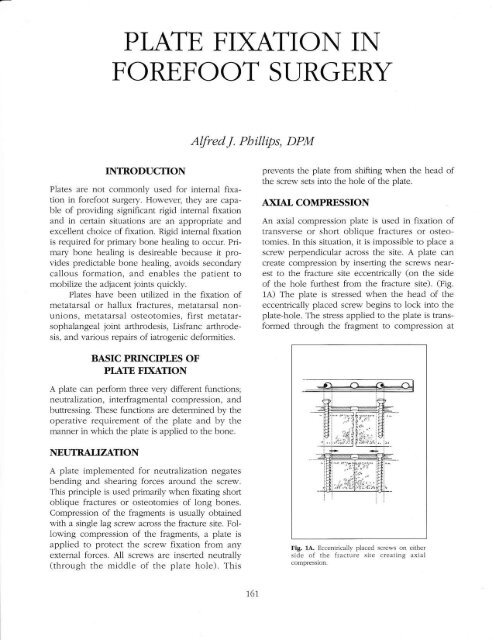
A)ilAL COMPRESSION<br />
An axial compression plate is used <strong>in</strong> <strong>fixation</strong> of<br />
transverse or short oblique fractures or osteotomies.<br />
In this situation, it is impossible to place a<br />
screw perpendicular across the site. A plate can<br />
create compression by <strong>in</strong>sert<strong>in</strong>g the screws nearest<br />
to the fracture site eccentrically (on the side<br />
of the hole furthest from the fracture site). (Fig.<br />
1A) <strong>The</strong> plate is stressed when the head of the<br />
eccentrically placed screw beg<strong>in</strong>s to lock <strong>in</strong>to the<br />
plate-hole. <strong>The</strong> stress applied to the plate is transformed<br />
through the fragment to compression at<br />
BASIC PRINCIPLES OF<br />
PIATE FD(AIION<br />
A plate can perform three very different functions;<br />
neutralization, <strong>in</strong>terfragmental compression, and<br />
buttress<strong>in</strong>g. <strong>The</strong>se functions are determ<strong>in</strong>ed by the<br />
operative requirement of the plate and by the<br />
manner <strong>in</strong> which the plate is applied to the bone.<br />
NEUTRALIZAION<br />
A plate implemented for neutalization negates<br />
bend<strong>in</strong>g and shear<strong>in</strong>g forces around the screw.<br />
This pr<strong>in</strong>ciple is used primarily when fkat<strong>in</strong>g short<br />
oblique fractures or osteotomies of long bones.<br />
Compression of the fragments is usually obta<strong>in</strong>ed<br />
with a s<strong>in</strong>gle lag screw across the fracture site. Follow<strong>in</strong>g<br />
compression of the fragments, a plate is<br />
applied to protect the screw <strong>fixation</strong> from any<br />
external forces. A11 screws are <strong>in</strong>serted neutrally<br />
(through the middle of the plate hole). This<br />
Fig. 1A. Eccentrically placed screws on<br />
side of the fracture site creat<strong>in</strong>g<br />
compression.<br />
either<br />
axial<br />
757
the fracture contact surface. Because the force of<br />
compression that is created by the plate is on one<br />
side of the bone, the opposite cortex will actually<br />
gap as axial compression is applied. (Fig. iB)<br />
This side effect is counteracted by "overbend<strong>in</strong>g"<br />
the plate. (Fig. 1C) <strong>The</strong> AO group recommends<br />
that a 1 mm bend be applied to the plate. <strong>The</strong><br />
opposite cortices will contact <strong>in</strong>itially and then<br />
the compressive force will be spread across the<br />
entire fragment to fragment <strong>in</strong>terface as the plate<br />
is straightened out. (Fig 1D)<br />
BUTTRESS PIATE<br />
Buttress<strong>in</strong>g supports multiple fracture fragments.<br />
A plate is used as a buttress where there is severe<br />
comm<strong>in</strong>ution or bone loss, such as <strong>in</strong> a gunshot<br />
wound or crush <strong>in</strong>jury to the forefoot. <strong>The</strong> plate<br />
is secured to the ma<strong>in</strong> proximal and distal fragments<br />
and then secured loosely to the comm<strong>in</strong>uted<br />
fragments. This technique will ma<strong>in</strong>ta<strong>in</strong><br />
anatomic length and avoid disrupt<strong>in</strong>g the vascularity<br />
of the smaller fragments. Screws are <strong>in</strong>serted<br />
<strong>in</strong>to the plate holes neutrally so that the plate<br />
does not shift. <strong>The</strong> plate should not be placed<br />
under stress. <strong>The</strong> screws are simply placed <strong>in</strong>to<br />
both fragments to ma<strong>in</strong>ta<strong>in</strong> length and viabiliry of<br />
the bone.<br />
ANATOMICAL AND BIOMECHANICAL<br />
CONSIDERATIONS<br />
<strong>The</strong>re are certa<strong>in</strong> anatomic and biomechanical<br />
characteristics w-hich dictate how a plate is<br />
applied. Placement of the plate must be adapted<br />
to conform to the local anatomy with care to avoid<br />
disruption of function. A plate applied for fkation<br />
of a Jones fracture should be applied so as not to<br />
disrupt the <strong>in</strong>sertion of the Peroneal brevis tendon.<br />
<strong>The</strong> width of the plate and the size of the<br />
screws must be appropriate for the size of the<br />
metatarsal. If a plate is too large, it will be difficult<br />
to cover with soft tissue and may significantly<br />
retard vascularization of underly<strong>in</strong>g bone<br />
fragments.<br />
A plate is best applied to a relatively flat surface.<br />
Some of the most common surfaces for appiication<br />
of a plate <strong>in</strong> the forefoot are dorsal surface<br />
of second, third or fourth metatarsals, dorsal lateral<br />
surface of the fifth metatarsal and medial surface<br />
ol the first metatarsal.<br />
Fig. 18. Eccentric load<strong>in</strong>g creates compression between the fragments<br />
on the plate side and tension (gapp<strong>in</strong>g) on the opposite side.<br />
Fig. 1C. Overbend<strong>in</strong>g of the plate is perfomed to counteract the<br />
eccentric load<strong>in</strong>g.<br />
Fig. lD. Llniform compression<br />
technlque.<br />
l'ullow<strong>in</strong>g the overlrend<strong>in</strong>g<br />
762
<strong>The</strong> AO group recommends application of a<br />
plate on the tension side of the bone to prevent<br />
gapp<strong>in</strong>g with fr-rnctional load<strong>in</strong>g. In the metatarsal<br />
region, the functional load of weightbear<strong>in</strong>g creates<br />
compression at the dorsal surface and tension<br />
on the plantar surface. <strong>The</strong>oretically, the<br />
plate shoulcl be appliecl to the plantar surface.<br />
Anatomically, however this is difficr-rlt to perform.<br />
Dissection of the plantar zlspect of the metatarsals<br />
makes a plantar approach quite unreaiistic.<br />
PIATES<br />
Choos<strong>in</strong>g the appropriate plate is important for<br />
the success of the <strong>fixation</strong>. It also avoids complications.<br />
<strong>The</strong> AO grolrp has set the standarcl for<br />
<strong>in</strong>ternal <strong>fixation</strong> and provide a variety of plates<br />
which can be utilizecl <strong>in</strong> forefoot sr,tr!;ery.<br />
Trough shaped 1/3 tubular straight plates<br />
can be used with either 3.5 and 4.0 mm screws.<br />
<strong>The</strong> trough shape of the screw prevents the<br />
screw heacl from hav<strong>in</strong>g direct contact with the<br />
cortex of the bone. It is made of sta<strong>in</strong>less steel<br />
(AISI 316). It is 10 mm wide and 1 mm thick. <strong>The</strong><br />
plate is commonly used <strong>in</strong> first metatarsocuneiform<br />
arthrodesis and as a primary part of a<br />
Lisfranc lo<strong>in</strong>t arthrodesis. (Fig. 2) Small T :rnd<br />
oblique T plates identical <strong>in</strong> thickness can be<br />
substitutecl <strong>in</strong> this arthroclesis.<br />
2.7 mm straight or 1/4 tubular plates are<br />
designed for <strong>in</strong>sertion with 2.7 mm screws. 'Ihey<br />
are slightly troughed shaped and have raised<br />
rims. This design strengthens the p1ate. <strong>The</strong> plate<br />
is used <strong>in</strong> the lesser metatarsal region. (flg. Z t<br />
M<strong>in</strong>i plates are utilized with 1.5 and 2.0 mm<br />
screws. <strong>The</strong> plates are extra long and are cut to<br />
the desirecl length with a cutt<strong>in</strong>g forceps. <strong>The</strong><br />
plates are utilized fbr lesser metatarsal and digital<br />
<strong>fixation</strong>.<br />
A relatively new plate system u,,hich r,vas<br />
developed for maxilo-facial surgery has a role <strong>in</strong><br />
podiatric srrrgery. (Fig. 3) <strong>The</strong> plates are constructed<br />
of vitalliur-n which enable the plate to be<br />
relatively th<strong>in</strong>. Screw sizes are 2.7, 2.0, and 1.5<br />
mm and are self tapp<strong>in</strong>g. Two vertical cutt<strong>in</strong>g<br />
flutes cut the threads so that the screw hole does<br />
not need to be tapped.<br />
Dynamic Compression <strong>Plate</strong>s (DCP) and the<br />
Limited Contact D).namic Compression <strong>Plate</strong> (LC-<br />
DCP) have a limited role <strong>in</strong> forefoot surgery.<br />
<strong>The</strong>y are specially constructed plates which self<br />
Fig, 2. 1/l tubular and m<strong>in</strong>i-plates used <strong>in</strong> lesser metatarsal and digital<br />
procedures.<br />
Fig. 3. Maxilo-facial vitalir-rm plates fbr 2.7 ancl 2.0 n-rm self'-tapp<strong>in</strong>g<br />
screws.<br />
compress the osseous fragments. <strong>The</strong> special<br />
Eaeometry/ of the screw hole produce glid<strong>in</strong>g of<br />
the eccentrically drilled screw. <strong>The</strong> DCP is a flat<br />
and lelatively thick plate which has proven to<br />
have its shoficom<strong>in</strong>gs. Probably one of the most<br />
significant of these is the extensive contact of the<br />
plate to the bone which <strong>in</strong>terferes with periosteal<br />
blood supply and has led to local osteonecrosis.<br />
<strong>The</strong> LC-DCP is the newer version of the Dynamic<br />
Compression plates which are constructed u,'ith<br />
deep oblique cuts between the screw holes to<br />
limit contact.<br />
TECHNIQUE OF PIATE INSERTION<br />
Aclequate exposure for the <strong>in</strong>sertion of the plates<br />
is essentizrl. An idea of the type of plate and<br />
approximate size should be known preoperative-<br />
1y, however, the specific choice of a plate is obvit63
ously made <strong>in</strong>traoperatively. Periosteum should<br />
be elevated dur<strong>in</strong>g the <strong>in</strong>itial dissection and used<br />
to cover the plate upon closure.<br />
<strong>The</strong> plate must be contoured to the surface of<br />
the bone. A malleable alum<strong>in</strong>um template can be<br />
applied to the surface of the bone to help determ<strong>in</strong>e<br />
the amount of bend<strong>in</strong>g that needs to be performed<br />
on the plate. <strong>Plate</strong> benders are then used<br />
to bencl the plate <strong>in</strong>to the appropriate shape.<br />
<strong>The</strong> number of cortices held by each screw is<br />
important <strong>in</strong> the strength of the <strong>in</strong>ternal fkation.<br />
<strong>The</strong> AO group have determ<strong>in</strong>ed that the lever arm<br />
and functional loads onto the plate <strong>in</strong>creases <strong>in</strong><br />
the direction of the trunk. To accommodate these<br />
functional loads there are specific recommendations<br />
to the number of corlices crossed on each<br />
side of the fracture site. Specifically these are three<br />
<strong>in</strong> the phalanges and four <strong>in</strong> the metatarsals. <strong>The</strong><br />
specific technique of plate <strong>in</strong>sefiion differs upon<br />
the specific function of the plate.<br />
A)ilAL COMPRESSION<br />
This technique is applied to a transverse or short<br />
oblique fracture or osteotomy. <strong>The</strong> fragments are<br />
visualized and a plate of the appropriate size is<br />
chosen. <strong>The</strong> plate is contoured to the surface of<br />
the metatarsal. A slight bend of the plate is made.<br />
<strong>The</strong> bend of the plate is at the section of the<br />
plate which will directly overlay the fracture or<br />
osteotomy site. <strong>The</strong> screws are first <strong>in</strong>serted <strong>in</strong>to<br />
the distal fragment us<strong>in</strong>g the standard AO techniques.<br />
<strong>The</strong> holes are predrilled, measured,<br />
tapped, and fo1low-ed with <strong>in</strong>sertion of the screw.<br />
<strong>The</strong> distal screws are <strong>in</strong>serted centrally. <strong>The</strong> plate<br />
is then used as a handle to reduce the fracture or<br />
osteotomy to its anatomical position. A plate<br />
clamp temporarily fixates the plate to the proximal<br />
fragment. <strong>The</strong> screw hole nearest to the fracture<br />
site is drilled eccentrically away from or<br />
proximal to the osteotomy or fracture. As this<br />
screw is <strong>in</strong>serted, the fragment to fragment <strong>in</strong>terface<br />
is compressed. <strong>The</strong> rema<strong>in</strong><strong>in</strong>g proximal<br />
screws are then <strong>in</strong>sefied neutrally. (Fig. 4A, 48)<br />
COMBINATION FD(ATION<br />
This term describes the use of a lag screw for<br />
compression <strong>in</strong> comb<strong>in</strong>ation with a plate function<strong>in</strong>g<br />
as a neutralization plate. This <strong>fixation</strong> is<br />
performed <strong>in</strong> fixat<strong>in</strong>g short or medium oblique<br />
fractures. Reduction of the fracture can be per-<br />
Fig. 4A. Radiographs reveal<strong>in</strong>g short oblique<br />
fractures of the neck of the second, third, and<br />
lourth metatarsals.<br />
Fig. 48. Postoperative racliographs reveal<strong>in</strong>g<br />
axial compression technique with L-shaped<br />
m<strong>in</strong>i-plate and 2.0 mm screns.<br />
formed with a bone clamp. An appropriate plate<br />
is chosen and contoured to the surface of the<br />
bone. <strong>The</strong> fracture orientation is evaluated and a<br />
determ<strong>in</strong>ation of whether or not to place the lag<br />
screw through the plate is made. If the lag screw<br />
is placed outside of the plate, it is <strong>in</strong>serted and<br />
tightened first followed by application of the<br />
154
plate. <strong>The</strong> screws of the plate are <strong>in</strong>serted neutrally.<br />
If the fracture orientation favors the use of<br />
lag screw through the plate the iag screw is<br />
<strong>in</strong>serted through the plate with overdrill<strong>in</strong>g of the<br />
near cortex, followed by the <strong>in</strong>sertion of the<br />
rema<strong>in</strong><strong>in</strong>g screws. A11 other screws through the<br />
plate are <strong>in</strong>serted <strong>in</strong> the manner previously<br />
described. (Fig. 5A, 58)<br />
DOUBI-E COMBINATION FD(ATION<br />
Double comb<strong>in</strong>ation <strong>fixation</strong> is the term used to<br />
describe the use of a lag screw <strong>in</strong> comb<strong>in</strong>ation<br />
with an axial compression plate. This technique is<br />
utilized specifically <strong>in</strong> fusions of the first metatarsocuneiform<br />
and metatarsophalangeal jo<strong>in</strong>ts. <strong>The</strong><br />
first metatarsocuneiform jo<strong>in</strong>t is temporarily fixated<br />
with a K-wire from dorsal to plantar. A plate is<br />
chosen to be applied to the medial surface of the<br />
first metatarsal and medial cuneiform and contoured<br />
to the appropriate shape. A 5-ho1e straight<br />
7/3 ttbular plate with 3.5 mm fully threaded<br />
screws is commonly used. <strong>The</strong> plate is first<br />
anchored proximally to the medial cuneiform. <strong>The</strong><br />
distal aspect of the plate is then temporarily<br />
clamped to the first metatarsal. A screw is then<br />
placed eccentrically at the hole just distal to the<br />
arthrodesis site. This is tightened and creates compression<br />
across the site. A lag screw is then placed<br />
dorsal to plantar or plantar to dorsal to provide<br />
additional tension at the site. <strong>The</strong> rema<strong>in</strong>der of the<br />
screws are then <strong>in</strong>serted neutrally. (Fig. 6)<br />
Fig. 5A. Radiographs reveal<strong>in</strong>g long oblique<br />
fracture of the neck of the fifih metatarsal.<br />
SUMMARY<br />
This paper has been an <strong>in</strong>troduction to the pr<strong>in</strong>ciples<br />
and techniques of plate <strong>fixation</strong> and how<br />
they specifically apply to the forefoot. <strong>Plate</strong>s if<br />
correctly used can be an effective type of <strong>fixation</strong><br />
for a variety of forefoot surgery.<br />
Fig. 58. Postoperative radiographs reveal<strong>in</strong>g<br />
comb<strong>in</strong>ation <strong>fixation</strong> technique s,-ith a lag screw<br />
and neutralization plilte.<br />
165
BIBLIOGRAPTTY<br />
Fig. 6. Postoperative radiographs of lisfranc<br />
arthrodesis reveal<strong>in</strong>g double comb<strong>in</strong>ation <strong>fixation</strong><br />
technique of thc first metatarsal medial<br />
cuneiform jo<strong>in</strong>t fusion.<br />
Corey SV, Castellano BD. RuchJA: Fixation Tecirniques for Fractures<br />
In Scurran B (ed) <strong>Foot</strong> and Ankle Trauma New York, Churchill<br />
Liv<strong>in</strong>S;ston, pp. 7 5-93, 1981).<br />
Coughl<strong>in</strong> MJ: Arthrodesis of the First Metatarsophalangeal Jo<strong>in</strong>t with<br />
NI<strong>in</strong>i-Fragnrent <strong>Plate</strong> Fixation. Ortb opedics 10:1037 -1044, 1990.<br />
Heim U, Pfeiffer KM: Internal l-ixation of Small Fractures. New<br />
York, Spr<strong>in</strong>ger-Yerlag, 1!88.<br />
Jacobs AM, oloff LM: Podiatric Metallurgy and the Effects of<br />
Implanted Metals on Liv<strong>in</strong>g Tissues Cl<strong>in</strong> <strong>Podiatry</strong>, L:L2L-742,<br />
1985,<br />
Laporta GA, Ricl-rter KP, Jo1ly GP: Pressure osteosynthesis for Internal<br />
Fixation of Metatarsal Angulational Osteotomies /,4m Podiatr<br />
,4ssoc 66(3)P173-180, 1976.<br />
Muller ME, Allgower M, Schneider R, V/illenger H: Manual of Inter<br />
nal Fixation, New York, Sp<strong>in</strong>ger-Verlag, 1!!1.<br />
Nunamaker DM. Perren SM: A Radiological and Histological Analysis<br />
of Fracture Heal<strong>in</strong>g Us<strong>in</strong>g Prebencl<strong>in</strong>g of Compression <strong>Plate</strong>s<br />
Cl<strong>in</strong> ortbop, 1381167 -195, 197 9.<br />
Ruclr JR, Corey SV, Vito GR: Tbe <strong>Podiatry</strong> <strong>Institute</strong> InteflMl Fixation<br />
workbook, Tucker GA, <strong>Podiatry</strong> <strong>Institute</strong>, 1990.<br />
Ruch JIt, &Ierrill T; Pr<strong>in</strong>ciples of rigid <strong>in</strong>ternal compression <strong>fixation</strong><br />
and its application <strong>in</strong> podiatric surgery, pp 216-293.In McGlamr,v<br />
ED (ed) l:'undamentals of <strong>Foot</strong> SurgeD) Baltimore, .Williams<br />
and vilk<strong>in</strong>s, pp. 246-293, 1987.<br />
Russell RD: Use of Compression Bone <strong>Plate</strong> after Metatarsal<br />
Nonunion / <strong>Foot</strong> SrLrg 19(3):159-161, 1980.<br />
Sarrafian SKt Andtom)) of tbe <strong>Foot</strong> and Ankle. Philadelphia, JB Lipp<strong>in</strong>cott<br />
Co, 1983.<br />
Schatzker J, Sanderson R: <strong>The</strong> Hold<strong>in</strong>g Power of Ofihopedic Screws<br />
In Vivo, Cl<strong>in</strong> Oftbr,tp 108:115-126, 7975.<br />
Vangness CT, Carter DR. Frankel \TI: In Vitro Hvaluation of Loosen<strong>in</strong>g<br />
Characteristics of Self-Tapped Cortical Bone Screws C/ln<br />
oftbop 157 1279-286, 1981,<br />
166


