High resolution CT in Interstitial Lung Diseasesby Dr
High resolution CT in Interstitial Lung Diseasesby Dr
High resolution CT in Interstitial Lung Diseasesby Dr

Create successful ePaper yourself
Turn your PDF publications into a flip-book with our unique Google optimized e-Paper software.
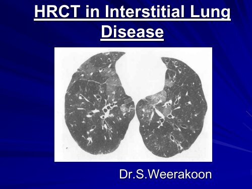
HR<strong>CT</strong> <strong>in</strong> <strong>Interstitial</strong> <strong>Lung</strong><br />
Disease<br />
<strong>Dr</strong>.S.Weerakoon
In Diffused <strong>Lung</strong> Disease<br />
CXR<br />
Limitations<br />
Cannot Clearly Depict the Alteration of<br />
lung parenchyma<br />
Radiographic Pattern of Diffuse<br />
<strong>Lung</strong> Disease is Often Non Specific<br />
Inability of Resolv<strong>in</strong>g Small Differences<br />
<strong>in</strong> Density
HR<strong>CT</strong> improves<br />
Sensitivity<br />
Specificity<br />
Diagnostic Accuracy<br />
HR<strong>CT</strong> images of the lung correlate closely<br />
with the macroscopic appearance of<br />
pathologic specimen
Scan Collimation<br />
Narrow<br />
Collimation<br />
Technique<br />
Volume<br />
Averag<strong>in</strong>g<br />
With<strong>in</strong> the<br />
Section<br />
Spatial<br />
Resolution<br />
•Resolution is Optimized by Us<strong>in</strong>g Th<strong>in</strong> Sections -<br />
1 – 1.5 mm are obta<strong>in</strong>ed from the apex to lung<br />
bases with the patient sup<strong>in</strong>e.<br />
•Sections are Usually Separated by 1 – 4 cm. S<strong>in</strong>ce<br />
Most Infiltrative <strong>Lung</strong> Diseases are Diffuse.<br />
•<strong>Lung</strong> Nodules may be missed Unless HR<strong>CT</strong> is<br />
Supplemented with Contigous Imag<strong>in</strong>g
Anatomy Of <strong>Lung</strong><br />
The Secondary Pulmonary Lobule is the<br />
Smallest Unit of the <strong>Lung</strong><br />
The Secondary Pulmonary Lobule is Wrapped<br />
with connective Tissue Septae.<br />
It conta<strong>in</strong>s Ve<strong>in</strong>s / Lymphatics Vessels. A<br />
Secondary Bronchus & Accompany<strong>in</strong>g Artery<br />
Situated Centrally
Bronchi & Pulmonary Arteries run &<br />
Branch Together Through Out the <strong>Lung</strong><br />
They Taper Slightly as They Travel<br />
Radialy<br />
At any Given Level the Diameter of the<br />
Bronchus is the Same of its<br />
Accompany<strong>in</strong>g Pulmonary Artery
Patterns Of Diffuse <strong>Lung</strong><br />
Disease<br />
Reticular and short lenear<br />
pattern<br />
HR<strong>CT</strong> Patterns<br />
Nodular density<br />
Ground glass density/<br />
consolidations<br />
Cystic spaces and areas<br />
of decreased lung<br />
density
Reticular And Short Lenear<br />
Pattern<br />
Due to thicken<strong>in</strong>g of<br />
<strong>in</strong>terstitial fibre network/<br />
pulmonary lymphatics by<br />
Fluid<br />
Fibrosis<br />
Cells<br />
Other materials….<br />
Conditions associated<br />
with Dialatation of<br />
pulmonary ve<strong>in</strong>s.
Cont’d Reticular and …<br />
Interlobular Septal Thicken<strong>in</strong>g<br />
Normal<br />
only few septae should be seen<br />
Abnormal<br />
presence of numerous clearly visible<br />
<strong>in</strong>terlobular septae, almost always <strong>in</strong>dicates<br />
and <strong>in</strong>terstitial abnormality<br />
Abnormally thickened septea outl<strong>in</strong>e part or<br />
entire lobule.
Septal thicken<strong>in</strong>g<br />
1.Smooth<br />
Pul. Oedema<br />
Lymphangitic CA<br />
Leukaemia<br />
Amyloidosis<br />
Lymphocytic<br />
<strong>in</strong>terstitial pneumonia<br />
Alveolarmicrolithiasis
2. Nodular ( Beaded)<br />
Lymphangitic Spread of CA<br />
Sarcoidosis, Silicosis<br />
CWP<br />
Amyloidosis<br />
Milliary TB
Interface Sign
Parenchymal Bands<br />
Asbestosis<br />
Sarcoidosis<br />
Silicosis<br />
Tuberculosis
Intra Lobular Septal Thicken<strong>in</strong>g
Peribroncho Vascular <strong>Interstitial</strong><br />
Thicken<strong>in</strong>g
Peribroncho Vascular <strong>Interstitial</strong><br />
Thicken<strong>in</strong>g
Honey Comb<strong>in</strong>g<br />
Extensive <strong>in</strong>terstitial & Alveolar Fibrosis<br />
Alveolar disruption & Bronchiectasis<br />
Honey Comb <strong>Lung</strong>
Honey Comb<strong>in</strong>g<br />
Subpleural,Posterior /<br />
lower lobe<br />
predom<strong>in</strong>ance<br />
• IPF – 60%<br />
• Collagen disease<br />
• Hypersensitivity pneumonitis<br />
• Asbestosis<br />
• <strong>Dr</strong>ug <strong>in</strong>duced fibrosis<br />
• Sarcoidosis<br />
Central / Upper lobe<br />
predom<strong>in</strong>ance<br />
• Sarcoidosis<br />
• Hypersensitivity pneumonitis<br />
• Radiation<br />
• IPF<br />
• Collagen disease<br />
• <strong>Dr</strong>ug <strong>in</strong>duced fibrosis
Nodules / Nodular<br />
Nodules<br />
Discrete densities rang<strong>in</strong>g from 2mm to 10mm <strong>in</strong><br />
diameter.<br />
With<strong>in</strong><br />
Interstitium<br />
Air spaces
Distribution<br />
Perilymphatic<br />
Centrilobular<br />
Random
Reticular Nodular pattern <strong>in</strong> Sarcoidosis
Nodules / Nodular<br />
Perilymphatic
Perylymphatic Distribution<br />
Sarcoidosis<br />
Lymphangitic CA<br />
Lymphoproliferative Disordes<br />
Amyloidosis
Nodules / Nodular<br />
Centrilobular<br />
• Hypersensitivity<br />
pneumonitis<br />
• Sarcoidosis<br />
•Langerhan cell<br />
histiocytosis<br />
•Silicosis
Centrilobular<br />
Nodules / Nodular<br />
Sarcoidosis<br />
Respiratory<br />
bronchiolitis<br />
<strong>in</strong>terstitial<br />
lung<br />
disease
Nodules / Nodular<br />
Random<br />
• MiliaryTB<br />
• Haematogenous<br />
spread of CA<br />
• Milliary fungal<br />
<strong>in</strong>fections.<br />
• Dissem<strong>in</strong>ated viral<br />
<strong>in</strong>fections.
Large Nodules <strong>in</strong> Sarcoidosis
Large Nodules / Masses<br />
Nodules < 1 cm<br />
Masses > 3 cm
Increased <strong>Lung</strong> Density<br />
Ground glass patterns<br />
Hazy <strong>in</strong>crease <strong>in</strong> the density of lung<br />
parenchyma – Air space patterns<br />
E.g. Fibros<strong>in</strong>g Alveolitis (Active phase)<br />
DIP<br />
BOOP<br />
Pneucystis Carr<strong>in</strong>i <strong>in</strong>fection<br />
Diffuse pulmonary H’ge
DIP
AIP
Alveolar protenosis<br />
Pneumocystis carr<strong>in</strong>i <strong>in</strong>fection<br />
ARDS<br />
Pul.Oedema<br />
Pul.Haemorrhage<br />
Mycoplasma pnumonia<br />
NSIP/UIP
Increased<br />
<strong>Lung</strong> Density<br />
Consolidation
Cont’d… Increased <strong>Lung</strong> Density<br />
Consolidation<br />
Increased lung<br />
attenuation with<br />
obliteration of<br />
pulmonary vessels
Decreased <strong>Lung</strong> density / Cystic<br />
spaces
Destruction of Alveolar walls of distal air<br />
spaces.<br />
Panlobular Emphysema<br />
Centrilobular Emphysema<br />
ParaseptaL Emphysema<br />
Lymphangiomyomatosis<br />
Langerhans cell Histiocytosis
L<br />
I<br />
P
Decreased <strong>Lung</strong> density / Cystic<br />
spaces
Bibliography<br />
IMAGING OF THE DISIEASES OF THE<br />
CHEST –David.M.Hansell,Peter<br />
Armstrong,David.A.Lynch,H.Page<br />
McAdams<br />
<strong>High</strong> Resolution <strong>CT</strong> of the <strong>Lung</strong>-W.Richard<br />
Webbs,Nestor L.Mǖller,David.P.Naidich<br />
Radiol Cl<strong>in</strong> N Am 43(2005)<br />
American Journal of Respiratory & Critical<br />
Care Medic<strong>in</strong>e. Vol.165 2004
Thank You …!